final program - isfp - international society for fertility
TRANSCRIPT

THE 2nd WORLD CONGRESS ON FERTILITY PRESERVATION
Sponsored by The International Society for Fertility Preservation and University of Kansas Medical Center Continuing Education
The International Society for Fertility Preservation
www.isfp-fertility.org
Speaker presentations are available on-lineafter completing a brief evaluation at:https://www.surveymonkey.com/s/ISFPWorldCongress2
December 8–10, 2011Miami Beach, Florida
FINAL PROGRAM

Message from the Congress Chair:
On behalf of the International Society of Fertility Preservation, I would like to extend my warm
welcome to you all.
The International Society of Fertility Preservation (ISFP) was founded in 2007 to promote progress in
fertility preservation through international networking, sharing information and new discoveries. The
Society is growing fast, both in quality and quantity, and has established itself as a leading
international organization for fertility preservation.
The World Congress is one of the most important functions of the ISFP, and I am delighted and
honored to serve as Organizing Chair of the 2nd
ISFP World Congress in Miami Beach, FL, USA. As
you can see, the scientific program is filled with current and important topics that will be presented by
world-renowned experts. In addition, we are offering a hands-on workshop on vitrification as a pre-
Congress course. I am also very pleased with the number and quality of abstracts submitted this year.
All of these reflect the significance of the World Congress.
I would like to thank each of you for participating in the 2nd
ISFP World Congress on Fertility
Preservation. It would not be possible to have a successful meeting without the support of sponsors,
exhibitors, the planning committee, CME staff, speakers, and most of all, the attendees. I hope all of
you enjoy the 2nd
ISFP World Congress in Miami Beach.
Sincerely,
S. Samuel Kim, M.D.
President, ISFP
Congress Chair, The 2nd
ISFP World Congress on Fertility Preservation

The Second ISFP World Congress on
Fertility Preservation
Contributors These organizations have generously supported this congress
and we acknowledge them with sincerest appreciation.
Platinum Level:
Ferring Pharmaceuticals
Bronze Level:
Walgreens
Lance Armstrong Foundation
Cornell Center for Reproductive Medicine (Zev Rosenwaks)
KU Department of OB/GYN (Carl Weiner)
Cleveland Clinic Department of OB/GYN (Tommaso Falcone)
Cook Medical
Catholic University of Louvain (Jacques Donnez)
University of Kansas Reproductive Endocrinology (Samuel Kim)
Xytex Cryo International (Michael Tucker)
Yale Fertility Center (Pasquale Patrizio)
IVI (Antonio Pellicer)
Dexeus University (Pedro Barri)
ReproTech, Ltd.
Reproductive Biology Associates (Zsolt Peter Nagy)
California Cryobank
The Second ISFP World Congress on
Fertility Preservation
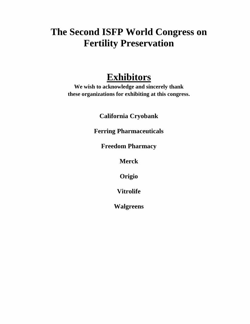
Exhibitors We wish to acknowledge and sincerely thank
these organizations for exhibiting at this congress.
California Cryobank
Ferring Pharmaceuticals
Freedom Pharmacy
Merck
Origio
Vitrolife
Walgreens
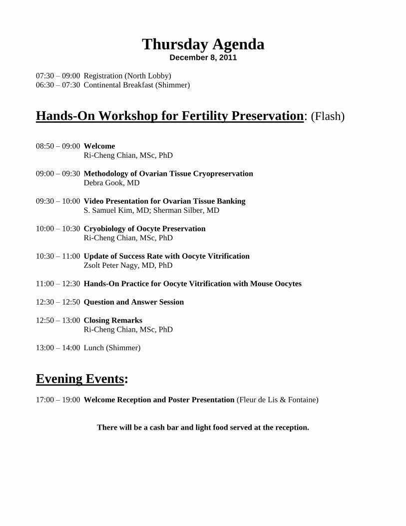
Thursday Agenda December 8, 2011
07:30 – 09:00 Registration (North Lobby)
06:30 – 07:30 Continental Breakfast (Shimmer)
Hands-On Workshop for Fertility Preservation: (Flash)
08:50 – 09:00 Welcome
Ri-Cheng Chian, MSc, PhD
09:00 – 09:30 Methodology of Ovarian Tissue Cryopreservation
Debra Gook, MD
09:30 – 10:00 Video Presentation for Ovarian Tissue Banking
S. Samuel Kim, MD; Sherman Silber, MD
10:00 – 10:30 Cryobiology of Oocyte Preservation
Ri-Cheng Chian, MSc, PhD
10:30 – 11:00 Update of Success Rate with Oocyte Vitrification
Zsolt Peter Nagy, MD, PhD
11:00 – 12:30 Hands-On Practice for Oocyte Vitrification with Mouse Oocytes
12:30 – 12:50 Question and Answer Session
12:50 – 13:00 Closing Remarks
Ri-Cheng Chian, MSc, PhD
13:00 – 14:00 Lunch (Shimmer)
Evening Events:
17:00 – 19:00 Welcome Reception and Poster Presentation (Fleur de Lis & Fontaine)
There will be a cash bar and light food served at the reception.
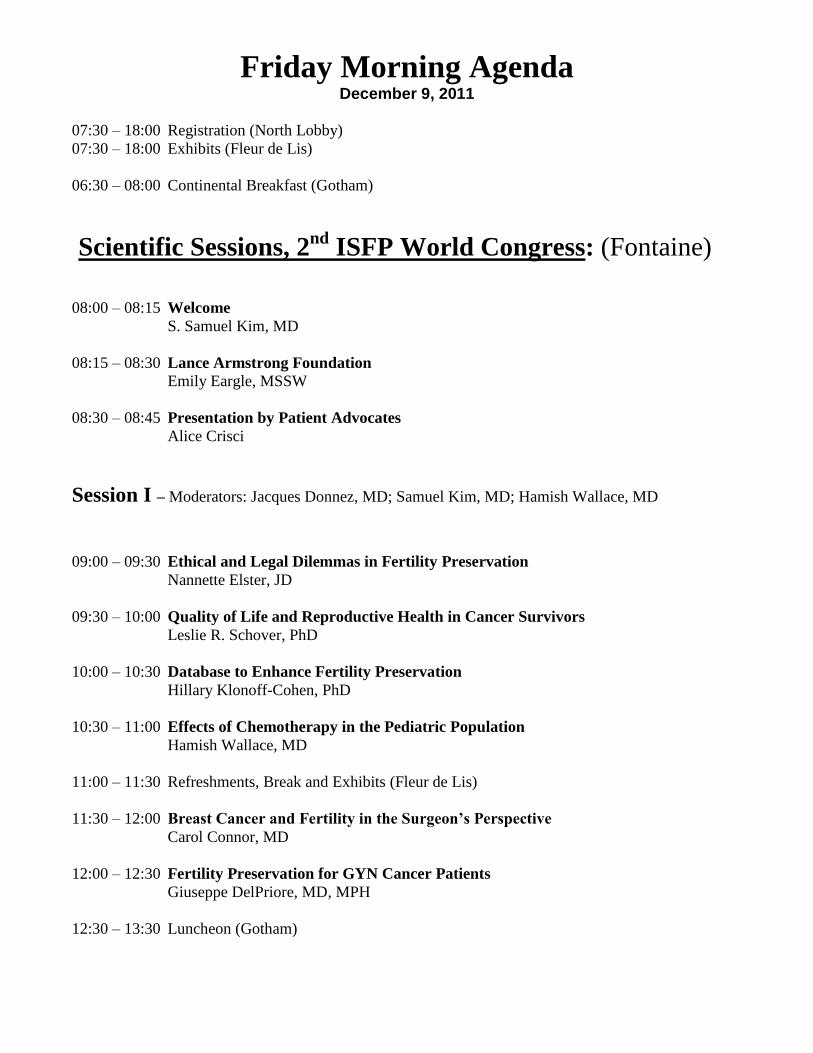
Friday Morning Agenda December 9, 2011
07:30 – 18:00 Registration (North Lobby)
07:30 – 18:00 Exhibits (Fleur de Lis)
06:30 – 08:00 Continental Breakfast (Gotham)
Scientific Sessions, 2nd
ISFP World Congress: (Fontaine)
08:00 – 08:15 Welcome
S. Samuel Kim, MD
08:15 – 08:30 Lance Armstrong Foundation
Emily Eargle, MSSW
08:30 – 08:45 Presentation by Patient Advocates
Alice Crisci
Session I – Moderators: Jacques Donnez, MD; Samuel Kim, MD; Hamish Wallace, MD
09:00 – 09:30 Ethical and Legal Dilemmas in Fertility Preservation
Nannette Elster, JD
09:30 – 10:00 Quality of Life and Reproductive Health in Cancer Survivors
Leslie R. Schover, PhD
10:00 – 10:30 Database to Enhance Fertility Preservation
Hillary Klonoff-Cohen, PhD
10:30 – 11:00 Effects of Chemotherapy in the Pediatric Population
Hamish Wallace, MD
11:00 – 11:30 Refreshments, Break and Exhibits (Fleur de Lis)
11:30 – 12:00 Breast Cancer and Fertility in the Surgeon’s Perspective
Carol Connor, MD
12:00 – 12:30 Fertility Preservation for GYN Cancer Patients
Giuseppe DelPriore, MD, MPH
12:30 – 13:30 Luncheon (Gotham)
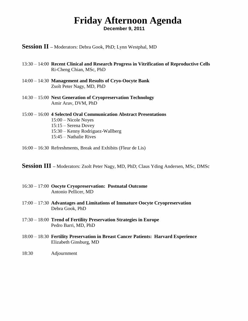
Friday Afternoon Agenda December 9, 2011
Session II – Moderators: Debra Gook, PhD; Lynn Westphal, MD
13:30 – 14:00 Recent Clinical and Research Progress in Vitrification of Reproductive Cells
Ri-Cheng Chian, MSc, PhD
14:00 – 14:30 Management and Results of Cryo-Oocyte Bank
Zsolt Peter Nagy, MD, PhD
14:30 – 15:00 Next Generation of Cryopreservation Technology
Amir Arav, DVM, PhD
15:00 – 16:00 4 Selected Oral Communication Abstract Presentations
15:00 – Nicole Noyes
15:15 – Serena Dovey
15:30 – Kenny Rodriguez-Wallberg
15:45 – Nathalie Rives
16:00 – 16:30 Refreshments, Break and Exhibits (Fleur de Lis)
Session III – Moderators: Zsolt Peter Nagy, MD, PhD; Claus Yding Andersen, MSc, DMSc
16:30 – 17:00 Oocyte Cryopreservation: Postnatal Outcome
Antonio Pellicer, MD
17:00 – 17:30 Advantages and Limitations of Immature Oocyte Cryopreservation
Debra Gook, PhD
17:30 – 18:00 Trend of Fertility Preservation Strategies in Europe
Pedro Barri, MD, PhD
18:00 – 18:30 Fertility Preservation in Breast Cancer Patients: Harvard Experience
Elizabeth Ginsburg, MD
18:30 Adjournment

Saturday Morning Agenda December 10, 2011
07:30 – 10:30 Registration (North Lobby)
07:30 – 18:00 Exhibits (Fleur de Lis)
06:30 – 08:30 Continental Breakfast (Gotham)
Scientific Sessions, 2nd
ISFP World Congress: (Fontaine)
Session IV – Moderators: Bruno Salle, MD, PhD; Antonio Pellicer, MD
08:00 – 08:30 The Future of Translational Research in Fertility Preservation
Mary Zelinski, PhD
08:30 – 09:00 DNA Damage / Repair with Ovarian Tissue Cryopreservation
S. Samuel Kim, MD
09:00 – 09:30 Ovarian Tissue Transplantation: Danish Model and Outcome
Claus Yding Andersen, MSc, DMSc
09:30 – 10:00 Strategies to Improve Post-Transplant Survival of Ovarian Tissue
Jacques Donnez, MD, PhD
10:00 – 10:30 Refreshment, Break and Exhibits (Fleur de Lis)
Session V – Moderators: Pasquale Patrizio, MD; Jehoshua Dor, MD
10:30 – 11:00 Medical Options for Fertility Preservation
Dror Meirow, MD
11:00 – 11:30 Risks of Cancer Cell Reintroduction after Ovarian Transplantation
Marie-Madeleine Dolmans, MD, PhD
11:30 – 12:00 Potential Challenges of Whole Ovary Transplant
Pasquale Patrizio, MD, MBE; Bruno Salle, MD, PhD
12:00 – 12:30 The Future Role of Social Egg Banking
Dror Meirow, MD; Pasquale Patrizio, MD
12:30 – 13:30 Luncheon (Gotham)

Saturday Afternoon Agenda December 10, 2011
Session VI – Moderators: Zev Rosenwaks, MD; Dror Meirow, MD; Chii-Ruey Tzeng, MD, PhD
13:30 – 14:30 4 Selected Oral Communication Abstract Presentations
13:30 – Deepa Bhartiya
13:45 – Yoni Cohen
14:00 – Mahmoud Salama
14:15 – Michael Grynberg
14:30 – 15:00 ART for Fertility Preservation
Glenn Schattman, MD
15:00 – 15:30 From Pluripotent Stem Cells to Germ Cells
Outi Hovatta, MD
15:30 – 16:00 Refreshment, Break and Exhibits (Fleur de Lis)
16:00 – 16:30 Current Status of In Vitro Growth and Maturation
Evelyn Telfer, PhD
16:30 – 17:00 IVM: An Old Technology in a New Era?
Johan Smitz, MD, PhD
17:00 – 17:30 Mechanism of DNA Damage with Chemotherapy / Radiotherapy
David Albertini, PhD
17:30 – 18:00 Non-Traditional Animal Models for Advancing Fertility Preservation Studies in
Humans
Pierre Comizzoli, PhD
18:00 – 18:30 Prize Presentation and Closing Ceremony
Presentations by ISFP Officers:
Pedro Barri, MD
Jacques Donnez, MD
S. Samuel Kim, MD
Pasquale Patrizio, MD
Antonio Pellicer, MD

Friday Morning Abstracts
09:00 – 09:30 Ethical and Legal Dilemmas in Fertility Preservation
Nannette Elster, JD
09:30 – 10:00 Quality of Life and Reproductive Health in Cancer Survivors
Leslie R. Schover, PhD
10:00 – 10:30 Database to Enhance Fertility Preservation
Hillary Klonoff-Cohen, PhD
10:30 – 11:00 Effects of Chemotherapy in the Pediatric Population
Hamish Wallace, MD
11:30 – 12:00 Breast Cancer and Fertility in the Surgeon’s Perspective
Carol Connor, MD
12:00 – 12:30 Fertility Preservation for GYN Cancer Patients
Giuseppe DelPriore, MD, MPH

Ethical and Legal Dilemmas in Fertility Preservation
Nanette Elster, JD, MPH
Recent scientific advances have made the once remote possibility of conception following
cancer treatment more feasible, however, these advances are not without risks including legal and
ethical risks. This presentation will discuss some of the legal and ethical challenges inherent in
fertility preservation including informed consent for both children and adults, disposition of
gametes and embryos after death as well as accessibility of fertility preservation services.

NOTES

Quality of Life and Reproductive Health in Cancer Survivors
Leslie R. Schover, PhD
Cancer treatment may interfere with becoming a parent because of direct damage to
gametogenesis in males and females, damage to uterine capacity to carry a pregnancy, damage to
the ability to have functional sexual intercourse, or lessening of physical attractiveness to a
potential mate. Young cancer survivors may also forego having children out of anxieties such as
fear that fertility preservation would cause a dangerous delay in starting cancer treatment or that
fertility-sparing modifications of cancer treatment would be less successful in controlling cancer.
Women have exaggerated fears about a pregnancy causing a cancer recurrence and survivors of
both genders worry about birth defects and lifetime cancer risk in any child born after a parent’s
cancer treatment. Other worries are passing on a genetic risk for inherited cancer syndromes or
dying prematurely and being unable to protect a young child. Survivors who are infertile but want
to be social parents face barriers to adopting a child after cancer and often know little about
options of donated gametes or embryos. The majority of young survivors do want biological
children, however, particularly those childless at the time of cancer diagnosis. Our own interview
data show that as long as an average of 10 years after cancer treatment, women diagnosed before
age 40 remain very distressed about unfulfilled desires to have a child. Despite the efforts to
standardize communications from oncologists about fertility risk and options for fertility
preservation, at least half of patients still are not getting the information they need to make a
rational decision. Patients who get more information feel more comfortable with their decisions
about fertility preservation, even though a majority still do not choose to cryopreserve gametes.
For men, both teenagers and men over 40 may be good candidates to bank sperm and should not be
ruled out because of age. Teens may need to be counseled separately from their parents so that
everyone’s opinions and needs can be heard and a family decision made. However, we need to
focus more attention on the needs of the thousands of young survivors who did not preserve
fertility and need to make decisions on their options for parenthood after cancer.

NOTES

Database to Enhance Fertility Preservation
Hillary Klonoff-Cohen, PhD
Objective: Currently, no comprehensive research database exists to evaluate the different
fertility preservation strategies. The largest national data sets (e.g., NCHS, CDC, and SEER)
contain no questions pertaining to infertility caused by cancer or other fertility-compromising
treatments. Moreover, the Society of Assisted Reproductive Technology (SART) database
does not address fertility preservation procedures. Creation of a Fertility Preservation
Database will provide information on the prevalence, demographic characteristics of patients,
procedural types, efficacy, safety and long-term effects, and success rates (e.g., pregnancy
and birth outcomes) of fertility preservation.
Methods: We conducted two feasibility studies. The first study ascertained the prevalence of
fertility preservation options for cancer patients at 16 clinics within 100 miles of UC San
Diego. The purpose of the second feasibility study was to determine whether we could
acquire an adequate sample of cancer and non-cancer patients who choose to undergo
fertility preservation by contacting every SART-affiliated fertility clinic in California. Clinic
Directors provided the total number of fertility preservation procedures performed in 2009.
Results: In the first feasibility study, of the 11 clinics that responded, 8 (73%) reported
performing fertility preservation; 1 (9%) reported having had an inquiry about it, while 2
(18%) reported not having done any. Of the 54 patients seen at these fertility clinics, the vast
majority (> 40 patients, 75%) had a cancer diagnosis, and of these, most (> 26, 65%) had
breast cancer. All patients underwent embryo cryopreservation (except one case where the
patient died). Forty women (74%) still had embryos in storage and three (5.5%) had
attempted pregnancy using frozen embryo transfers, resulting in one successful pregnancy. In
our second study, out of 44 fertility clinics, 21 responded; seven of which did not perform
fertility preservation. In 2009, a total of 77 cancer patients were treated with fertility
preservation procedures at the responding clinics in California. To be maximally
conservative, we assumed that all the non-responding sites treated zero cancer patients. This
would imply that the 44 clinics in California (rather than 21 responders) treated the 77 cancer
patients with fertility preservation. Based on a total of 395 fertility clinics within the US,
using the same ratio would translate to 691 cancer patients per year in 2009.
Conclusions: From this feasibility study, we conclude that there will be ample eligible
cancer patients available for participation in a registry to elucidate medical, social, ethical
and legal issues of fertility preservation for physicians and patients.

NOTES
Fertility Preservation Options for Children with Cancer
Hamish Wallace, MD
With increasing numbers of survivors from cancer at a young age the issue of fertility
preservation has assumed greater importance. This lecture will describe normal ovarian
function and summarises what is known about the effect of chemotherapy and radiotherapy
on the ovary and uterus. The value of an assessment of ovarian reserve for the individual
patient using AMH will be discussed. Recent prospective studies on AMH during
chemotherapy in children will be reviewed.
To date, there have been at least 17 pregnancies worldwide after othotopic
reimplantation of frozen-thawed ovarian cortex. The success rate is unclear as the
denominator (the number of women in whom frozen-thawed ovarian tissue has been
reimplanted) is unknown. There have been no pregnancies reported following the
reimplantation of ovarian tissue harvested pre-pubertally, but with the accepted age related
decline from birth in the number of non-growing follicles, young children are potentially
ideal candidates for this procedure
All young patients with cancer or leukaemia should have their fertility prognosis
discussed before treatment begins. Sperm and embryo cryopreservation should be considered
standard practice and be widely available for those at significant risk of infertility. The
Edinburgh experience of ovarian cryopreservation will be presented. Risks and benefits from
reimplantation of frozen/thawed ovarian cortical strips will be discussed. For pre-pubertal
girls ovarian tissue cryopreservation should be considered if the risk of a premature
menopause is high, but for the pre-pubertal boy there are no established techniques in current
practice.

NOTES
Breast Cancer and Fertility in the Surgeon’s Perspective
Carol S. Connor, MD
Objective: Review the surgeon’s role in the evaluation and treatment of newly diagnosed
young breast cancer patients and the challenges to integration of fertility preservation into
surgical therapy.
Methods/Results: Fertility preservation is an important option for young women with newly
diagnosed breast cancer. The surgeon is often the first specialist to see a newly diagnosed
breast cancer patient, perform the initial evaluation, provide appropriate referrals to other
specialists, and coordinate the treatment plan. The principles of surgical management of
young breast cancer patients will be reviewed, with special attention to the management of
young patients receiving neoadjuvant therapy. Clinical and biologic factors that contribute to
treatment decisions in young breast cancer patients will be presented, including a summary of
fertility preservation options based upon these factors. A case study will be presented that
demonstrates the need for involvement of the surgeon in a multidisciplinary plan for fertility
preservation.
Conclusion: Education of breast surgeons regarding the options for fertility preservation in
young patients with breast cancer could potentially improve patient referral to reproductive
specialists and facilitate the coordination of care in these often complex clinical scenarios.

NOTES

Fertility Preservation for GYN Cancer Patients
Giuseppe DelPriore, MD, MPH
Fertility preservation for all cancer patients has become an important quality-of-life
outcome for oncologist and infertility specialists. Several overseeing and regulatory entities
have similarly weighed in encouraging consideration of fertility preservation as part of the
treatment planning for cancer patients. And finally, the public and the courts have adopted
fertility preservation as standard of care in certain circumstances.
Gynecologic cancers including breast cancer may have special population issues
similar to those affecting pediatric patients. In other cancers the interaction with fertility
between the disease and its treatment, may be less apparent although not less important. For
gynecologic cancers the impact on fertility is inescapable. Participants should know the latest
oncologic principles related to this field.
Cervical cancer is the most common gynecologic cancer seen in many areas of the
world. In developing countries it surpasses all other cancers including breast. Unlike breast,
the peak age of incidence is relatively young, corresponding to the reproductive age period.
Fortunately the benefits of chemotherapy have been demonstrated in almost all settings of
cervical cancer care but especially in fertility preservation when used for neoadjuvant or
adjuvant therapy.
Ovarian cancer which is thought of as the most lethal gynecologic cancer nevertheless
still allows fertility preservation to be considered during its treatment. Coordination with
surgeon, chemotherapist, and infertility specialists is especially important in this cancer.
Endometrial cancer is a common cancer in Western cultures. It is often thought of as
an indolent cancer allowing for fertility preservation to be considered. However risks do exist
and must be recognized and dealt with.
Although the intersection of fertility in cancer care is complex, this presentation will
be a practical guide limited to the most relevant clinical updates.

NOTES

Friday Afternoon Abstracts
13:30 – 14:00 Recent Clinical and Research Progress in Vitrification of Reproductive
Cells Ri-Cheng Chian, MSc, PhD
14:00 – 14:30 Management and Results of Cryo-Oocyte Bank
Zsolt Peter Nagy, MD, PhD
14:30 – 15:00 Next Generation of Cryopreservation Technology
Amir Arav, DVM, PhD
16:30 – 17:00 Oocyte Cryopreservation: Postnatal Outcome
Antonio Pellicer, MD
17:00 – 17:30 Advantages and Limitations of Immature Oocyte Cryopreservation
Debra Gook, PhD
17:30 – 18:00 Trend of Fertility Preservation Strategies in Europe
Pedro Barri, MD, PhD
18:00 – 18:30 Fertility Preservation in Breast Cancer Patients: Harvard Experience
Elizabeth Ginsburg, MD

NOTES

Recent Clinical and Research Progress in Vitrification of Reproductive
Cells
Ri-Cheng Chian, MSc, PhD
Cryobiology is the branch of biology involving the study of the effects of low
temperatures on organisms, in which most often for the purpose of achieving
cryopreservation. The cryobiology is core of fertility cryopreservation. The principal
application for human fertility cryopreservation was begun with sperm freezing, and then
with embryo and oocyte as well as gonadal cryopreservation. Although the advanced
knowledge and medical achievements have been obtained in the field of fertility
cryopreservation, especially with recent development of oocyte and ovarian tissue
cryopreservation, the field of cryobiology can be considered as relatively new branch of
biology.
Many factors affect the successful cryopreservation of the cells. The first, it may
depend on the cell type, cell size, cell growth phase, cell water content, cell lipid content and
the composition of the cells as well as cell density. The second, it may depend on the
composition of freezing or vitrification medium, cooling rate, storage temperature and
duration of storage, warming rate and recovery medium. The third, it may be the most
important to supplemented cryoprotectant into aqueous solution.
The mechanism of cryoprotectants action can be considered as lowering the freezing
point and preventing ice crystal formation of intracellular and extracellular solutes. It has
been considered that there may be minor or server toxicity of cryoprotectants. This toxicity
of cryoprotectants is related directly to its concentration to be used, and the cell exposure
temperature and time. Although some theories of cryobiology developed, it may not be
applied to all types of cells. Theoretical considerations are needed for further development in
the field of cryobiology.
The development of an effective oocyte cryopreservation system has a significant
impact on clinical practice of assisted reproduction. With modified slow freezing method,
particularly increased sucrose concentration in suspending solution, the improved survival
and pregnancy rates have been obtained. However, super-rapid cooling of human oocytes has
resulted in relatively higher survival rate. Pregnancies achieved with cryopreservation of
oocytes regardless of slow freezing or vitrification do not appear to be associated with
adverse pregnancy outcomes, indicating that cryopreservation of oocytes represent a novel
option and efficient method for female fertility preservation. It seems that cryopreservation
of mature stage oocytes has better results than freezing immature stage oocytes, because
oocyte maturation rate will be significantly reduced when the oocytes were cryopreserved at
immature stage followed by IVM. Although a couple of thousands live births obtained from
the cryopreserved oocytes and appeared no difference in congenital anomalies compared
with naturally conceived infants, more live birth data and long-term monitoring are required
to assure the safe and expeditious development of oocyte cryopreservation technology.
In addition, the updated information for cryopreservation of ovarian tissues followed
by transplantation will also be discussed in this presentation.

NOTES

Management and Results of Cryo-Oocyte Bank
Zsolt Peter Nagy, MD, PhD
The efficiency of oocyte cryopreservation has dramatically increased in recent years,
mainly due to the employment of the vitrification technique. There are several potential
benefits of oocyte cryopreservation, including fertility preservation for medical and social
reasons; ethical/moral consideration or legislative restrictions. However, one of the first
applications of egg freezing relates to oocyte donation. Oocyte donation has been a well-
established practice for decades to treat IVF patients with advanced reproductive age or other
conditions. Since its introduction, it was always the practice to perform it through fresh
donation, however this is associated with many disadvantages, including long waiting times,
limited donor choice, and difficulty of synchronization. A donor egg cryo-bank, on the other
hand, can eliminate all these difficulties. Recent publications and our own experience with
over 500 recipient cycles demonstrate that using the vitrification technique, high survival
rates (90% or more) of donor eggs can be achieved followed by high fertilization (>70%) and
satisfactory embryo development (>50% good quality embryos). Clinical pregnancy rates are
consistently above 50% (70% when two embryos and 50% when a single embryo is
transferred). Evaluation of children born after the use of vitrified eggs does not show higher
rates of birth defects which helps to reassure the safety aspects of the technique. Thus our
experience with cryo-egg donation is highly positive. It provides similar outcomes to fresh
donation, without the difficulty of synchronization, and with several benefits including
immediate access to a large variety of donors, increased safety by possible quarantining eggs,
as well as economic benefits to recipient making the treatment more affordable.

NOTES
Next Generation of Cryopreservation Technology
Amir Arav, DVM, PhD
Imagine a world without liquid nitrogen storage. What unifies the tissue and cell
cryobanking is that they store the samples in liquid nitrogen. Storage of cryopreserved
samples, under liquid nitrogen, is very demanding in terms of maintenance, storage space,
storage equipment and costs. An alternative that would minimize costs, storage and
maintenance has been gaining a foothold in the field of cell preservation in recent years - the
dry storage. In nature, many plants and animals can enter the state of anhydrobiosis by
accumulating disccharides such as trehalose in their cells to as much as 50% of their dry
weight. Following Nature’s lead, trehalose is being used these days during the process of
freeze-drying in vitro as well. Drying of cells can be achieved by either convective-drying or
freeze-drying. Freeze-drying (lyophilisation), the more commonly used technique, was
known for hundreds of years as a method for meat and vegetable preservation among the
people who lived in very high altitudes, like in the Andes mountain dwellers of South
America. In more recent times, freeze-drying is used for preparation of food products such as
instant coffee, tea and soup, fish food and even ice cream for NASA astronauts. It is also
widely used to prepare pharmaceuticals, viral, bacterial, fungal or yeast products in a dry and
convenient form for handling, transporting and long-term storage. Freeze-drying is achieved
by sublimation of the ice after freezing the sample to subzero temperatures. The process is
damaging to the cellular membrane and some degree of chromosomal damage may also take
place due to endogenous nucleases.
To date, embryonic development after intracytoplasmic sperm injection (ICSI) with
freeze-dried sperm heads has been reported in humans and hamster, cattle, pigs, rhesus
macaque and cats, and live offspring were reported in mice, rabbits, rat and fish . We have
recently demonstrated the use of sheep freeze-dried somatic cells for somatic cell nuclear
transfer. We utilized the directional freezing technology to freeze-dry somatic cells which
were kept at room temperature for 3 years. These cells were rehydrated and then used to
direct embryonic development following nuclear transfer into in vitro matured enucleated
oocytes. Finally, human hematopoietic stem cells that were lyophilized and rehydrated with
water were viable and have maintained their clonogenic capacity showing that they were able
to develop into all blood lineages. This was the first report to show cells that have undergone
complete lyophilization and following rehydration have maintained not only their viability
but also their functionality.

NOTES

Oocyte Cryopreservation: Postnatal Outcome
Antonio Pellicer, MD
OBJECTIVE: The number of vitrified live births has augmented lately in parallel with the
increasing use of this technique. There are few published data on perinatal outcome of these
pregnancies. The aim of this study was to compare the obstetric and perinatal outcome in
pregnancies and children conceived after oocyte vitrification and standard IVF treatments
conducted with fresh oocytes.
DESIGN: Retrospective cohort study.
PATIENTS AND METHODS: Maternal and neonatal data were recorded for all patients
who had delivered babies achieved after undergoing an IVF treatment carried out with
vitrified and fresh oocytes. A cohort of 395 pregnancies (516 live births) following oocyte
vitrification was compared with another cohort of 390 pregnancies (500 live births) achieved
using fresh oocytes (control group). Cases and controls were matched according to the
number of fetuses (singleton vs. twins) and the source of oocytes (own vs donated oocytes).
A live birth was defined as any pregnancy with at least one live born delivered beyond 25
weeks’ gestation. We assessed the incidence of gestational diabetes (GD), gestational
hypertension (GH), preterm premature rupture of membranes (PRM) and preterm delivery
(PD) in both groups. Birth outcomes were also studied. Comparisons between groups were
performed by Student´s t-test.
RESULTS: Gestational age at delivery was 38.2 ± 2.5 weeks and 38.0 ± 2.3 weeks for
vitrified and fresh oocytes groups, respectively (p=NS). The mean birth weight was 2.811 ±
0.664 g and 2.810 ± 0.661g for vitrified and fresh oocytes groups, respectively (p=NS).
There were 4 major birth defects in the vitrification group (0.7%) and 2 in the control group
(0.8%) (p=NS). In the ovum donation group singleton pregnancies, the relative risk of
suffering PRM was RR=0.725 (CI95%0.291-1.822 GD: RR=0.998
(CI95%0.561-0.739), GH: RR=0.830 (CI95%0.493-1.425) and PD: RR=1.388 (CI95%0.651-
2.958 between fresh and vitrified groups (NS). The obstetric outcome for multiple
pregnancies derived from ovum donation was as follows: PRM was RR=1.072(CI95%0.480-
2.395) GD: RR=0.741 (CI95%0.304-1.807), GH: RR=0.007 (CI95%0.350-0.160) and PD:
RR=0.007 (CI95%0.814-3.141) respectively (NS). In the group of singleton pregnancies
developed from own oocytes no any patient presented premature delivery risk and none of
the patients suffered from gestational diabetes though the relative risk were not calculated.
The relative risks of suffering other obstetric outcomes were as follows GH: RR=0.925
(CI95%0.248-3.455) and PD: RR=0.279 (CI95%0.068-1.143) for vitrified and fresh oocyes
(NS). In the group of multiple pregnancies developed from own oocytes cycles the obstetric
risks were: PRM RR=0.944(CI95%0.055-16.32 GD: RR=0.488 (CI95%0.098-2.433, GH:
RR=0.588 (CI95%0.0862- 4.009) and PD: RR=0.375 (CI95%0.078-1.812) respectively (NS).
In all cases RR CI95% included 1 and in consequence were not significant. No differences
were found (p=NS) when analysing separately data for singletons and twins, and ovum
donation vs own oocytes cycles.
CONCLUSIONS: Obstetric and perinatal outcomes in oocyte vitrification are similar to
those achieved using fresh oocytes. These findings are reassuring regarding the safety of the
cryopreservation procedure.

NOTES

Advantages and Limitations of Immature Oocyte Cryopreservation
Debra Gook, PhD
For many young women with malignant disease the urgency to commence cytotoxic
treatments prohibits the option to undergo ovarian stimulation and collection of mature
oocytes for cryopreservation. For these women collection of immature oocytes, whether in
the form of ovarian cortex containing preantral follicles or following aspiration of
spontaneous antral follicles, provides the only option available at present to preserve fertility.
The advantage of collection at any time in the cycle and/or during other operative
procedures, together with the reduced cost compared to a stimulation cycle, may seem
attractive. However, what is the clinical efficiency of these procedures? The question is
whether these technologies, as applied today, provide the patient with a realistic opportunity
to achieve a pregnancy or are they just providing hope?
The benchmark for comparison is mature oocyte cryopreservation. Recent advances
in mature oocyte cryopreservation have seen equivalent rates of fertilization, embryo
development and implantation rates to those of fresh oocytes. Vitrification of mature oocytes
in an open system using the method reported first by Katayama [1]results in extremely high
survival of oocytes from young women (donated oocytes) and also infertile women.
Although survival is lower with controlled rate freezing, under optimal conditions it can
produce equivalent outcomes to fresh oocytes.
In-vitro maturation of oocytes from hCG primed ovaries has been practised with little
change for over 10 years, primarily for women with ovaries of polycystic morphology but
also, more recently, for fertility preservation patients in conjunction with vitrification.
Maturation prior to vitrification gives an indication that at least some mature oocytes will be
available for clinical use, but subsequent embryo development and implantation rates appear
reduced compared to outcomes with mature oocytes from stimulated cycles. Therefore, is it
more beneficial for fertility preservation patients to vitrify oocyte cumulus complexes
pending improvements in IVM technology?
The potential contained within a small area of ovarian tissue, due to the number of
follicles/gametes present, is obviously higher than using other approaches. However, the
attrition which occurs due to cryopreservation and post graft ischemia or through follicle
culture is extremely high. As with in-vitro maturation, pregnancies have been achieved with
ovarian tissue cryopreservation and grafting but embryo quality from aspirated follicles
within this tissue also appears to be compromised.
So, should we be more persuasive with oncologists in influencing a delay in therapy
to collect mature oocytes for cryopreservation in order to achieve a realistic prospect of
fertility preservation?

NOTES

Trend of Fertility Preservation Strategies in Europe
Pedro Barri, MD, PhD
In Europe there is no single network for fertility preservation and the approach has so far
been at national level. However all the strategies should be under the guidelines of the European
Tissue directives 2004/23/EG and 2006/86/EC.
We will present different situations of different countries, from those who have set up their
own national networks to those that work on the basis of individual Centres.
In order to compile this information we have used documents from the ESHRE Special
Interest Group on Fertility Preservation, which covers practically all information about Europe, and
also from the Spanish Fertility Society, which presents the strategy followed in this country.
We started by analysing the situation of legal cover and access to the techniques in several
European countries and then gathered the information through a questionnaire sent to 28 countries.
The response from the 23 countries that answered showed that in 6 countries (Germany, Denmark,
Bulgaria, Sweden, the Netherlands and Norway) there are national fertility preservation programmes,
while in 3 countries (Finland, France, Switzerland) programmes are in preparation. Three countries
(Germany, Denmark and Norway) have centralised activity registers.
In Spain, 64 hospitals offer this service but only 22% of the centres offer all of the
cryopreservation strategies for semen, oocytes, embryos and ovarian tissue. The remaining centres
outsource some activity.
This year in Spain there have been 59 cases of oocyte vitrification, 54 cases of embryo
freezing, 23 cases of freezing of ovarian tissue and 649 cases of semen being frozen in order to
preserve the fertility of oncology patients.
Probably in this year (2011), a number of national registers will be consolidated in Europe
and global figures for activity and results will become available.

NOTES
Fertility Preservation in Breast Cancer Patients: Harvard Experience
Elizabeth Ginsburg, MD
Objective: To review the experience with fertility preservation in breast cancer patients at
Brigham & Women's Hospital, Harvard Medical School
Materials and Methods: A review of the past 2 year experience in our institution and Dana
Farber Cancer Institute was undertaken.
Results: Women undergoing fertility preservation prior to chemotherapy are very likely to
be successful at banking eggs and/ or embryos, however after chemotherapy cancellation
rates are high. National data show that fertility counseling does not appear to be occurring
adequately in oncology practice.
Conclusions: Discussion of impact of cancer treatment is not performed as often as it should
be, and referrals for fertility preservation likely lag behind need. Patients with breast cancer
who present for fertility preservation prior to chemotherapy have excellent responses to
ovulation induction and are highly likely to bank eggs and/ or embryos successfully.
Support: None

NOTES
Saturday Morning Abstracts
08:00 – 08:30 The Future of Translational Research in Fertility Preservation
Mary Zelinski, PhD
08:30 – 09:00 DNA Damage / Repair with Ovarian Tissue Cryopreservation
S. Samuel Kim, MD
09:00 – 09:30 Ovarian Tissue Transplantation: Danish Model and Outcome
Claus Yding Andersen, MSc, DMSc
09:30 – 10:00 Strategies to Improve Post-Transplant Survival of Ovarian Tissue
Jacques Donnez, MD, PhD
10:30 – 11:00 Medical Options for Fertility Preservation
Dror Meirow, MD
11:00 – 11:30 Risks of Cancer Cell Reintroduction after Ovarian Transplantation
Marie-Madeleine Dolmans, MD, PhD
11:30 – 12:00 Potential Challenges of Whole Ovary Transplant
Pasquale Patrizio, MD, MBE; Bruno Salle, MD, PhD
12:00 – 12:30 The Future Role of Social Egg Banking
Dror Meirow, MD; Pasquale Patrizio, MD

NOTES
The Future of Translational Research in Fertility Preservation
Mary B. Zelinski, PhD
Two theoretical possibilities for preserving female fertility from side-effect damage
caused by chemo- or radiotherapy are currently under investigation using the nonhuman
primate model for translational research in fertility preservation. First, reducing or
eliminating the gametotoxic effects of cancer therapies on the ovary in vivo remains
challenging. Recently, intraovarian infusion of an agonist of sphingosine-1-phosphate,
FTY720, prior to ovarian X-irradiation was shown to protect a cohort of preantral follicles
from radiation damage in macaques, allowing birth of live, healthy offspring. Similarly,
exposure of human ovarian cortex to anti-apoptotic agents in vitro prior to, or in vivo during
xenotransplantation preserved primordial follicles, thus supporting the feasibility of in vivo
protection. Other agents, such as tamoxifen and imatinib, inhibit preantral follicle and/or
oocyte destruction caused by chemotherapy drugs in rodents. Further translational research
will be necessary to identify the most potent fertoprotective agents along with their safety,
and to develop clinically acceptable delivery targeted to the ovary. Second, preventing
exposure to gametotoxic effects by removing the gametes or ovary prior to therapy, and
returning gametes or embryos for fertility after eradicating the cancer is being investigated in
the nonhuman primate using in vitro follicle culture as well as ovarian cortex
cryopreservation/transplantation. It is now possible to grow primate primary and secondary
follicles to the antral stage in the encapsulated 3D culture system. Future studies will
optimize the protocol to yield more mature oocytes capable of fertilization and embryonic
development, determine whether cultured follicles are equivalent in structure and function to
those developed in vivo, and to understand the basic biology of the primate follicle.
Systematic study of vitrification methods using macaque ovarian cortex revealed
cryoprotectants that preserve primordial, primary as well as secondary follicles. Macaque
follicles isolated from vitrified ovarian tissue can survive, grow, form an antrum and produce
steroids in 3D culture, indicating functional preservation of cryopreserved follicles, and the
utility of 3D follicle culture as a bioassay to screen cryopreservation methods prior to in vivo
studies involving tissue transplantation. Future directions include development of novel
vitrification protocols compatible with closed system storage, vitrification of individual
follicles, improving heterotopic transplantation of ovarian cortex and achieving live offspring
from vitrified- thawed tissue via transplantation and/or 3D follicle culture. Research in
nonhuman primates will continue to provide an evidence-based foundation for safely
producing meiotically and developmentally competent oocytes from fresh or cryopreserved
tissue to enhance clinical fertility preservation options for female cancer patients.

NOTES

DNA Damage / Repair with Ovarian Tissue Cryopreservation
S. Samuel Kim, MD
The cancer survival rate has increased dramatically during last two decades.
Currently, more than 11 million cancer survivors are living in the US. Unfortunately,
aggressive cancer treatment can result in infertility. Loss of fertility after cancer therapy can
profoundly impact on quality of life and cause significant distress to young cancer patients.
Of note, four percent of the newly diagnosed cancer patients in the US are under age 35.
There are a few options to preserve fertility in young female cancer patients including
embryo cryopreservation, oocyte cryopreservation, and ovarian tissue cryopreservation.
Ovarian tissue cryopreservation is the only option for pre-pubertal girls and for those who
cannot delay cancer treatment. As of October 2011, seventeen babies have been born after
transplantation of cryopreserved ovarian tissue.
Cryopreservation of human ovarian tissue by slow freezing technique has been
successful since 1994 and is currently considered as a standard method. Although over 70%
of primordial follicles survive (morphologically) after slow freezing and rapid thawing,
significant ultra-structural damage can be detected by TEM. Many investigators have
explored strategies to optimize follicle survival after ovarian tissue cryopreservation.
Theoretically, vitrification can be an ideal method as it can eliminate ice formation that is the
main cause of cryoinjury. However, high concentration of cryoprotectants and devitrification
can be problematic. For the successful pregnancy outcome, the genomic integrity of oocytes
subjected to ovarian tissue cryopreservation should be thoroughly investigated.
Therefore, we investigated DNA damage, apoptosis, autophagy after slow freezing
and vitrification of bovine ovarian tissue using four biomarkers, H2AX, RAD 51, cPARP,
and LC3B. In addition, we assessed DNA damage and apoptosis in ovarian tissue after
treatment with four different vitrification solutions (40% DMSO, 40% PROH, 40% EG,
VPII) for 10 min and 60 min. We noticed that oocytes within primordial and primary follicles
generate an acute DNA repair process in response to DNA damage induced by tissue
cryopreservation. Apoptosis of follicles was more severe after vitrification compared to slow
freezing. Interestingly, there was no sign of DNA damage and repair even after prolonged
exposure to cryoprotectants with high concentration (up to 60 min). However, exposure to
these cryoprotectants induced chromatin condensation.
Although benefits and efficacy of vitrification of ovarian tissue should be further
investigated, our results of this study show that cellular and biochemical damage to follicles
appears to be more severe with vitrification compared to slow freezing technology.

NOTES
Ovarian Tissue Transplantation: Danish Model and Outcome
Claus Yding Andersen, MSc, DMSc
Girls and women suffering from disease that require treatment with gonadotoxic
drugs may as a side effect loose the ovarian function. When the ovaries are depleted of
follicles many women experience profound effects on the physical and psychological status.
Menstrual cycles ceases and pregnancies will be unobtainable. To young girls it may further
imply that a normal pubertal development fails.
Cryopreservation of ovarian tissue is a new method, which has been developed in an
attempt to circumvent the long-term ablative effect on reproductive performance by
gonadotoxic treatment. Removing one whole ovary or part of an ovary from women in their
reproductive years prior to treatment and cryopreserving the tissue can retain a viable pool of
follicles. When the women have been cured and is considered fit, the thawed ovarian tissue
may be transplanted to women who entered menopause.
Laboratory of Reproductive Biology at University Hospital of Copenhagen is the only
center in Denmark offering cryopreservation of ovarian tissue as a treatment in close
collaboration with three fertility clinics round the country. Totally, more than 500 girls and
women have had ovarian tissue cryopreserved in Denmark. The youngest girl was half a year
old and the oldest 38 years. We have currently cryopreserved ovarian tissue from around 100
girls younger than 18 years of age. The ovarian tissue is excised at the local hospital and
transported on ice to our laboratory, where cryopreservation and storage is performed. In
case of transplantation the frozen tissue will transported to the local hospital for the
operation. This transport model has been validated and has now been used for more than 250
cases.
In Denmark, a total of 19 women (13 having their tissue transported prior to
cryopreservation) have experienced transplantation of frozen/thawed ovarian tissue a total of
26 times (7 women having tissue transplanted twice). All women regained ovarian function
and none have experienced relapse as a consequence of the transplantation. Over a period of
20 – 25 weeks levels of FSH gradually return to pre-menopausal levels and menstrual cycles
are regained. The longevity of the tissue depends on the age of the woman at tissue retrieval
and the amount of tissue transplanted. Most women experience return of ovarian function for
several years with just a fraction of tissue from one ovary being replaced. Recently, one child
has had ovarian tissue transplanted for natural induction of puberty; this case will be
presented in detail.
Seven women have been pregnant; in most cases following natural conception. Two
women have delivered three healthy babies as a result of transplanted frozen/thawed ovarian
tissue. In the latter two cases the tissue was transported 4—5 hours prior to cryopreservation.
The presentation will review our experiences and results with transplantation of
cryopreserved ovarian tissue.

NOTES

Strategies to Improve Post-Transplant Survival of Ovarian Tissue
Jacques Donnez, MD, PhD
The different cryopreservation options available for fertility preservation in cancer
patients are embryo cryopreservation, oocyte cryopreservation and ovarian tissue
cryopreservation.
The only established method of fertility preservation is embryo cryopreservation, but
this requires the patient to be of pubertal age, have a partner, and be able to undergo a cycle
of ovarian stimulation.
Cryopreservation of ovarian tissue is the only option available for prepubertal girls,
and for woman who cannot delay the start of chemotherapy.
More or less 50 cases of orthotopic reimplantation of cryopreserved ovarian tissue
have so far been reported and 17 live births have been achieved, yielding a pregnancy rate of
more than 25%. In our department, eight women have undergone orthotopic reimplantation
of cryopreserved tissue either once or twice. Restoration of ovarian function, proved by
follicular development and estradiol secretion, occurred in all cases. A time interval of 3.5 to
5 months was observed.
In order to improve the survival of the graft, angiogenic factors and/or antioxidants
could be delivered. Furthermore, antioapoptotic factors could be also administered to
improve follicle survival. In animal models, angiogenic factors and antioxidants have been
exogenously delivered in the animal or directly in the ovarian tissue prior grafting.
Additionally, they can be host-delivered through the induction of granulation tissue. This
option has been tested both in humans and animals.
Several antioapoptotic factors, such as kit ligand, vitamin C, growth differentiation
factor-9 have been shown to improve survival rate and development of preantral follicles in
in vitro culture experiments. Such factors could be locally applied in the ovarian tissue before
its transplantation.

NOTES

Medical Options for Fertility Preservation
Dror Meirow, MD
Abstract not in hand at time of printing.
NOTES

NOTES

Risks of Cancer Cell Reintroduction after Ovarian Transplantation
Marie-Madeleine Dolmans, MD, PhD
Reversing treatment-related premature ovarian failure using auto-transplantation of
frozen-thawed ovarian tissue harvested before chemo-radiotherapy is becoming an
increasingly realistic prospect for clinical application, since more than 15 live births have
already been reported with this technique. Our objective is to offer young patients at risk of
premature ovarian failure after treatment, safe fertility preservation options.
One major concern raised by the transplantation of ovarian cortical fragments in
cancer patients is the potential risk that the cryopreserved ovarian tissue might harbor
malignant cells that could induce a recurrence of the disease after re-implantation.
Hematological malignancies and breast cancer are the most frequent indications for
ovarian tissue cryopreservation. Both carry the risk of ovarian metastasis.
We therefore decided to conduct a study to evaluate the presence of breast cancer
cells and leukemic cells in human cryopreserved ovarian tissue from patients with advanced
breast cancer disease and chronic myeloid leukemia or acute lymphoblastic leukemia.
In each case, histology, polymerase chain reaction for disease-specific markers and
xenografting were used to test the frozen-thawed ovarian tissue. Results show that malignant
cells may be present in ovarian tissue from leukemic patients and give rise to tumor
development in mice after xenografting (n=5/12, acute leukemia). For the mice grafted with
ovarian tissue from patients with advanced breast cancer, PCR and MGB2-gene sequencing
were positive on the ovarian tissue of 5 out of 10 patients, but none of the xenografted mice
developed tumor masses during the 6-month grafting period.
Although the malignant potential of these cells is not yet known, the current study
demonstrates that conventional histology and IHC need to be associated with more sensitive
screening methods, like PCR and sequencing, before ovarian tissue transplantation can be
contemplated.
Research in this field has to continue, in order to develop different possibilities for
fertility preservation that will allow us to propose the most appropriate option to patients,
according to disease.

NOTES
Potential Challenges of Whole Ovary Transplant
Pasquale Patrizio, MD, MBE; Bruno Salle, MD, PhD
Patients with cancer who desire to preserve their future reproductive potential but
require immediate gonadotoxic treatments (chemo and/or radiotherapy), are left with few
options, all experimental, for fertility preservation. These options include: a)
cryopreservation of ovarian tissue as cortical strips; b) dual cryopreservation of both ovarian
cortical tissue and preservation, after in vitro maturation (IVM), of immature oocytes
extracted from small antral follicles visible within the ovarian cortex at the time of the
harvest; c) cryopreservation of one whole ovary; d) in vitro folliculogenesis.
Ovarian tissue cryopreservation and transplantation as orthotopic allografts has
shown reproductive success. Typically, it takes about 4 to 5 months for resumption of
endocrine function as evidenced by return of menses or by normalization of FSH and
estradiol. However, the re-transplanted cortical pieces only retain ovarian function for short
time. One reason is that the amount of cryopreserved/thawed cortical tissue re-transplanted
during a graft is limited. Another reason is that the cortical tissue is grafted without a
vascular anastomosis and is, therefore, completely dependent for its survival on the
development of a new vasculature; a process which requires at least a week. By the time neo-
vascularization occurs, the grafts will have already sustained significant ischemic damage
resulting in massive loss of primordial follicles, ultimately responsible for the limited
functional lifespan of the graft.
The strategy of whole human ovary cryopreservation has a major potential advantage
over the cortical strips: it allows for immediate perfusion of the transplanted organ thereby
reducing the ischemic damage, thus theoretically resulting in long-term resumption of both
ovarian and endocrine function. However, whole ovary cryopreservation may not be a
realistic option for many patients due to inherent technical challenges and difficulties. The re-
transplantation process requires a very experienced microvascular surgeon due to the small
diameter of the ovarian artery (about 0.4 mm). This difficulty is further exacerbated by
inadequate length of the vascular pedicle (preferable to be 3 or more centimeters). If the
microvascular anastomosis fails, then the whole organ survival is irreversibly compromised,
thus preventing a second attempt at transplantation. This is in contrast to failure of
transplanted cortical strips, when another attempt can be performed with the remaining,
additional frozen cortical strips.
Should attempts at whole ovary cryopreservation be abandoned? A short answer is
no, since there are still potential benefits with whole ovary preservation.
An issue that remains unresolved is the handling of ovarian tissue containing
metastasis from systemic cancers such as leukemia. In this setting, whole ovary
cryopreservation has an advantage over cortical strips. Patients with malignancies at high risk
of ovarian metastasis could have an ovary removed, perfused in vitro to stimulate
folliculogenesis and then frozen for future in vitro use. Whole ovary cryopreservation could
be a valuable research tool for perfecting in vitro folliculogenesis by excising small amounts
of cortical tissue, over time, for the in vitro experiments.

NOTES

The Future Role of Social Egg Banking
Dror Meirow, MD; Pasquale Patrizio, MD, MBE
In the last three decades, an increasing trend for women from western countries in
delaying child-bearing to a later age has been reported. Demographic studies from both
Europe and the United States have shown that the age at first pregnancy as well as the
number of pregnancies in women over age 35 has been rising since 1980. The confluence of
these two epidemiologic trends has led to the need for better and more widely available
strategies for postponing fertility.
Oocytes cryopreservation has recently been proposed for women that wish to
postpone their reproductive plans at later age for career or social reasons. The utilization of
oocyte preservation in this setting is appealing also from an ethical perspective, allowing
maintenaince of reproductive autonomy and rights, thus avoiding the stigma of childlessness
or resorting to oocytes donation to fullfil the desire of motherhood at a later age.
Our experience on oocyte cryopreservation for social reasons include 27 cycles (in 21
patients) for a total of 231 oocytes (average oocytes per patient 8.5), of which 134
cryopreserved by slow freezing and 97 by vitrification. The median age was 37 (age range
31-42). According to job classification the patients that cryostored oocytes were: 8
businesswomen; 4 M.D.; 2 psychologists; 3 teachers; 1 lawyer; 1 minister; 1 student; and 1
chemist. However, despite increasing reports about the safety (hundreds of documented
births and reassuring data on obstetrical and neonatal safety), the 2009 practice committee
opinion of the American Society for Reproductive Medicine (ASRM) still considers oocyte
cryopreservation an experimental procedure requiring an investigational IRB-approved
protocol. However, since experimental procedures cannot be advertised and are not covered
by insurance plans, patients are not properly informed of this option and cannot benefit from
it.
As laboratory techniques for oocyte cryopreservation continue to improve and
evidence of high survival and pregnancy rates comparable to those obtained with fresh
oocytes continue to accumulate, it is anticipated that soon ASRM will remove the label of
experimental.

NOTES

Saturday Afternoon Abstracts
15:00 – 15:30 From Pluripotent Stem Cells to Germ Cells
Outi Hovatta, MD
16:00 – 16:30 Current Status of In Vitro Growth and Maturation
Evelyn Telfer, PhD
16:30 – 17:00 IVM: An Old Technology in a New Era?
Johan Smitz, MD, PhD
17:00 – 17:30 Mechanism of DNA Damage with Chemotherapy / Radiotherapy
David Albertini, PhD
17:30 – 18:00 Non-Traditional Animal Models for Advancing Fertility Preservation
Studies in Humans
Pierre Comizzoli, PhD

NOTES

From Pluripotent Stem Cells to Germ Cells
Outi Hovatta, MD
Obtaining gametes using pluripotent stem cells as a source would be highly desired.
The process would give us a lot of new information regarding gametogenesis, particularly in
human where the early stages of this process can only be reached in vitro. There is a lack of
human oocytes for research, for somatic cell nuclear transfer experiment, and for clinical
purposes. Sperm from stem cell would add our knowledge on human spermatogenesis and if
successful from autologous induced pluripotent stem cells in a clinically safe manner,
possibly in treatment of couples with an azoospermic male partner.
Oocytes able to parthenogenetic activation and blastocyst formation in vitro have
been obtained from mouse embryonic stem cells (mESC). Oocyte-like cells from other cells
types, such as stem cells in skin have been reported, but no functionality of such
morphologically oocyte-like cells has been proven. Fertilization would be the first real
functional proof. Postmeiotic male germ cells have been obtained in embryoid bodies from
mouse and human embryonic stem cells. These cell have at highest been spermatid-like, with
no functionality by fertlization proven.
Improvements in these cultures are underway. We are testing three-dimensional
cultures in which we get several testicular cell types from human embryonic stem cell lines
in media used for testicular cells.
Induced pluripotent stem cells (iPSC) can be obtained from several human adult cell
types. We have established them from skin fibroblasts using non-replicating lentiviral vectors
with the Cre-Lox recombination that allows their removal from the cells. We also get these
cells using non-integrating Sendai viruses. Together with our collaborative partners in
Stanford University we differentiated iPSC starting in embryoid bodies. We selected VASA-
positive cells, and continued differentiating up to postmeiotic germ cells. Synaptonemal
complex 3 positive cells were obtained, showing that skin-derived iPSC can differentiate to
germ cell lineage and undergo meiosis. More research is needed to get mature germ cell.
Possible clinical use of iPSC-differentiated cells is a long way ahead.

NOTES
Current Status of In Vitro Growth and Maturation
Evelyn E. Telfer, PhD
The ability to develop human oocytes from the earliest follicular stages through to
maturation and fertilisation in vitro would revolutionise fertility preservation practice. This
has been achieved in mouse where in vitro grown (IVG) oocytes from primordial follicles
have resulted in the production of live offspring. However, developing IVG systems to
support complete development of human oocytes has been more difficult because of
differences in scale of timing and size. Successes in growing human oocytes in vitro are
being made in a step wise manner and the challenge now is to obtain complete oocyte
development in vitro.
Our lab has been working on a multi-step culture system to support growth and
development of bovine and human oocytes from primordial through to fully grown using
fresh and cryopreserved ovarian cortical tissue. Our recent work has shown that human and
bovine primordial follicles can be activated in vitro within ovarian cortical pieces and grow
to multilaminar preantral (secondary) stages within 6 days (Step 1). These preantral follicles
can be isolated and have the potential to grow to the antral stage (Step 2) within a total
culture period of 10 days. A further step that involves growing oocyte-granulosa cell
complexes on collagen membranes (step 3) results in fully grown oocytes which can be
placed into maturation medium for IVM. This multi-step approach makes the complete in
vitro development of oocytes from human tissue a practical and viable prospect. This
presentation will focus on the approaches being taken to optimise IVG of human oocytes and
strategies for assessment of subsequent growth rate and competence of IVG oocytes will be
discussed.

NOTES

IVM: An Old Technology in a New Era?
Johan Smitz, MD, PhD
Women with a normal antral follicle count can yield immature oocytes after a short-
course HP-hMG stimulation with maximally 450 IU as a total dose. Improvements of the
clinical and laboratory aspects of IVM treatment should increase the implantation potential of
IVM-derived embryos as these are still suboptimal. Outcomes between different centers are
still very variable, which is due to differences in IVM cycle treatment (stimulation with
gonadotrophines or not, hCG triggering or not, variable lab procedures, mixed transfers).
According to some authors, Human chorionic gonadotrophin (hCG) priming before
immature oocyte retrieval in patients with PCOS would lead to an increased maturation rate
of the collected oocytes, nonetheless prospective studies on the effect of hCG-priming in
IVM treated women with PCOS are still insufficient to conclude. Giving hCG-priming
induces also recovery of in vivo matured oocytes, which are than confounding the real result
from the IVM procedure. HCG triggers meiotic reinitiation and leads to the interruption of
communication within the oocyte-cumulus complex (OCC) and might therefore compromise
subsequent oocyte and embryonic developmental potential The heterogeneity of oocyte
maturation stages at oocyte retrieval after an HCG bolus results in differential fertilization
schedules interfering with regular working schemes in the IVF laboratory leading to
dyssynchronous developmental stages complicating the embryo transfer and cryopreservation
procedures.
High survival rates of vitrified IVM-derived embryos have been reported enhancing
the outcomes after IVM treatment. The presentation will discuss the IVM results derived
exclusively from GV oocytes with a compacted cumulus cell mass, retrieved from very small
antral follicles <10 mm without hCG trigger.

NOTES

Mechanism of DNA Damage with Chemotherapy / Radiotherapy
David Albertini, PhD
Non-cancerous somatic cells deploy cell cycle checkpoints to correct abnormalities in
chromosome balance or DNA structure. While oocytes draw upon elements of a DNA damage
repair response (DDR) in resolving homologous recombination events during meiotic
prophase, their ability to sustain the DDR throughout the course of oogenesis and into
embryogenesis has not been thoroughly investigated. This lecture will review new findings on
the expression and localization of DDR components during the growth and maturative stages
of oogenesis in a variety of mammals under both normal conditions of follicular development
and in response to DNA damage induced by gamma radiation or chemotherapeutic agents. Our
results suggest that a constitutive DDR pathway exists in mammalian oocytes that can be
upregulated under conditions of extreme genotoxic stress. The implications of these findings
with respect to maternal aging and cancer survivorship will be discussed.

NOTES

Non-Traditional Animal Models for Advancing Fertility Preservation
Studies in Humans
Pierre Comizzoli, PhD
Sustaining viable populations of any wildlife species requires a combination of
adequate habitat protection as well as a good understanding of environmental and biological
factors (including reproductive mechanisms) that ensure species survival. Thousands of
species are under threat of extinction due to habitat loss/degradation, over-exploitation,
pollution, disease, alien species invasions and urban sprawl. This has served as incentive for
intensive management of animal populations, both ex situ (in captivity) and in situ (living in
their natural habitat). Assisted reproductive technologies developed for addressing human
infertility and enhancing livestock production have shown encouraging promise for wildlife
species. However, species-specific physiological variations and a lack of fundamental
knowledge have limited how these tools can be used to help rapidly re-build sustainable
populations of endangered species. Despite limitations, there is enormous potential in
applying human-related fertility preservation strategies to wild animals, especially
approaches that could assist managing or ‘rescuing’ the genomes of genetically valuable
individuals. Indeed, one of the highest priorities in wildlife ex situ management is sustaining
all existing genetic diversity to (1) preserve heterozygosity to avoid inbreeding depression
and (2) ensure species integrity and the persistence of genomic adaptability to environmental
changes. There are specific components of the rapidly emerging field of fertility preservation
in men and women that are highly compatible with preserving valuable genomes of
individuals or populations of threatened wildlife. Besides the more ‘classical’ approaches
focusing on sperm and oocyte freezing, strategies associated with gonadal tissue
cryopreservation and in vitro culture are especially attractive for better protecting and
extending fertility for rare and endangered individuals. Likewise, lessons learned over the
last decades in wildlife reproductive biology (either from wild or captive populations) are
highly relevant to the advancement of human health and fertility. Additionally, studies
conducted at the molecular or cellular level always are related to physiological investigations
in wild individuals or entire populations and take into account the interactions with the
environment. The substantial amount of scholarly knowledge generated by multispecies and
comparative approaches therefore is critical to better understand and mitigate complex issues
affecting human beings (fertility, contraception, impact of the environmental changes).
Comparative approaches in fertility preservation could benefit to the intensive and practical
management of gene diversity in endangered species and lead to translational tools for
human reproductive medicine.

NOTES

Overview: Fertility preservation is a substantial quality of life issue for young cancer survivors. As a
consequence, the demand for fertility preservation has dramatically increased. The aim of the
Congress is to update current scientific and clinical development of fertility preservation
strategies. The Congress will not only propagate the current knowledge but also provide an
opportunity for networking.
Target Audience: This congress is designed for reproductive endocrinologists, hematology-oncologists,
gynecologic oncologists, psychologists, basic scientists, clinical researchers, oncology nurses,
REI nurses, oncology social workers and others interested in fertility preservation.
Certificates: Certificates of Attendance are in your registration packets.*
The top white copy is your official "Certificate of Attendance" to keep for your records.
You must turn in the yellow copy of your certificate to the registration desk.
You will not be sent any other type of certificate after the program.
* On-site or A/R Registrations: Certificates will not be issued to those participants whose registration
fees are paid on site, or to those whose registration fees have not yet been paid. A “Verification of
Attendance” form will be in your registration packet instead. Complete this form and turn it in to the
registration desk. A certificate will be sent to you once payment is received and verified at the KUMC
Continuing Education office.
Objectives:
At the completion of this symposium, participants should be able to:
Educate medical professionals to facilitate FP referrals.
Define the ethical and legal issues related to FP.
Explain the impact of cancer treatment on fertility and reproduction.
Evaluate the current status of gamete cryopreservation.
Assess the strategies and values of emerging technology in ovarian transplantation.
Recognize the basic physiology and effects of cryopreservation.
Analyze physiology and current techniques of folliculogenesis and IVM.
Identify new fertility preservation technologies.
Accreditation:
All participants are required to sign attendance rosters at the beginning of each day. A certificate
of completion will be provided to all activity participants based on documentation of actual
attendance time, meeting minimum attendance requirements specific to the activity and payment
in full. If you are not paid in full, your certificate will be mailed to you upon receipt of payment.
Nurses:
Up to 15.5 contact hours will be awarded to all individuals based on documentation of actual
attendance time, meeting minimum attendance requirements specific to the activity, and payment
in full.
University of Kansas School of Nursing is accredited as a provider of continuing nursing
education by the American Nurses Credentialing Center’s Commission on Accreditation.
Accredited status does not imply endorsement by the provider or ANCC of any commercial
products displayed in conjunction with this activity.
Physicians:
This activity has been planned and implemented in accordance with the Essential Areas and
policies of the Accreditation Council for Continuing Medical Education through the joint
sponsorship of the KU Medical Center Office of Continuing Medical Education and International
Society for Fertility Preservation. The KU Medical Center Office of Continuing Medical
Education is accredited by the ACCME to provide continuing medical education for physicians.
The KU Medical Center Office of Continuing Medical Education designates this live activity for
a maximum of 15.5 AMA PRA Category 1 Credits™
. Physicians should claim only the credit
commensurate with the extent of their participation in the activity.
Scientific Chair:
S. Samuel Kim, MD, President, International Society for Fertility Preservation; Director,
Reproductive Endocrinology, University of Kansas Medical Center, Kansas City, Kan., USA
Scientific Committee: Tommaso Falcone, MD, Chair of Obstetrics and Gynecology, Cleveland Clinic, Cleveland,
Ohio, USA
Zsolt Peter Nagy, MD, PhD, Reproductive Biology Associates, Atlanta, Ga., USA
Pasquale Patrizio, MD, Director Yale Fertility Center and REI Medical Practice, Yale Fertility
Center, Yale University, New Haven, Conn., USA
Zev Rosenwaks, MD, Director, The Ronald O. Perelman and Claudia Cohen Center for
Reproductive Medicine and Infertility, Weill Cornell College of Medicine, New York, N.Y.,
USA

Faculty:
David Albertini, PhD, University of Kansas School of Medicine, Kansas City, Kan., USA
Claus Yding Andersen, MSc, DMSc, Copenhagen University Hospital, Copenhagen,
Denmark
Amir Arav, DVM, PhD, Core Dynamics, Ness Ziona, Israel
Pedro Barri, MD, PhD, Institut Universitari Dexeus, Barcelona, Spain
Ri-Cheng Chian, MD, MSc, PhD, McGill University, Montreal, Canada
Pierre Comizzoli, PhD, Smithsonian National Zoological Park, Washington, D.C., USA
Carol Connor, MD, University of Kansas Hospital, Kansas City, Kan., USA
Alice Crisci, Fertile Action, Inc., Manhattan Beach, Calif., USA
Giuseppe Del Priore, MD, MPH, Indiana University Medical Center, Indianapolis, Ind.,
USA
Marie-Madeleine Dolmans, MD, PhD, Université Catholique de Louvain, Brussels,
Belgium
Jacques Donnez, MD, Cliniques Universitaires, Université Catholique de Louvain, Brussels,
Belgium
Emily Eargle, MSSW, Lance Armstrong Foundation, Austin, Tex., USA
Nanette Elster, JD, DePaul University College of Law, Chicago, Ill., USA
Elizabeth Ginsburg, MD, Brigham & Women’s Hospital, Harvard Medical School, Boston,
Mass., USA
Debra Gook, PhD, Royal Women’s Hospital, Parkville, Victoria, Australia
Outi Hovatta, MD, Karolinska University Hospital, Stockholm, Sweden
S. Samuel Kim, MD, University of Kansas Medical Center, Kansas City, Kan., USA
Hillary Klonoff-Cohen, PhD, University of California San Diego, La Jolla, Calif., USA
Dror Meirow, MD, Sheba Medical Center, Tel-Aviv University, Jerusalem, Israel
Zsolt Peter Nagy, MD, PhD, Reproductive Biology Associates, Atlanta, Ga., USA
Pasquale Patrizio, MD, MBE, Director, Yale Fertility Center and REI Medical Practice,
Yale University, New Haven, Conn., USA
Antonio Pellicer, MD, Instituto Valenciano de Infertilidad, Valencia, Spain
Zev Rosenwaks, MD, Weill Cornell College of Medicine, New York, N.Y., USA
Bruno Salle, MD, PhD, Hospices Civils de Lyon, Bron, France
Glenn Schattman, MD, New York Presbyterian Hospital, Weill Cornell College of
Medicine, New York, N.Y., USA
Leslie R. Schover, PhD, MD, Anderson Cancer Center, University of Texas, Houston, Tex.,
USA
Sherman Silber, MD, The Infertility Center of St. Louis, St. Louis, Mo., USA
Johan Smitz, MD, PhD, Free University of Brussels, Brussels, Belgium
Evelyn Telfer, PhD, University of Edinburgh, Edinburgh, United Kingdom
Hamish Wallace, MD, Royal Hospital for Sick Children, University of Edinburgh,
Edinburgh, United Kingdom
Mary Zelinski, PhD, Oregon National Primate Research Center, Oregon Health & Science
University, Beaverton, Ore., USA

Faculty and Planning Committee Disclosure Information
Disclosure of Relevant Financial Arrangements
As a provider accredited by the Accreditation Council for Continuing Medical Education (ACCME)
and the American Nurses Credentialing Center (ANCC), the University of Kansas Medical Center
Continuing Education must ensure that the health and well-being of the public is more important than
any economic interest, and that activity content is effective in improving practice, independent of
commercial interests, and based on valid content. Individuals with control over the content of this
activity are required to disclose to the learners any relevant financial relationships within the past 12
months with any proprietary entities producing, marketing, re-selling, or distributing healthcare goods
or services related to the content of the activity (with the exemption of non-profit or governmental
organizations and non-healthcare related companies). This includes any relevant financial
arrangements involving their spouse/partner. Relevant financial relationships may include
employment, management position, independent contractor (including contracted research),
consulting, speaking and teaching, membership on advisory committees or review panels, board
membership, etc. The intent of this disclosure is not to prevent an individual with a relevant financial
relationship from being a planning committee member, a teacher, or an author of CME/CNE having
control of, or responsibility for, the development, management, presentation, or evaluation of the
CME/CNE activity, but rather to assist the provider in the identification and resolution of conflict of
interest prior to the activity and to provide the learners with the information they need to determine
whether these interests or relationships influenced the content of the activity.
The following presenters do not have any relevant financial relationships with any proprietary
entities producing, marketing, re-selling, or distributing healthcare goods or services consumed by,
or used on patients related to the content of their presentations:
David Albertini, PhD Clays Yding Anderson, MSc, DMSc
Pedro Barri, MD, PhD Deepa Bhartiya, PhD
Ri-Cheng Chian, MD, MSc, PhD Yonni Cohen, MD
Pierre Comizzoli, PhD Carol Connor, MD
Marie-Madeleine Dolmans, MD, PhD Jacques Donnez, MD
Jehoshua Dor, MD Serena Dovey, MD
Nanette Elster, JD Elizabeth Ginsberg, MD
Debra Gook, PhD Michael Grynberg, MD
Outi Hovatta, MD Hillary Klonoff-Cohen, PhD
Dror Meirow, MD Nicole Noyes, MD
Antonio Pellicer, MD Mahmoud Salama, MBBCh, MSc
Bruno Salle, MD Glenn Schattman, MD
Sherman Silber, MD Johan Smitz, MD, PhD
Evelyn Telfer, PhD Kenny Rodriguez-Wallberg, MD, PhD
Chii-Ruey Tzeng, MD, PhD Hamish Wallace, MD
Lynn Westphal, MD Mary Zelinski, PhD
The following presenters have disclosed relevant financial relationships with proprietary entities
producing, marketing, re-selling, or distributing healthcare goods or services consumed by, or used
on patients related to the content of their presentations:
Amir Arav, DVM, PhD –He has received salary from Coredynamics for his role as CEO.
Giuseppe DelPriore, MD, MPH- He has received honorarium from Precision Therapeutic
for his role as a speaker, and from Covidien and Boehringer Ingleheim for his participation
on the advisory council.
Leslie Schover, MD, PhD- She stands to receive potential royalties from NCI for her role as Co-
Principle Investigator.
Faculty and Planning Committee Disclosure Information (continued)
The following planning committee member does not have any relevant financial relationships with
any proprietary entities producing, marketing, re-selling, or distributing healthcare goods or
services consumed by, or used on patients related to the content of the activity:
Tomasso Falcone, MD
The following presenter/planning committee member does not have any relevant financial
relationships with any proprietary entities producing, marketing, re-selling, or distributing
healthcare goods or services consumed by, or used on patients related to the content of his
presentation or related to the content of the activity:
Zev Rosenwaks, MD
The following presenter/planning committee members have disclosed relevant financial
relationships with proprietary entities producing, marketing, re-selling, or distributing healthcare
goods or services consumed by, or used on patients related to the content of their presentation(s) or
related to the content of the activity:
S. Samuel Kim, MD- He has received royalties from Cambridge University Press for his role as an
editor.
Zsolt Peter Nagy, MD, PhD – He has received honorarium from EMD Serono for his role as a
speaker, from Origio for his participation on the Advisory Board, and he holds shares from
MEB and is the Scientific Director of the Board of MEB.
Pasquale Patrizio, MD, MBE- He has received honorarium from EMD Serono for his role on the
Speakers Bureau.
At press time, the following presenter had not disclosed if she has any relevant financial
relationships with proprietary entities producing, marketing, re-selling, or distributing healthcare
goods or services consumed by, or used on patients related to the content of the activity:
Nathalie Rives, PhD
A disclosure announcement will either be placed by the sign in sheets at the registration
table and/or the moderator will make an announcement from the podium regarding her
disclosure status.

Faculty and Planning Committee Disclosure Information (continued)
A conflict of interest, or a potential for bias, exists if an individual/entity in a position to benefit
financially from the success of a continuing education activity is also in a position to influence its
content, design, or implementation. For this continuing education activity, conflict of interest was
resolved and successful resolution of conflict of interest will be validated through the implementation
of the following mechanisms.
· Relevant financial relationships were disclosed and resolved prior to everyone’s participation
in the planning, development, and implementation of this activity.
· Prior to participating in this activity, everyone in a position to influence its content, design or
implementation received our terms and conditions regarding conflict of interest expectations
and they agreed to comply.
· Speakers were selected based upon a review of their qualifications and an assessment of their
ability to present the best available evidence accepted in health care practice.
· Clinical content was validated by a review of the activity for fair balance and bias, appropriate
patient treatment recommendations, and whether scientific studies cited in the activity conform
to standards accepted by the scientific community.
· Oversight will be maintained by monitoring the planning, development, and implementation of
this continuing education activity.
· Disclosure of relevant financial relationships will be provided to the participants prior to the
activity.
· Participants will evaluate the activity’s success in resolving conflict of interest and providing
an activity free of bias.
Product Disclosures
This activity will include information about off-label use of a product(s) for a purpose other than that
for which it was approved by the Food and Drug Administration (FDA). Speakers will disclose to the
program participants if they are addressing unlabeled and/or unapproved uses. Speakers will clearly
acknowledge the unlabeled identification or the investigational nature of drug products and/or devices
to the learners.
Non-Endorsement of Products
The University of Kansas School of Nursing’s accredited provider status refers only to
continuing nursing education activities and does not imply that there is real or implied
endorsement of any product, service, or company referred to in this activity nor of any
company subsidizing costs related to the activity.