final report of the cosmetic ingredient review expert...
TRANSCRIPT
Final Report of the Cosmetic Ingredient Review Expert Panel
Safety Assessment of Coal Tar
March 16, 2004
The 2004 Cosmetic Ingredient Review Expert Panel members are: Chairman, Wilma F. Bergfeld, M .D., F.A.C.P.; Donald
V. Belsito, M.D.; Curtis D. Klaassen, Ph.D.; James G. M arks, Jr ., M.D ., Ronald C. Shank, Ph.D.; Thomas J. Slaga, Ph.D.;
and Paul W. Snyder, D.V.M., Ph.D. The CIR Director is F. Alan Andersen, Ph.D. This final safety assessment was
prepared by M elody A. Chen, Scientific Analyst and Writer.
Cosmetic Ingredient Review1101 17th Street, NW, Suite 310 " Washington, DC 20036-4702 " ph 202.331.0651 " fax 202.331.0088 " [email protected]

Copyright 2004
Cosmetic Ingredient Review
1101 17th Street, NW, Suite 310
Washington, DC 20036
1
Final Safety Assessment of Coal Tar
Abstract: Coal Tar is a semisolid byproduct obtained in the destructive distillation of bituminous coal which functions in
cosmetic products as a cosmetic biocide and denaturant — antidandruff agent is also listed as a function, but this is considered
an over-the-counter (OTC) drug use. Coal Tar is a nearly black, viscous liquid, heavier than water, with a naphthalene-like odor
and a sharp burning taste, produced in coking ovens as a byproduct in the manufacture of coke. Crude Coal Tar is composed
of 48% hydrocarbons, 42% carbon, and 10% water. In 2002, Coal Tar was reported to FDA to be used in four formulations,
all of which appear to be OT C drug products. Coal Tar is monographed by the Food and Drug Administration as Category I
(safe and effective) for over-the-counter drug ingredient for use in the treatment of dandruff, seborrhoea, and psoriasis. Coal
Tar is absorbed through through the skin of animals and humans and is systemically distributed. In short term studies, mice
fed Coal Tar in their diet found their diet unpalatable, but no adverse effects were reported other than weight loss; rats injected
with Coal Tar experienced malaise in one study and decreased water intake and increased liver weights in another; rabbits
injected with Coal Tar residue experienced eating avoidance, respiratory difficulty, sneezing, and weight loss. In a subchronic
neuro toxicity study using mice, a mixture of phenols, cresols, and xylenols at concentrations approximately equal to those
expected in Coal Tar extracts produced regionally selective effects, with a rank order of striatum > cerebellum > cerebral cortex.
Coal Tar applied to the backs of guinea pigs increases ep idermal thickness. Painting female rabbits with tar decreases the
absolute and relative weights of the ovaries and decreased the number of interstitial cells in the ovary. Four therapeutic Coal
Tar preparations used in the treatment of psoriasis screened using the Ames test were mutagenic. Urine and blood from patients
treated with Coal Tar were geno toxic in bacterial assays. Coal Tar was genotoxic in a mammalian genotoxicity assay and
induced DNA adducts in various tissue types. Chronic exposure of mice significantly decreased survival and liver neoplasms
were seen in a significant dose-related trend; in other studies using mice lung tumors and perianal skin cancers were found. Coal
Tar was comedogenic in three small clinical studies. Folliculitis is associated with the prolonged use of some tars. Several
published reports describe cases of contact sensitivity to Coal Tar. Po lycyclic aromatic hydrocarbons which make up Coal Tar
are photosensitizers and cause phototoxicity by an oxygen dependent mechanism. A retrospective study of the reproductive
toxicity of Coal Tar in humans compared exposed women to controls and found little difference in spontaneous abortion and
congenital disorders. Cancer epidemiology studies of patients who have received Coal Tar therapy of one form or other have
failed to link treatment with an increase in the risk of cancer. While the CIR Expert Panel believes that Coal Tar use as an
antidandruff ingredient in OTC drug preparations is adequately addressed by the FDA regulations, the Panel also believes that
the appropriate concentration of use of Coal Tar in cosmetic formulations should be that level that does not have a biological
effect. Additional data needed to make a safety assessment include product types in which Coal Tar is used (other than as an
OT C drug ingredient), use concentrations, and the maximum concentration that does not induce a biological effect.
INTRODUCTION
Coal Tar (CAS No. 8007-45-2) is a thick liquid or semi-solid
obtained as a byproduct in the destructive distillation of
bituminous coal. In the United States, Coal Tar may be used
as an active ingredient (treatment of dandruff, seborrhoea, and
psoriasis) in OT C drug products. Coal Tar is listed as an
antidandruff agent, cosmetic biocide, and denaturant in
cosmetics in the International Cosmetic Ingredient Dictionary
and Handbook (Gottschalck and M cEwen, 2004).
CHEMISTRY
DEFINITION AND STRUCTURE
According to International Agency for Research on Cancer
(IARC, 1985) and the Environmental Protection Agency
(EPA, 1994), crude Coal Tar is composed of 48%
hydrocarbons, 42% carbon, and 10% water. It is composed of
approximately 10,000 compounds, of which about 400 have
been identified. One hundred of these 400 compounds are
polycyclic aromatic hydrocarbons (PAHs) only 17 of which
have been chemically and toxicologically characterized.

2
Synonyms for Coal Tar include Picis Carbonis and Pix
Carbonis, (Budavari, 1989; Lewis, 1993; RTECS, 2001).
Jackson (2003) stated that Crude Coal Tar is refined or
processed for use in over-the-counter drug products by alcohol
extraction (USP coal tar) or vegetable o il solubilization. There
are also coal tars refined by patented and proprietary filtration
and solubilization processes which are also used in over-the-
counter drug products.
The USP does not resolve whether products labeled as Coal
Tar are refined or crude, stating only that such material may
be processed further, while products labeled as Coal Tar
topical solution are clearly refined (Committee on Revision of
the United States Pharmacopeial Convention, 1995).
PHYSICAL AND CHEM ICAL PROPERTIES
Coal Tar is a nearly black, viscous liquid, heavier than water,
with a naphthalene-like odor and a sharp burning taste
(Gennaro, 1990). The composition of Coal Tar is variable,
but generally it consists of 2 to 8% light oils (benzene,
toluene, xylene); 8 to 10% middle oils (phenols, cresols, and
naphthalene); 8 to 10% heavy o ils (naphthalene and
derivatives); 16 to 20% anthracene oils (mostly anthracene);
and about 50% pitch (Gosselin e t al., 1984). Coal Tar is
“practically insoluble” in water; however “all or almost all”
dissolves in benzene or nitrobenzene (Budavari, 1989).
METHOD OF M ANUFACTURE
Coal Tar is produced in coking ovens as a byproduct in the
manufacture of coke (see Figure 1). In this process, coal is
subjected to destructive distillation and is transformed into an
amorphous mass of coke, which in turn is used in the
manufacture of steel. The gases produced are condensed,
forming a liquid. Upon removal of ammonia, a black viscous
product, crude Coal Tar, is left. Crude Coal Tar may be
heated at various temperatures to yield fractional distillates as
shown in Figure 2 (Lin and Moses, 1985).
Lerner and Lerner (1960) noted that the term “crude coal tar”
was not very specific. For example, when crude Coal Tar is
ordered without further specification in the eastern part of the
United states, it is prepared from bituminous (soft) coal,
whereas when it is ordered on the west coast, it comes from
the oils of natural gas. Some commercial dermatologic tars are
derived from anthracite (hard) coal.
According to the Code of Federal Regulations
(21CFR358.703), the Coal Tar used for medicinal purposes is
“obtained as a byproduct during the destructive distillation of
bituminous coal at temperatures in the range of 900 deg. C to
1,100 deg. C. It may be further processed using either
extraction with alcohol and suitable dispersing agents and
maceration times or fractional distillation with or without the
use of suitable organic solvents”.
ANALYTICAL METHODS
Crude Coal Tar, refined Coal Tar, and over-the-counter drug
products containing Coal Tar are analyzed by one of two
methods: high pressure liquid chromatography with either UV
or fluorescence detection (HPLC/UV, HPLC/FD), and gas
chromatography/mass spectroscopy (GC/MS) (EPA, 1994;
Litofsky, 1999).
Figure 1. Derivation of crude Coal Tar (Lin and Moses,
1985).
Figure 2. Fractional distillation of Coal Tar (Lin and
Moses, 1985).

3
USE
CO SM ETIC
As noted earlier, the International Cosmetic Ingredient
Dictionary and Handbook (Gottschalck and McEwen, 2004)
gives the functions of Coal Tar in cosmetic products as a
cosmetic biocide and denaturant — antidandruff agent is also
listed as a function, but this is considered an over-the-counter
(OTC) drug use. For more information on the OTC drug use,
see the NON-COSM ETIC use section.
Industry reports to the Food and Drug Administration (FDA)
give current categories with products containing Coal Tar
(FDA, 2002), but as noted above and in Table 1, these uses
are over-the-counter (OTC) drug uses in which coal tar
functions as an antidandruff ingredient.
According to the Bureau of Alcohol, Tobacco, and Firearms
regulations in the Code of Federal Regulations (CFR), 10
pounds of Coal Tar may be added as a denaturant to 100
gallons of ethanol to make SD Alcohol 38-B, which has uses
listed as hair and scalp preparations; lotions and creams;
deodorants; perfumes and perfume tinctures; toilet waters and
colognes; dentifrices; mouthwashes; shampoos; and soap and
bath preparations (27CFR21.65). As 1 gallon of ethano l =
6.59 pounds, the approximate maximum concentration of use
for Coal Tar used as a denaturant is 10 pounds Coal Tar/659
pounds of ethanol = 1.5% (CTFA 2002).
Currently, there appear to be no uses of Coal Tar as a
denaturant or as a cosmetic biocide.
In Europe, Coal Tar is in the list of substances which must not
form part of the composition of cosmetic products (EEC
Cosmetics Directive, 1999). Coal Tar is not included on the
list of prohibited ingredients that are marketed in Japan
(Ministry of Health, Labor, and W elfare [MHLW ], 2001a)
or on the list of restricted ingredients for cosmetic products
that are marketed in Japan (MHLW , 2001b).
Coal Tar is used in shampoos as a keratolytic or exfoliative in
the treatment of dandruff (Wilkinson and Moore, 1982).
According to the International Cosmetic Ingredient Dictionary
and Handbook, Coal Tar functions as an antidandruff agent,
cosmetic biocide, and denaturant in cosmetics (Gottschalck
and McEwen, 2004).
NO N-COSMETIC
Coal Tar is monographed by the FDA as a Category I (safe
and effective) for over-the-counter drug ingredient for use in
the treatment of dandruff, seborrhoea, and psoriasis (FDA,
1982; 1986; 1991). FDA regulations specify the
concentration for Coal Tar for external drug products in the
control of dandruff at 0.5 to 5 percent (21CFR358.710). FDA
re-reviewed Coal Tar in 2001 in response to a citizens
petition. This review, including more recent epidemiology
studies, confirmed Coal Tar as a Category I (Safe and
Effective) OTC drug ingredient (FDA, 2001a; FDA, 2001b).
Therefore, while the International Cosmetic Ingredient
Dictionary and Handbook (Gottschalck and McEwen, 2004)
gives a function of Coal Tar in cosmetics as an antidandruff
agent, industry reports of Coal Tar use in cosmetics are
actually in OTC preparations (as an antidandruff agent) at
concentrations from 0.06 - 7% (CTFA, 2002).
Coal Tar is used in the treatment of chronic skin diseases, such
as psoriasis , often with ultraviolet radiation. It reportedly
suppresses hyperplasia by decreasing ep idermal synthesis of
DNA (Gennaro, 1990).
Coal Tar is also used for waterproof coatings, wood
impregnation, road surfaces, and as a chemical feedstock for
the production of benzene, toluene, xylene, phenol, etc.
(L’Epee et al., 1983).
Table 1. Coal Tar Product Formulation and Concentration of Use Data
Product Category (Total number of Formulations in Category, FDA, 2002)
Formulations containing Coal Tar(FDA 2002)
Current concentration of use (CTFA 2002)
Bath preparations
Bath Oils, Tablets, and Salts (143) - .06 - 5%*
Hair preparations (non-coloring)
Shampoos (884) 3* 1 - 7%*
Tonics, Dressings, etc. (598) 1* 5%*
Total uses/ranges for Coal Tar 4* 0.06 - 7%*
* Over-the-counter (OTC) drug uses (treatment of dandruff, seborrhoea, and psoriasis); There are no reported cosmetic uses of coal tar as a cosmeticbiocide or denaturant.
4
GENERAL BIOLOGY
ABSORPTION, DISTRIBUTION, M ETABO LISM,
EXCRETION
The information on absorption, distribution, metabolism, and
excretion of Coal Tar is gleaned from studies for which
gathering these data were not the purpose of the study. Studies
involving various animal species and humans are described.
In many cases penetration of Coal Tar through the stratum
corneum was detected by measuring enzyme induction rather
than Coal Tar or one of its constituents, e.g. aryl hydrocarbon
hydroxylase (AHH) induction.
Mice
Das et al. (1985) irradiated SKH hairless mice with UVB to
induce squamous cell carcinoma (SCC). Mice were given a
single topical application of USP Coal Tar solution (1
ml/100g) 24 hours before being killed. Coal Tar treatment
resulted in a 14-fold induction of AHH and 7-ethoxycoumarin
O-deethylase (ECD) activities.
Das et al. (1986) divided twenty four male athymic nude mice
with skin engrafted from one human specimen into three
groups of eight animals. Group 1 mice were treated topically
with 0.1 ml of Crude Coal Tar to the human grafted skin.
Group 2 received 0.1 ml of Crude Coal Tar at a site opposite
to the grafted skin. Group 3 received 0.1 ml of acetone on
both the engrafted human and mouse skin. All animals were
killed 24 hours after the application of Crude Coal Tar. The
skin was scraped, then minced and homogenized. AHH and
ECD levels were measured with a spectrophotofluorimeter and
ethoxyresorufin deethylase (ERD) levels were measured with
a spectrofluorometer.
Group 1 mice showed 3.9 and 3.5; 3.2 and 2.9; and 1.1 and
1.2 fold increases in mouse and human epidermal AHH, ERD
and ECD activities respectively. Group 2 mice showed 27 .8
and 6.4; 12.8 and 3.3; and 1.7 and 2.6 fold increases in mouse
and human epidermal AHH, ERD and ECD activities,
respectively. Topical application of Coal Tar either onto
human transplanted skin or to mouse skin also resulted in
substantial induction of hepatic and pulmonary AHH and ERD
activities (Das et al., 1986).
Weyand et al. (1991) maintained B6C3F1 mice on 0.25%
Coal Tar adulterated diets for 15 days. PAH metabolites
excreted in the urine of animals ingesting a control or
adulterated gel diet were determined by using HPLC with
fluorescence detection. 1-Hydroxypyrene (1-OH-P) was the
major fluorescent metabolite excreted by all groups of animals
maintained on a Coal Tar diet. The amount of 1-OH-P
excreted in urine paralleled the pyrene content of Coal Tar
samples.
Rats
Bickers and Kappas (1978) applied 1% Coal Tar solution
(0.05 ml) to the skin of six neonatal rats. Twenty-four hours
later, AHH activity (production of 3-OH benzo[a]pyrene
(BP)/min/mg protein) was measured in the skin and liver.
After topical application, there was a greater than 10-fold
induction of skin AHH activity compared to the controls.
Hepatic AHH activity also increased “markedly” indicating
substantial percutaneous absorption had occurred.
Bickers et al. (1982) applied 100 :l of a Coal Tar solution
(USP) to 6-8 neonatal rats. In another experiment in which
they studied maternal and prenatal enzymes 48 hours prior to
the expected date of delivery, the backs of pregnant animals
were shaved and Coal Tar solution (USP) was applied to the
shaved area. Treatment and other details are given in Tables
2 and 3 . After 24 hours, animals were killed. In each
experiment, tissues for 6 animals were pooled for single
determinations. Skin and liver were removed. The epidermis
and dermis were separated and AH H activity was assayed
spectrophotofluorometrically.
Application of Coal Tar solution to neonatal rats induced skin
and liver AHH 15- and 8- fold, respectively. AHH induction
in isolated epidermis and dermis was 10 and 18- fold over the
corresponding control values (Table 2). The authors also
tested whether the vapors from the Coal Tar solution applied
on the experimental animals might be inducing cutaneous
AHH activity in the controls. Data supporting this hypothesis
are shown in Table 4. W hen pregnant rats were treated with
Coal Tar, the skin and liver AHH activity of the mothers was
induced to a higher extent (3.8 and 4.8-fold for skin and liver
respectively) than that achieved in the fetuses (2.0 and 1.9-
fold for skin and liver respectively). In further studies, several
constituents of Coal Tar were analyzed for their ability to
induce AHH, with acridine, anthracene, and benzo[a]pyrene
reaching statistical significance as shown in Table 4 (Bickers
et al., 1982).
Mukhtar and Bickers (1982) treated neonatal rats (type
unspecified) with skin applications of 100 :l of Coal Tar
(USP) solution. Control rats were treated with acetone.
Twenty-four hours after treatment, rats were killed. AHH
activity was determined spectrophotofluorometrically.
Following topical application of Coal Tar, there was an
approximately 10-fold induction of AHH and ECD activities
in the skin. Hepatic AHH and ECD activities were induced
6.2 and 2 .9 fold, respectively. Coal Tar application also
resulted in the induction of hepatic cytosolic glutathione-S-
transferase activities in neonatal rats.
Mukhtar et al. (1986b) divided neonatal Sprague-Dawley rats
into 6 groups of 20 each. Group 1 rats, the control group,
received a single topical treatment of acetone. Group 2
received UVB alone.

5
Table 2. Effect of cutaneously applied Coal Tar on AH H in neonatal rats (Bickers et al., 1982).
TreatmentAHH (p mole 3-OH BP/min/mg protein)
Whole skin Epidermis Dermis Liver
Controla 0.24±0.03 0.35±0.02 0.42±0.03 23.22±1.41
Coal Tarb 3.69±0.42d 3.58±0.51d 7.82±0.81d 192.73±5.82d
Coal Tar fumes c 0.51±0.06d 0.62±0.04d 0.86±0.06d 39.47±1.57d
aFour-day old neonatal rats were treated with topically applied acetone (100 :l) and kept in a separate room from other experimental animals.bAnimals were treated with 100 :l Coal Tar solution (USP) 24 hours prior to sacrifice.c Animals were treated with 100 :l acetone and housed in cages adjacent to Coal Tar treated animals 24 hours prior to sacrifice. Data represents mean±SD
of 3 experiments.dResults are significantly different from respective controls (p<0.05)
Table 3. Effect of cutaneously applied Coal Tar on maternal and fetal AHH in pregnant rats (Bickers et al., 1982)
AHH (p mol 3-OH BP/min/mg protein)
Skin Liver
Control Treatment Control Treatment
Maternal a 1.01±0.11 3.82±0.26b 3.24±0.31 15.72±0.86b
Fetal a 0.24±0.01 0.47±0.03b 0.45±0.05 0.85±0.07b
a Sperm positive pregnant rats at 19 days of gestation (2 days before expected delivery) were shaved and treated with 500 :l Coal Tar solution (USP). Twenty-four hours later (16 Hours before expected delivery), mothers were killed by decapitation. Unborn rats from control and Coal Tar-treated motherswere removed and washed. Skin and liver supernatant fractions were prepared and used as the enzyme source. Data represent mean±SD of 3experiments in each of which one mother and a minimum of 8 neonates was used.
b Results statistically different from controls (p<0.05)
Table 4. Effect of cutaneously applied Coal Tar constituents on AHH activity in neonatal rats (Bickers et al., 1982)
Constituenta
AHH (p mole 3-OH BP/min/mg protein)
Skin Liver
Control Treated Control Treated
Benzene 0.51±0.03 0.54±0.04 22.15±4.12 24.81±5.12
Naphthalene 0.53±0.05 0.57±0.07 23.12±3.12 25.89±4.81
Acridine 0.57±0.05 1.23±0.02b 23.17±3.33 27.80±6.67
Anthracene 0.53±0.04 1.43±0.11b 21.16±1.67 58.67±9.67b
Benzo[a]pyrene 0.59±0.04 5.23±0.40b 27.83±8.33 214.50±9.33b
aEach was dissolved in acetone or benzene and administered topically to 4-day old rats in a single dose (100 mg/kg body weight). Animals were killed 24 hourslater. Data represent mean ±SD of 3-4 experiments in each of which a minimum of 6 neonates was studied.
bResults are significantly different from respective controls (p<0.05).

6
Group 3 received a single topical application of Crude Coal
Tar alone (10 ml/kg). Group 4 received UVB followed
immediately by Crude Coal Tar. Group 5 received UVB
followed by acetone. Group 6 received Crude Coal Tar
followed by UVB. AHH, ERD and ECD were determined as
in Das et al. 1986. The quantitation of phenolic BP
metabolites was based on comparison with the fluorescence of
a standard solution of 3-hydroxybenzo[a]pyrene. The effect of
exposure of animals to UVB and Crude Coal Tar, alone and
combined, on cutaneous AHH, ERD, and ECD activities is
given in Table 5. A single topical application of Coal Tar
resulted in significant induction of AHH, ERD, and ECD.
When UVB exposure was added, the result was additive and
synergistic.
Treatment of animals with Crude Coal Tar alone resulted in
352% increased formation of benzo[a]pyrene (BP)
metabolites. Topical application of Crude Coal Tar followed
by exposure to UVB resulted in the highest enhancement of
BP metabolite formation. BP metabolite formation increase
was 834%, 322% , and 373% , respectively, as compared with
the control group, and groups treated with UVB alone, and
with UVB followed by acetone (Mukhtar et al., 1986b).
Pigs
VanRooij et al. (1995) killed healthy domestic pigs (75-100
kg) and retrieved the ears. Five ears were used. Each ear was
cut transversely, distal from the bifurcation of the vena
auricularis lateralis. Cannulas were inserted into the vena
auricularis intermedius and arteria auricularis.
The preparation was perfused with phosphate-buffered saline
until the perfusate was clear. After 30 minutes of perfusion,
industrial Coal Tar was applied on a square area of 6 x 4 cm2
with an average dose of 11 mg/cm2. The range in absorbed
amounts of 10 PAH s through pig ear skin during 200 minutes
after Coal Tar application is given in Table 6 (VanRooij et al.,
1995) .
Hum ans
Bickers and Kappas (1978) studied the induction of AHH by
Coal Tar. Nine patients with psoriasis or atopic dermatitis
applied 100 :l of a 20% Coal Tar solution to clinically
unaffected skin in the lower lumbar region. A second skin
area left untreated or treated with the vehicle only served as a
control. Twenty-four hours later, a 6 mm punch biopsy was
obtained from both sites. The skin samples were
homogenized and AHH activity was measured via a
spectrophotometer. A two- to fivefold increase in AHH
activity was seen in the treated areas compared to the control
sites.
�erníková et al. (1983) selected 28 patients (2 females, 26
males) who required Coal Tar treatment on an area larger than
two-thirds of the body surface. Approximately 1-6 g of Coal
Tar in a paste was spread on the skin in one application.
Urine analysis was carried out before and after the treatment,
and in some cases during the treatment. The presence of
acridine, which is present in Coal T ar, in the urine was
demonstrated by mass spectrographic analysis. The authors
concluded that the presence of acridine demonstrated
absorption of a Coal Tar component through the skin.
Table 5. Effect of UVB and Crude Coal Tar (CCT), Alone and Combined in Neonatal Rats (Mukhtar et al., 1986b)
AHH Activity ERD Activity ECD Activity
Treatment pmol3-OHBP/min/mg
Protein
PercentIncrease over
Control
pmolRF/min/mg
Protein
PercentIncrease over
Control
pmol7-HC/min/mg
Protein
PercentIncrease over
Control
Control 1.75±0.12 1.92 ± 0.14 0.34±0.01
UVB alone 3.97±0.34a 127 4.64±0.27a 142 0.89±0.07a 162
CCT alone 7.85±0.66 350 17.20±2.41b 796 3.47±0.25b 921
UVB+CCT 8.00±0.64b 358 17.89±2.84b 832 3.67±0.27b 979
UVB+acetone 3.27±0.21a 87 3.59±0.31a 87 0.82±0.08a 141
CCT+UVB 16.74±1.06c 858 24.30±2.97c 1166 4.52±0.34c 1229
a Statistically significant from control (p<0.01)b Statistically significant from control and from UVB (p<0.01)c Statistically significant from control, from UVB and from UVB + CCT (p<0.01)

7
Table 6. Absorbed amounts of PAHs through pig ear skin (VanRooij et al., 1995)
PAH Amount Absorbed (pmole/cm2)
Fluorene 222-2377
Phenanthrene 334-1623
Anthracene 47-302
Fluoranthene 23-193
Pyrene 26-193
Benzo[b]fluoranthene <0.1-13
Benzo[k]fluoranthene <0.1-1
Benzo[a]pyrene <0.1-13
Indeno[123-cd]pyrene <0.1-1
Dibenzo[ah]anthracene <0.1-<2
To study the inducibility of AHH, Hukkelhoven et al. (1984)
conducted a study using human volunteers. A circled area of
about 3 cm diameter of the scalp of each volunteer (number
not given) was marked with ink. This area received 5
applications of Coal Tar (0.5 ml) with an interval of 12 hours.
During this period, volunteers did not wash their hair. The
first application was at about 2300 h. In the morning,
volunteers were asked to wash their hair thoroughly to remove
exogenously absorbed Coal Tar. Hair follicles were then
plucked from the study site while control follicles were
plucked from the other side of the scalp. For the measurement
of AHH activity, hair follicles were incubated for 1 hour.
Fluorescence was determined with a Perkin-Elmer 650-40
fluorometer.
Even after extensive washing of the hair after the last Coal Tar
application, some Coal Tar remained associated with the hair.
When the hair follicles plucked from the Coal Tar region were
incubated, a relatively high background fluorescence was
obtained. No fluorescence was extracted from the control hair
follicles. It was also studied whether the effect of Coal Tar on
AHH activity was limited to the treated scalp region. AHH
activity was measured outside the marked area in 3 persons
before and after application of Coal Tar.
The results showed similar enzyme activities before and after
treatment indicating the effect of Coal Tar on AHH activity is
restricted to the trea ted skin surface (Hukkelhoven et al.,
1984).
Storer et al. (1984) gave five volunteers 85 g of a 2%-Crude
Coal Tar in petro latum preparation and instructed them to
apply the medication to the trunk and extremities. The
application was to be done at night and removed 8 hours later
on each of 2 successive days. Blood samples were taken
before the study and after completion of the second 8 hour
application. Blood samples were analyzed by gas
chromatography and mass spectrometry. Values obtained
from the first blood sample were subtracted from those for the
final blood samples. PAHs found in the volunteers’ blood
include naphthalene, biphenyl, acenaphthene, fluorene,
phenanthrene, anthracene, fluoranthene, pyrene, and
benzo[b]thiophene. Absorption of PAHs in Crude Coal Tar
occurred in a variable manner. PAH levels in blood ranged
from undetectable amounts to 100.0 parts per billion.
Van Cantfort et al. (1986) studied BP metabolism by the
incubation of epidermal blisters from 19 volunteers with 0.35
:Ci [14C]BP at 32°C for 24 or 48 hours. The survey found
large variations in basal epidermal activity. Next, 11
volunteers were treated with Coal Tar (one or three
applications at 24 hour intervals) . This resulted in a 2 to 8-fold
increase in BP metabolism. This induction was not increased
with repeated Coal Tar application.
Merk et al. (1987) evaluated the effect of human exposure to
a Crude Coal Tar. AHH activity was measured as well as the
metabolism of BP and benzo[a]pyrene-7,8-diol (BP 7,8-diol).
Twelve healthy volunteers were studied before and after
shampooing their hair daily for 4 days with the Crude Coal
Tar-containing shampoo. Hair follicles were plucked and
incubated in the appropriate solutions for each assay. For the
AHH study, specific basal enzyme activity ranged from 0 .6 to
8.9 fmol/h/hair follicle.

8
Coal tar application caused a 50-148% increase in AHH
activity in 10 of the 12 individuals. The remaining 2
individuals, who manifested the highest basal levels of enzyme
activities, showed a decrease in enzyme activity after use of
the shampoo. For the BP study, 628 fmol of BP derivatives
were detected after a 90 minute incubation. The metabolites
formed were as follows: BP 9,10-diol, 91 fmol; BP 4,5-diol,
64 fmol; BP 7,8-diol, 55 fmol; BP 1,6 -quinone, 171 fmol; BP
3,6-quinone, 174 fmol; 9-hydroxy-BP, 18 fmol; and 3-
hydroxy-BP, 55 fmol. The authors concluded that the BP 7,8-
diol study showed that human hair follicle enzymes are
capable of converting BP 7,8-diol to tetro ls (Merk et al.,
1987).
Jongeneelen et al. (1988) treated five female patients suffering
from eczematous dermatitis on the arms and legs for several
days with an ointment containing 10% Coal Tar. During the
treatment, the ointment was removed daily with arachis oil,
and a fresh dose of approximately 40 g of ointment was
applied for the next 24 hours. The patients collected spot
urine samples, one before the start of the application and two
samples per day (morning and evening) during the first 3 days
of treatment. After the treatment was started, the
concentration of 1-OH-P rapidly increased to about 100-1000
times the background level.
Arnold et al. (1993) investigated the effects of topical
application of isoquinoline (a component of Coal Tar) on
human skin. Of interest was the level of induction of ornithine
decarboxylase (ODC) following tape stripping as an indicator
for potential PK C inhibition in vivo . The subjects included 18
volunteers with no history of skin diseases and 17 psoriasis
patients who had received no therapy for 2 and/or 4 weeks
prior to the study. In each patient, two symmetrical
comparab le lesions designated left and right were selected.
For the volunteers, Crude Coal Tar containing 0.2 %
isoquinoline was applied in an area of 3 cm2 and covered with
gauze. The treated site and a contralateral site were tape
stripped after 16 hours followed by a second application of the
Coal Tar to the treated site. A biopsy was taken from both
sites after 8 hours.
For psoriasis patients, 2 jars, one with Vaseline®
album/lanette wax cream (50%/50%) with 0.2% isoquinoline
and one with basecream only were randomly assigned to the
left or right lesion. Patients were asked to use the creams
twice daily. In their last trial week, the patients applied the
creams on 2 uninvolved areas. On day 21, the different areas
were tape stripped, followed by application of the assigned
cream, and biopsied after 8 hours. Biopsies were
homogenized, and ODC measurements were taken via
scintillation counting. The authors concluded that application
of 0.2% isoquinoline or even Crude Coal Tar did not have any
significant influence on ODC induction (Arnold et al., 1993).
Hansen et al. (1993) studied the urinary excretion patterns of
1-OH-P and "-naphthol in urine in 2 patients. Each subject
was treated once a day with Coal Tar pitch covering >50% of
the skin. After 1 week, the urinary concentration of 1-OH-P
and "-naphthol increased approximately 100 times. However,
after 3 weeks, the urinary concentration decreased to
approximately the pre-experiment levels, even though the
treatment remained unchanged.
VanRooij et al. (1993) applied a dose of 2.5 mg/cm2 Coal Tar
ointment on 24 cm2 skin to the forehead, shoulder, volar
forearm, palmar site of hand, groin, and ankle of 4 male
volunteers. After 45 minutes, the remaining ointment was
removed using tissues with 1 ml TIV-plus cleaner (DEB
Nederland NV), followed by washing with warm water and
soap. The disappearance of the remaining compounds was
monitored up to 55 hours, taking 8-18 measurements per skin
site in triplicate, using a fiberoptic luminoscope, that enables
the measurement of the fluorescence of chemical substances
on and in the upper layers of the skin. HPLC separation of the
Coal Tar combined with fluorescence detection was applied
to estimate the contribution of 11 polycyclic aromatic
hydrocarbons (PAHs) to the luminescence signal as measured
by the luminoscope. The percentage of each PAH is listed in
Table 7.
Table 7. Levels of PAH in Pharmaceutical Coal Tar
(VanRooij et al. 1993).
PAH Level (%) *
Naphthalene 0.13
Fluorene 0.28
Phenanthrene 0.91
Anthracene 0.24
Fluoranthene 1.07
Pyrene 0.81
Benzo[b]fluroanthene 0.60
Benzo[k]fluroanthene 0.31
Benzo[a]pyrene 0.72
Dibenzo[ah]anthracene 0.16
Idenol[123-cd]pyrene 0.38
* Mean value of two measurements
These authors also applied 2.5 mg/cm2 Coal Tar ointment for
three 6 h periods to either the volar forearm, hand (both
palmar and dorsum), neck, trunk, or calf of eight male
volunteers. Sites were covered with plastic and co tton. After
6 hours, TIV-plus cleaner was used in combination with soap
to clean the surface. All urine voided 24 hours before the
application to 3 days after application was sampled.

9
The total 1-OH-P excreted ranged from 5.0 to 23.8 nmol.
There were significant differences in the total excreted amount
of 1-OH-P between individuals, but no significant differences
in the extent of urinary 1-OH-P excretion after Coal Tar
application between the various skin sites. The time in which
half the total 1-OH-P was excreted differed significantly
between the volunteers ranging from 8.2 to 18.9 hours
(VanRooij et al., 1993).
Santella et al. (1994) used Coal Tar treated psoriasis patients
as a model population to test a newly developed ELISA for
the urinary excretion of BP and related PAHs. Urine samples
were collected from 57 patients and 53 untreated volunteers.
Patients applied either an ointment or gel-based Coal Tar
product, or both, to the entire body surface at least once a day,
followed by UVB treatment. Precise dosages were not
possible because treatments were self applied with variable
efficiency. The estimated exposure was 20-100 g of tars/day.
Twenty-four hour urine samples were collected from all
subjects and frozen. 1-Hydroxypyrene (1-OH-P) was
analyzed by HPLC with a fluorometer. PAH metabolites were
analyzed by competitive ELISA.
Urinary PAH metabolites were elevated in patients (mean 730
± 1370 :mol equivalents of BP/mol creatinine) compared
with untreated volunteers (110 ± 90 :mol/mol, P<0.0001).
Urinary levels of 1-OH-P were also elevated in treated
patients (mean 547 ± 928 :mol/mol creatinine) compared with
volunteers (mean 0.14 ± 0.17 :mol/mol, P<0.0001). The
authors indicated there was a good correlation (r=0.717,
n=96, P<0.0001) between the PAH-ELISA data and the 1-
OH-P levels in all subjects (Santella et al. 1994).
van Schooten et al. (1994) assessed the urinary excretion of 1-
OH-P to assess the internal dose of polycyclic aromatic
hydrocarbons after acute dermal application of a Coal Tar
shampoo. A single use of the shampoo resulted in increased
1-OH-P excretion in all 11 volunteers on day 1 compared to
pre-experiment samples. The mean increase was 10 times
pre-experiment values. On day 2, the concentration was still
raised; the mean increase was 5 times pre-experiment values.
Viau and Vysko�il (1995) had one male volunteer suffering
from psoriasis of the scalp and undergoing treatment with a
Coal Tar shampoo provide all his micturitions between two
applications (interval of 4 days). A single treatment with the
Coal Tar shampoo resulted in at least a 10-fold increase in the
excretion of 1-OH-P. The excretion reached its maximum
approximately 12 hours after the treatment and corresponds to
3.45 :mol/mol creatinine.
The use of 1-OHP as a marker for PAH exposure has been
criticized because: 1-Hydroxypyrene levels fluctuate and
decline after initial exposure which prevents constant
monitoring of PAH exposure from coal tar (Hansen, 1993); 1-
Hydroxypyrene is not a suitable marker for low urinary
excretion rates which would result from topical applications
(Jacob, 1989; Grimmer, 1990); and 1-Hydroxypyrene levels
vary dramatically among exposed individuals, especially those
who smoke, even when the exposure rate is constant (Jacob,
1989; Grimmer 1990).
HAIR GROW TH
Ritschel et al. (1975) studied the influence of Coal Tar on hair
growth in New Zealand rabbits. W hite and black rabbits with
two rectangular patches shaved on their backs were used. In
the first experiment, the left side was kept as a control and on
the right side 5% Coal Tar was applied once a day for three
days. On the fourth day, the tar was removed and hairs were
epilated and measured for length. The rate of hair growth for
the white rabbit with Coal Tar was significantly increased but
not for the black rabbit.
For the second experiment the black rabbits were shaved and
killed. A lipid extract and an aqueous extract using a 0.9%
sodium chloride solution were prepared from the skin and
hair, respectively. On the shaved backs of white rabbits 8
areas were marked on each side, the left side the test without
tar, the right side for the test with tar. Two rabb its each were
used for the lipid extracts and the aqueous extracts. The
following areas were used: control, solvent blank, skin extract,
and hair extract. The solvent blanks and extracts were
injected intracutaneously (0.5 ml dose) into the designated
areas. Tar application was done once a day for three days.
The intracutaneous administration of a 0.9% sodium chloride
solution with and without Coal Tar had a stimulatory effect on
hair growth. A slight positive effect occurred when aqueous
skin and hair extracts were used together with Coal Tar.
In the third experiment, the influence of intracutaneous
administration of 0.9% sodium chloride solution and aqueous
extract of skin with and without application of tar on the rate
of hair growth was studied. The extracts were the same as
described for the second experiment. Again, a highly
significant difference occurred with the Coal Tar treatment. A
significant increase was also observed for the intracutaneous
administration of 0.9% sodium chloride solution, which was
further increased when followed by Coal Tar application
(Ritschel et al., 1975).
INFECTION
Stone and W illis (1969) reported the effect of tar on bacterial
infection. The hair was clipped from the backs of 12 adult,
white, male rabbits. Two grams of Coal Tar U.S.P. mixed with
hydrophilic ointment was applied to one side and an equal
amount of base was applied to the opposite side. Sites were
covered with plastic film, covered by gauze, and removed
after 24 hours. A micrococcus suspension (0.2 ml) was
injected into the superficial dermis at both sites. The areas
were again covered by tar and the base. After 24 hours, the

10
sites were uncovered and induration was measured. Yellow
staining and a mild erythema were present on the tar site. The
average diameters of the indurations were 12.7 mm at the tar
sites and 4.3 mm at the control sites.
ANIMAL TOXICOLOGY
SHORT-TERM ORAL TO XICITY
Weyand et al. (1991) conducted palatability experiments using
B6C3F1 mice. Eight groups of 5 male mice were maintained
on a control basal gel diet for 14 days after which seven of the
groups were switched to an adulterated diet containing 0.1,
0.2, 0.5, 1, 2, 5, or 10% Coal Tar. Animals were maintained
on either control or adultera ted diets for another 15 days. A
ninth group of mice received a pellet diet during the 29 day
study. Animals on the control and 0.1, 0 .2, 0.5 , and 1% diets
were killed on day 29.
No difference in body weight was detected between animals
fed a control gel diet or a standard pellet diet. Animals given
2, 5, or 10% Coal Tar adulterated diets refused to eat and
rapidly lost weight. Animals receiving the 0.5 or 1%
adulterated diets initially refused to eat the diets but eventually
tolerated the diets. Animals on the 0 .5% diet regained lost
body weight; however, those on the 1% diet continued their
weight loss. In contrast, animals fed the 0.1 or 0.2%
adulterated diets readily consumed the diet and had weight
gains similar to the controls (Weyand et al., 1991).
Culp and Beland (1994) fed male B6C3F1 mice (8 per dose
group) NIH-31 meal containing 0, 0.10, 0.25 , 0.50, 1.0, or 2.0
g Coal Tar per 100 g meal (or % Coal Tar) for 28 days. A
dose-related decrease in food consumption and body weight
was found. The average body weights and food consumption
over the 28 day period for the 1 and 2% dose groups were
significantly less than the controls (p<0.05). Significant
differences were no t found for the remaining dose groups.
SUBCHR ONIC ORAL TOXICITY
Pinsky and Bose (1988) conducted a study to determine if
chronic exposure to the principal constituents of the aqueous
fraction of Coal Tar extracts could lead to neurological
damage. Forty-two pigmented mice of the Belknap strain
were divided into groups of 6. Six experimental groups
received mixtures of artificial Coal Tar extracts in their
drinking water, a seventh (control) group received tap water.
The solutions were composed as follows: pyridine at 0.2
:l/ml; pyridine at 2.0 :l/ml; a mixture of phenols, cresols, and
xylenols at concentrations approximately equal to those
expected in Coal Tar extracts with the pyrid ine amounts used
above (designated MXT1LO and MXT1HI); and mixtures of
pyridine, phenol, cresols, and xylenol at concentrations
proportional to those found by Perov (1972) in Coal Tar
extracts and again containing pyridine in the above amounts
(designated MXT2LO and MXT2HI). After 3 months of
exposure, the mice were decapitated and their brains and
livers removed and dissected.
There were no significant differences in water consumption
between any of the groups, including the controls. Weight
gain, general health, and behavior were also similar in all
groups. There was no difference in vertical climbing ability
between any of the groups. The MXT2HI mixture produced a
significant increase in lipid peroxidation, relative to that seen
with tap-water, in the striatum, cerebellum, and liver. The 2.0
:l/ml pyridine mixture induced statistically significant
increases in the levels of lipid peroxidation in the striatum and
cerebellum. The striatum was less sensitive to the 2.0 :l/ml
pyridine mixture than to the MXT2HI mixture while the
reverse was true for the cerebellum. The authors concluded
that artificial mixtures of the aqueous fraction of Coal Tar
could exert a distinct regionally selective neurotoxicity in
pigmented mice, with a rank order of regional sensitivity being
striatum > cerebellum > cerebral cortex (Pinsky and Bose,
1988).
SHORT-TERM PARENTERAL TO XICITY
Jorstad (1925) injected 12 male and 10 female rats with 1 cc
of Coal Tar subcutaneously at the same place (location not
given) every seventh day. After the third injection, the rats
had definite signs of malaise. They lost weight, their coats
became yellow and ruffled.
Watson (1935) injected two groups of 12 rats with 0.375 cc of
Coal Tar mixed with 50% rat tissue extract or 50% paraffin
wax over a period of 12 weeks. For the rat tissue extract
group, the numbers of animals alive on the 200, 300, and 400th
day were 7, 7, and 3 respectively. For the paraffin wax group,
the numbers of animals alive on the 200, 300, and 400th day
were 11, 10, and 7, respectively.
Simonds and Curtis (1935) injected 5% Tar residue (in heavy
liquid petrolatum) into 32 healthy rabbits of both sexes,
chiefly rufous reds, into a vein of the ear usually once a week.
Their weights ranged from 2.5 to 5 kg. Depending on the
constitution and local reactions to the first injection, each
animal was given an injection of 0.2 or 0 .4 cc of the Tar-oil
mixture. If an animal became undernourished and appeared
sick, the injections were stopped. One animal received one
injection of 1 cc anthracene intravenously.
All the animals refused to eat for a day or two following the
injections. The majority of the rabbits had signs of respiratory
difficulty and sneezing. All animals died at times ranging from
8 days to over a year after the first injection. A few days
before death, a serosanguineous fluid escaped from the nose
and sometimes the mouth. There was marked weight loss in
all animals that lived less than 9 months (average of 0.8 kg).

11
The decrease was proportionally greatest following the first
injection. Of the 32 rabbits, 15 had no proliferation of either
the bronchial or alveolar epithelium, 4 had very slight
epithelial proliferations, and 13 had marked epithelial
proliferation with metaplasia (Simonds and Curtis, 1935).
Evelo et al. (1989) divided nine one month old male Wistar
rats into three groups. Two groups were injected with 5 or 25
mg/day of a Coal Tar suspension in olive oil for eight days.
After this period, animals were killed and the livers were
isolated. The animals were observed daily. No visible changes
in appearance or behavior were noted; however when the
animals treated with 25 mg/day were handled, contact with the
belly appeared to be painful. The intake of water was
significantly decreased for both treated groups (30.5±6.1 and
22.6±3.1 ml/day for the 5 and 25 mg Coal Tar treated rats
respectively) compared to that of the control animals
(17.5±2 .9 ml/day).
When the livers were isolated, macroscopic observation of the
abdomen revealed the black residue of the injected Coal Tar.
Small fat particles on the liver were similarly blackened. On
the liver of one animal from the 5 mg group, a hollow 7 mm
cyst was present. The mean liver weights were 13.2±0.6,
15.4±2.5, and 16.6±1.7 mg for the control, 5, and 25 mg for
the treated groups respectively. The increased liver weight for
the 25 mg treated groups was statistically significant (p<0.05)
(Evelo et al., 1989).
CHRO NIC ORAL TOX ICITY
Culp et al. (1998) compared the effects in mice of two Coal
Tar mixtures to that of benzo[a]pyrene (BP) after 2 years of
feeding. Mixture 1 was a composite from seven manufactured
gas plant waste sites. Mixture 2 was a composite from two of
the seven waste sites plus a third site having a very high BP
content. Coal Tar diets were prepared by freezing the mixtures
in liquid nitrogen and blending with the appropriate amount of
NIH-31 meal. BP diets were prepared by dissolving the
appropriate amount of BP in acetone and mixing the solution
with the NIH-31 meal. Female B6C3F1 mice were divided
into 14 dose groups of 48 mice each. The dose groups
consisted of 0.01, 0.03, 0.1, 0.3, 0.6, and 1.0% Mixture 1;
0.03, 0.1, and 0.3% Mixture 2; and 5, 25, and 100 ppm BP.
An additional group was fed untreated meal to serve as a
control. All mice, including those that died during the
experiment, were examined. The results of the gross
examinations are given in the Carcinogenicity section.
Mice fed 0.6 or 1 .0% Mixture 1 ate significantly less than the
control mice. Similarly, a significant decrease in food
consumption was seen in mice fed 0.3% Mixture 2. None of
the mice fed 0.6 or 1.0% Mixture 1 survived the treatment
period. Only 21% of the mice in the 0.3% M ixture 1 group
survived the 2-year period, a difference that was significantly
different from the control group (p<0.0001). The survival for
the mice in the 0.0, 0.01, 0.03, and 0 .1% Mixture 1 groups
ranged from 63 to 71%. In mice fed Mixture 2, there was
significantly decreased survival in the 0.3% dose group
compared to the control. All the mice fed 100 ppm BP were
removed from the study due to morbidity or death. A
significant number of mice in the 25 ppm BP group also died
early (Culp et al., 1998).
DERMAL IRRITATION
Sarkany and Gaylarde (1976) applied preparations of Coal Tar
(5-10%) and its components to 2-8 regions on the clipped
backs of albino male guinea pigs. Applications were made
daily for 3 or 4 days. Animals were killed 24 hours after the
final application and biopsies were taken. Increases in
epidermal thickness between 12.4 and 180% were observed.
Foreman et al. (1979) applied 45-55 mg of crude Coal Tar to
the dorsal flanks of hairless hamsters daily for 9 days. In
another study the intervals were 1, 2, 4, and 6 day periods.
After the specified length of treatment, animals were killed
and the skin from the test and control sites were excised. The
measurement of epidermal thickness was performed using a
Quantimet 720 Image Analysing Computer. Epidermal
thickness increased significantly (p<0.05) by Day 2, reaching
an almost two-fold increase by Day 9. Comedo formation was
also noted in many of the sections studied.
REPRODUCTIVE ANDDEVELOPMENTAL TOXICITY
Bacelar (1932) painted 11 female rabbits with tar and
compared them to 2 controls. The absolute and relative
weights of the ovaries were increased among the control
rabbits than among those painted with tar. The author
speculated that painting with tar decreased the number of
interstitial cells in the ovary.
GENOTOXICITY
B AC TER IA L SY STEM S
Coal Tar
Koppers Company, Inc. (no date) examined seven Coal Tar
distillate fractions for mutagenic activity using the Ames
Salm onella/mammalian microsome reverse mutation assay. S.
typhimurium strains TA98 and TA1537 were used with and
without S9. The doses ranged from 0 .00001 to 6.0 mg/plate
and differed for each distillate fraction under study. Also, the
number of experiments performed using each distillate
fraction differed. Only two of the seven Coal Tar distillate
fractions tested were mutagenic to TA98 in the presence of
S9. The other five caused slight increases in the number of

12
TA98 revertants when S9 was present, but these were not
dose-related or reproducible. Likewise, only one Coal Tar
fraction showed an increase in the number of revertants in
TA1537, but the increase was neither dose related nor
reproducible.
Saperstein and W heeler (1979) evaluated four therapeutic
Coal Tar preparations used in the treatment of psoriasis (Zetar
® Emulsion, Estar ®, Lavatar, and Coal Tar Solution USP)
using the Ames Salm onella/microsome mutagenicity test. The
products were screened in strains TA98, TA100, and TA1538
with S9 fraction. Tar preparations were tested at doses ranging
between 10 and 200 :g tar/plate. All of the Coal Tar
preparations were mutagenic within the dose range tested.
Strain TA98 was the most sensitive to the mutagenic effects of
the tar, but a significant increase in his+ revertants was seen
in all three strains.
Fysh et al. (1980) tested the mutagenicity of four commercial
shampoos (Zetar Emulsion®, Tersa Tar ®, Pentrax ®, and
Polytar ®) using S. typhimurium strain TA100 in the presence
and absence of S9. Each sample was extracted with hexane,
and dissolved in dimethylsulfoxide for the mutagenesis assay.
It was decided that 5 :l of each shampoo extract gave close to
optimal results (maximal mutagenicity and minimal toxicity).
If the background rates are subtracted (85-118
revertants/plate), the revertant rates ranged between
approximately 200 and 400/plate.
Santella et al. (1994) analyzed urinary mutagens from healthy
volunteers and from psoriasis patients who applied an
ointment or a gel-based Coal Tar product (or both) to their
entire body surface at least once a day, followed by UVB
treatment. Analysis was carried out on S. typhimurium strain
TA100 in the presence of S9 and $-glucuronidase. Induced
revertants/25 ml of urine for controls ranged from 0 to 480
(mean, 134±99 revertants/25 ml) while in patients the range
was 0-670 (mean, 153±134 revertants/25 ml).
Coal Tar M etabolites
Wheeler et al. (1981) collected urine samples from 12
nonsmoking and 2 smoking psoriasis patients and from 4 non
smoking volunteers (controls). Patients were treated with 1%
Crude Coal Tar U.S.P. or 1 to 10% Crude Coal Tar in
petrolatum in the evening. The following morning, patients
received Coal Tar baths followed by UV light (mainly 290-
320 nm UVB). Urine samples were extracted by adsorption
and assayed in the Ames Salmonella/Microsome test using
strain TA98 with S9. Ten of the 12 nonsmoking psoriatics had
at least one mutagenic urine that was 3 to 10-fold higher than
control urines. Typical values for nonsmoking psoriatics
treated with Crude Coal Tar ranged from 42 to 496 his+/20 ml
of urine after the subtraction of spontaneous his+ counts. Two
nonsmoking normal volunteers were found to excrete
mutagenic urines. The values for smoking psoriatic patients
ranged from 213 to 1100 his+/20 ml urine.
These authors also applied Crude Coal Tar for 3 consecutive
days to 3 male psoriatic patients, followed by exposure to UV
rays for 2 minutes on the first day, 3 minutes on the second,
and 4 minutes on the third. Urine was collected by each
patient starting from 6 hours after the first therapeutic
application of Coal Tar up to 36-48 hours after the last
application. Urinary extracts were tested for mutagenicty in
the Ames plate incorporation assay using S. typhimurium
strains TA98 and TA100 in the presence of S9 both in the
presence and absence of $-glucuronidase. Mutagenicity of the
urinary extracts was detectable at 6-7 hours after the first
application of Coal Tar. In all patients, elevated peak values,
ranging from 37,600 to 129,300 induced revertants/g
creatinine were reached after approximately 50 hours from the
start of the therapy.
Clonfero et al (1986) also tested the Coal Tar preparation in
the Ames plate incorporation assay using the following dose
levels: 500 :g, 100 :g, 10 :g, and 1 :g of Coal Tar in 100 :l
of DM SO per p late in the presence or absence of S9.
Salm onella typhimurium strains TA98 and TA100 were used.
No direct mutagenic activity was found, while after addition
of S9 mix, the Coal Tar was mutagenic on strain TA100 and
TA98 of S. typhimurium. A 2-fold increase over the
spontaneous revertant level was ob tained with the following
doses of Coal Tar/plate: 10 :g/plate on TA98 and 16 :g/plate
on TA100 (Clonfero et al., 1986).
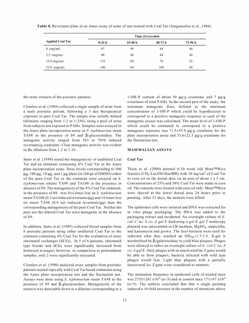
Jongeneelen et al. (1986) conducted the Ames mutagenicity
test using urine from 5 female eczema patients treated with an
ointment containing 10% Coal Tar for several days.
Salm onella typhimurium strain TA98 was used in the presence
of S9 and $-glucuronidase. The mutagenicity of the urine
samples could not be assayed because of the toxic properties
of the urine extract, even after dilution. However, the authors
also used rats receiving topical Coal Tar treatment. The 24
hour urine samples from 3 rats were pooled for the
mutagenicity study using S. typhimurium strain TA98 in the
presence of S9 and $-glucuronidase. There was evidence of a
dose-dependent uptake through the skin. The results are
shown in Table 8.
Clonfero et al. (1987) monitored three male psoriatic patients
during their therapy which consisted of a daily application of
Crude Coal Tar for 3 consecutive days followed by exposure
to UV rays. Urine samples were collected starting from 6
hours after the first Coal Tar application up to 36-48 hours
after the last. Various doses of urine extracts were tested in the
Ames plate incorporation assay using strains TA98 and
TA100 of S. typhimurium in the presence of S9 and $-
glucuronidase. The psoriatic patients had 64,462 induced
revertants/g of creatinine compared to 279 for a group of 5
control subjects. In addition, there was a correlation found
between the total urine PAH levels and mutagenic activity of
13
Table 8. Revertants/plate in an Ames assay of urine of rats treated with Coal Tar (Jongeneelen et al., 1986).
Applied Coal Tar
Time of excretion
0-24 h 25-48 h 48-72 h 72-96 h
0 (mg/rat) 47 49 44 44
2.5 (mg/rat) 90 66 44 44
12.6 (mg/rat) 135 101 74 52
53.0 (mg/rat) 146 141 109 69
the urine extracts of the psoriatic patients.
Clonfero et al. (1989) collected a single sample of urine from
a male psoriatic patient, following a 3 day therapeutical
exposure to pure Coal Tar. The sample was serially diluted
(dilutions ranging from 1:2 to 1:256), using a pool of urine
from subjects not exposed to PAHs. Samples were assayed in
the Ames plate incorporation assay on S. typhimurium strain
TA98 in the presence of S9 and $-glucuronidase. The
mutagenic activity ranged from 563 to 7034 induced
revertants/g creatinine. Clear mutagenic activity was evident
in the dilutions from 1:2 to 1:16.
Sarto et al. (1989) tested the mutagenicity of undiluted Coal
Tar and an ointment containing 4% Coal Tar in the Ames
plate incorporation assay. Dose levels corresponding to 500
:g, 100 :g, 10 :g, and 1 :g/plate (in 100 :l of DMSO) either
of the pure Coal Tar or the ointment were assayed on S.
typhimurium strains TA98 and TA100 in the presence or
absence of S9. The mutagenicity of the 4% Coal Tar ointment,
in the presence of S9, was five times less on S. typhimurium
strain TA100 (0.3 net induced revertants/:g) and 10 times less
on strain TA98 (0.9 net induced revertants/:g) than the
corresponding mutagenicity of the pure Coal Tar. Neither the
pure nor the diluted Coal Tar were mutagenic in the absence
of S9.
In addition, Sarto et al. (1989) collected blood samples from
6 psoriatic patients using either undiluted Coal Tar or the
ointment containing 4% Coal Tar for the evaluation of sister
chromatid exchanges (SCEs). In 5 of 6 patients, chromatid
type breaks and SCEs were significantly increased from
historical averages; however, in comparison to pretreatment
samples, only 2 were significantly increased.
Clonfero et al. (1990) analyzed urine samples from psoriatic
patients treated topically with Coal Tar based ointments using
the Ames plate incorporation test and the fluctuation test.
Assays were done using S. typhimurium strain TA98 in the
presence of S9 and $-glucuronidase. Mutagenicity of the
extracts was detectable down to a dilution corresponding to a
1-OH -P content of about 50 :g/g creatinine and 7 :g/g
creatinine of total PAHs. In the second part of the study, the
minimum mutagenic dose, defined as the minimum
concentration of 1-OH-P which could be hypothesized to
correspond to a positive mutagenic response in each of the
mutagenic assays was calculated. The mean level of 1-OH-P
which could be estimated to correspond to a positive
mutagenic repsonse was 71.5±55.9 :g/g creatinine for the
plate incorporation assay and 33.6±22.5 :g/g creatinine for
the fluctuation test.
MAM MALIAN ASSAYS
Coal Tar
Thein et al. (2000) painted 6-10 week old Muta™Mice
females (CD2-LacZ80/HazfBR) with 10 mg/cm2 of Coal Tar
or corn oil on the dorsal skin, on an area of about 1 x 3 cm.
Concentrations of 25% and 50% Coal Tar were made in corn
oil. The controls were treated with corn oil only. Muta™Mice
were shaved at the lower dorsal area 24 hours prior to
painting. After 32 days, the animals were killed.
The epidermal cells were isolated and DNA was extracted for
in vitro phage packaging. The DNA was added to the
packaging extract and incubated. An overnight culture of E.
coli C lac Z rec A gal E harboring a gal K gal T multicopy
plasmid was innoculated in LB medium, MgSO4, ampicillin,
and kanomycin and grown. The host bacteria were used for
infection after they reached an OD709=1.7-1.9. X-gal is
metabolized by $-galactosidase to yield blue plaques. Phages
were allowed to infect an overnight culture of E. coli C lac Z
rec A gal E . Only phages with an inactivated lac Z gene would
be able to form plaques; bacteria infected with wild type
phages would lyse. Light blue plaques with a partially
inactivated lac Z gene were considered as mutants.
The mutantion frequency in epidermal cells of treated mice
was 2735±281 x10-6 (n=3) and in control mice 171±97 x10-6
(n=5). The authors concluded that that a single painting
induced a 16-fold increase in the number of mutations above

14
the background level (p<0.002) in epidermal cells (Thein et
al., 2000).
Coal Tar M etabolites
Granella and Clonfero (1992) compared the sensitivity of the
pla te incorporation, macro-scale f luctuation, and
microsuspension tests in detecting mutagens in urine of 3 male
psoriatic patients who applied Coal Tar ointments for 4
consecutive days. Urine samples were filtered, eluted, and
dried to obtain a 250 X concentrate for the plate and
fluctuation tests and a 100 X concentrate for the
microsuspension assay. S. typhimurium strain TA98 with S9
and $-glucuronidase was used. A urine extract was considered
positive if it produced at least a doubling of the number of
spontaneous revertants, or in the case of the fluctuation assay,
a O2 statistical significance with p<0.05 .
All samples were mutagenic. Minimum mutagenic doses of
extracts were 1.5-12.5, 0.4–3.1, and 0.5-2.5 ml of urine for the
plate, fluctuation, and microsuspension tests respectively
(Granella and Clonfero, 1992).
Giles et al. (1996) investigated the metabolic activation of
benzo[g]chrysene (B[g]C), a moderately carcinogenic PAH
present in Coal Tar, in mouse skin. B[g]C (0.5 :mol) was
applied to the dorsal skin of male Parkes mice. Control mice
received acetone only. Groups of four animals were killed 6
hours and 1, 2, 4, 7, or 21 days after treatment and the treated
areas of skin were removed and frozen. DNA was isolated for32P-postlabelling. Resolution of 32P-labelled adducts was
performed on TLC sheets and visualized by autoradiography.
Seven principal adduct spots were consistently detected.
Maximum levels of 6.55 fmol adducts/:g of DNA were
detected 24 hours after treatment. There was a loss of 58% of
the damage by day 4, followed by a slower removal of adducts
over subsequent days (Giles et al., 1996).
DNA ADD UCTS
The formation of DNA adducts as a result of Coal Tar
exposure is well recognized --- Table 9 summarizes the
available data.
CARCINOGENICITY
The carcinogenicity of Coal Tar was first reported when a
variety of metastasizing skin cancers occurred in mice painted
with Coal Tar (Yamagiwa and Itchikawa, 1917). The ability
of tar to cause cancer is generally attributed to its PAH
content. According to Lin and Moses (1985), inducible
microsomal enzymes, such as aryl hydrocarbon hydroxylase,
convert these compounds into active forms which then bind to
DNA and RNA, thus exerting their carcinogenic influences of
DNA and RNA.
RAT - SUBCUTANEOUS
Jorstad (1923) injected Coal Tar beneath the epidermis of an
embryonic rat and described how the epithelial cells migrated
towards the drop to form a collar. When injected into
subcutaneous tissue, Coal Tar caused dense masses of
connective tissue cells to develop. These masses became
larger when more tar was added until they were sarcomatous.
MOUSE - ORAL
In the study by Culp et al. (1998) (see Chronic Oral Toxicity
section for details), the tumorigenicity of two Coal Tar
mixtures was compared to benzo[a]pyrene (BP) after 2 years
of feeding in mice. Liver neoplasms occurred in all dose
groups fed Mixtures 1 and 2, but not in the control group. A
s i g n i fi c a n t d o s e - r e la t e d t re n d w a s o b s e rv e d .
Alveolar/bronchiolar adenomas, carcinomas, or both were
present in the control group and in all groups of mice fed
Mixtures 1 and 2. W ith Mixture 1, the incidence in the 0.3,
0.6, and 1.0% dose groups was significantly increased
compared to the control group and a significant dose-related
trend was observed. A significant dose-related trend was also
found with Mixture 2 and the frequency was significantly
increased in the 0.1 and 0.3% dose groups. The predominant
lung lesions were adenomas, with only 15 carcinomas
detected. Papillomas and/or carcinomas of the forestomach
squamous epithelium were observed in all groups of mice
treated with Mixtures 1 and 2 but no neoplasms were detected
in the control group.
MOUSE - DERMAL
Murphy and Sturm (1925) studied the influence of external
application of Coal Tar on the incidence of lung tumors in
mice. The tar product used had for its base the residue from
a coke oven in which the crude tar had been distilled at a
temperature of approximately 377°C. The mice came from a
stock with an extremely low cancer incidence. For each
experiment, each mouse received Coal Tar on 12 areas, each
less than a centimeter in diameter, painted in rotation such that
36 applications were distributed over 83 days. Each site was
painted three times with a month in between. In the first
experiment, 20 mice were painted according to the above
scheme. As a control, 22 mice from the same stock were kept
under the same laboratory conditions.
The lung tumor rate for the treated group in this experiment
was 66.6%. N one of the control animals showed developed
tumors. In the second experiment, an unspecified number of
mice were subjected to the same system of applications as that
in the first experiment. The lung tumor rate was 60%;
however, 1 mouse had a tumor of the uterus. In the third
experiment, 40 mice were painted in the same fashion as in the

15
Table 9. Studies on Coal Tar and DNA adducts
Reference Test system Results
Mukhtar et al. 1986c male SENCAR mice Topical application of 0.5 ml of Crude Coal Tar resulted in the formation of 278 fmol and410 fmol BPDE-I-dGa adducts per mg DNA in the epidermis and lung, respectively.
Schoket et al. 1988a male Parkes mice The application of 150 :l of 20% pharmaceutical grade Coal Tar solution resulted in ~0.4fmol total adducts/:g DNA 24 hours after treatment. When given multiple treatments, asteady accumulation of adducts was seen in skin DNA.
Schoket et al. 1988b Adult and fetal humanskin samples
A single dose of pharmaceutical grade Coal Tar (equivalent to 30 mg Crude Coal Tar)resulted in 0.2 to >0.6 fmol adduts/:g DNA.
Schoket et al. 1990 male Parkes mice skin Application of 45 mg of Coal Tar ointment resulted in 0.5 fmol/:g DNA in the skin 1 dayafter the five treatments.
Human skin biopsysamples
Biopsies taken 24 hours after 5 treatments with Coal Tar ointment contained adductsranging from 0.1 to 0.39 fmol/:g DNA.
Zhang et al. 1990 Human skin biopsysamples
Topical application of 10-50 mg/kg/day of Coal Tar for at least 7 days produced adductlevels ranging from 0.18 to 9.4 adducts/108 nucleotides.
Weyand et al. 1991 B6C3F1 mice A dose related increase in DNA adduct levels was observed in the lung tissue of mice fed0.1, 0.2, 0.5, and 1% Coal Tar diets. Adduct levels were 4 times greater in mice fed a 0.5or 1% diet relative to those fed a 0.1 or 0.2% diet.
Paleologo et al. 1992 Human lymphocytes Psoriatic patients treated with Coal Tar products had mean adduct levels of 0.257±0.162fmol BPDE/:g DNA in their white blood cells during treatment.
Pavanello and Levis 1992 Human lymphocytes No significant differences were found in the amounts of total DNA adducts betweenpsoriatic patients treated with Coal Tar and controls or between psoriatic pateints beforeand after Coal Tar treatment.
Hughes et al. 1993 Male Parkes mice When a group of PAHsb were applied to mouse skin, the pattern of adducts formed in theskin resembled that elicited by the application of Coal Tar solution
Pfau et al. 1993 Male Parkes mice A major DNA adduct formed in the skin of mice treated with pharmaceutical grade coaltar was found to be derived from benzo[a]pyrene rather than benzo[b]fluoranthene
Pavanello and Levis 1994 Human lymphocytes In psoriatic patients treated with crude Coal Tar, there was no correlation between thelevels of PAH-DNA adducts and the exposure to Coal Tar. According to the authors, CoalTar treatment should not be considered a potential genetic and carcinogenic risk forpsoriatic patients.
Culp and Beland 1994 Male B6C3F1 mice Adduct levels were greater in Coal Tar fed mice compared to those fed benzo[a]pyrene. InCoal Tar fed mice, adduct levels were in the order of lung>liver<forestomach.
Santella et al. 1995 Human lymphocytes PAH-DNA adducts were elevated in Coal Tar treated psoriasis patients compared tocontrols (6.77±12.05 adducts.108 nucleotides).
Giles et al. 1997 Male Parkes mice When 0.5 :mol of benzo[c]chrysene, a component of Coal Tar was applied to the skin ofmice, a maximum adduct level of 0.89 fmol adducts/:g of DNA was detected in the skin.
Godschalk et al. 1998 Human skin When eczema patients were topically treated with Coal Tar, median aromatic DNA adductlevels significantly increased in the skin from 2.9 to 63.3 adducts/108 nucleotides.
Human lymphocytes Median aromatic DNA adduct levels were significantly increased in lymphocytes from0.33 to 0.89 adducts/108 nucleotides.
Pavanello et al. 1999 Human lymphocytes Psoriatic patients treated with Coal Tar had 0% of their BPDE-DNA adduct levelsexceeding the 95 percentile control subject value.
Koganti et al. 2000 Female CD-1 mice In animal feeding studies, 7H-benzo[c]fluorene, a PAH in Coal Tar, is a potent lungadductor.
a BPDE-I-dG = benzo[a]pyrene diol epoxide-I-deoxyguanosineb Concentrations of benzo[a]pyrene, benzo[b]fluoranthene, benzo[j]fluoranthene, benzo[k]fluoranthene, benzo[ghi]perylene, dibenz[a,h]-anthracene,indenol[1,2,3-cd]pyrene, benz[a]anthracene, and cyclopental[cd]pyrene equivalent to those present in 30 mg of Coal Tar were used

16
first experiment. For controls, a group of 16 untreated mice
from the same stock were kept under the same conditions.
While none of the control mice had any tumors, the tumor
incidence for the painted animals was 78.3% (Murphy and
Sturm, 1925).
Watson and Mellanby (1930) studied the effects of
pretreatment of the skin and diet on Coal Tar induced
carcinogenesis, but much experimental detail is lacking in the
report.
Pretreatment with tannic acid (saturated aqueous solution) or
by application of homologous fat (ether extracted from mouse
carcases) followed by Coal Tar application was done for 120
days in 70 animals for each group. The animals were
followed for one year. The authors concluded that the rate of
growth of tumors was unaffected by tannic acid pretreatment,
but accelerated by fat pretreatment, compared to controls.
Pretreatment with olive oil was done in another group of 70
animals for 90 days. According to the authors, animals in the
olive oil + Coal Tar group showed a greater tendency to
develop tumors than the control group. This is a different
measure than above, where rate of growth was determined.
Pretreatment with ether (to remove fatty substances in the
skin) prior to Coal Tar treatment was done in another group of
60 mice for 120 days. Animals were followed for 360 days.
The authors interpreted the results to suggest that the rate of
tumor growth was inhibited by ether pretreatment. Lung
nodules in tumor-bearing animals were reduced in the ether
pretreatment group, as well.
In another experiment, a diet rich in butter was compared to
control diet. Animals (70 in each group) were treated with
Coal Tar daily for 120 days and followed for 360 days. More
animals in the butter diet group had tumors than the controls,
and those in the butter diet had a higher proportion with lung
nodules than the control group (Watson and Mellanby, 1930).
Seelig and Cooper (1933) tested the effect of light on cancer
in mice. Three sets of mice were used in this experiment: (1)
125 male mice for tarring in the dark; (2) a similar group of
125 mice for tarring in the light; and (3) a similar group of 50
which were kept as untarred controls in the dark. Coal Tar
(distillate at 370°-440°C was collected) was applied to the
interscapular region at the root of the neck, over an area about
1.5 cm in diameter. The time intervals between applications
were varied to meet the problem of high death rates in the
mice. Applications continued until the death of the last tarred
mouse. A necropsy was performed on all mice that died. Only
growths that infiltrated the subcutaneous muscle layers were
considered carcinomas. The last mouse in Group 1 died 23
weeks after the first application and the number of carcinomas
in the group was 11. In contrast, the last mouse in Group 2
died 32 weeks into the experiment and there were only 5 cases
of carcinomas in the group. When the last tarred mouse died,
only 58% of the control mice had died and none of the Group
3 mice had carcinomas. The authors concluded that the
absence of light did not compromise the health of mice and
that white light was not a necessary factor in the development
of tar cancer in mice.
Berenblum (1948) painted 12 white mice twice weekly for 41
weeks with undiluted Coal Tar on a small area of the skin in
the interscapular region. Papillomas appeared in the treated
areas in seven of the mice. Of these seven, four subsequently
developed malignant growths, which were found to be
squamous cell carcinomas. There were no metastases.
Christie and McCallum (1958) studied the carcinogenicity of
brown Coal Tar. Included in their experiments were two
treatments (8 mice each, C3H strain) where (1) brown Coal
Tar ointment, identical with black Coal Tar ointment APF, but
substituting brown Coal Tar for black and (2) black Coal Tar
ointment were applied on the back and flank of the mice once
per day, six days per week. In the brown Coal Tar ointment
group, no papillomata, warts or deranged keratinisation
occurred in 8 months of painting. In the black Coal Tar
ointment group, five animals developed warty papillary
growths from 6-8 months, but no malignant tumors were
obtained.
Wright et al. (1985) performed painting assays on Charles
River CD-1 mice (numbers not given). Industrial Coal Tar
from the National Bureau of Standards (NBS) and the Coal
Tar portion of a pharmaceutical stock material were diluted
1:1 by weight in methylene chloride and applied to the shaved
backs of the mice in 50 :l volumes. Two weeks after
initiation, 50 :l of phorbol myristate acetate were applied to
the treated area twice weekly for 6 months. Both the NBS
Coal Tar and the Coal Tar isolate from the pharmaceutical
stock produced tumors in all the mice after 169 days.
Mukhtar et al. (1986a) applied a single dose of 200 :l of
crude Coal Tar in 0.2 ml of acetone to 20 female SENCAR
mice. The mice were shaved and depilated before the
experiment. Skin tumor formation was recorded weekly and
tumors greater than 1 mm in diameter were included in the
cumulative total only if they persisted for 2 weeks or more.
The first appearance of tumors occurred at 6 weeks. After 8
weeks, there were 46 cumulative tumors in the group of mice.
At the termination of the experiment after 11 weeks, 100% of
the mice had tumors and the average number of tumors/mouse
was 3.3.
Warshawsky et al. (1993) painted Coal Tar (in toluene such
that it contained 0.0006% benzo[a]pyrene) on the clipped
backs of male C3H/HEJ mice twice weekly for up to 66
weeks. Observations of the skin were recorded. Lesions
persisting for at least one week which were a minimum of 1
mm3 in size were considered papillomas. The times and

17
appearance of tumors and time for their progression to
malignancy were noted. A necropsy was conducted on all
animals. Tumors were present in 51% of the mice. The latent
period was 73 weeks.
Phillips and Alldrick (1994) attempted to determine whether
treatment with Coal Tar followed by dithranol was
tumorigenic. Female CD-1 mice were treated topically 5 times
a week with 1.5% Coal Tar ointment (50 mg per treatment) for
2 weeks and then with 0.1% dithranol cream (50 mg per
treatment) three times weekly for 40 weeks. Other groups
received either Coal Tar or dithranol alone. Hair on the mice
was shaved and the material was applied to the whole of the
shaved area. Tumors >1 mm were scored. The animals were
killed by cervical dislocation and subjected to post-mortem
examination. None of the mice treated with Coal Tar or
dithranol alone developed tumors during the experiment.
However, four mice that had received Coal Tar followed by
dithranol developed papillomas and 12 mice had enlarged
cervical lymph nodes. Five mice that received d ithranol only
also had enlarged lymph nodes, though this condition was not
seen in any of the mice treated with Coal Tar only.
MOUSE - MUCOSAL
Shabad (1927) applied Coal Tar to the rectum of 78 juvenile
white mice 2-3 times a week. A thin smooth round wooden
stick with a blunt tip was dipped in Coal Tar and carefully
inserted 1.5-2 cm into the rectum. Of the 37 animals that
survived the first 6 months of the test, cancer of the perianal
skin developed in 21 (56.7%).
RABBIT - DERMAL
Itchikawa and Baum (1925) conducted three experiments
using rabbits (strain unspecified). In the first experiment 6
rabbits had Coal Tar painted on their ears in addition to being
fed 10 grams of anhydrous lanolin mixed in the food. In the
second experiment, 6 rabbits had Coal Tar painted on their
ears in addition to being fed 0.5 cc of a 2% watery solution of
potassium arsenite mixed in the food. In the third experiment,
6 rabbits had Coal Tar painted on their ears. The painting
covered the entire inner surface of rabbits’ ears and was
performed usually twice a week for 10 weeks. After the
painted area dried, the coal tar was easily removed with
tweezers, and the ears were repainted. The appearance of
folliculo-epithelioma and its transformation into the early
stage of carcinoma appeared around the same time in all the
animals, but the alteration of the early stage into advanced
carcinoma took place sooner in the animals that were fed on
arsenic and lanolin than in those that were painted with Coal
Tar alone.
Kligman and K ligman (1994) applied ~10 mg of crude Coal
Tar on albino rabbit ears at 0.001, 0.01, 0.1 and 1.0% with 5
weekday applications for 3 weeks and at 10, 25, and 100%
three times weekly for up to 15 weeks. Crude Coal Tar,
whether applied at 10, 25, or 100% was carcinogenic. W ith
10% Coal Tar, tumors developed as early as 1-2 weeks
following 3 weeks of treatment. With longer term treatment,
the number of tumors increased. No results were reported for
Coal Tar concentrations at 1.0% or less.
These authors also applied ~10 mg of crude Coal Tar on
albino rabbit ears at 0.001, 0.01, 0.1 and 1.0% with 5 weekday
applications for 3 weeks and at 10, 25, and 100% three times
weekly for up to 15 weeks. The vehicle was petrolatum.
Crude Coal Tar, whether applied at 10, 25, or 100% was
comedogenic. The threshold for comedogenicity was less than
0.1% with 3 weeks application (Kligman and Kligman, 1994).
CLINICAL ASSESSMENT OF SAFETY
DERMAL IRRITATION
Burden et al. (1994) patch tested 74 psoriasis patients with
Coal Tar 3% pet. A positive reaction was followed by dilution
series testing down to a concentration of 0.1%. Of the 74
patients, 13 reported that they were allergic to coal tar;
however, in actuality only 2 had a positive patch test reaction
confirmed by serial dilutions.
PHOTO SENSITIZATION AND PHO TOTOXICITY
Several of the polycyclic aromatic hydrocarbons which make
up coal tar are photosensitizers and cause phototoxicity by an
oxygen dependent mechanism (Gould et al. 1995). Ultraviolet-
A (UVA) light contains the part of the light spectrum that
activates coal tar (Arno ld 199 7). Studies on
photosensitization and phototoxicity of Coal Tar are
summarized in Table 11.
FOLLIC ULITIS
Folliculitis is associated with the prolonged use of some tars
(Lerner and Lerner, 1960).
COM EDOGENICITY
Kaidbey and Kligman (1974) applied 25% crude Coal Tar to
the backs of adult males for 3 times a week for 3 weeks.
Biopsy specimens were obtained at 7, 14, and 21 days and
sporadically thereafter. Caucasians and African Americans
differed in their responses. In Caucasians, the response was
primarily inflammatory. In African Americans, the
inflammatory phase was largely absent. Instead, small open
comedones began to appear after 14 days and were obvious by
the end of the treatment period.

18
Table 11. Studies on Coal Tar photosensitization and phototoxicity.
Reference Summary
Starke and Jillson 1961 Case Report - A 37 year old male had eczema and weeping to patch tests of Coal Tar. A photopatchdeveloped an even more eczematous reaction.
Everett et al. 1961 When Coal Tar solution and ultraviolet light were applied to the skin of 21 persons, erythema andtanning were more prominent at the control sites in all patients
Everett and Miller 1961 When Coal Tar solution (USP) and ultraviolet light were applied to the skin of five adult males,erythema was greater in the tar-free areas than the tar-exposed areas
Kaidbey and Kligman 1974b The intensity of the phototoxic response to Coal Tar was strongly influenced by the vehicle, withvanishing cream producing the greatest reaction (erythema and minimal edema)
Kaidbey and Kligman 1975 When 40 :l of a 25% Coal Tar distillate was applied to the skin of adult males irradiated with UVAalone, the epidermis was largely unaffected except for occasional intercellular edema.
Kaidbey and Kligman 1977 Volunteers who applied a 5% concentration of four different crude Coal Tars followed by UVAexposure experienced whealing followed by raised lesions.
Kroon 1983 40 out of 194 patients photopatch tested with Waxtar (Coal Tar 5%) had phototoxic reactions. 4 patientsdeveloped pigmentation 7 days after irradiation.
Diette et al. 1983, 1985 The time between tar removal and UVA irradiation was important in determining the minimalphototoxic dosea and the minimal smarting doseb in human volunteers.
Berne and Fischer 1987 Compared to Balnetar® and Spiritus carbonis detergens, 5% crude Coal Tar gave the strongest erythemaphototoxicity. Its action spectra ranged from 330 to 400 nm.
a Minimal phototoxic dose: the minimal UVA dose required to induced delayed erythema b Minimal smarting dose: the minimal UVA dose required to induce an immediate smarting reaction
Mills et al. (1978) applied 15% crude Coal Tar in Hydrophilic
Ointment USP to the upper backs of 5 adult males. The agent
was applied to 8 cm squares of skin thrice weekly. Before
renewing the applications, one-half of each square was
subjected to UVA radiation. A combination of open and
closed comedones was present in each of the 5 subjects
receiving Coal Tar without radiation. Comedones and papules
increased in size and number when irradiated.
CASE REPORTS
Table 12 summarizes case reports involving Coal Tar.
CLINICAL TESTING
Wright et al. (1992) divided 15 healthy volunteers (11 women,
4 men) into 3 groups of 5, subjecting them to different modes
of application of Berniter®, a 0.5% coal tar preparation for
the scalp. In application mode 1, Berniter® was applied twice
a week over a period of 8 weeks; in mode 2, it was applied on
a daily basis for 8 weeks. The preparation was administered
by wetting the scalp with water, spreading and massaging the
preparation into the scalp, rinsing the hair and scalp with
water, spreading and massaging the preparation into the scalp
again, and allowing the preparation to work for 5 minutes. In
application mode 3, Berniter® was applied every other day
over a period of 4 weeks. Here the preparation was applied
under occlusion (a plastic cap) and allowed to work for 15
minutes. The test subjects’ 24-hour urines were tested for
low-molecular phenol bodies before and after treatment. In
addition, multiple chemical analyses were undertaken (GOT,
GPT, Gamma-GT , AP, LDH, cholinesterase, total bilirubin,
etc.) as well as creatinine clarance, gel-electrophoresis protein
excretion pattern, and alanine aminopeptidase activity.
No pathological findings were noted in any of the test
subjects. The results of the blood and urine studies, as well as
of the multiple chemical analyses, were all within normal
limits (Wright et al., 1992)
CLINICAL TESTING FOLLOW-UP
Kaaber (1976) reported on 326 patients treated with Coal Tar
for atopic dermatitis. Patients were followed for 42 years to
determine if they had developed cancer. These 326 patients
had been treated in the Department of Dermatology, Finsen
Institute, Copenhagen, Denmark from 1930 to 1939 for atopic
dermatitis. The follow up period for each patient was
calculated from the first examination until the date of death or

19
Table 12. Coal Tar case reports.
Reference Summary
Hodgson, 1948 A 62 year old male using Coal Tar to treat pruritus developed epithelioma.
Simon and Brandt, 1953 A 60 year old female with a history of psoriasis had a positive reaction to a patch test with 2% crude Coal Tarin an oxycholesterol-petroleum ointment base.
A 64 year old female had a positive reaction to a patch test with 3% crude Coal Tar in petrolatum witherythema and edema occurring within 48 hours.
Alexander and Macrosson, 1954 A 31 year old male using a 20% Coal Tar in emulsifying base ointment to treat psoriasis developed squamousepithelioma.
Person and Rogers III, 1976 A 53 year old male using the Goeckerman regimen to treat psoriasis developed bullous pemphigoid.
A 53 year old male using 2% crude Coal Tar to treat psoriasis developed bullae.
A 66 year old male using the Goeckerman regimen to treat psoriasis developed a bulla.
Durkin et al., 1978 A 32 year old male psoriatic developed melanoma after 16 years of treatment with UV light and Coal Tar.
Koerber et al., 1978 A 53 year old female using Coal Tar creams to treat psoriasis developed bullous pemphigoid.
Gonçalo et al., 1984 A 25 year old female with psoriasis had a positive reaction to a patch test with Coal Tar.
A 17 year old female with psoriasis had a positive reaction to a patch test with Coal Tar.
Kokkini and Giotaki, 1984 A 45 year old female psoriatic developed pancytopenia due to hypoplastic and ineffective bone marrowfollowing 5% Coal Tar cream application for 6 months.
Riboldi et al., 1986 A 54 year old female with psoriasis had a positive reaction to a patch test with Coal Tar (blistering).
Illchyshyn et al., 1987 A 31 year old female with eczema had a positive reaction to patch tests with 1% and 5% Coal Tar inpetrolatum.
McGarry and Robertson, 1989 A 67 year old male with scrotal psoriasis developed squamous cell carcinoma after uing a 10% crude Coal Tarointment.
Cusano et al., 1992 A 57 year old male had a positive reaction to a patch test with Coal Tar (5% pet).
Landro et al., 1992 A 29 year old male with psoriasis had a positive reaction to a patch test with 0.05% Coal Tar.
Ibbotson et al., 1995 A 29 year old female asthmatic had severe symptomatic bronchoconstriction which was likely due to inhalationof Coal Tar vapor from the application of Coal Tar bandages. An inhalation challenge test with vapor from theCoal Tar bandages produced a 28% reduction in forced expiratory volume.
Clark and Sheretz, 1998 A psoriasis patient had positive allergic reactions to Coal Tar diluted to 1% and 5% when given patch tests.
until cancer was diagnosed. The expected cancer rated was
calculated based on the cancer registry data on the incidence
of cancer in Denmark. The values were then compared to the
results of the follow up of the 326 patients. There was no
sufficient deviation in the expected values versus the observed
values for the 326 patients treated for atopy.
Maughan et al. (1980) performed a 25 year follow-up study on
305 patients with atopic dermatits or neurodermatits who
received Coal Tar and UV treatment (Goeckerman therapy)
from 1950 through 1954. These patients were located and
contacted by either a follow-up questionnaire or telephone in
order to determine if any malignancy of the skin or other
cancer had developed since the initial treatment. The numbers
of persons in whom skin cancers were expected to develop
were calculated using data from the Third National Cancer
Survey. Of the 305 patients, 13 reported skin cancer had
developed after the initial Goeckerman therapy. Eight patients
had basal cell carcinomas, one had a squamous cell
carcinoma, two had an unknown skin malignancy, and two had
malignant melanomas. The maximum number of people in the
study who would have been expected to have developed non-
melanoma skin cancers if they had lived in any of the regions
reported in the Third National Cancer Survey was 18.8 for the

20
Dallas-Fort Worth area. Since 11 patients developed non-
melanoma skin cancer, no significant increase in the incidence
of skin cancer was seen in the population of patients when
compared to the expected occurrence of skin cancer if the
population were to have resided in the D allas-Fort Worth area.
In a later study, Pittlekow et al. (1981) performed another 25
year follow-up analysis of patients receiving Coal Tar and UV
treatment from 1950-1954. Patients were instructed to report
any cancerous lesions of the skin that had been removed since
the initial Goeckerman treatment. Of 260 patients, 20 (7.7%)
had one or more reported carcinomas of the skin. One patient
reported the development of a malignant melanoma; the
remaining 19 reported either basal cell or squamous cell
carcinomas or unknown varieties of cutaneous malignant
neoplasms. Comparing the group of patients in which skin
cancer subsequently developed with the group in which it did
not, there was no appreciable difference in the median number
of days of Coal Tar treatment.
The Pittlekow study also contains the results from a review by
the National Psoriasis Foundation on the medical histories of
15,000 of its members. The results from that review found no
suggestion of an increased risk to skin cancer. The results of
this survey also showed no increase in skin cancer from
patients who had been treated with Coal Tar.
Larko (1982) conducted a study in Sweden on 85 psoriasis
patients extensively treated with UVB light and topical Coal
Tar. The results were compared to 385 individuals in a control
group. The incidence of premalignant/malignant skin lesions
was only 5.96% in the 85 treated psoriasis patients, but was
10.1% in the 385 control individuals. The authors conclude
that it is unlikely that treated psoriatic patients will have an
increased incidence of malignant skin lesions from their
treatment with topically applied Coal Tar and UVB light.
Jones et al. (1985) compared the observed incidence of
carcinoma in 719 psoriatics treated with Tar with that
expected in the general population. The 719 patients (305
males and 414 females) had received topical Tar therapy
intermittently over a 10 year period between 1953 and 1973.
There were 19 cases of cancer in the male patients which was
very similar to that expected from the general population. In
the case of female patients, the 12 cases found was less than
expected. Of particular interest were the rates of cancer in the
skin and b ladder, neither of which showed a significant
increase in the patient population. The authors concluded that
long-term Tar therapy was not associated with an increase in
cutaneous or non-cutaneous malignancy in patients with
psoriasis.
Bhate (1993) published the results of an epidemiology study
on the use of Coal Tar in the treatment of psoriasis. Between
1977 and 1983, 2,247 patients were treated with Coal Tar at
the Department of Dermatology, Royal Victoria Infirmary,
UK where they were examined for the prevalence of both skin
and internal malignancies. These results were compared
against 4,494 matched control individuals. The results of this
extensive epidemiological study were that there was no
difference in the age of onset of skin cancers between
psoriatics and controls. There was also no evidence of a
cumulative therapeutic risk. Finally, there was no difference
in the prevalence of non-skin cancers between psoriatics and
controls.
Jemec (1994) published results from 88 patients treated
extensively with Coal Tar in Denmark from 1917 to 1937.
Cancers were identified through the Danish Cancer Registry
and relative risk of cancer was calculated. No overall risk of
cancer for the treated patients was found in the 88 patients
compared to the incidence of cancer in the general population.
The authors go on to state that these data provide further
support for the safety of Coal Tar in the management of
dermatological disease.
EPIDEMIOLOGY
Franssen et al. (1999) performed a retrospective study on the
reproductive toxicity of Coal Tar in humans. Women were
chosen based on past history of psoriasis or dermatitis. Out of
103 pregnancies there were 59 in which no Coal Tar had been
used, 21 in which it was uncertain whether or not Coal Tar
had been used, and 23 in which Coal Tar was definitely used.
Of the 59 in which no Coal Tar had been used, 19% of the
pregnancies ended in spontaneous abortion and 5% in a
congenital disorder. In the 23 cases in which Coal Tar was
used during pregnancy, 26% resulted in spontaneous abortion
and 4% in a congenital disorder. T he authors note the small
sample size of patients exposed to Coal Tar and the lack of
uniformity regarding the time and duration of exposure.
In a study completed in Yugoslavia, Vlajinac et al. (2000)
found that the use of tar for cosmetic purposes was a risk
factor for basal cell carcinoma. Participants (200 confirmed
cases of basal cell carcinoma and 399 control subjects) were
interviewed by physicians and given questionnaires regarding
various risk factors. The use of tar was found to be a
significant risk factor (p=0.0064). However, the authors also
used oral methotrexate and photochemotherapy (PUVA) and
the effects from Coal Tar cannot be separated out as the sole
cause.
RISK ASSESSMENTS
The International Agency for Research on Cancer (IARC)
reviewed PAHs as well as Coal Tars in 1984 (IARC 1985).
IARC concluded: “There is sufficient evidence for the
carcinogenicity in experimental animals of coal-tars,
creosotes, creosote oils, anthracene oils and coal-tar pitches.”
IARC evaluated the limited epidemiological studies generated

21
on the therapeutic use of Coal Tar up to that point in time, but
could not draw any conclusions. The therapies were mixed in
most studies and therefore effects from Coal Tar alone could
not be evaluated.
In a review of the use of dermatologic drugs during
pregnancy, Bologa et al. (1992) suggested limiting the use of
Coal Tar to the second and third trimesters, once
organogenesis is complete. In a review of the use of OTC
drugs during pregnancy, Conover and Rayburn (1992),
however, believe the risk of congenital anomalies in children
of women treated with Coal Tar during pregnancy is unlikely.
Pion (1995) examined the question of whether the use of Coal
Tar in dermatological practice was a cause of concern. From
his extensive review of both animal and human data, he stated
that “conclusive evidence for the carcinogenicity of coal tar
used in dermatologic practice is lacking.”
Hess (1999) used BP data as a surrogate to perform a
quantitative risk assessment on Coal Tar as an ingredient.
Rinse-off hair care products contain 1% refined Coal Tar that
were stated to include a maximum level of 50 mg/kg BP.
After a typical application of 12 g, it was estimated that 1% of
the product remains after rinsing. The lifelong exposure to BP
was estimated to be not higher than 0.001 ng/cm2 of exposed
skin surface area per day, which was below the BP no-effect
level. The author concluded that the safety of cosmetic usage
of coal tar was further confirmed by extensive
epidemiological data showing that no carcinogenic
consequences have emerged from its well-established and
intense use in dermatological therapy.
Fehling (2000) stated that an absorbed dose of 52.12 mg/day
was the no significant risk level (NSRL) for Coal Tar
containing products.
Jackson (2003) generated a quantitative risk assessment for 8
Coal Tar containing products, one soap bar, two ointments,
and 5 shampoos by chemically analyzing the PAH content of
these products. The life time averaged absorbed dose for each
of these Coal Tar containing products was significantly below
the 0.0600 :g/day NSRL for benzo[a]pyrene, a known
genotoxic chemical.
Coal Tar itself is on California’s Propo sition 65 list of
substances known to be carcinogenic; soots, tars, and mineral
oils (untreated and mildly treated oils and used engine oils)
are also listed (OEHHA 2003).
SUMMARY
Coal Tar is a semisolid byproduct obtained in the destructive
distillation of bituminous coal, produced in coking ovens as a
byproduct in the manufacture of coke. Coal Tar is a nearly
black, viscous liquid, heavier than water, with a naphthalene-
like odor and a sharp burning taste. Crude Coal Tar is
composed of 48% hydrocarbons, 42% carbon, and 10% water.
Coal Tar is an active ingredient in OTC drug products and is
described as an antidandruff agents, cosmetic biocides, and
denaturant in cosmetics. Coal Tar is monographed by the
Food and Drug Administration (FDA 1991) as a Category I
(safe and effective) for OT C drug ingredient for use in the
treatment of dandruff, seborrhoea, and psoriasis. While Coal
Tar was reported in 2002 to FDA to be used in four
formulations, these uses are OTC. No uses of Coal Tar as a
cosmetic biocide or denaturant have been reported. The
concentration of use of Coal Tar was reported as fo llows:
Shampoos (OTC) (1-7%), Bath soaps and detergents (OTC)
(0.06-5%), and Lotions (OTC)(5% ).
Mice, rats, and humans given a single dose of Coal Tar
solution orally resulted in the induction of one or more of aryl
hydrocarbon hydroxylase (AHH), ethoxyresorufin deethylase
(ERD), and 7-ethoxycoumarin O-deethylase (ECD) activities
in the skin and liver (liver studies not performed on humans).
The absorption of Coal Tar through skin and its systemic
distribution is confirmed by these AHH, ERD, and ECD
induction studies. The absorption of specific Coal Tar
polycyclic aromatic hydrocarbons (PAH s) was demonstrated
in a pig ear skin penetration study. The presence of acridine,
and other Coal Tar PAHs, in urine of patients or healthy
volunteers treated with Coal Tar demonstrated absorption of
Coal Tar.
In short term animal studies mice fed Coal Tar at
concentrations over 2% in their diet found their diet
unpalatable, but no adverse effects were reported other than
weight loss.
Injection of 1 ml Coal Tar 3x over 21 days produced malaise
in rats. Injection of a 5% Tar residue in rabbits produced
eating avoidance, respiratory difficulty, sneezing, and weight
loss. Injection of 5 mg/kg day-1 of a Coal Tar suspension for
8 days in rats decreased water intake, resulted in black
residues in the liver, and increased liver weights.
In a subchronic neurotoxicity study using mice using 2.0 :l/ml
of a mixture of phenols, cresols, and xylenols at
concentrations approximately equal to those expected in Coal
Tar extracts, regionally selective toxicity was found, with a
rank order of striatum > cerebellum > cerebral cortex.
Mice given a chronic exposure to 0.3% Coal Tar in the diet
had a significantly decreased survival. Liver neoplasms were
seen in a significant dose-related trend.
When Coal Tar was applied to the backs of guinea pigs,
increases in epidermal thickness were seen.
22
Painting female rabbits with tar decreases the absolute and
relative weights of the ovaries and decreased the number of
interstitial cells in the ovary. A retrospective study of the
reproductive toxicity of Coal Tar in humans compared 23
exposed women to 59 controls and found little difference in
spontaneous abortion and congenital disorders. Conflicting
advice has been given to pregnant women — one suggestion
was to avoid use of Coal Tar to the 2nd or 3rd trimester, after
major organogenesis; another opined that the risk of
congenital abnormalities in children of women treated with
Coal Tar during pregnancy is unlikely.
When four therapeutic Coal Tar prepara tions (Zetar ®
Emulsion, Estar ®, Lavatar, and Coal Tar Solution USP) used
in the treatment of psoriasis were screened using the Ames
Salm onella/microsome mutagenicity test, all of the Coal Tar
preparations were mutagenic within the dose range tested.
Strain TA98 was the most sensitive to the mutagenic effects
of the tar. Urine and blood from patients treated with Coal
Tar were genotoxic in bacterial assays. Coal T ar was
genotoxic in plate incorporation, macro-scale fluctuation, and
microsuspension assays.
In a mammalian genotoxicity assay (Muta™ Mice), the
mutation frequency in treated animals was 2735±281x10-6
(n=3) and in control mice 171±97x10-6 (n=5), a 16 fold
increase in the number of mutations above the background
level in epidermal cells.
Coal Tar induces DNA adducts in various tissue types. A
lowest observed effect level of 0.1% Coal Tar (oral) produced
DNA adducts in mouse lung tissue.
In a carcinogenicity experiment, five groups of 95 mice were
painted with Coal Tar for four months. The percentage of
animals with tumors in the lungs was 45.1, 53.7, 50.0, 55.6,
and 57.1, respectively. After being treated with 200 :l Crude
Coal Tar in 0.2 ml acetone for 11 weeks, 100 % of the 20
female SENCAR mice had tumors. After Coal Tar was
applied to the rectum of 78 juvenile white mice for 6 months,
of the 37 animals that survived , cancer of the perianal skin
developed in 21 (56 .7%).
Out of 74 psoriasis patients, 13 reported that they were
allergic to Coal Tar; but only 2 had a positive patch test.
Several of the polycyclic aromatic hydrocarbons which make
up Coal Tar are photosensitizers and cause phototoxicity by
an oxygen dependent mechanism. Coal Tar was comedogenic
in three small clinical studies. Several published reports
describe cases of contact sensitivity to Coal Tar. Folliculitis
is associated with the prolonged use of some tars.
Follow-up studies have been performed on patients who have
received Coal Tar therapy of one form or other: 326 patients
with atopic dermatitis or neurodermatitis treated with Coal Tar
were followed for 42 years; 305 patients who received Coal
Tar and UV treatment were followed for 25 years; 206
patients receiving Coal Tar and UV treatment were followed
for 25 years; 85 psoriatic patients treated with Coal Tar and
UV treatment were compared to 385 matched controls; 719
psoriatics who received Coal Tar therapy intermittently were
studied 10 - 25 years later; 2,247 psoriatics treated with Coal
Tar were compared to 4,494 matched controls; cancers
identified from a cancer registry in 88 patients treated
extensively with Coal Tar from 1917 to 1937 were compared
to the general population. In none of these follow-up studies
was an increase in the risk of cancer found.
One retrospective study of women who used Coal Tar at the
time of their pregnancies. It was unclear if the percentage with
spontaneous abortions or births with congenital disorders was
higher in the Coal Tar group compared to controls. A case-
control study of 200 individuals with basal cell carcinoma
compared to 399 controls identified use of tar for cosmetic
purposes as a risk factor, but it was not clear how oral
methotrexate and photochemotherapy may have confounded
the results.
DISCUSSION
The data presented in this report indicate that Coal Tar can
produce adverse biological effects. While the CIR Expert
Panel believes that Coal Tar use as an antidandruff
ingredient in OTC drug preparations is adequately
addressed by FDA regulations, the Panel also believes that
the appropriate concentration of use of Coal Tar in cosmetic
formulations should be that level that does not have a
biological effect. The Panel was concerned that information
on the maximum concentration that could be used in
cosmetic formulations that would not have a biological
effect was not available.
Section 1, paragraph (p) of the CIR Procedures states that
“A lack of information about an ingredient shall not be
sufficient to justify a determination of safety.” In
accordance with Section 30(j)(2)(A) of the Procedures, the
Expert Panel informed the public of its decision that the
data on this ingredient were not sufficient for determination
whether the ingredient, under relevant condition of use, was
either safe or unsafe. Additional data needed to make a
safety assessment are product types in which Coal Tar is
used (other than as an OTC drug ingredient), use
concentrations, and the maximum concentration that does
not induce a biological effect.
CONCLUSION
The available data are insufficient to support the safety of
Coal Tar for use in cosmetic products as described in this
safety assessment.

23
REFERENCES
Alexander, J. O. and K. I. Macrosson. 1954. Squamous epitheliomaprobably due to tar ointment in a case with psoriasis. Br. Med. J.2:1089.
Ames, B. N. and L. S. Gold. 1999. Paracelsus to Parascience-TheEnvironmental Cancer Distraction.Http://marshall.pjdoland.com/article.php?id=73&print=1
Arnold, W. P. 1997. Tar. Clin. Dermatol. 15(5):739-44.
Bacelar, J. 1932. Ovarian changes in rabbits painted with tar. Annalesd’Anatomie Pathologique 9:565-568.
Berenblum, I. 1948. Liquor picis carbonis (BP), a carcinogenic agent. Br.Med. J. 2:601.
Berne, B. and T. Fischer. 1987. The UV erythema action spectra of threecoal tar preparations. Clin. Exp. Dermatol. 12(6):400-402.
Bhate, S. M. , G. R. Sharpe, J. M. Marks, S. Shuster and W. M. Ross.1993. Prevalence of skin and other cancers in patients with psoriasis.Clin. Exp. Dermatol. 18: 401-404.
Bickers, D. R. and A. Kappas. 1978. Human skin aryl hydrocarbonhydroxylase: Induction by coal tar. J. Clin. Invest. 62(5):1061-1068.
Bickers, D.R., D. Wroblewski, T. Dutta-Choudhury, et al. 1982. Inductionof neonatal rat skin and liver aryl hydrocarbon hydroxylase by coal tarand its constituents. J. Invest. Dermatol. 78(3):227-229.
Bologa, M., A. Pastuszak, N. H. Shear, et al. 1992. Dermatologic drugs inpregnancy. Clin. Dermatol. 9(4):435-351.
Bos, R. P. and F. J. Jongeneelen.1988. Nonselective and selective methodsfor biological monitoring of exposure to coal-tar products. In:Methods for Detecting DNA Damaging Agents in Humans:Applications in Cancer Epidemiology and Prevention, IARCSceintific Publications No, 89. H. Bartsch, K. Hemminki, and IKO’Neill (eds), Lyon, France, International Agency for Research onCancer, p 389-395.
Budavari, S. (ed.) 1989. The Merck Index. An Encyclopedia of Chemicals,Drugs, and Biologicals, 11th edn. Rathway, NJ: Merck & Co., Inc.2421.
Burden, A. D., H. Muston, and M. H. Beck. 1994. Intolerance and contactallergy to tar and dithranol in psoriasis. Contact Dermatitis31(3):185-186.
�erníková, M., H. Dubský, and J. Horá�ek. 1983. Detection of acridine inhuman urine after topical coal-tar treatment. J. Chromatog. 273:202-206.
Christie, G. S. and N. E. W. McCallum. 1958. The carcinogenicity ofbrown coal tar. Australasian J. Pharmacy 39(458):116-118.
Clark, A. R. and E. F. Sheretz. 1998. The incidence of allergic contactdermatitis in patients with psoriasis vulgaris. Am. J. ContactDermatitis 9(2):96-9.
Clonfero, E., M. Zordan, D. Cottica, et al. 1986. Mutagenic activity andpolycyclic aromatic hydrocarbon levels in urine of humans exposed totherapeutic coal tar. Carcinogenesis 7(5):819-23.
Clonfero, E., D. Cottica D, P. Venier, et al. 1987. Mutagenic activity andpolycyclic aromatic hydrocarbon (PAH) levels in urine of humans. In:Occupational and Environmental Chemical Hazards. V. Foa, E. A.Emmett, M. Maroni, and A. Colmbi, eds, Chichester, Ellis HorwoodLimited, 438-443.
Clonfero, E., M. Zordan, P. Venier, et al. 1989. Biological monitoring ofhuman exposure to coal tar. Urinary excretion of total polycyclicaromatic hydrocarbons, 1-hydroxypyrene and mutagens in psoriaticpatients. Int. Arch. Occup. Environ. Health 61:363-8.
Clonfero, E., F. Jongeneelen, M. Zordan, et al. 1990. Biologicalmonitoring of human exposure to coal tar. Urinary mutagenicityassays and analytical determination of polycyclic aromatichydrocarbon metabolites in urine. In: Complex Mixtures and CancerRisk, H. Vainio, M. Sorsa, and A. J. McMichael, Eds. IARC ScientificPublications No. 104, Lyon, International Agency for Research onCancer p215-222.
Committee of Revision of the United States Pharmacopeial Convention.1995. The United States Pharmacopeia 23rd edition. Rockville:United States Pharmacopeial Convention, Inc. p413.
Conover, E. A. and W. F. Rayburn. 1992. Over-the-counter drugs duringpregnancy. Drug Therapy in ObGyn. 3:45-55.
Cosmetic, Toiletry, and Fragrance Association (CTFA). 2002.Concentration of use of Coal Tar. Unpublished data submitted by theCTFA. 1
Culp, S. J. and F. A. Beland. 1994. Comparison of DNA adduct formationin mice fed coal tar or benzo[a]pyrene. Carcinogenesis 14(2):247-52.
Culp, S. J., D. W. Gaylor, W. G. Sheldon, et al. 1998. A comparison of thetumors induced by coal tar and benzo[a]pyrene in a 2-year bioassay.Carcinogenesis 19(1):117-124.
Cusano, F., M. Capozzi, and G. Errico. 1992. Allergic contact dermatitisfrom coal tar. Contact Dermatitis 27(1):51-2.
Das, M., D. R. Bickers, R. M. Santella, et al. 1985. Altered patterns ofcutaneous xenobiotic metabolism in UVB-induced squamous cellcarcinoma in SKH-1 hairless mice. J. Invest. Dermatol. 84(6):532-6.
Das, M., P. Asokan, P. S. Don, et al. 1986. Carcinogen metabolism inhuman skin grafted onto athymic nude mice: a model system for thestudy of human skin carcinogenesis. Biochem. Biophys. Res.Commun. 138(1):33-9.
Diette, K. M., R. W. Gange, R. S. Stern, et al. 1983. Coal tarphototoxicity: kinetics and exposure parameters. J. Invest. Dermatol.81(4):347-50.
Diette, K. M., R. W. Gange, R. S. Stern, et al. 1985. Coal tarphototoxicity: characteristics of the smarting reaction. J. Invest.Dermatol. 84(4):268-71.
Durkin, W., N. Sun, J. Link, et al. 1978. Melanoma in a patient treated forpsoriasis. Southern Med. J. 71(6):732-3.
Environmental Protection Agency (EPA). 1994. Toxicological Profile forPolycyclic Hydrocarbons (PAHs). Washington, DC: EnvironmentalProtection Agency:
1 Available for review from the Director, CIR, 1101 17th Street, NW, Suite310, Washington, DC 20036
24
European Economic Community.1999. EEC Cosmetics Directive76/768/EEC, as amended through the 26th Adapting CommissionDirective 2002/34/EC, Annexes 1-VII. Brussels: EEC. [Documentaccessible at: http:dg3.eudra.org/F3/home.html].
Evelo, C. T. A., J. Atema,J. H. J. Copius-Peereboom, et al.1989. Liverdamage after coal-tar treatment does not prevent induction ofglutathione S-transferases in rat liver. Arch. Toxicol. Suppl. 13: 112-116.
Everett, M. A., E. Daffer, and C. M. Coffey 1961. Coal tar and ultravioletlight. Arch. Dermatol. 84:163-166.
Everett, M. A. and J. V. Miller 1961. Coal tar and ultraviolet light. III.Cumulative effects. Arch. Dermatol. 84:937-40.
Fehling, K. A., M. A. Bono, S. M. Hays, and D. J. Paussenbach. 2000.Identification of Proposition 65 No Significant Risk Level for CoalTar Shampoos, Poster Presentation, Annual Meetings of the Societyof Toxicology, March , 2000.
Food and Drug Administration (FDA). 1982. Advanced notice ofproposed rule making, OTC drug products for the control of dandruff,seborrheic dermatitis and psoriasis. Federal Register 47(233): 54646-54684.
FDA. 1986. Dandruff, seborrheic dermatitis and psoriasis drug productsfor over-the-counter human use; tentative final monograph. FederalRegister 51(146):27346-60. Compilation of OTC drug regulations.
FDA. 1991. Final monograph, dandruff, seborrheic dermatits and psoriasisdrug products for over-the-counter human use. Federal Register 56(233): 63554-63569.
FDA. 2001a. Letter from J. Woodcock to P. Gottesfeld. Re: Docket No.
99F-1210, Comments No. CP1, C1 and C2. January 24, 2001.
FDA. 2001b. Letter from D. E. Baker to P. Gottesfeld. Re: Docket No.00F-1210, Comments No. CP1, C1 and C2. February 27, 2001.
FDA. 2002. Frequency of use of cosmetic ingredients. FDA database.Washington, DC: FDA.
Foreman, M. I., S. Picton, G. A. Lukowiecki, et al. 1979. The effect oftopical crude coal tar treatment on unstimulated hairless hamster skin.Br. J. Dermatol. 100(6):707-15.
Franssen, M. E., G. J. van der Wilt, P. C. de Jong, et al. 1999. Aretrospective study of the teratogenicity of dermatological coal tarproducts [letter]. Acta Dermatol. Venereol. 79(5):390-1.
Fysh, J. M., L. S. Andrews, L. R. Pohl, et al. 1980. Differing degrees ofcoal-tar shampoo-induced mutagenesis in the Salmonella/liver testsystem in vitro. Pharmacology 20(1):1-8.
Gennaro, A. R. 1990. Remington’s Pharmaceutical Sciences, 18th edn.Easton, PA: Mack Publishing Co. 764.
Giles, A. S., A. Seidel, and D. H. Phillips. 1996. Covalent DNA adductsformed in mouse epidermis by benzo[g]chyrsene. Carcinogenesis17(6)1331-6.
Giles, A. S., A. Seidel, and D. H. Phillips. 1997. Covalent DNA adductsformed by benzo[c]chrysene fjord-region diol epoxides reacted withDNA and polynucleotides. Chem. Res. Toxicol. 10(11):1275-84.
Godschalk, R. W., J. U. Ostertag, E. J. Moonen, et al. 1998. AromaticDNA adducts in human white blood cells and skin after dermalapplication of coal tar. Cancer Epidemiol. Biomarkers Prev.7(9):767-73.
Goeckerman, W. H. 1925. The treatment of psoriasis. Northwest MedicineXXIV (January-February): 229-231
Gonçalo, S, I. Sousa, and A. Moreno. 1984. Contact dermatitis to coal tar.Contact Dermatitis 10(1):57-8.
Gosselin, R. E., R. P. Smith, and H. C. Hodge.1984. Clinical Toxicologyof Commercial Products, 5th edn. Baltimore, MD: Williams &Wilkins. II-191.
Gottschalck, T. E. and G. N. McEwen Jr. eds. 2002. InternationalCosmetic Ingredient Dictionary and Handbook. 10th edn. Washington, DC: CTFA.
Gould, J. W., M. G. Mercurio, and C. A. Elmets. 1995. Cutaneousphotosensitivity diseases induced by exogenous agents. J. Am. Acad.Dermatol. 33(4):551-573.
Granella, M. and E. Clonfero. 1992. Sensitivity of different bacterialassays in detecting mutagens in urine or humans exposed topolycyclic aromatic hydrocarbons. Mutat. Res. 268(1):131-7.
Grimmer, G. 1990. Reply to Jongaleenen Letter to the Editor. Cancer Lett.51:175-179.
Grubler, M., R. Klein, and M. Foxx. 1970. Chemical standardization andquality assurance of whole crude coal tar U. S. P. utilizing GLCprocedures. J. Pharm. Sci. 59:830-834.
Hansen, A. M., O. M. Poulsen, and T. Menne. 1993. Longitudinal study ofexcretion of metabolites of polycyclic aromatic hydrocarbons in urinefrom two psoriatic patients. Acta Dermatol. Venereol. 73(3):188-190.
Hess, R. 1999. Safety assessment of refined coal tar used in rinse-off haircare products. J. Toxicol. Cutan. Ocular Toxicol. 18(2):161-166.
Hodgson, G. 1948 Epithelioma following the local treatment of prurius aniwith liquor picis carbonis. Br. J. Dermatol. 60:282.
Hughes, N. C., W. Pfau, and A. Hewer. 1993. Covalent binding ofpolycyclic aromatic hydrocarbon component of coal tar to DNA inmouse skin. Carcinogenesis 14(1):135-44.
Hukkelhoven, M. W. A. C., L. W. M. Vromans, F. N. A. M. Van Pelt et al.1984. In vivo induction of aryl hydrocarbon hydroxylase in humanscalp hair follicles by topical application of a commercial coal tarpreparation. Cancer Lett. 23:135-143.
Ibbotson, S. H., S. C. Stenton, and N. B. Simpson. 1995. Acute severebronchoconstriction precipitated by coal tar bandages. Clin. Exp.Dermatol. 20(1):58-9.
Illchyshyn, A., P. H. Carwright, and A. G. Smith. 1987. Contactsensitivity to newsprint: a rare manifestation of coal tar allergy.Contact Dermatits 17(1):52-53.
International Agency for Research on Cancer (IARC). 1985. Polynucleararomatic hydrocarbons, Part 3, Risk to Chemicals to Humans,Industrial Exposures...Coal Gasification, Coke Production. IARCMonographs on the Evaluation of the Carcinogenic Risk toChemicals to Humans, Vol. 34. IARC: Lyon, France.
Itchikawa, K. and S. M. Baum. 1925. The rapid production of cancer inrabbits by coal tar. J. Cancer Res. 9:85-104.
25
Jackson, E. M. 2003. Coal Tar: Use, Genotoxicity, Risk Assessment andEpidemiology, presented to the Cosmetic Ingredient Review ExpertPanel, February 6, 2003. 1
Jemec, G. B. E. and A. Øserlind. 1994. Cancer in patients treated withcoal tar: a long term follow up study. J. Europ. Acad. Dermatol.Venereol.. 3:153-156.
Jacob J., H. Brune, G. Gettbarn et al. 1989. Urinary and faecal excretionof pyrene and 1-hydroxypyrene by rats after oral, intraperitoneal,intratracheal and intrapulmonary application. Cancer Lett. 46:14-20.
Jones, S. K., R. M. Mackie, D. J. Hole, et al. 1985. Further evidence of thesafety of tar in the management of psoriasis. Br. J. Dermatol. 113:97-101.
Jongeneelen, F. J., R. P. Bos, R. B. Anzion, et al. 1986. Biologicalmonitoring of polycyclic aromatic hydrocarbons. Metabolites in urine.Scand. J. Work Environ. Health 12(2):137-43.
Jongeneelen, F. J., R. P. Bos, and P. T. Henderson. 1988. Metabolites ofpolycyclic aromatic hydrocarbons in urine of exposed workers.Toxicological Environ. Chem. 16(4):295-307.
Jorstad, L. H. 1923. A study of the behavior of coal tar on tissues.Proceed. Soc. Exp. Biol. Med. 21:67-70.
Jorstad, L. H. 1925. The behavior of coal tar in adult and embryonictissue. J. Cancer Res. 9:232-253.
Kaaber, K. 1976. Occurrence of malignant neoplasms in patients withatopic dermatitis. Acta Dermatovener (Stockholm). 56:445-447.
Kaidbey, K. H. and A. M. Kligman. 1974a. A human model of coal taracne. Arch. Dermatol. 109(2):212-215.
Kaidbey, K. H. and A. M. Kligman. 1974b. Topical photosensitizers.Influence of vehicles on penetration. Arch. Dermatol. 110(6):868-870.
Kaidbey, K. H. and A. M. Kligman. 1975. Further studies ofphotoaugmentation in humans: phototoxic reactions. J. Invest.Dermatol. 65(5):572-5
Kaidbey, K. H. and A. M. Kligman. 1977. Clinical and histological studyof coal tar phototoxicity in humans. Arch. Dermatol. 113(5):592-5.
Kirby, P. E. 2003. Review of the Genotoxicity of Coal Tar. Report toJackson Research Associates, Inc. Sumner, WA. January 24, 2003. 1
Kligman, A. M. and L. H. Kligman. 1994. Carcinogens showcomedogenic activity: a potential animal screen for tumorigenicsubstances. Cancer Lett. 87(2):171-8.
Kochevar, I. E., R. B. Armstrong, J. Einbinder, et al. 1982. Coal tarphototoxicity: active compounds and action spectra. Photochem.Photobiol. 36(1):65-70.
Koerber, Jr., W. A., N. M. Price, and W. Watson. 1978. Coexistentpsoriasis and bullous pemphigoid: a report of six cases. Arch.Dermatol. 114(11):1643-6.
Koganti, A., R. Siingh, K. Rozett, et al. 2000. 7H-Benzo(c)fluorene: amajor DNA adduct forming component of coal tar. Carcinogenesis21(8):1601-1609.
Kokkini, G. and H. Giotaki. 1984. Pancytopenia associated with coaltreatment. Haematologica 69(4):454-7.
Koppers, Company, Inc. (no date). Ames Salmonella/microsome assay ofseven coal tar distillate fractions with attachments. NTIS Report No.OTS0516153.
Kosuge, T., H. Zenda, N. Nukaya, et al. 1982. Isolation and structuraldetermination of mutagenic substances in coal tar. Chem. Pharm.Bull. 30:1535-1538.
Kroon, S. 1983. Standard photopatch testing with Waxtar, para-aminobenzoic acid, potassium dichromate and basalm of Peru.Contact Dermatitis 9(1):5-9
Landro, A. D., R. Valsecchi, and T. Cainelli. 1992. Contact allergy todithranol. Contact Dermatitis 26: 49-50.
Larko, O. and G. Swanbeck. 1982. Is UVB treatment of psoriasis safe? Astudy of extensively UVB treated psoriasis patients compared with amatched control group. Acta Dermatovener (Stockholm). 62:507-512.
L’Epee, P., H. J. Lazarini, and J. Doignon. 1983. Encyclopaedia ofOccupational Health and Safety, Vol. 2 p 2147-2149.
Lerner, M. R. and A. B. Lerner. 1960. Dermatologic Medications, 2nd Ed.P169-173. Year Book Publishers, Chicago, Illinois.
Lewis, R. J. Sr. 1993. Hazardous Chemicals Desk Reference, 3rd edn. NewYork, NY: Van Nostrand Reinhold. 341-342
Lin, A. N., K. Moses. 1985. Tar revisited. Int. J. Dermatol. 24(4):216-8.
Litofsky, I. B. 1999. A review of analytical methodology for thedetermination of polycyclic aromatic hydrocarbons in coal tarproducts. J. Cutan. Ocular Toxicol. 18: 151-156.
Lowe, N. J., J. Breeding, and M. S. Wortzman. 1982. Thepharmacological variability of crude coal tar. Br. J. Dermatol.107:475-479.
Maughan, W. Z., S. A. Muller, H. O. Perry, et al. 1980. Incidence of skincancers in patients with atopic dermatitis treated with coal tar. A 25-year follow-up study. J. Am. Acad. Dermatol. 3(6):612-5.
McGarry, G. W. and J. R. Robertson. 1989. Scrotal carcinoma followingprolonged use of crude coal tar ointment. Br. J. Urol. 63(2):211.
Merk, H. F., H. Mukhtar, I. Kaufmann et al. 1987. Human hair folliclebenzo[a]pyrene and benzo[a]pyrene 7,8-diol metabolism: effect ofexposure to a coal-tar containing shampoo. J. Invest. Dermatol.88(1):71-6.
Mills, O. H., M. Porte, and A. M. Kligman. 1978. Enhancement ofcomedogenic substances by ultraviolet radiation. Br J Dermatol98(2):145-50.
Ministry of Health, Labor, and Welfare (MHLW). March 2001a.Unofficial translation of MHW Ordinance No. 331, Attached Table 1[Negative List]. Ministry of Health, Labor and Welfare,Pharmaceutical and Medical Safety Bureau, Inspection and GuidanceDivision, 2-2, 1-chome, Kasumigaseki, Chiyoda-ku, Tokyo 100-8045,Japan.
MHLW. March 2001b. Unofficial translation of MHW Ordinance No.331, Attached Table 2 [Restricted List]. Ministry of Health, Labor,and Welfare, Pharmaceutical and Medical Safety Bureau, Inspectionand Guidance Division, 2-2, 1-chome, Kasumigaseki, Chiyoda-ku,Tokyo 100-8045, Japan.
26
Mukhtar, H. and D. R. Bickers. 1982. Evidence that coal tar is a mixedinducer of microsomal drug-metabolizing enzymes. Toxicol. Lett.11(3-4):221-227.
Mukhtar, H., P. Asokan, M. Das, et al. 1986c. Benzo(a)pyrene diolepoxide-I-DNA adduct formation in the epidermis and lung ofSENCAR mice following topical application of crude coal tar. CancerLett. 33(3):287-94.
Mukhtar, H., M. Das, and D. R. Bickers. 1986a. Skin tumor initiatingactivity of therapeutic crude coal tar as compared to other polycylicaromatic hydrocarbons in SENCAR mice. Cancer Lett. 31(2):147-51.
Mukhtar, H., B. J. DelTito Jr., P. M. Matgouranis, et al. 1986b. Additiveeffects of ultraviolet B and crude coal tar on cutaneous carcinogenmetabolism: possible relevance to the tumorigenicity of theGoeckerman regimen. J. Invest. Dermatol. 87(3):348-53.
Murphy, J. B. and E. Sturm. 1925. Primary lung tumors in mice followingthe cutaneous application of coal tar. J. Exp. Med. 42:693-700.
OEHHA 2003. State of California Environmental Protection AgencyOffice of Environmental Health Hazard Assessment (OEHHA) SafeDrinking Water and Toxic Enforcement Act of 1986: Chemicalsknown to the state to cause cancer or reproductive toxicity, July 11,2003.http://www.oehha.org/prop65/prop65_list/files/71103LISTA.pdf
Paleologo, M., F. J. van Schooten, S. Pavanello, et al. 1992. Detection ofbenzo[a]pyrene-diol-epoxide-DNA adducts in white blood cells ofpsoriatic patients treated with coal tar. Mutat. Res. 281(1):11-6.
Pavanello, S., D. Favretto, F. Brugnone, et al. 1999. HPLC/fluorescecedetermination of anti-BPDE-DNA adducts in mononuclear whiteblood cells from PAH-exposed humans. Carcinogenesis 20(3):431-5.
Pavanello, S. and A. G. Levis. 1992. Coal tar therapy does not influence invitro benzo[a]pyrene metabolism and DNA adduct formation inperipheral blood lymphocytes of psoriatic patients. Carcinogenesis13(9):1569-73.
Pavanello, S. and A. G. Levis. 1994. Human peripheral blood lymphocytesas a cell model to evaluate the genotoxic effect of coal tar treatment.Environ. Health Perspec. 102:95-9.
Perov, O. V. 1972. Changes in antioxidative activity of cells and functionsof the nucleus by the action of subtoxic doses of phenol-pyridinemixtures from coal tar. Bull. Exp. Biol. Med. 73:267-270.
Person, J. R. and R. S. Rogers III. 1976. Bullous pemphigoid andpsoriasis: does subclinical bullous pemphigoid exist? Br. J. Dermatol.95(5):535-40.
Pfau, W., S. Lecoq, and N. C. Hughes. 1993. Separation of 32P-labellednucleoside 3'5'-bisphosphate adducts by HPLC. IARC Sci. Pub.(124):233-42
Phillips, D. H. and A. J. Alldrick. 1994. Tumorigenicity of a combinationof psoriasis therapies. Br. J. Cancer 69(6):1043-5.
Pickering, R. W. 1999. A toxicological review of polycyclic aromatichydrocarbons. J. Cutan. Ocular Toxicol. 18(2):101-135.
Pinsky, C. and R. Bose. 1988. Pyridine and other coal tar constituents asfree radical-generating environmental neurotoxicants. Mol.Cell.Biochem. 84(2):217-222.
Pion, I. A. , K. L. Koenig and H. W. Lim. 1995. Is dermtologic usage ofcoal tar carcinogenic? Dermatologic Surgery. 21:227-231.
Pittelkow, M. R., H. O. Perry, and S. A. Muller. 1981. Skin cancer inpatients with psoriasis treated with coal tar. A 25-year follow-upstudy. Arch. Dermatol. 117(8):465-8.
Registry of Toxic Effects of Chemical Substances (RTECS). 2001. Coaltar entries. RTECS database. Bethesda, MD: National Library ofMedicine.
Riboldi, A., P. D. Pigatto, M. O. Innocenti, et al. 1986. Contact dermatitisto coal tar in psoraisis. Contact Dermatitis 14(3):187-188.
Ritschel, W. A., P. E. Ring, and E. G. Siegel. 1975. Influence of coal taron hair growth in rabbits. Sci. Pharm. 43:73-80.
Santella, R. M., M. G. Nunes, R. Blaskovic, et al 1994. Quantitation ofpolycyclic aromatic hydrocarbons, 1-hydroxypyrene, andmutagenicity in urine of coal tar-treated psoriasis patients anduntreated volunteers. Cancer Epidem. Biomarkers Prevention3(2):137-140.
Santella, R. M., F. P. Perera, T. L. Young, et al. 1995. Polycyclic aromatichydrocarbon-DNA and protein adducts in coal tar treated patients andcontrols and their relationship to glytathione S-transferase genotype.Mutat. Res. 334(2):117-24.
Saperstein, M. D. and L. A. Wheeler. 1979. Mutagenicity of coal tarpreparations used in the treatment of psoriasis. Toxicol. Lett. 3:325-29.
Sarkany, I. and P. M. Gaylarde. 1976. Effect of coal tar fractions onguinea-pig and human skin. Clin. Exp. Dermatol. 1:51-58.
Sarto, F., M. Zordan, R. Tomanin, et al. 1989. Chromosomal alterations inperipheral blood lymphocytes, urinary mutagenicity and excretion ofpolycyclic aromatic hydrocarbons in six psoriatic patients undergoingcoal tar therapy. Carcinogenesis 10(2):329-334.
Schoket, B., A. Hewer, P. L. Grover, et al. 1988a. Covalent binding ofcomponents of coal tar, creosote and bitumen to the DNA of the skinand lungs of mice following topical application. Carcinogenesis9(7):1253-8.
Schoket, B., A. Hewer, P. L. Grover, et al. 1988b. Formation of DNAadducts in human skin maintained in short-term organ culture andtreated with coal-tar, creosote or bitumen. Int. J. Cancer 42(4):622-6.
Schoket, B., I. Horkay I, Á. Kósa, et al. 1990. Formation of DNA adductsin the skin of psoriasis patients, in human skin in organ culture, andin mouse kin and lung following topical application of coal-tar andjuniper tar. J. Invest. Dermatol. 94: 241-6.
Seelig, M. G. and Z. K. Cooper. 1933. Light and tar cancer: anexperimental study with a critical review of the literature on light as acarcinogenic factor. Surgery, Gynecology and Obstetrics 56:752-761.
Shabad, L. M. 1927. Coal tar as a carcinogen when applied to the rectumof white mice. Vestnik Rentgenologii i Radiologii 5:171-179.
Simon, C. R. and R. Brandt. 1953. Eczematous hypersensitivity to coaltar. AMA Arch. Dermatol.Syphilol. 67:584-586.
Simonds, J. P. and J. S. Curtis. 1935. Lesions induced in the lungs byintravenous injection of tar. Arch. Pathol. 19(3):287-302.
Starke, J. C. and O. F. Jillson. 1961. Photosensitization to coal tar. Acause of psoriatic erythroderma. Arch. Dermatol. 84:935-936.
Stone, O. J. and C. J. Willis. 1969. The effect of tar on infection. Toxicol.Appl. Pharmacol. 15:677-678.

27
Storer, J. S., I. DeLeon, L. E. Millikan, et al. 1984. Human absorption ofcrude coal tar products. Arch. Dermatol. 120(7):874-7.
Tenant, R. W., B. H. Margolin, M. D. Sheby et al. 1987. Prediction ofchemical carcinogenicity in rodents from in vitro genetic toxicityassays. Science. 236:933-941.
Thein, N., P. Moller, H. Amtoft, et al. 2000. A strong genotoxic effect inmouse skin of a single painting of coal tar in hairless mice and inMuta™Mouse. Mutat. Res. 468(2):117-24.
Van Cantfort, J., T. Lorand, J. E. Gielen, et al. 1986. Human epidermalblister: a convenient tissue for toxicological and genetic studies ofbenzo(a)pyrene metabolism. Arch. Dermatol. Res. 278(4):324-8.
VanRooij, J. G. M., J. H. C. De Roos, M. M. Bodelier-Bade, et al. 1993.Absorption of polycyclic aromatic hydrocarbons through human skin:Differences between anatomical sites and individuals. J. Toxicol.Environ. Health 38(4):355-368.
VanRooij, J. G. M., E. Vinke, J. De Lange, et al. 1995. Dermal absorptionof polycyclic aromatic hydrocarbons in the blood-perfused pig ear. J.Appl. Toxicol. 15(3):193-200.
van Schooten, F.-J., E. J. C. Moonen, E. Rhijnsburger, et al. 1994. Dermaluptake of polycyclic aromatic hydrocarbons after hairwash with coal-tar shampoo. Lancet 344(8935):1505-6.
Viau, C., A. Vyskocil. 1995. Patterns of 1-hydroxypyrene excretion involunteers exposed to pyrene by the dermal route. Sci. Total Environ.163(1-3):187-90.
Vlajinac, H. D., B. J. Adanja, Z. F. Lazar, et al. 2000. Risk factors forbasal cell carcinoma. Acta Oncol. 39(5):611-6.
Warshawsky, D., W. Barkley, and E. Bingham. 1993. Factors affectingcarcinogenic potential of mixtures. Fundam. Appl. Toxicol.20(3):376-82
Watson, A. F. 1935. Comparative studies on carcinogenesis in rats. Am. J.Cancer 25:753-762.
Watson, A. F. and E. Mellanby. 1930. Tar cancer in mice: I. Thetechnique of the comparative experiment. Br. J. Exp. Pathol. 11:267-281.
Weyand, E. H., Y. Wu, S. Patel, et al. 1991. Urinary excretion and DNAbinding of coal tar components in B6C3F1 mice following ingestion.Chem. Res. Toxicol. 4(4):466-73.
Wheeler, L. A., M. D. Saperstein, and N. J. Lowe. 1981. Mutagenicity ofurine from psoriatic patients undergoing treatment with coal tar andultraviolet light. J. Invest. Dermatol. 77(2):181-5.
Wilkinson, J. and R. Moore (eds). 1982. Harry’s Cosmetology, 7th ed.New York: Chemical Publishing. 499.
Wright, C. W., D. W. Later, R. A. Pelroy, et al. 1985. Comparativechemical and biological analysis of coal tar-based therapeutic agentsto other coal-derived materials. J. Appl. Toxicol. 5(2):80-88.
Wright, M. C., A. Kaufhold, F. Hevert, et al. 1992. Studies on the possiblenephrotoxicity of a coal tar-containing dermatic. Hautarzt 43:483-486.
Yamagiwa, K. and K. Ichikawa. 1917. Experimental study of thepathogenesis of carcinoma. J. Cancer Res. 3:1-29.
Yoshikawa, K. 1996. Anomalous nonidentity between Salmonellagenotoxicants and rodent carcinogens. Environ. Health Perspect.104:40-46.
Zhang, Y. J., Y. Li, V. A. DeLeo, et al. 1990. Detection of DNA adductsin skin biopsies of coal tar-treated psoriasis patients:immunofluorescence and 32P postlabeling. Skin Pharmacol.3(3):171-9.