final_project_of_m_pharm_2012
TRANSCRIPT

INTRODUCTION :
Very recent the mysterious fever is an alarming disease in India. It is now identified that the
“ARBOVIRUS” are responsible for the disease, according to the ICMR,virus unit,Kolkata.
There are many viruses, which are arthropod born, cause’s viral disease in human. The number
of arbo virus is growing almost daily, the name “ARBO virus” comes from Arthropod Born
virus. This implies that after a vertebrate is infected the virus is multiply in the vectors body, and
after lapse of time termed as intrinsic incubation period, by bite to another vertebrate. But this
information excludes those viruses that can transmit by arthropods but do not multiply in the
vector. There are 322 different (approx) are suspected to being arthropod born but conclusive
evidence is that, their transmission is relatively few. At present the number of the group are
classified on an immunological basis and any two or more virus shown to be related but not are
considered to form a group. Some of well known disease are dengue, chikunguniya, Japanese
encephalitis (JE) etc. dengue and JE virus are belongs to flavivirus genus.
Dengue is a timing illness, in other words, the progression to clinical manifestations may differ
among infected individuals, which has caused variation in time points of specimen sampling.
Currently, many of the descriptive events or associated factors related to dengue or dengue
pathogenesis are predominantly derived from the specimens obtained at the appearance of
clinical signs of dengue. Because of the lack of early time point in patient samples and suitable
or satisfactory animal models, a comprehensive picture of the events cumulating in DHF/DSS
pathogenesis, such as the role of enhancing antibodies, the requirement for specific sequence of
infection, the types of cells infected, as well as the nature and source of the mediators responsible
for increased vascular permeability, is unresolved and constantly in debate. In this paper, I
summarize or discuss what has been reported thus far on the permissive cells for dengue virus
1

infection both in vitro and in vivo and propose a new potential permissive cell type that has been
neglected frequently and deserves much more attention.
Dengue virus has four serotypes, and each of them can cause a spectrum of diseases ranging
from asymptomatic, mild febrile (dengue fever, DF) to a life-threatening illness, dengue
hemorrhagic fever (DHF)/dengue shock syndrome (DSS). Approximately 50 to 100 million
people contract dengue fever annually, and about 200,000 to 500,000 of these are DHF/DSS,
which has a mortality rate about 1%–5%, mainly in children under 15 years of age. Clinically,
DF and DHF/DSS have several common features: viremia lasting for 5 to 8 days, fever
persisting for 2 to 7 days, headache, myalgia, bone/joint pain, and rash, often accompanied
by leucopenia. Occasionally variable degrees of thrombocytopenia and cutaneous
hemorrhage are observed. More severe cases with incapacitating bone/joint pain (“break-bone-
fever”) are common among adults. The pathological hallmarks that determine disease severity
and distinguish DHF from DF and other viral hemorrhagic fevers are plasma/vascular leakage
resulting from increased vascular permeability and abnormal hemostasis. Factors and biomarkers
that can be used to identify those individuals at risk for DHF/DSS are not known.
Epidemiological evidence suggests that preexisting immunity to dengue virus can enhance
disease upon sequential infections. Although intense efforts have been made to identify the
etiology of DHF/DSS, the potential mechanisms involved in the pathogenesis of DHF/DSS
remain an enigma; in large part due to the lack of a satisfactory animal model that recapitulates
the clinical sequelae of human dengue virus infection. Currently, there are no effective vaccines
or therapeutic drugs available to prevent or treat dengue virus infection. The importance of the
dengue, in particular the more severe and potential dire consequences including death in
2

DHF/DSS, has caught the attention of public concerns, and the NIAID/NIH has listed dengue
virus as a Category A priority biothreat pathogen.
OBJECTIVE OF THE STUDY:
Dengue has been recognized as one of the most important vector-borne emerging infectious
diseases globally. Though dengue normally causes a self-limiting infection, some patients may
develop a life-threatening illness, dengue hemorrhagic fever (DHF)/dengue shock syndrome
(DSS). The reason why DHF/DSS occurs in certain individuals is unclear. Studies in the
endemic regions suggest that the preexisting antibodies are a risk factor for DHF/DSS. Viremia
and thrombocytopenia are the key clinical features of dengue virus infection in patients. The
amounts of virus circulating in patients are highly correlated with severe dengue disease,
DHF/DSS. Also, the disturbance, mainly a transient depression, of hematological cells is a
critical clinical finding in acute dengue patients. However, the cells responsible for the dengue
viremia are unresolved in spite of the intensive efforts been made. Dengue virus appears to
replicate and proliferate in many adapted cell lines, but these in vitro properties are extremely
difficult to be reproduced in primary cells or in vivo. This paper summarizes reports on the
permissive cells in vitro and in vivo and suggests a hematological cell lineage for dengue virus
infection in vivo, with the hope that a new focus will shed light on further understanding of the
complexities of dengue disease.
LITERATURE SURVAY:
3

1. Dengue Viruses:
Dengue viruses, similar to other flaviviruses, possess a positive single-stranded RNA genome
packaged inside a core protein, which is surrounded by an icosahedral scaffold and encapsidated
by a lipid envelope. Its 11kb genome functions similar to mRNA, encoding a polyprotein which
upon translation is cleaved into three structural proteins (C, prM/M, and E) and seven
nonstructural proteins (NS1, NS2A, NS2B, NS3, NS4A, NS4B, and NS5) by viral or host
proteases. Since dengue viral genome can function as mRNA, if the viral RNA can be delivered
into a cell’s cytoplasm through biologically active vesicles, translation and genome synthesis can
occur accordingly.
Figure 1.
The dengue viral genome. Noncoding regions with their terminal
structures are indicated by black lines. The single open reading frame
encodes a poly-protein that is processed by the viral NS2B-NS3 protease
and cell proteases to the mature viral proteins. Structural proteins are C,
prM, and E. Nonstructural proteins are 1, 2A, 2B, 3, 4A, 4B, and 5. The
genome is not drawn to scale.
2. Dengue Viremia:
4

Viremia is a common clinical manifestation in several severe viral infections. However, dengue
viremia is unique because in endemic regions, where majority of the population has
demonstrable neutralizing antibody to all four dengue serotypes, viremia still occurs in some of
these populations upon bitten by mosquitoes carrying infectious dengue virus. The reasons why
certain individuals developed clinical illness are not known, although an individual’s genetic
background, the interval between reinfection, sequence of infection by specific serotype, and
quality of immune responses may partially account for the differences. Since identifying the
permissive cell lineage(s) in vivo may uncover the underlying mechanisms leading to DHF/DSS
and aid in vaccine and antiviral drug development, the source(s) of circulating virus in acute
dengue patients has been the central focus for several decades. In spite of the efforts made to
identify these cell(s), the question remains elusive.
3. In Vitro Studies:
In vitro, numerous primary cell lineages and established cell lines have been studied for their
relative permissiveness for dengue virus infection, including endothelial and fibroblast cells,
myeloid-derived cells, and lymphocytes
Historically, dengue virus has been isolated from polymorph nuclear leukocytes (PMNs),
adherent cells presumed to be phagocytes monocytes or macrophages, and nonadherent
leukocytes from dengue patients. Additionally, since this virus is delivered to its host via
mosquito bites to the skin, the human Langerhans cells, skin cells with a morphology and
function similar to that of dendritic cells, have been suggested to be a potential target for dengue
5

virus infection. Several in vitro studies utilizing myeloid-derived dendritic cells have been
documented, which suggest the permissive cells upon contact with dengue virus are monocytoid-
derived DC-SIGN bearing DCs and mannose receptor bearing macrophages. In this regard,
however, other evidence suggests that Langerhans and/or dendritic cells are probably
implementing their normal immune functions, such as taking up antigens for processing and
presenting them to the adaptive immune cells, instead of serving as the reservoir cell for dengue
virus. In addition, it should be noted that atypical lymphocytes, which may be cells closely
related to CD19+ B cells, since there is a correlation between these two cell populations, have
been regularly reported to be found in increasing frequency, circulating in the peripheral blood of
naturally dengue-virus-infected human patients. This uncharacterized cell lineage has been
suggested as a potential host cell for the replication of dengue virus in infected patients. As a
whole, only a small subpopulation of cells in peripheral blood appears to be infected by dengue
virus, but the phenotype of this subpopulation has yet to be fully characterized.
4. Skin Innate Immune Cells:
Dengue disease is introduced to its hosts by the bite of mosquitoes carrying infectious virus.
The first obstacle that the mosquito encounters is the physical barrier of the skin, which is
composed of several layers of keratinocytes interspersed with a network of capillaries (Figure
2). Keratinocytes are on the outermost epidermal layer of the skin, are endowed with Toll-
like-receptors (TLR), and may be considered a component of the primary innate immune
system. Langerhans cells mainly reside in the thin layer of the epidermis, which does not
contain capillaries, while dendritic cells are predominantly in the thicker dermis layer, which
is filled with capillaries. Although Langerhans cells, in general, have the same phenotype as
dendritic cells, and is impossible to distinguish activated Langerhans cells from dendritic
6

cells by morphological appearance, numerous studies indicate that biological activities are
discernible between these two cell types. Many interesting questions can be asked. How does
dengue virus interact with skin cells during mosquito probing prior to penetration? How deep
does the mosquito fascicle penetrate into the skin? How does dengue virus behave upon
contacting epidermal and dermal innate immune cells after the mosquito fascicle penetrates?
And how does dengue virus get deposited and disseminated during the engorgement period
while the mosquito imbibes the blood? The answers to these questions can elucidate how the
fates of the cells on or in the skin are orchestrated.
Figure 2:
A schematic diagram of the skin. A cartoon drawing based upon the
textbook descriptions of the thickness of outer skin layers. Only layers
relevant to the subject are shown. LC, Langerhans cells; DC, dendritic
cells; Capillary, green and red internetworks.
7

5. Mosquito Imbibing:
Gordon and Lumsden, the authors of a historical in vivo frog's web paper in 1939, observed
that the mosquito’s proboscis is flexible and predominantly imbibes blood directly from the
capillary and only occasionally from the pools formed in the tissues by the leakage of blood
from the capillary previously lacerated by the mosquito's proboscis. This study is later
confirmed in mice ear and human beings implementing the same experimental designs. The
dimensions of an Aedes aegypti fascicle are typically 1.8mm in length with an internal radius
of 10micro meter and typically engorge a blood meal of 4.2micro liter in 141s. It is estimated
that during imbibing, approximately 50% (~0.9mm) of the fascicle penetrates into skin,
suggesting that the location of blood drawn from is the capillary-rich dermis layer,
implicating that pathogens may be directly injected into the blood.
8

Figure 3
9

6. Dendritic and Langerhans Cells:
Mosquito probing, penetration, and feeding on the surface of the skin is easily interrupted by
the movement of the host. Unsuccessful imbibing may result in a certain amount of virus
deposited on the outermost layers of skin, where keratinocytes, Langerhans, and dendritic
cells may encounter the virus. The delicate alarm system of the skin can sense the probing of
the mosquito and the penetration of the fascicle, potentially initiating a signaling cascade and
the activation of defense mechanism. Thus, if these dendritic cells are permissive as others
suggested, we would anticipate quite high incidence of the dengue cases in endemic regions
during the rainy season. The critical role of these antigen presenting cells (APCs) is to ingest
foreign particles including viruses, process these materials while migrating to the regional
lymph nodes. Here, the APCs can present the foreign proteins to other immune cells, such as
T cells, to initiate the cascade of the adaptive immune responses, including antibody
production. Dendritic cells, therefore, may be more important for the induction of the host’s
defense. Importantly, it is of benefit to the host that the virus be engulfed and processed in
order to generate an adequate immune response against the invading pathogen and protect the
host from further infection. Since such phagocytic cells are the first line of defense in our
body, this may perhaps explain why a majority of dengue cases are asymptomatic.
Interestingly, apoptotic keratinocytes and dendritic cells are observed in human skin explants
when dengue virus is directly injected into the epidermis with a fine needle. Furthermore,
others have observed that mosquitoes can deposit high doses of virus extravascularly as they
probe and feed on the host, while only a small amount of virus is injected directly into the
blood. Considering the fact that a majority of dengue virus infections are asymptomatic, this
evidence suggests that the role of dendritic cells at the site of fascicle penetration is to
10

eliminate or temporarily contain the intruders and thereby prevent or reduce the
dissemination of dengue virus. However, the role of keratinocytes and dendritic cells in
clearance of dengue virus remains to be further investigated.
7. Monocytes/Macrophages:
Since dengue viral antigens are detectable in adherent cells obtained from the peripheral
blood of dengue patients, monocytes and/or macrophages have been an assumptive target cell
for more than three decades. With the high level of interest in the pathogenesis of DHF/DSS,
intensive efforts have been made to identify the infected monocytes and/or macrophages in
the peripheral blood of infected patients, and some suggestive successes have been
documented. However, dengue is a timing disease. Specimens collected from dengue
patients are often after the onset of clinical manifestations; therefore, the intervals prior to
symptoms developed are different among individuals and are likely at the peak of dengue
viremia, and autopsy samplings are always at the convalescent stage or later. Within the
context, identifying a cell that is positive for dengue viral antigens in collected specimens
requires meticulous investigations and cautious interpretations. Although recently researchers
are attempting to address the issue with small animal models, such as the AG129 mice
experimentally infected with dengue virus, the major pitfall of this model is that mice have a
defective interferon response, which has been shown to play a very critical role in controlling
virus replication and proliferation. Consequently, dengue viral RNA or antigens are observed
in almost all the cells and organs that have been investigated. Within the same content, this
same group investigated the autopsy tissues from patients who died of dengue virus infection.
The authors showed that human tissues and the corresponding mice AG129 tissues were
positive for dengue virus NS3 antigen, concluding that these cells propagated virus.
11

However, the phenotypic markers of the cells that were positive for dengue viral antigen
were not confirmed, and thus a conclusion was drawn based upon the similarities between
humans and mice. Also, a new finding suggests that liver sinusoidal CD31+ endothelial cells
in AG129 mice are positive for dengue viral antigen and can support the antibody-mediated
infection . However, evidence indicates that there are many differences in immunological and
antiviral responses between humans and mice . Thus, clarifications of the role of monocytes
and macrophages in dengue virus infection in vivo are urgently needed. This notion is also
applied to the paper published by Jessie et al. , in which the cell phenotype markers in those
cells positive staining for either dengue viral antigens or RNA, were not confirmed.
In addition, Durbin et al. has performed an extensive phenotyping of PBMCs during acute
dengue illness, and the results suggest that quite a few immune cells with various cell surface
markers are positive for viral antigens, prM or NS3. Recently, in a study with AG129 mice,
dengue antigens are seen in CD31+ liver cells stained with the same antibody . However,
these observations can be explained by several factors. One of such alternative explanation is
platelet-leukocyte aggregation, which has been documented to occur in a number of
physiological and pathological states and has been implicated in contributing to
inflammation. Another possibility is that multiple cell types can be stained with the same cell
markers; for example, megakaryocytes and platelets can be stained with CD31-specific
antibody. Whether the virus actively replicates in these cells was not shown, and thus the
dengue viral antigen detected in these cells may be the result of engulfed materials or
undigested protein residue via in vivo deposition of virus-antibody complexes rather than
direct infection. However, if stainings included a specific marker for platelets and/or
megakaryocytes, it may help distinguish the phenotype of the dengue virus infected cells.
12

Although these studies demonstrated that dengue viral antigens or RNA were observed in
certain cell populations, the definitive phenotype was not determined. Therefore, in vivo, the
cell(s) accounting for viremia during dengue virus infection remains an enigma.
8. Historical Observations:
Retrospective literature reviews reveal that in bone marrows aspirated during the recovery
stage or immediately after death, phagocytic clasmatocytes contain normoblastic,
lymphocytic, granulocytic, erythrocytic, and platelet-like remnants in their cytoplasm.
Infected leukocytes (or monocytes) are frequently present on the last day, at the end of
viremia, or the day after the disappearance of the virus from the plasma, suggesting that
leukocytes may play an essential phagocytic role in the clearance of circulating virus.
Recently, the phagocytic phenomenon has been confirmed in dengue hemorrhagic nonhuman
primate model. Due to difficulties and inconsistencies in identifying the cell lineages
responsible for dengue viremia at the acute stage, monocytes and/or macrophages are
gradually being assumed as the main cells for dengue virus propagation for the following
reasons:
(i) Like the cells that can propagate the virus, they can adhere to cell culture flasks ,
(ii) They are capable of phagocytosis , and
(iii) Infrequently observed dengue viral antigens in cells with a similar morphology in tissues
obtained postmortem.
13

These observations then led to the postulated hypothesis of antibody-dependent enhancement
(ADE) in an attempt to explain the epidemiological observation in which secondary infection
with subsequent heterologous dengue serotypes is a risk factor for DHF/DSS. The ADE
theory is used to explain the severe dengue virus infection; antibody to the first infection may
not be sufficient enough to neutralize a heterologous infection, and this partial cross-reacting
antibody (or subneutralizing antibody) may promote Fc-bearing cells such as monocytes and
macrophages to opsonize the virus, leading to increased virus production.
However, studies have shown that some hematopoietic cells have the adherence and
phagocytic property as well, and consequently reports on the ADE hypothesis are in debate.
In support of this view, in the presence of subneutralizing antibody, a low percentage of
dengue virus infected monocytes and/or macrophages can be observed in vitro. On the
contrary, some reports indicate that monocytes and/or macrophages have a different role—to
protect against dengue virus replication. Evidences in support of this view include:
(i) monocytes/macrophages undergo apoptosis in contact with dengue virus,
(ii) they are capable of phagocytosis,
(iii) they phagocytose infected apoptotic cells or apoptotic bodies, and
(iv) they upregulate immune responses through autocrine or paracrine cytokine
mechanisms.
An interesting discrepancy abounds. If monocytes and/or macrophages are the cells
accounting for viremia during acute infection, why is it so difficult to detect the viral antigens
in peripheral blood cells obtained from acute dengue patients? The aforementioned scenario
14

—protective against dengue virus may account for the answer. With the evidence available in
vivo to date, it is more reasonable to assume that the presence of dengue viral antigens within
monocytes in samples obtained towards the end of the acute infection period may be the
result of phagocytosis and viral clearance. Interestingly, a recent report also suggests a
prominent role of monocytes and/or macrophages in the control of dengue virus in infected
mice. Unfortunately, the role of monocytes and/or macrophages in dengue virus infection has
drawn the center attention for more than three decades, yet the importance they play in the
pathogenesis of DHF/DSS is still unclear. Recently, an immunocompetent nonhuman primate
model recapitulating the dengue hemorrhagic is available; the mystified issue on the role of
monocytes and/or macrophages in dengue virus infection may be further delineated and
hopefully resolved.
9. Biological Characteristics in Cells Infected by Dengue Virus:
The reason why dengue viruses are capable of infecting a wide range of immortalized cell
lines, such as myeloid-originated, B, T, fibroblast, and endothelial cells but yet
comparatively poor at replicating in primary cells is currently unknown. Perhaps, it is likely
that cell factors that are altered in immortalized cell lines contribute to this differential
permissiveness. Immortalized cell lines are normally transformed with viruses, such as SV40
or EBV, which encode viral gene products that have an effect on interferon-signaling.
Interestingly, among the cell mediator repertoire, interferon expression appears to be a very
crucial element limiting the propagation of dengue virus. In addition, defects in interferon
signaling pathway has been shown in cancer cells, such as lymphoma and leukemia and
established immortalized cell lines . This line of evidence may, to some extent, explain why
cell lines, such as Vero and K562 cells, which lack a functional interferon system, are highly
15

permissive to dengue virus infection. In addition, activation of interferon-stimulated genes
are the constant findings in cells with relatively poor permissive for dengue virus, and in
specimens obtained in dengue-virus-infected humans and rhesus monkeys . Within the same
content, it is interesting to review what has been investigated in paucity of dengue animal
models.
10. In Vivo Dengue Patients:
Studies over the years with specimens collected from the peripheral blood of dengue patients
reveal that virus can be recovered or detected in a variety of cells. However, a general
consensus concerning which cell lineages are involved in dengue viremia has never been
conclusive, partly due to the variation of timing in specimen collection. Upon admission to
the hospital with clinical symptoms, patients are always several days after the infection and
frequently at the peak or downturn in viremia. By that time, a complex network of immune
responses initiated and is in the action of viral clearance. Perhaps, this may explain why
immune cells are commonly associated with the detection and/or isolation of virus in dengue
patients.
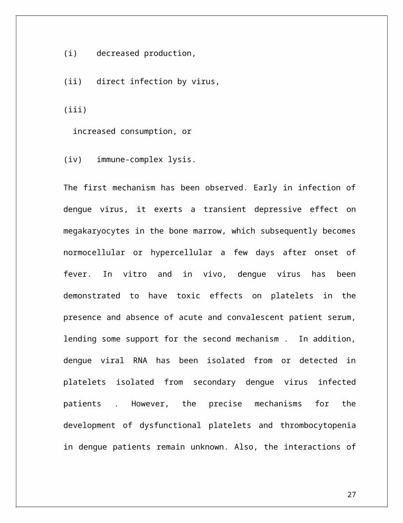
Platelets in Dengue:
One of the important clinical hallmarks in dengue virus infection in patients is platelet
dysfunction, which occurs throughout the acute phase, and/or thrombocytopenia, which
frequently occurs at the defebrile stage, thus this is a subject of interest, especially in
understanding the possible mechanisms leading to the observed phenomena. There are a few
proposed mechanisms that may explain platelet dysfunction and/or thrombocytopenia:
(i) decreased production,
16

(ii) direct infection by virus,
(iii) increased consumption, or
(iv) immune-complex lysis.
The first mechanism has been observed. Early in infection of dengue virus, it exerts a
transient depressive effect on megakaryocytes in the bone marrow, which subsequently
becomes normocellular or hypercellular a few days after onset of fever. In vitro and in vivo,
dengue virus has been demonstrated to have toxic effects on platelets in the presence and
absence of acute and convalescent patient serum, lending some support for the second
mechanism . In addition, dengue viral RNA has been isolated from or detected in platelets
isolated from secondary dengue virus infected patients . However, the precise mechanisms
for the development of dysfunctional platelets and thrombocytopenia in dengue patients
remain unknown. Also, the interactions of dengue virus with platelets, including entry and
possible virus production, have not been investigated.
Platelets may be a critical element in early dengue virus infection, which may partially
account for the dysfunction of platelets. Subsequent systematic investigations, with
biological assays and electron microscopy, reveal that dengue viral RNA, either the positive
stranded genome or negative stranded template, and the presence of mature virus-like
particles, are consistently observed in platelets isolated from dengue confirmed patients
during the acute phase of infection. A micrograph of dengue virus-like particles within
platelets isolated from confirmed dengue patients is depicted (Figure 4). Typical clustering
of dengue virus-like particles surrounded by a vesicle was observed in platelets (Figure
4(a)), and occasionally single or isolated dengue virus-like particles were observed [133].
17

Infrequently, dengue virus-like particle with a fuzzy morphology were observed associated
with or released from platelets (Figure 4(b)). However, we could not rule out the possibility
that these dengue virus-like particles containing platelets are in the category of
megakaryocyte-derived microparticles.
(a) (b)
Figure 4: Dengue virus-like particles in platelets isolated from confirmed dengue
patients. Platelets were isolated from confirmed dengue patients at the acute stage and subjected to electron microscopy.
(a) Dengue virus-like particles were observed inside vesicle compartment (red arrow) and a particle appeared to be on its
way budding out into the vesicle (blue arrow). (b) A single fuzzy virus-like particle was released from platelet (red arrow
head). Red circle indicates the enlarged area. Insert is the platelets.
In addition, immunofluorescent staining of platelets isolated from confirmed dengue patients
reveals that viral antigens can be observed not only in platelets, but also in cells with the similar
morphology as proplatelets, while some dengue viral antigens were observed in presumably the
micromegakaryocytes. This observation is consistent with early reports by Nelson et al., who
originally observed the presence of immature and nonplatelet forming megakaryocytes
18

circulating in dengue patients and by Bhamarapravati and Boonyapaknavik, who noted that
positive staining for dengue viral antigen in human tissues was demonstrated only in the
lymphoid-like cells. Interestingly, the nucleated micromegakaryocytes, which are similar in size
and morphology to lymphocytes, have been well documented. The presence of
micromegakaryocytes, as opposed to megakaryocytes, suggests that production of platelets from
bone marrow increases in response to dysfunctional or low numbers of platelets in the circulation
of acute dengue patients.
Although platelets do not have a nucleus, they possess functional spliceosomes that are able to
process pre-mRNAs into mature mRNA, from which proteins can be translated and processed .
In vitro experiments were set up to investigate the susceptibility of platelets to support dengue
virus production, which may directly contribute to the platelet dysfunction. A low level of
dengue virus production could occur in infected platelets with the peak occurring at 18 hours
post infection (Figure 5), suggesting that dengue virus is capable of replicating in platelets and
dengue viral antigens may be expressed on the surface of platelets. Alternatively, the moderate
viremia changes may result from the transient ability of platelets reproduction in culture
conditions, which may have the capacity of capturing and releasing dengue viruses in later hours.
Perhaps, this may account for the rise of platelet-associated antibodies (PAIgM/IgG) during
acute dengue virus infection and the increased incidence of phagocytosis of platelets from
patients with secondary infections by human macrophages. In addition, administration of
intravenous immunoglobulin, which saturates phagocytosis and impedes antibody production,
lacked efficacy when used to treat severe thrombocytopenic patients with secondary dengue
virus infection . As a whole, these evidences suggest that dengue virus may take a ride and
19

experience ongoing maturation within platelets produced from infected progenitor
megakaryocytes.
Figure 5:
Transient replication of dengue virus in platelets. Platelets were isolated from a healthy donor and experimentally infected
with dengue virus serotype 2 (strain 16681) at an MOI of 0.01. RNA was isolated from supernatants and pellets at
indicated time and subjected to real-time qRT-PCR for dengue viral RNA.
Platelets are anucleate cells that have hemostatic and inflammatory functions and are
composed of a concentrate of megakaryocyte membrane, cytoplasm, granules, and
organelles. Platelets circulate throughout blood vessels during which they monitor the
integrity of the vascular system. All functional platelet responses must be tightly regulated
to ensure that the formation of blood clots is of sufficient size to seal off the damaged area,
20

while not disrupting blood flow to vital organs by causing vessel occlusion. With the
observation that dengue viral antigens are associated with proplateletes or
micromegakaryocytes in blood during acute dengue virus infection , it is likely that a
platelet lineage parental cell, megakaryocytes, may be involved in the production of dengue
virus during acute infection. In addition, platelets contain several key elements related to
dengue virus infection, such as DC-SIGN as well as complement and Fc receptor, which
have been implicated in virus uptake. It is also possible that a unique receptor or coreceptor
is required for viral binding and entry into platelets. However, this particular receptor or
coreceptor may not be evenly distributed or allocated in platelets since platelets are
demarcated from the membrane of megakaryocytes, which may result in heterogeneous
populations of platelets. This heterogeneity of platelet alloantigen referred to as human
platelet alloantigen (HPA) polymorphism in the literature, and how it contributes to dengu
virus infection and dengue disease severity warrants further investigation.
A deadly switch:
In the infectious form of the virus, the envelope protein lays flat on the surface of the virus,
forming a smooth coat with icosahedral symmetry. However when the virus is carried into
the cell and into lysozomes, the acidic environment causes the protein to snap into a
different shape, assembling into trimeric spike, present in the tip of the spike, which has
several hydrophobic amino acid region.. this insert into the lysozomal membrane and cause
the virus membrane to fuse with lysozome. This release the RNA into the cell and the
infection starts.
21

Figure:6
VECTOR:
Figure: 7
Scientific Classification:
Kingdom: Animalia
Phylum: Arthropoda
Class: Insecta
Order: Diptera
Family: Culicidae
Genus: Aedes
22

Subgenus: Stegomyia
Binomial name: Aedes aegypti.
Characteristics of Aedes mosquito:
The yellow fever mosquito, Aedes aegypti is a mosquito that can spread the dengue fever,
Chikungunya and yellow fever viruses, and other diseases. The mosquito can be recognized by
white markings on legs and a marking in the form of a lyre on the thorax. The mosquito
originated in Africa but is now found in tropical and subtropical regions throughout the world.
Breeding place:
Aedes mosquito lay their eggs in clear, clean water and not in contaminated water. As they fly
only a few meters their breeding place is usually in our house itself.
Lifecycle of Dengue Virus:
Under optimal conditions, the egg of an Aedes mosquito can hatch into a larva in less than a day.
The larva then takes about four days to develop in a pupa, from which an adult mosquito will
emerge after two days. Three days after the mosquito has bitten a person and taken in blood, it
will lay eggs, and the cycle begins again.
23

figure: 8
Pathogenesis of Dengue virus:
Dengue viruses are members of the family Flaviviridae genus Flavivirus. They are small
enveloped viruses containing a single-strand RNA genome of positive polarity . Dengue viruses
infect a wide range of human and nonhuman cell types in vitro. Viral replication involves the
following steps:
Attachment to the cell surface
Entry into the cytoplasm
Translation of viral proteins
Replication of the viral RNA genome
Formation of virions (encapsidation)
24

Release from the cell
Binding of dengue virions to cells, which is mediated by the major viral envelope (E)
glycoprotein, is critical for infectivity . The determination of the three-dimensional structures of
the dengue E glycoprotein and the intact virion has facilitated the understanding of this process.
Dengue viruses bind via the E glycoprotein to viral receptors on the cell surface, which may
include heparan sulfate or the lectin DC-SIGN; they can also bind to cell surface
immunoglobulin receptors in the presence of antibodies to the E glycoprotein or membrane
precursor (pre-M) protein, as described further below.
Following fusion of viral and cell membranes in acidified endocytic vesicles, the viral RNA
enters the cytoplasm. The viral proteins are then translated directly from the viral RNA as a
single polyprotein, which is cleaved to yield the three structural and seven nonstructural proteins.
Cleavage of several of the viral proteins requires a functional viral protease encoded in the
nonstructural protein NS3. The nonstructural protein NS5 is the viral RNA-dependent RNA
polymerase, which assembles with several other viral proteins and several host proteins to form
the replication complex. This complex transcribes the viral RNA to produce negative-strand viral
RNA, which serves as the template for the production of the viral genomic RNA.
Incidence of Dengue:
The Incidence of this fever is variable and depends on the geographical region and the density of
mosquito-borne diseases in a region. The worldwide incidence is estimated to be 50 to 100
25

million cases of dengue fever (DF) and several hundred thousand cases of dengue hemorrhagic
fever (DHF) per year.
Management of Dengue:
Supportive care with analgesics, fluid replacement, and bed rest is usually sufficient.
Acetaminophen may be used to treat fever and relieve other symptoms. Aspirin, Nonsteroidal
anti-inflammatory drugs (NSAIDs), and corticosteroids should be avoided. Management of
severe dengue requires careful attention to fluid management and proactive treatment of
hemorrhage.
Single-dose methylprednisolone showed no mortality benefit in the treatment of dengue shock
syndrome in a prospective, randomized, double-blind, placebo-controlled trial. The Novartis
Institute for Tropical Diseases (NITD) in Singapore is carrying out research to find inhibitors of
dengue viral target proteins to reduce the viral load during active infection.
Laboratory Diagnosis:
Mac- ELISA for detection of Dengue IgM.
Serological tests like haemagglutination inhibition (HI) is widely used for the purpose.
Compliment fixation (CF) test is also helpful
Application of PCR technology
Immunofluroscence microscopy
Virus isolation
26
Symptoms consider for study:
Acute fever lasts for 2-7 days
Headach, backache, myalgia
Rash , petechie
Nausea ,vomiting
Thrombocytopenia
Complications :
Dengue fever can result in the following complications:
dengue haemorrhagic fever
dengue shock syndrome
Dengue hemorrhagic fever
Dengue hemorrhagic fever is a potentially fatal complication of dengue that can cause an
enlarged liver and, in severe cases, can lead to shock (a sudden drop in blood pressure). This is
called dengue shock syndrome (see below).
27

Symptoms of dengue hemorrhagic fever are the same as those for dengue, but there are
sometimes also:
tiny spots of blood on the skin
larger patches of blood under the skin
bleeding from your gums and nose
a weak pulse and clammy skin
sweatiness
a tender abdomen (tummy) and body
discomfort (malaise)
loss of appetite
fatigue (tiredness)
sore throat and cough
Four different strains of the dengue virus can cause this complication. If you have previously
been infected with one strain of dengue and are infected again with a different strain of the virus,
this can cause dengue hemorrhagic fever.
Previous immunity (the body’s ability to resist infection) to a different strain of dengue virus
plays a role in this serious complication.
You are also at an increased risk of getting dengue hemorrhagic fever if you are female
28

and under 12 years of age.
The main feature of treatment for dengue haemorrhagic fever is keeping the patient’s fluids at
the right level to prevent dehydration.
Dengue shock syndrome (DSS)
This is a complication of dengue hemorrhagic fever in which the symptoms above can be
accompanied by symptoms of shock.
Symptoms of shock include:
a sudden drop in blood pressure
cold, clammy skin
a weak rapid pulse
dry mouth
irregular breathing
dilated pupils
reduced flow of urine
Mortality rates can be as high as 40% if this serious complication is not treated. If it is treated,
the mortality rate is 1-2%.
29

If you have any symptoms of dengue, dengue hemorrhagic fever or dengue shock syndrome,
seek immediate medical help to prevent the disease progressing.
MAC-ELISA:
Enzyme linked immunosorbent assay(ELISA) uses antibodies or antigen and other different
immunogens. The ELISA technique was conceptualized and devoloped bt two sweidish
scientists, Peter Perlmann(principal investigator) and Eva Engvall at Stockholm University.
Engvall and perlmann publish teir first paper in ELISA in 1971 and demonstrated its quantitative
value using alkaline phosphatase as the enzyme(24,25) .It is very easy and very sensative, can
detect picogram level of sample and relies on monoclonal antibody. The MAC-ELISA test for
dengue diagonosis involves the exposure of IgM antibodies in human serum to antihuman IgM
are previously bound to the solid phase. This test is to aid in the diagonosis of human exposure to
dengue virus. It is not intended to screen blood or blood components, and is for professional
invitro diagonosis use .
MAC-ELISA has become widely used in past few yers. It is a simple ,rapid test that requirs very
little sophisticated,equipment. MAC-ELISA is based on detecting the dengue spcific IgM
antibodies in the test serum by capturing them out of solution using anti human IgM that was
previously bound to the solid phase. If the IgM antibody from patient’s serum is dengue specific,
30

it will bind the dengue derived recombinant antigen that is added in subsiquent step and can be
detected by addition of an enzyme labelled anti dengue antibody, which may be human or
monoclonal antibody. As substrate is added to give a colour reaction.
31

RT-POLYMERASE CHAIN REACTION(RT-PCR)
Rverse transecription polymerase chain reaction (RT-PCR) , a process of “ amplification” ,is
varient of polymerase chain reaction (PCR), a laboratory technique commonly used to generate
many copies of DNA fragmant. In RT-PCR however RNA strand is first reverse transcriptase,
and the resulting cDNA is amplified using traditional or real time PCR. Reverse transcription
PCR is not to be confused with real time PCR, which is also some times abbriviated as RT-PCR.
Reverse transcriptase was discovered by Howard Temin at the University
of Wisconmsin-Madism and independently by Devid Baltimore at 1970 at MIT. Both the group
shared the nobel prize in physiology and medicine.
MATERIALS AND METHODS:
EXP-1 MAC-ELISA
Objective : To detect the IgM antibody is patients sample.
Principle of assay:
IgM antibody(ab) in the patient’s blood are captured by anti-human IgM that are coated on solid
surface. In the next step DEN antigen is added which binds to captured IgM, if the IgM and
antigen are homologous. Unbound antigen is removed during washing step. In the subsiquent
32

step, Biotinylated Flavivirus cross reactive monoclonal antibody (HX-B) is added followed by
Avidin-HRP. Subsequently substrate/ chromogen is added and watched for the devolopment of
colour. The reaction is stopped by 1N H2SO4 . the intensity of colour/optical density is measured
at 450nm. OD reading are directly proportional to the amount of dengue virus specific IgM
antibodies in the sample.
Requirments:
A. Reagents:-
Antihuman IgM coated test strip
Dengue antigen
Biotinylated Flavivirus cross reactive monoclonal antibody (HX-B)
Controls (positive + negative)
Avidin – HRP
Wash buffer concentrated (10x)
TMB substrate (20x)
1N H2SO4
Distilled water
33

B. Material Required:
1. Glass ware: pipettes, cylinders, flasks,testtube for sample dilution,
bread box, tissue paper roll, aluminium foil.
2. Adjustable micro pipettes and tips.
3. ELISA washer/ ELISA reader
4. Incubator/ laminar air flow unit.
Procedure:
1. All reagents were brought to room temparature.
2. Antigen, HX-B and controls were reconstituted using distilled water.
3. Dilute serum sample, were dilute 1:100 with sample dilution buffer.
4. Coated/post coated wells were washed thrice with wash buffer.
5. 50µl of dilute samples were taken to the appropriate wells and 50µl of
reconstituted +ve control and –ve control were added to respective wells.
6. The plate was incubated at 370c for 1 hr in a humid atmosphere.
7. After the incubation, plate was washed five times with wash buffer and was
trapped on a tissue paper to remove the existing moisture.
34

8. 50µl reconstituted HX-Bwas added to each well.
9. Plate was kept in a humid box and incubated at 370c for 1 hr.
10. At the end of incubation, washing and trapping procedure was replaced.
11. The 50µl of avidin- HRP , was added to each well.
12. The plate was kept in a humidified box for incubation at 370c for 30 min.
13. At the end of incubation, the plate was washed for 5 times with wash buffer
and was trapped on a tissue paper.
14. Then 100µl of dilute substrate was added to each well.
15. The plate was incubated in dark at room tempareture and observed for the
devolopment of colour, which usually devolop in 3-7 min.
16. 100µl of 1 N H2SO4 was used to stop the reaction.
17. At 450nm the absorbence was measured within 10 min.
Interpretation of result:
1. If OD value of sample tested exceeds the OD value of negative control by a factor of 4,
the sample should be considered as positive.
2. If the OD of sample tested is less then the negative control by a factor 4, the sample
should be considered as negative.
3. If the OD value of sample tested is more then the OD of negative control by a factor 4 but
less then OD of negative control by a factor 4 . It is connsidered as indeterminate.
35

Observation and results:
Measured mode: Absorbence
Measured weave length: 450 nm
Read mode: normal
Unit: Optical Density (OD)
Raw data:
Sample no. Absorbance at
450 nm
A1 (-ve control) 0.252
B1 0.093
C1 0.118
D1 0.923
E1 0.072
F1 0.215
G1 0.231
H1 0.192
A2 0.122
B2 0.015
C2 0.214
D2 0.203
36

E2 0.224
F2 0.029
G2 0.197
H2 0.042
H6 (+ve control) 1.114
Calculation of results:
Cut off value = Optical density of –ve control X 4
OD of negative control X 4 = 0.063 x 4
Now the samples that have OD = 0.252
Greater then 0.252 indicate +ve results of dengue IgM positive so positive samples are D(0.923).
37

RT –PCR for Dengue Genome Detection:
Purification of viral RNA from serum of patients.
Kit content:-
1. Q 1 Aamp mini spin columns =50
2. Collection tubs(2ml)=200
3. Buffer AVL*=31ml
4. Buffer Aw1*(concentrate)=19ml
5. Buffer Aw2+ (concentrate)=13ml
6. Buffer AVE+=2x3ml
7. Carrier RNA (poly A) = 310µg.
contains chaotropic salt which is an irritant. Not compatible with disinfection reagents which contains bleach.
Safety information:-
When working with chemicals, always wear a suitable lab coat, disposable gloves and protective goggles. For more information please consult the appropriate material safety data.
Caution:-Do not add bleach or acidic sol directly to water containing buffer AVL or buffer Aw1.
Buffer AVL and Aw1 contain guanidine salts, which can form highly reactive compound when combined with bleach. If liq containing these buffers is spilt, clear with suitable laboratory detergent and water. If the split affected area first with laboratory detergent and water and then with 1%(v/v)sodium hypochlorite.
Buffer AVL:-
Contains guanidine thiocyanate: harmful risk and safety phrases.
Buffer AWI:-
Contains guanidine hydrochloride harmful, irritant risk and safety phrases.
Storage of kit
38

Q1 Aamp minis pin columns should be stored dry at room temperature (15˚C-25˚C).Storage at higher temperature should be avoided. All solution should be stored at room temperature unless otherwise stated Q1Aamp mini spin coloums and all buffer and reagents can be stored under these condition until the expiration date on the kit box without showing any reduction in performance.
Lyophilizer carries RNA can be stored at room temperature (12-25c) until the expiration date on the kit box. Carried RNA should be dissolved in buffer AVE, dissolve carrier RNA should be immediately added to buffer AVL. This Solution should be prepared fresh, and is stable at 2˚C-8˚C for up to 48 hours. Buffer AVL carrier RNA develops a precipitate. When stored at 2˚C-8˚C that must be re-dissolved by warming up to 80˚C before use.
Principal:-
Purification of virul RNA for reliable use in amplification technology viral RNA can be purified from plasma(treated with anticoagulants other then heparin),serum and other than cell-free body flied. Samples may be fresh or frozen, but if frozen, should not be thawed more than once. Repeated freeze-thawing of plasma sample may be freshor frozen, should not be thawed more than once. Repeated freeze plasma samples will lead to reduced viral titers and should be avoided for optimal sensitive. Cryoprecipitate accumulate when samples are subjected to repeated freeze-thawing cycles. This may lead to clogging of the Q1Aamp members when using the vacuum protocol.
Procedure:-
The kit combination the selective binding properties of a silica gel-based membrane with the speed of micro spin or vacuum technology and is ideally suited for simultaneous processing of multiple samples. The sample is first tested under highly denaturidge Conditions to inactivate RNA s and to ensure isolation of intact viral RNA. Buffering condition then adjusted to provide optimum binding of RNA to the Q1Aamp membrane and the sample is loaded onto the Q1Aamp mini spin columns. The RNA binds to the membrane and contaminants are efficiently washed away in two steps using two different wash buffers. High quality RNA is eluted in a special RNA s free buffer, ready for direct use or safe storage. The purified RNA is free of protein, nucleases and other membrane guarantees extremely high recovery of peers, intact RNA is just twenty minutes without the use of phenol/chloroform extraction or alcohol precipitation. All buffer are reagents are RNA s free.
Cellular DNA contamination:-
39

Q1Aamp viral RNA mini kits are not designed to separate viral RNA from cellular DNA, and both will purified in parallel if present in the sample to avoid co purification of cellular DNA, the use of cell free body fluids for preparation of viral RNA is recommended. Samples containing cells such as cerebrospinal fluids bone marrow, urine and most swabs should first be filtered or centrifuged for 10 min at 1500xg and the supematent used. If RNA and DNA have been isolated in parallel, the eluted can be DNA s digested using RNA s free DNA s following by heat treatment(15-min,70˚C) to inactive the DNA s.
Lyses:-
The sample is first lysed the highly denaturing conditions provided by buffer AVL to inactive RNA s and to ensure isolated of intact viral RNA. Carries RNA, added to buffer AVL, improves the binding of viral RNA to the Q1Aamp members especially in the case of low-titles samples, and limits possible degradation of the viral RNA due to any residual RNA s activity.
Sample volumes:-
Q1Aamp mini spin columns can bind RNA greater than 200 nucleotides in length. Actual yield will depend on sample size, sample storage and virus title. The procedure is optimized for use with 140 ul sample, but sample up to 280 µl can be used small samples should be adjusted to 140ul with phosphate-buffered saline (PBS) before loading, and samples with a low viral title should be concentrated to 140µl, the amount of lyses buffer and other reagents added to the sample before loading must be increased proportionally, but the amount of buffer AW1 and AW2 used in the wash steps usually do not need to be increase. If the initial sample volis increase application with required multiple loading steps. There is no danger of over loading the Q1Aamps mini will be unaffected.
Carrier RNA:-
Carrier RNA serves two purposes. Firstly it enhances binding of viral nucleic acid to the Q1Aamps mini membrane, especially if there are very few target molecules in the sample.
Secondary:-
The addition of large amount of carrier RNA reduces the chance of viral RNA degradation in the rare events that RNA s molecules escape denaturation by the chaotropic slats and detergents in buffer AVL. If carrier RNA is not added to buffer AVL this may lead to reduce viral RNA recovery.
The amount of lyophilized carries RNA provided is sufficient for the volume of buffer AVL RNA has been adjusted so that the Q1Aamp viral RNA mini kit can be used as a genetic purification system compatible with much different amplification system and is suitable for a wide range of RNA viruses.
40

Different amplification system vary is efficient depending on the total amount of nuclear a present in the reaction. Elutes from this kit contains both viral nucleic acid and carried RNA and of carries RNA will greatly exceed and of viral nucleic acid calculations of how much elute to add to downstream of carries RNA added to obtain the highest levels of sensitively in amplification reaction, it may be necessary to adjust the amount of carries RNA added to buffer.
Addition of internal controls:-
Using the q1amp viral RNA mini protocols is combinations commercially available of an internal control into the purification procedure. Internal control RNA or DNA should be added together with carried RNA to the lysis of buffer. For optimal purification efficiency, internal control molecules should be longer than 200 nucleotides as smaller molecules are not efficiently recovered. Refer to the manufacturer’s instructions in order to determine the optimal concentration. Using concentration other than that recommended may be reducing amplification efficiency.
Spin Procedure:-
The Q1Aaps viral RNA mini purification procedure is carried out in three steps using Q1Aamps mini spin columns in a std. mrcrocentrifuge. Mini spin columns fit into most std microcentrifuge tubes. In the spin protocol, due to the volume of filter, 2ml collection tubes (Provided) are required to support the Q1aamp mini spin column during loading and wash steps. Eluted RNA can be collected in standard 1.5ml microcentrifuge tubes(not provided).These tubes must be RNA s are to avoid degradation of viral RNA by RNA s.
Determination of yield:-
Yields of viral RNA isolated from biological samples are normally less than 1µg and therefore difficulty to determine photo metrically. Quantitative RT-PcR is recommended for determination of view RNA yield.
Determination of viral RNA length:-
The six distribution of viral RNA purified using Q1Aamp spin column can be checked by deraturing agarose gel electrophoresis followed by hybridization with a virus specific labeled probe and autoradiography.
41

Equipments and reagents to be supplied by uses:-
When working with chemicals,always wear a suitable lab coat,disposable gloves, and protective goggles.
Ethanol (39%-100%)
1.5ml microcentrifuge tubes.
Sterile, RNA s-free preventing pipet tips (pipet tips with aerosol barriers for preventing cross contamination are recommended
Micro-certrifuge (With rotor for 1.5 ml and 2 ml tubes)
Important notes:-
All steps of Q1Aamp viral RNA mini protocols should be performed quickly and at room temperature. After collection and centrifugation, Plasma (untreated and treated anticoagulants other then heparin) or serum can be stored at 2˚C-8˚C for up to 6hrs. For long term storage, freezing at -20˚C to 80˚C in a aliquots is recommended. Frozen plasma or serum or samples must not be thawed more than once. Repeated free Zing and thawing leads to denaturation and precipitation of proteins, causing reduced viral titers and subsequently reduced yields of isolated viral RNA. In addition, cryo precipitates formed by freeze-thawing will cause clogging of Q1Aamp membrane. If cryo precipitates are visible, they can be pelleted by briefly centrifuging at 6800 xg for 3 min. The cleared supernatent should be removed, without distributing the pellet, and processed immediately. This step will not reduce viral titer.
The Q1Aamp viral RNA mini procedure isolates all RNA molecules larges than 200 nucleotides. Smaller RNA molecules will not bind quantitatively under the condition used.
Preparation of Reagents:-
Addition of carried RNA to buffer AVL*add 310µl buffer AVE to the tube containing 310ug lyophilized carries to obtain a solution of 1µg/ml. Dissolute the carries RNA thoroughly, divide it into conveniently sized aliquots, and stored it at -20˚C. Do not freeze thaw the aliquots of caries RNA more than 3 times.
42

Check buffer AVL for precipitate, and if necessary incubate at 80˚C until the precipitate is dissolved. Calculate the volume of buffer AVL-carrier RNA mix needed per batch of samples by selecting the number of samples to be simultaneously processed from table,
Table :- for large number of samples, volumes can be calculated using the following sample calculation:
n x 0.56 ml = y ml.
Y ml x 10µl/ml = z µl.
Where:-
n:- numbers of sample to the processed simultaneously.
y:- Calculated volume of buffer AVL.
z:- Volume of carried RNA-buffer AVE to add to buffer AVL.
Gently mix by inverting the tubes 10 times. To avoid foaming, do not vortex.
Table:-1
Volumes of buffer AVL and carried RNA-Buffer AVE mix required for the Q1Aamp viral RNA procedure
No. of Sample Volume of Buffer AVL(ML) Vol. Carried RNA-AVE(µl)
1. 0.56 5.6
2. 1.12 11.2
3. 1.68 16.8
4. 2.24 22.4
5. 2.80 28.0
6. 3.36 33.6
43

7. 3.92 39.2
8. 4.48 44.8
9. 5.04 50.4
10 5.60 56.0
11. 6.16 61.6
12 6.72 67.2
13. 7.28 72.8
14. 7.84 78.4
15. 8.40 84.0
16. 8.96 89.6
17. 9.52 95.2
18. 10.08 100.8
19. 10.64 106.4
20. 11.20 112.0
21. 11.76 117.6
22. 12.32 123.2
23. 12.88 128.8
24. 13.44 134.4
Note:-
The sample pre parathion procedure is optimized for 5.6 ug of carried RNA per sample. If less carried RNA has been shown to be better for amplification system, transfer only the required amount of dissolved carries RNA to the tubes containing of dissolve carries RNA to the tubes containing buffer AVL.(use of less than 5.6 µg carries RNA per sample must be validated for each particular sample type and downstream assay).
Buffer AVL-carries RNA should be preparation fresh and suitable at 2˚C-8˚C for up to 48 hours. This sol develops a precipitate when stored at 2-8 c that must be re dissolved by warming at 20˚C
44

before use. Do not warm buffer AVL-carrier RNA solution more than 6 times. Do not incubate at 80˚C for more than 5 min. Frequent warming and extended incubation will cause degradation of carries RNA and eventually false negative RT-PCR result. This is particularly the case with low titles samples.
Buffer AW1*:-
Buffer Aw1 is supplied as a concentrate. Before using for the first time, add the appropriate amount of ethanol(96-100%) as indicated on the bottle and in table-2. Buffer Aw1 is stable for 1 years when stored closed at noon temperature, but only until the kit expiration date.
Table-2:- Preparation of Buffer AW1
No.of prepas Aw1.concentrate Ethanol Final Volume
50 19ml 25ml 44ml
250 95ml 125ml 220ml
Buffer Aw2:-
Buffer AW2 is supplied as a concentrated. Before using for the first time , add the appropriate amount of ethanol(96-100%) to buffer AW2 concentrate as indicated on the leottle and in the table-3.Buffer Aw2 is stable for 1 year when stored closed at room temperature, but only until the kit expiration date.
No of. prepr Aw2 concentrate Ethanol Final Volume
50 13ml 30ml 43ml
250 66ml 160ml 226ml
Contains sodium azide as a preservative.
45

Handling of Q1Aamp Mini Spin Columns:-
Owing to the sensitivity of nucleic acid amplification, technologies, the following precautions are necessary when handling Q1Aamp mini spin columns to avoid cross contamination then sample preparation.
Carefully apply the sample or solution to the Q1Aamp Mini spin columns. Pipet the sample into the Q1Aamp spine column without wetting the rim of the column.
Change pipet tips between all liquid transfer steps. The use of aero sol barriers is tips os also recommended.
Avoid touching of Q1Aamp membrane with puppet tip. After all pulse-overtaxing steps, briefly centrifuge, 1.5ml micro centrifuge tube to remove
drops from inside of the lid. Where gloves throughout the procedure. In case of contact b/n gloves and sample, change
gloves immediately.
Steps of Spine procedure for purification of viral RNA:- Thing to do before starting:-
Equilibrate sample to room temperature(15˚C-25˚C).Equilibrate Buffer AVE to room temperature for elution.Check the Buffer AW1 and Buffer AW2 have been prepared.Add carries RNA reconstitute in Buffer AVE to buffer AVL.
Procedure:-
1. Pipet 560µl of prepared AVL containing carries RNA into a 1.5ml micro centrifuge tube.[if the sample volume is large then 140µl,increase the amount of Buffer AVl-carrier RNA proportionally(eg. A280 µl sample will required 1120 µl buffer AVL- carries RNA) and use a large tube.
2. Add 140ul plasma, serum, urine, cell-culture supermatent, pr cell free body fluid to the buffer AVL-carries RNA in the micro centrifuge tube. Mix buy pulse-overtaxing for 15S.
To ensure efficient lyses, it is essential that the sample is mixed thoroughly with buffer AVL yield a homogenous so. Frozen samples that have only been thawed once can also be used.
3. Incubate at room temperature (15˚C-25˚C) for 10 min viral particle lysis is complete after lyses for 10 min at room temperature. Long incubation times have no effect on the yield or quality of purified RNA. Potentially infectious agents and RNAse are inactivated in buffer AVL.
46

4. Briefly centrifuge the tube to remove drops from inside of the lid.
5. Add 560ul of ethanol (96-100%) to the sample & mix by pulse overtaxing for 15sec. After mixing briefly centrifuge the tube remove drops from inside the lid. Only ethanol should be used since other alcohols may result in reduced RNA yield purity. Do not use denatured alcohol, which contains other substances such as methanol or methyl ketone. If the sample volume is greater than 140ul, increase the amount of ethanol proportionately (eg: a280 µl) sample will required 1120 ul of ethanol). In order to ensure efficient binding, it is essential that the sample is mixed thoroughly with the ethanol to yield a homogenous sol.
6. Carefully apply 630 ul of sol from step 5 to the Q1Aamp mini spine column ( in a 2ml collection tube) without wetting the rim. Close the cap and centrifuge at 6000 x g(8000rpm) for 1 min. Place the Q1Aamp spin column in to a clean 2ml collection tube and discard the tube containing the filtrate. Close each spine column to avoid the cross contamination during centrifugation is performed at 8000 rpm in order to limit micro centrifuge noise. Centrifugation at full speed will not affect the yield or purity of viral RNA. If the sol has not complete passed through the membrane, centrifuge again at high speed until all of the sol has passed through.
7. Carefully open the Q1Aamp Mini spine column and repeat step 6. If the sample value was greater than 140µl, repeat this step until all of the lysate has been loaded onto the spin column.
8. Carefully open the Q1Aamp mini spine column and add 500 ul of buffer Aw1, close the cap and centrifuge at 8000 rpm for 1 min. Place the Q1Aamp mini spine column in a clean 2 ml collection tube (provided) and discard the tube containing the filtrate. It is not necessary to increase the volume of Buffer Aw1 even if the original sample volume was larger than 140 µl.
9. Carefully open the Q1Aamp mini spine column and add 500µl of buffer AW2. Close the cap and centrifuge at full speed 14,000 rpm for 3 min. Continue directly with step 11 or to eliminate any chance of possible buffer AW2 carry over perform step 10, and then continue with step 11.
47

Note:-
Residual Buffer Aw2 I the eluate may cause problems in downstream applications. Some centrifuge rotors may be vibrate upon deceleration, resulting in flow-through, containing Buffer Aw2, containing the Q1Aamp mini spine column removing the Q1Aamp mini spine column. In this case, the optional step should be performed.
Recommended:- Place the Q1Aamp mini spine column in a 2ml collection tube and discard the old collection tube with the fill rate. Centrifuge at full speed for 1 min.
Place the Q1Aamp mini spin column is a clean 1.5 ml micro centrifuge tube. Discard the old collection tube containing the filtrate. Carefully open the Q1Aamp spin column and add 60µl of buffer AVE equilibrate at room temperature. Close the cap and incubate at room temperature for 1 min centrifuge 8000 rpm at 1 min. A single elution with 60µl of buffer AVE is sufficient to elute at least 90% of the viral RNA from the Q1Aamp mini spine column. Performing a double elution using 2x 40µl of buffer AVE will yield by up to 10% elution with volumes of less than 30µl will lead to reduce yields & will not the final one of RNA in the elute. Viral RNA is stable up to 1 year when stored at 20˚C or-70˚C.
.
Dengue One Tube RT-PcR for Viral Detection:-
Aim:- To amplify the RNA by using single tube RT-PcR.
b. Required equipment:
Refrigerated centrifuge
Micro centrifuge
Vortex mixture
Powder free gloves
Centrifuge tubes
c. Procedure:
48

1. 500µl of serum and 200µl of chloroform was added in 2 ml sterile eppendrof tube.
2. The mixture was centrifuged at 12,000 rpm for 10 min at 4˚C.
3. The supernatant was taken out in a fresh sterile 2 ml eppendrof tube.
4. 500µl of ice cold is isopropanol was added to supernatant.
5. Then kept the eppendorf tube at-20˚C
6. Again the mixture was centrifuged at 12,000rpm for 15min at 4˚C.
7. Now the supernatant was discarded and the pallet was retained.
8. The pallet was washed 2 times with the ice cold 75% alcohol followed by
centrifugation.
9. The pallet was dissolved in DEPC water. Then kept it at 80˚C for storage.
NOTE:- A total of six samples were tested and RNA isolated from the sample were subjected to
RT-PCR for amplification
Primer:-
Primer D1 ( 5′-TCA ATA TGC TGA AAC GCC GAA ACC-3′)
Primer TS1 (5′-CGT CTC AGT GAT CCG GGG-3′)
Primer TS2 (5′-CGC CAC AAG GGC CAT GAA CAG-3′)
Primer TS3 (5′-TAA CAT CAT CAT ACA GAG C-3′)
Primer DEN4 (5′-TGT TGT CTT AAA CAA GAG AGG TC-3′)
Master mix for single reaction was prepared as follows:-
CONTENTS VOLUME
Nuclease free water 28.85 µl
49

10x RT-PCR buffer 5 µl
Primer TS1 1 µl
Primer TS2 1 µl
Primer TS3 1 µl
Primer DEN4 1 µl
dNTPs (10mM) 10.15 µl
RNase out 1 µl
PCR-enzyme mix 1 µl
AMV RT (10u/ µl) 1 µl
5x magic solution 10 µl
Each reaction tube contains 45 µl of master mix +5 µl of isolated RNA.
Six-reaction tubes were prepared for six cycles.
PCR-Cycle:-
TEMPARATURE TIME (min)
55˚C 30min
94˚C 2min
94˚C 30sec (40cycle)
55˚C 1min (40cycle)
72˚C 2min (40cycle)
72˚C 5min
4˚C infinite
50

Now these amplified products were subjected to Agarose Gel Electrophoresis.
AGAROSE GEL ELECTROPHORESIS:-
Aim:- To determine the presence and quantity of DNA.
Primer:- Solidification of the agarose forms in the agarose gels through which RNA fragments
are forced to move when placed in electric current.
Materials:-
Gel apparatus.
Water bath.
Microwave Oven.
Gel-loading buffer.
RNA sample.
Gel Casting Apparatus.
Racks for Eppendorf Tubes.
Agarose (1.5%)
10x TAE.
51

Ethidium Bromide.
Procedure:-
Preparing the Agarose Gel:-
1. 1.0gm agarose was taken & 50ml of the 1x TAE buffer was added to a 250ml flask.
2. This agarose solution was kept in microwave for 1min then swirl and repeated until the
agarose melted completely.
3. Then the solution was allowed to cool down slowly to 50˚C -55˚C by placing the flask in
a 55˚C water bath.
4. 5 µl of ethidium bromide was added slowly into agarose solution gradual swirling the
flask.
5. After that 5ml of warm agarose solution was poured into the gel tray.
6. Before removing the comb the agarose was kept for 30min.
7. Before pulling out the combination 5ml of 1x TAE was added to the surface of the gel.
Electrophoresis of DNA:-
1. On the electrophoresis tank the gel and the platform were placed.
2. 1x TAE buffer was added on to the electrophoresis tank to cover 1mm layer above the gel
surface.
3. In each sample 6 µl of gel loading dye was added and was mixed by gentle flicking.
4. 30 µl of each sample was quickly loaded into six wells, marker in one well and negative
control in one well. Then was started, keeping the lid of the tank closed tightly.
5. The electrophoresis was connected to a power supply of 100 volts for 1hr.
52

6. After that power pack was switched off and the power cords were disconnected.
7. Using gloves, the gel was transferred to an UV transilluminator and then turned on the
transilluminator and the ethidium bromide stained DNA fragment was examined.
Instrument of Agarose gel electrophoresis
Result:-
53

Figure: Agarose gel Electrophoresis Under UV light.
Discussion:-
A total of acute sample could be collected from the cases and screened for the presence for of
Dengue IgM ab by IgM captured ELISA (MAC-ELISA),using a kit (prepared by National
Institute of Virology, Pune, India), following the prescribed protocol. O.D-was measured at
450nm using ELISA reader ( Titestek Multirisk as plus, Lab system, Finland, Type-314) of the
six acute samples, dengue virus specific IgM antibodies were observed only in one sample.
Although all the samples did not tallied with any off our serotypes of Dengue Virus by RT-PCR
method, but only one was positive against Dengue Virus by ELISA method. It is worthy to
maintain that the viremic stage, is very difficult to detect the RNA in those sample conversely
the same sample were drawn on 6th to 7th day from the same individual only one was positive to
Dengue IgM antibody. The IgM antibody comes in the blood from 5th to 6th days of injection &
54

persists for some days in the blood. So the samples which are RT-PCR negative should be
confirmed by Mac-ELISA Method.
Although all the samples did not tallied with any of the four serotypes of Dengue Virus by
RT-PCR method. Out of the six sample tested, only one was positive against dengue virus by
ELISA method. It is worthy to maintain the vermic stage is short and if the samples are not
collected within the viremic stage, it is very difficult to detect the RNA in those samples
conversely the same samples were drawn on the 6 th-7th day from the same individual and one out
of six IgM positive to dengue virus, as the IgM antibody comes in the blood from 5th-6th days of
infection and persists for some days in the blood.
Results:
Total no. of sample collected = 162
Total no of +ve sample= 70
Total no. of female patients = 52
Total no. of male patients = 18
Table 1
Months No. of patients
January nil
February nil
55

March nil
April 3
May 3
June 4
July 9
August 10
September 12
October 14
November 11
December 4
Graphical representation of the result : month vs no of patients:
56

Table 2
Age group No. of patients
0-10 30
11-20 3
21-30 7
31-40 15
41-50 17
50+ 9
Graphical representation of result : Age group vs No of patients
Table 3:
57

Name of district No. of patients
Kolkata 55
Howrah 24
Hooghly 0
N.24.pargaras 0
S.24. parganas 0
Bardwan 0
Nadia 1
Birbhum 0
Bankura 0
Mednapure 0
Murshidabad 0
Malda 0
N.Dinajpur 0
S.dinaj pur 0
Jalpaiguri 0
Koachbihar 0
Darjeeling 0
Purulia 0
58

Graphical representation of result: name of district vs no of patients.
Tabale 4:
Duration of fever No of patients
1-2 days 0
3-4 days 35
5-6 days 20
7-8 days 0
9-10 days 0
11-15 days 5
16-25 days 0
30-45 days 10
60-90 days 0
91-120 days 0
59

Graphical Representation of result: No. of patient vs No of days
Table 5:
Year % Of the dengue cases
2007 20%
2008 33.30%
2009 53.40%
2010 60%
2011 66.60%
60

Graphical representation of thr dengue cases in last 5 years .
Discussion:
After performing the above test and analysing the result it has been found that , the peack time of the outbreak of Dengue is August to September Throughout the year 2011-2012. The female patient,s are more affected by the male , mainly the age group of upto 10 years.The reason of this is , the Dengue vector Aedes aegypti is a paradomestic mosquito and females are generally stay in home and affected by Degue. Throughout the WestBengal kolkata and Howrah mainly dengue cases are found . In nodia also some Denguw cases are found.
References:
1. S. B. Halstead, “Dengue,” The Lancet, vol. 370, no. 9599, pp. 1644–1652, 2007
2. WHO, Prevention and Control of Dengue and Dengue Hemorrhagic Fever, WHO,
61

SEARO, 1999
3. NIAID, Dengue Fever, 2005.
4. G. Clark, D. Gubler, and J. Dengue Fever, CDC Traveler's Information on Dengue Fever,
Centers for Disease Control, Atlanta, Ga, US.
5. S. B. Halstead, “Dengue,” The Lancet, vol. 370, no. 9599, pp. 1644–1652, 2007.
62