fine-tuning thermoresponsive functional poly(ε
TRANSCRIPT

Find more research and scholarship conducted by the School of Natural Sciences and Mathematics here. This document has been made available for free and open access by the Eugene McDermott Library. Contact [email protected] for further information.
School of Natural Sciences and Mathematics 2015-1 Fine-tuning Thermoresponsive Functional Poly(ε-caprolactone)s to Enhance Micelle Stability and Drug Loading
UTD AUTHOR(S): Elizabeth A. Rainbolt, Katherine E. Washington, Suchithra A. Senevirathne, Michael C. Biewer and Mihaela C. Stefan
©2015 The Royal Society of Chemistry. This article may not be further made available or distributed.
Rainbolt, E. A., J. B. Miller, K. E. Washington, S. A. Senevirathne, et al. 2015. "Fine-tuning thermoresponsive functional poly(ε-caprolactone)s to enhance micelle stability and drug loading." Journal of Materials Chemistry B 3(9): 1779-1787.

Journal ofMaterials Chemistry B
PAPER View Article OnlineView Journal | View Issue
Fine-tuning therm
aDepartment of Chemistry, University of Texa
[email protected] of Biochemistry, University o
Simmons Comprehensive Cancer Center, Da
utsouthwestern.edu
† Electronic supplementary informationDOI: 10.1039/c4tb02016b
Cite this: J. Mater. Chem. B, 2015, 3,1779
Received 8th December 2014Accepted 16th January 2015
DOI: 10.1039/c4tb02016b
www.rsc.org/MaterialsB
This journal is © The Royal Society of C
oresponsive functional poly(3-caprolactone)s to enhance micelle stability anddrug loading†
Elizabeth A. Rainbolt,a Jason B. Miller,b Katherine E. Washington,a
Suchithra A. Senevirathne,a Michael C. Biewer,a Daniel J. Siegwart*b
and Mihaela C. Stefan*a
Block copolymers synthesized by the ring-opening polymerization of g-2-[2-(2-methoxyethoxy)ethoxy]
ethoxy-3-caprolactone (ME3CL), g-2-methoxyethoxy-3-caprolactone (ME1CL), and 3-caprolactone (CL)
are reported. Previously, diblock copolymers of PME3CL-b-PME1CL displayed excellent
thermoresponsive tunability (31–43 �C) and self-assembled into micelles with moderate thermodynamic
stability. In this report, two strategies are employed to enhance thermodynamic stability of PME3CL/
PME1CL-type block copolymer micelles while maintaining their attractive thermoresponsive qualities:
modification of the end group position and alteration of hydrophobic block composition by using both
ME1CL and CL. These new thermoresponsive amphiphilic block copolymers showed lower critical
micelle concentration (CMC) values by one order of magnitude and formed thermodynamically stable
micelles. Furthermore they demonstrated good biocompatibility and up to 4.97 wt% doxorubicin loading,
more than double the amount loaded into the PME3CL-type polymeric micelles previously reported.
Introduction
In the development of drug delivery systems, synthetic polymersoffer versatility in terms of biocompatibility, self-assembly, andstimuli-responsive features.1–6 Poly(3-caprolactone)s are ofparticular interest due to their hydrolyzable backbones andarray of properties attainable by attaching functional groupsalong the polymer chain.6–12 Recently, by the ring-openingpolymerization (ROP) of g-2-[2-(2-methoxyethoxy)ethoxy]ethoxy-3-caprolactone (ME3CL), g-2-methoxyethoxy-3-caprolactone(ME1CL), and other g-substituted caprolactone (CL) monomers,amphiphilic block copolymers were synthesized and investi-gated for use as micellar vehicles for drug delivery by ourgroup.3,13–15 PME3CL served as the hydrophilic segment andimparted lower critical solution temperature (LCST) behavior tothe block copolymer in the form of a cloud point, the temper-ature at which a thermoresponsive polymer in water undergoesa coil-to-globular transition. For the hydrophobic segment,alkoxy-, benzyloxy-, and methoxyethoxy- substituted poly(3-caprolactone)s were explored.3,15 By adjusting the hydrophilic/
s at Dallas, Richardson, TX, USA. E-mail:
f Texas Southwestern Medical Center,
llas, TX, USA. E-mail: daniel.siegwart@
(ESI) available: Additional gures. See
hemistry 2015
hydrophobic block ratios and/or the functional groups on thehydrophobic block, the LCST behavior could be tuned in therange of 31–44 �C.15 Due to their combined thermoresponsiveand biodegradable properties, this family of copolymersconstitutes a new direction in synthetic polymeric micelles forcontrolled drug delivery applications.
In a drug carrier application, micelle-forming block copoly-mers with LCST behavior above 37 �C would encapsulate drugmolecules, inltrate the body, and accumulate in solid tumorsby the enhanced permeability and retention (EPR) effect.16
Warming the micelles above the cloud point, by localizedheating or mild hyperthermia would dehydrate the PME3CLshells, thusly deforming the micelles and triggering the releaseof their drug cargoes.14 While the thermoresponsive propertiesof PME3CL-b-PME1CL displayed excellent tunability, their crit-ical micelle concentrations (CMC) were moderate at best, andthus prompted the redesign of the polymer architecture.15
As documented in literature, the positioning of end groupscan inuence substantially the physical properties of polymers,such as self-assembly and LCST.17–22 Typically, to reduce theCMC of an amphiphile—and thereby increase its thermody-namic stability—efforts are focused on lengthening the core-forming segment and/or reducing its hydrophilicity.23 In thecase of the PME3CL-b-PME1CL copolymers, the hydrophobicblock was comprised of methoxyethoxy-functionalized capro-lactone units. Block copolymers featuring ME1CL in thehydrophobic block were posited to enhance micelles' drugloading capacity due to the possibility of hydrogen bonding
J. Mater. Chem. B, 2015, 3, 1779–1787 | 1779

Journal of Materials Chemistry B Paper
View Article Online
with encapsulated anticancer drugs like doxorubicin (DOX).While ME1CL is less hydrophilic than ME3CL, its side units aremore hydrophilic than an alkoxy-substituted caprolactone.However, the effects on drug encapsulation by incorporatingunsubstituted caprolactone within the hydrophobic blockalongside ME1CL had not been reported.
We hypothesized that improved micelle stability wouldresult from self-assembled diblock copolymers in which: (1) theROP initiator was relegated to the hydrophobic chain end, and(2) the core-forming block was a random copolymer of ME1CLand CL. Thus, a series of block copolymers containing CL,ME1CL, and ME3CL were synthesized by ring-opening poly-merization and investigated for use as micellar drug carriers.Their cloud points, CMCs, micelle sizes and stabilities wereexamined. Preliminary biological studies were performed toprobe the polymer cytotoxicity, drug loading, stability in serum-containing media, and micelle uptake by HeLa cells.
Results and discussion
In an effort to improve the micelle stability of PME3CL-b-PME1CL copolymers, unsubstituted 3-caprolactone was incor-porated into the hydrophobic block. It was hypothesized thatdifferent self-assembly behavior might result from a terpolymerin which the hydrophobic block was a random copolymer ofME1CL and CL. First, an experiment was performed to see if in acopolymerization of ME1CL and CL, one monomer would bemore active than the other, and a gradient copolymer would beformed. Briey, equimolar quantities of both ME1CL and CLwere mixed together and subjected to ring-opening polymeri-zation with stannous(II) 2-ethylhexanoate and benzyl alcohol asinitiator. Samples were taken periodically, and shown inFig. S1† are 1H NMR spectra of the raw polymer at time pointsfrom 5 to 60 minutes. Chemical shis of the methylene protonsadjacent to oxygen in the cyclic ME1CL and CL monomers differfrom those of the caprolactone units incorporated into thepolymer. Because those signals do not overlap, the number ofrepeat units of each CL and ME1CL (dH ¼ 4.06 ppm, 4.17 ppm)could be calculated by integration and comparison to the inte-gration of the benzylic protons of the initiator (dH ¼ 5.11 ppm).As shown in Table S1,† the units of ME1CL and CL per chain atthe given time points are comparable.
Block copolymer synthesis
With evidence pointing towards random copolymerization ofME1CL and CL rather than a gradient, four block copolymers(P1–P4) were synthesized with hydrophobic blocks comprised ofrandomly incorporated ME1CL and CL units. As reported in ourprior publication, a block copolymer with 76 mol% ME3CL and24 mol% ME1CL, displayed a cloud point of 38.5 �C.15 Thus asimilar hydrophilic : hydrophobic molar ratio for P1 was tar-geted.15 Briey, ME3CL was subjected to tin-catalyzed ROP andupon its consumption, ME1CL and CL were added simulta-neously to generate polymer P1, as illustrated in Scheme 1.Polymers P2–P4 were prepared by the copolymerization ofME1CL and CL, followed by addition of the ME3CL to generate
1780 | J. Mater. Chem. B, 2015, 3, 1779–1787
the hydrophilic block. For P2–P4, the benzyl group from theinitiator is situated at the hydrophobic chain end, as shown inScheme 1. This change in block copolymer structure from P1 toP2–P4 was hypothesized to facilitate the self-assembly of stablemicelles.
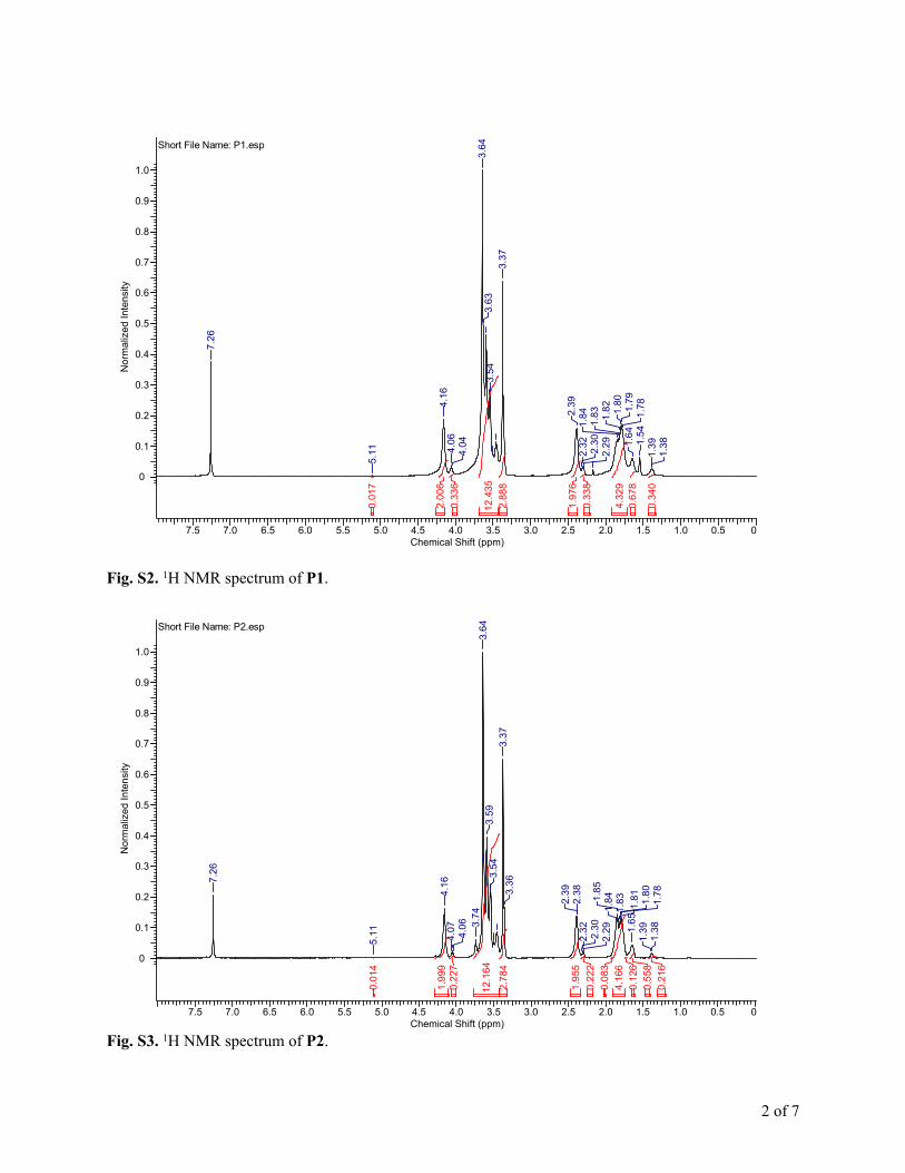
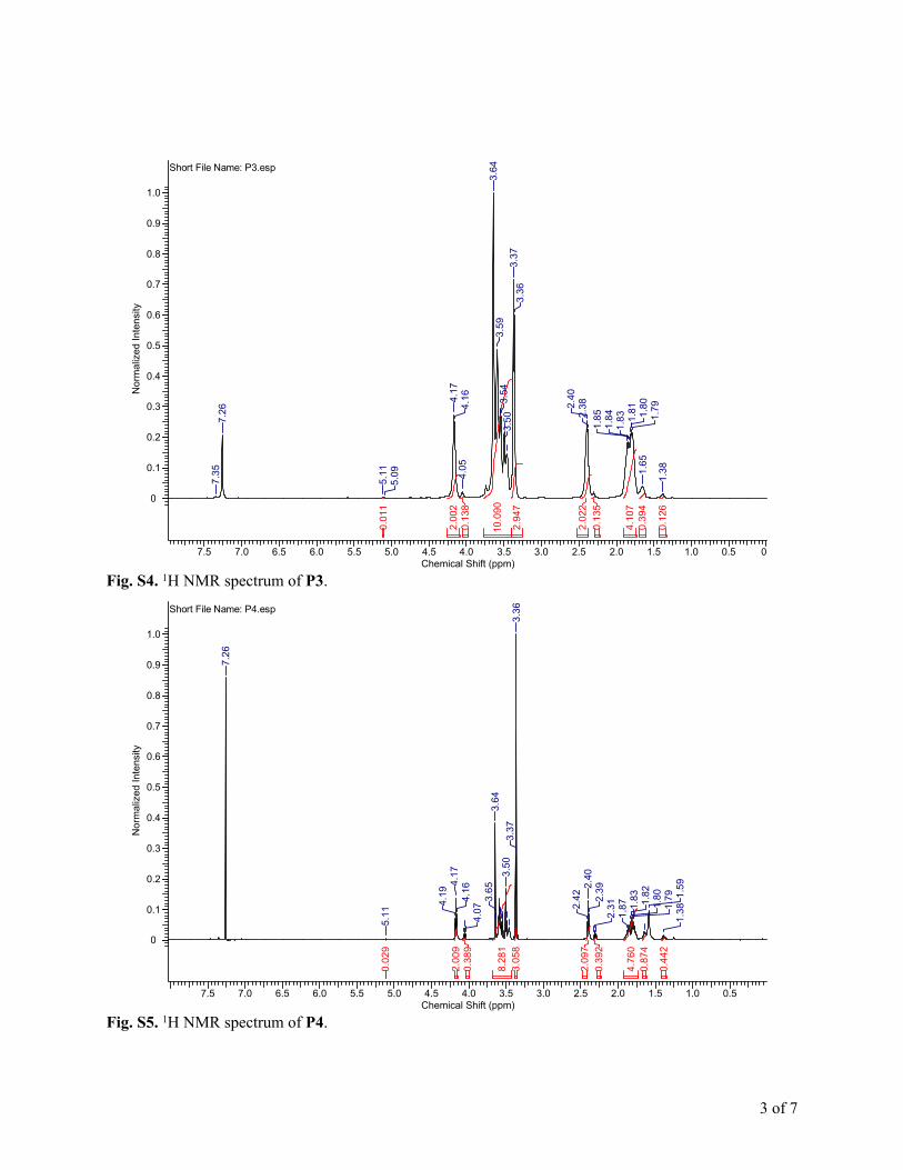
The 1H NMR spectra for P1–P4 are shown in Fig. S2–S5,† andthe polymer compositions are summarized in Table 1. The Mn
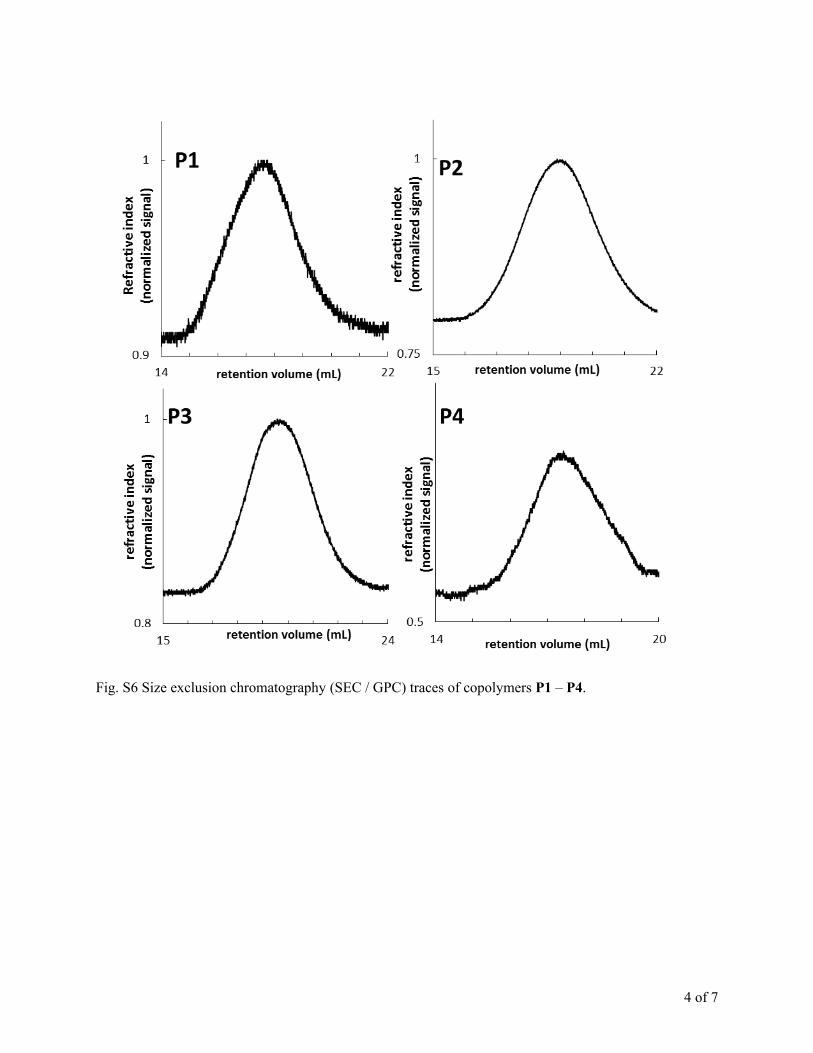
acquired from size exclusion chromatography (SEC) was muchlower than the one calculated by 1H NMR. In SEC, samples areeluted according to hydrodynamic volume, which does notnecessarily correlate with molecular weight. Typically the SEC iscalibrated with poly(styrene) standards. Two polymers ofcomparable molecular weight may have very different hydro-dynamic volumes depending on their makeup, and as a result,SEC has been shown to underestimateMn in some cases.3,24,25 Inthe case of terpolymers P1–P4, the SEC traces show monomodaldistributions, indicating the formation of block copolymersupon the addition of the second portion of monomer(s)(Fig. S6†). A more accurate picture of molecular weight iscalculated from 1H NMR by comparing the integration of thebenzylic protons on the end group to that of protons in therepeat units, as described above for the ME1CL/CL randomcopolymerization.
The terpolymers were expected to degrade over time inphosphate buffered saline (PBS) due to the hydrolysis of theirpolyester backbones. With the highestMn of the terpolymers, P2was selected to demonstrate the terpolymers' degradability insimulated physiological conditions, i.e. 37 �C, pH 7.4 PBS. Asshown in Fig. 1, over the course of ve days in the buffer, themolecular weight decreased by more than 60%.
Self-assembly and thermoresponsive behavior
Critical micelle concentration (CMC) was measured using thehydrophobic uorescent molecule, pyrene, as a probe.13,15 P1self-assembled into micelles in aqueous media above aconcentration of 1.39 � 10�2 mg mL�1 (Fig. 2). The CMC is inline with previously reported values,14,15 however, sinceimproved thermodynamic stability was a primary objective inthis study, P2–P4 were synthesized differently than P1 so thatthe benzyloxy from the initiator would be on the hydrophobicend of the polymer chain. This small change in backbonestructure yielded the desired effect, evidenced in the lower CMCvalue of P2, 4.39� 10�3 mg mL�1 (Fig. 2(e)). As the length of thehydrophobic block increased (Fig. 2(f)), the CMC decreased to1.34 � 10�3 mg mL�1 for P4, indicating improved thermody-namic stability by one order of magnitude over P1. Alsoobserved was reduced CMC when the benzyl group from theinitiator was on the end of the hydrophobic block (as in P2, P3,and P4) rather than the on the hydrophilic end (as in P1).Furthermore, the block terpolymers with hydrophobic blockscomposed of PME1CL-co-PCL (P1–P4, CMC � 10�3 mg mL�1)self-assembled into more stable micelles rather than those withjust PME1CL (CMC � 10�2 mg mL�1).15
Due to the PME3CL hydrophilic block, thermoresponsiveproperties were expected for each terpolymer. P1–P4 all dis-played LCST behavior, with cloud point decreasing as the mol%
This journal is © The Royal Society of Chemistry 2015

Scheme 1 Ring-opening polymerization (ROP) of g-2-[2-(2-methoxyethoxy)ethoxy]ethoxy-3-caprolactone (ME3CL), g-2-methoxyethoxy-3-caprolactone (ME1CL), and 3-caprolactone (CL), using benzyl alcohol initiator (BnOH) and stannous(II) 2-ethylhexanoate catalyst to generateamphiphilic block copolymers P1–P4.
Fig. 1 Demonstration of degradability of P2: decrease in Mn as afunction of time, performed in PBS solution (pH 7.4) at 37 �C over thecourse of 5 days.
Paper Journal of Materials Chemistry B
View Article Online
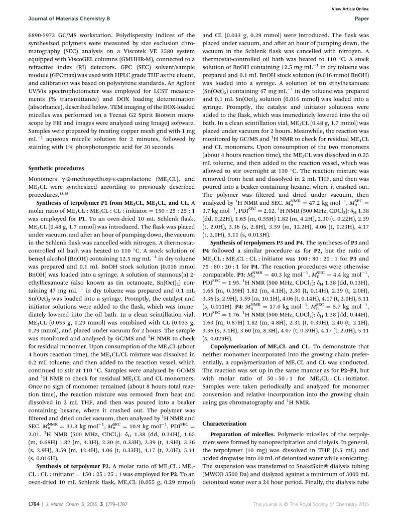
hydrophilic block decreased. This trend is in agreement withthe one observed in the PME3CL-b-PME1CL diblock copolymerspreviously studied.15 The cloud point for P1 with 71 mol%ME3CL was 44.3 �C; however, LCST behavior at lower tempera-ture between 34.3 and 38.5 �C was expected based on thethermal properties of the diblock copolymers with 60–75 mol%ME3CL content.15 Thus, polymers with a hydrophobic blockcomprised of CL and ME1CL have cloud points higher thanpolymers with hydrophobic blocks of ME1CL alone. Regardlessof the CL effect on LCST, P1–P4, with varying hydrophilic tohydrophobic block ratios, displayed LCST behavior between 30–54 �C, shown in Fig. 3(a–d).
The hydrodynamic diameters (Dh) of the micelles formed byP1–P4 ranged from 38–106 nm as estimated by DLS, shown inFig. 3(e�h). The target size for these micelles as drug carriersis in the range of 10–200 nm so as to exploit the EPR effect.26,27
The hydrodynamic diameter of empty micelles prepared fromP1, P2, P3, and P4 were about 55 nm, 39 nm, 80 nm, and 106nm respectively, with micelle dispersity indices of 0.320, 0.363,0.108, and 0.144. While the four sets of micelles closely matchthe targeted 10–200 nm window, the micelles from P3 and P4were substantially more uniform than those of P1 and P2, asindicated by their dispersity values of less than 0.200. Thisfavorably low dispersity of the P3 and P4 micelles was attrib-uted to their reduced CMC and relatively larger hydrophobicblock lengths.
Table 1 Summary of amphiphilic block copolymers P1–P4
PolymerMNMR
na
(kg mol�1)MSEC
nb
(kg mol�1) PDIbME3CL
a
(mol%)ME1CL
a
(mol%)
P1 33.3 10.9 2.01 71 13P2 47.2 7.0 2.12 79 12P3 40.3 4.4 1.95 42 51P4 17.0 5.7 1.76 31 53
a Calculated from 1H NMR, spectra shown in ESI Fig. S2–S5. b Estimatedc Critical micelle concentration (CMC) was measured using pyrene as atransmittance at 600 nm of aqueous polymer solution as a function omeasured by DLS.
This journal is © The Royal Society of Chemistry 2015
Doxorubicin encapsulation
One of the challenges in developing polymeric micellar drugdelivery systems is low drug loading capacity.1,28 Previouslyreported PME3CL-b-poly(g-alkoxy-CL) copolymers achieved 1–2.4 wt% doxorubicin (DOX) loading.3,14 To better encapsulateanticancer drug DOX, P1–P4 were designed with ME1CL units inthe hydrophobic block to provide sites with which DOX maynoncovalently interact. Drug loaded micelles were preparedwith a polymer : drug feed weight ratio of 10 : 1. The encapsu-lation efficiency (wt% EE) and drug loading capacity (wt% LC)were calculated using the equations below.
wt% EE ¼ wt of encapsulated DOX
wt of total DOX added� 100
CLa
(mol%)CMCc
(mg mL�1)LCSTd
(�C)Dh
e
(nm)Micelledispersitye
16 13.87 � 10�3 44.3 55.4 � 1.6 0.3209 4.39 � 10�3 54.2 38.5 � 0.2 0.3637 3.21 � 10�3 40.8 79.8 � 0.6 0.108
16 1.34 � 10�3 29.9 106.1 � 0.5 0.144
by size exclusion chromatography (SEC), traces shown in ESI Fig. S6.probe. d Cloud point was determined by monitoring the change in %f temperature. e Hydrodynamic diameter and micelle dispersity was
J. Mater. Chem. B, 2015, 3, 1779–1787 | 1781
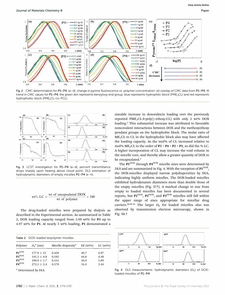
Fig. 2 CMC determination for P1–P4, (a–d), change in pyrene fluorescence vs. polymer concentration; (e) overlay of CMC data from P1–P4; (f)trend in CMC values for P1–P4, the green dot represents benzyloxy end group, blue represents hydrophilic block (PME3CL) and red representshydrophobic block (PME1CL-co-PCL).
Fig. 3 LCST investigation for P1–P4 (a–d), percent transmittancedrops sharply upon heating above cloud point; DLS estimation ofhydrodynamic diameters of empty micelles P1–P4 (e–h).
Journal of Materials Chemistry B Paper
View Article Online
wt% LC ¼ wt of encapsulated DOX
wt of polymer� 100
The drug-loaded micelles were prepared by dialysis asdescribed in the Experimental section. As summarized in Table2, DOX loading capacity ranged from 3.09 wt% for P3 up to4.97 wt% for P1. At nearly 5 wt% loading, P1 demonstrated a
Table 2 DOX-loaded terpolymer micelles
Polymer Dha (nm) Micelle dispersitya EE (wt%) LC (wt%)
P1DOX 177.9 � 15 0.429 49.7 4.97P2DOX 141.3 � 0.8 0.182 44.8 4.48P3DOX 199.9 � 1.7 0.153 30.9 3.09P4DOX 272.3 � 2.4 0.178 34.4 3.44
a Determined by DLS.
1782 | J. Mater. Chem. B, 2015, 3, 1779–1787
sizeable increase in doxorubicin loading over the previouslyreported PME3CL-b-poly(g-ethoxy-CL) with only 2 wt% DOXloading.3 This substantial increase was attributed to favorablenoncovalent interactions between DOX and the methoxyethoxypendant groups on the hydrophobic block. The molar ratio ofME1CL to CL in the hydrophobic block also may have affectedthe loading capacity. As the mol% of CL increased relative tomol% ME1CL in the order of P3 < P4 < P2 < P1, so did the % LC.A higher incorporation of CL may increase the void volume inthe micelle core, and thereby allow a greater quantity of DOX tobe encapsulated.3
The P1DOX through P4DOX micelle sizes were determined byDLS and are summarized in Fig. 4. With the exception of P1DOX,the DOX-micelles displayed narrow polydispersities by DLS,indicating highly uniform micelles. The DOX-loaded micellesexhibited hydrodynamic diameters more than double those ofthe empty micelles (Fig. S7†). A marked change in size fromempty to loaded micelles has been documented in severalreports, but P1DOX, P2DOX, and P3DOX micelles still fall withinthe upper range of sizes appropriate for micellar drugcarriers.26,29–31 The larger Dh for loaded micelles also wasobserved by transmission electron microscopy, shown inFig. S8.†
Fig. 4 DLS measurements: hydrodynamic diameters (Dh) of DOX-loaded micelles of P1–P4.
This journal is © The Royal Society of Chemistry 2015

Fig. 5 Temperature-induced change in size of DOX-loaded micellesplotted as % change in Dh vs. temperature for P1–P4.
Fig. 7 Fluorescence microscopy images of HeLa cells after treatmentwith P3DOX micelles at (a) 20� magnification, and (b) 40� magnifi-cation; DOX shown in red, stained HeLa cell nuclei shown in blue;scale bars represent 100 mm.
Paper Journal of Materials Chemistry B
View Article Online
The thermoresponsive behavior of the DOX-loaded micellesis illustrated in Fig. 5. Plotted is the % increase in Dh as afunction of temperature; each point represents the mean ofthree runs with vertical error bars signifying one standarddeviation. As the suspensions were heated to above the poly-mers' cloud points, the apparent Dh increased markedly. Thiswas attributed to the dehydration of the micelles' PME3CL-based shells and subsequent aggregation of the deformedmicelles and chains.
Investigation of biocompatibility and cell studies
Micelles of P3, displaying LCST behavior closest to but greaterthan 37 �C, were prepared and investigated for cytotoxicity,stability in serum-containing media, and cellular uptake usingHeLa cells. Biocompatibility was examined by standard assay inwhich MTT, a yellow tetrazolium dye, is reduced by mitochon-drial reductase in living cells to generate a purple formazan dye.The absorbance due to formazan in the micelle-treated cells wasnormalized to that of the control cells without micelle treat-ment, and was proportional to the amount of living cells. Theempty micelles demonstrated no inherent cytotoxicity atdosages up to 40 mg mL�1, and only contributed to a smallreduction in cell viability at 80 mg mL�1 (Fig. 6(a)).
PBS and fetal bovine serum (FBS) were used to test micellestability by incubating Nile Red-loaded micelles P3NR in FBS (0
Fig. 6 (a) Biocompatibility of empty polymer micelles P3; cell viabilitydetermined by MTT assay; (b) micelle stability of NR-loaded polymermicelles P3NR in serum-containing media.
This journal is © The Royal Society of Chemistry 2015
to 50 vol% in PBS) and monitoring changes in Nile Red (NR)uorescence, as described in the experimental section. NR is ahydrophobic molecule which most strongly uoresces inhydrophobic environments (e.g. a micelle core).32 Should themicelle disassemble, NR would be exposed to the aqueousenvironment and its emission intensity would drop dramati-cally. For P3NR, no signicant decrease in NR uorescence wasobserved at any concentration of FBS during a 48 hour period,indicating the micelles were not destabilized by the proteinmedia. The data is shown in Fig. 6(b), where NR uorescence isnormalized to the initial intensity at t ¼ 0 hours.
To conrm their potential utility in biomedical applicationsP3DOX micelles were added to human cervical cancer cells(HeLa) and incubated at 37 �C for 4 hours. The growth mediawas changed and the cells were incubated at 37 �C for anadditional 24 hours. Following incubation, the cells werewashed and stained with DAPI so that the cell nuclei could bevisualized by uorescence microscopy, shown in Fig. 7 and S9.†The red DOX signal appeared to be throughout the cell cyto-plasm, which suggested that the P3DOX micelles were internal-ized successfully by the HeLa cells.
ExperimentalMaterials
Nile Red (NR) was purchased from Chem-Impex International,Inc. Doxorubicin hydrochloride (DOX$HCl, 99%) waspurchased from AvaChem Scientic. All other commercialchemicals were purchased from Sigma-Aldrich Co. and wereused without further purication unless otherwise noted.Benzyl alcohol and stannous(II) 2-ethylhexanoate were puriedby vacuum distillation prior to use. All polymerization reactionswere conducted under puried nitrogen. The polymerizationglassware and syringes were dried at 120 �C for at least 24 hoursand then were cooled under nitrogen before use.
Analysis1H NMR spectra of the synthesized monomers and polymerswere recorded on a Bruker AVANCE III 500 MHz NMR instru-ment at 25 �C in CDCl3.
1H NMR data are reported in parts permillion as chemical shi relative to tetramethylsilane (TMS) asthe internal standard. GC/MS was performed on an Agilent
J. Mater. Chem. B, 2015, 3, 1779–1787 | 1783
Journal of Materials Chemistry B Paper
View Article Online
6890-5973 GC/MS workstation. Polydispersity indices of thesynthesized polymers were measured by size exclusion chro-matography (SEC) analysis on a Viscotek VE 3580 systemequipped with ViscoGEL columns (GMHHR-M), connected to arefractive index (RI) detectors. GPC (SEC) solvent/samplemodule (GPCmax) was used with HPLC grade THF as the eluent,and calibration was based on polystyrene standards. An AgilentUV/Vis spectrophotometer was employed for LCST measure-ments (% transmittance) and DOX loading determination(absorbance), described below. TEM imaging of the DOX-loadedmicelles was performed on a Tecnai G2 Spirit Biotwin micro-scope by FEI and images were analyzed using ImageJ soware.Samples were prepared by treating copper mesh grid with 1 mgmL�1 aqueous micelle solution for 2 minutes, followed bystaining with 1% phosphotungstic acid for 30 seconds.
Synthetic procedures
Monomers g-2-methoxyethoxy-3-caprolactone (ME1CL), andME3CL were synthesized according to previously describedprocedures.13,15
Synthesis of terpolymer P1 from ME3CL, ME1CL, and CL. Amolar ratio of ME3CL : ME1CL : CL : initiator ¼ 150 : 25 : 25 : 1was employed for P1. To an oven-dried 10 mL Schlenk ask,ME3CL (0.48 g, 1.7 mmol) was introduced. The ask was placedunder vacuum, and aer an hour of pumping down, the vacuumin the Schlenk ask was cancelled with nitrogen. A thermostat-controlled oil bath was heated to 110 �C. A stock solution ofbenzyl alcohol (BnOH) containing 12.5 mg mL�1 in dry toluenewas prepared and 0.1 mL BnOH stock solution (0.016 mmolBnOH) was loaded into a syringe. A solution of stannous(II) 2-ethylhexanoate (also known as tin octanoate, Sn(Oct)2) con-taining 47 mg mL�1 in dry toluene was prepared and 0.1 mLSn(Oct)2 was loaded into a syringe. Promptly, the catalyst andinitiator solutions were added to the ask, which was imme-diately lowered into the oil bath. In a clean scintillation vial,ME1CL (0.055 g, 0.29 mmol) was combined with CL (0.033 g,0.29 mmol), and placed under vacuum for 2 hours. The samplewas monitored and analyzed by GC/MS and 1H NMR to checkfor residual monomer. Upon consumption of the ME3CL (about4 hours reaction time), the ME1CL/CL mixture was dissolved in0.2 mL toluene, and then added to the reaction vessel, whichcontinued to stir at 110 �C. Samples were analyzed by GC/MSand 1H NMR to check for residual ME1CL and CL monomers.Once no sign of monomer remained (about 8 hours total reac-tion time), the reaction mixture was removed from heat anddissolved in 2 mL THF, and then was poured into a beakercontaining hexane, where it crashed out. The polymer wasltered and dried under vacuum, then analyzed by 1H NMR andSEC. MNMR
n ¼ 33.3 kg mol�1, MSECn ¼ 10.9 kg mol�1, PDISEC ¼
2.01. 1H NMR (500 MHz, CDCl3): dH 1.38 (dd, 0.34H), 1.65(m, 0.68H) 1.82 (m, 4.3H), 2.30 (t, 0.33H), 2.39 (t, 1.9H), 3.36(s, 2.9H), 3.59 (m, 12.4H), 4.06 (t, 0.33H), 4.17 (t, 2.0H), 5.11(s, 0.016H).
Synthesis of terpolymer P2. A molar ratio of ME3CL : ME1-CL : CL : initiator ¼ 150 : 25 : 25 : 1 was employed for P2. To anoven-dried 10 mL Schlenk ask, ME1CL (0.055 g, 0.29 mmol)
1784 | J. Mater. Chem. B, 2015, 3, 1779–1787
and CL (0.033 g, 0.29 mmol) were introduced. The ask wasplaced under vacuum, and aer an hour of pumping down, thevacuum in the Schlenk ask was cancelled with nitrogen. Athermostat-controlled oil bath was heated to 110 �C. A stocksolution of BnOH containing 12.5 mg mL�1 in dry toluene wasprepared and 0.1 mL BnOH stock solution (0.016 mmol BnOH)was loaded into a syringe. A solution of tin ethylhexanoate(Sn(Oct)2) containing 47 mg mL�1 in dry toluene was preparedand 0.1 mL Sn(Oct)2 solution (0.016 mmol) was loaded into asyringe. Promptly, the catalyst and initiator solutions wereadded to the ask, which was immediately lowered into the oilbath. In a clean scintillation vial, ME3CL (0.48 g, 1.7 mmol) wasplaced under vacuum for 2 hours. Meanwhile, the reaction wasmonitored by GC/MS and 1H NMR to check for residual ME1CLand CL monomers. Upon consumption of the two monomers(about 4 hours reaction time), the ME3CL was dissolved in 0.25mL toluene, and then added to the reaction vessel, which wasallowed to stir overnight at 110 �C. The reaction mixture wasremoved from heat and dissolved in 2 mL THF, and then waspoured into a beaker containing hexane, where it crashed out.The polymer was ltered and dried under vacuum, thenanalyzed by 1H NMR and SEC. MNMR
n ¼ 47.2 kg mol�1, MSECn ¼
3.7 kg mol�1, PDISEC ¼ 2.12. 1H NMR (500 MHz, CDCl3): dH 1.38(dd, 0.22H), 1.65 (m, 0.55H) 1.82 (m, 4.2H), 2.30 (t, 0.22H), 2.39(t, 2.0H), 3.36 (s, 2.8H), 3.59 (m, 12.2H), 4.06 (t, 0.23H), 4.17(t, 2.0H), 5.11 (s, 0.013H).
Synthesis of terpolymers P3 and P4. The syntheses of P3 andP4 followed a similar procedure as for P2, but the ratio ofME3CL : ME1CL : CL : initiator was 100 : 80 : 20 : 1 for P3 and75 : 80 : 20 : 1 for P4. The reaction procedures were otherwisecomparable. P3: MNMR
n ¼ 40.3 kg mol�1, MSECn ¼ 4.4 kg mol�1,
PDISEC ¼ 1.95. 1H NMR (500 MHz, CDCl3): dH 1.38 (dd, 0.13H),1.65 (m, 0.39H) 1.82 (m, 4.1H), 2.30 (t, 0.14H), 2.39 (t, 2.0H),3.36 (s, 2.9H), 3.59 (m, 10.1H), 4.06 (t, 0.14H), 4.17 (t, 2.0H), 5.11(s, 0.011H). P4: MNMR
n ¼ 17.0 kg mol�1, MSECn ¼ 5.7 kg mol�1,
PDISEC ¼ 1.76. 1H NMR (500 MHz, CDCl3): dH 1.38 (dd, 0.44H),1.63 (m, 0.87H) 1.82 (m, 4.8H), 2.31 (t, 0.39H), 2.40 (t, 2.1H),3.36 (s, 3.1H), 3.60 (m, 8.3H), 4.07 (t, 0.39H), 4.17 (t, 2.0H), 5.11(s, 0.029H).
Copolymerization of ME1CL and CL. To demonstrate thatneither monomer incorporated into the growing chain prefer-entially, a copolymerization of ME1CL and CL was conducted.The reaction was set up in the same manner as for P2–P4, butwith molar ratio of 50 : 50 : 1 for ME1CL : CL : initiator.Samples were taken periodically and analyzed for monomerconversion and relative incorporation into the growing chainusing gas chromatography and 1H NMR.
Characterization
Preparation of micelles. Polymeric micelles of the terpoly-mers were formed by nanoprecipitation and dialysis. In general,the terpolymer (10 mg) was dissolved in THF (0.5 mL) andadded dropwise into 10 mL of deionized water while sonicating.The suspension was transferred to SnakeSkin® dialysis tubing(MWCO 3500 Da) and dialyzed against a minimum of 3000 mLdeionized water over a 24 hour period. Finally, the dialysis tube
This journal is © The Royal Society of Chemistry 2015

Paper Journal of Materials Chemistry B
View Article Online
contents were ltered through a Nylon syringe lter (0.45 mmpore size), and a 1 mg mL�1 solution of polymeric micelles wasobtained.
Preparation of NR-loaded micelles. NR-loaded micelles wereprepared in a similar fashion as the blank ones. In general, theterpolymer (5 mg) was dissolved in THF (0.15 mL) in a smallvial. A stock solution of NR in THF (5.0 mg mL�1) was prepared,and an aliquot (0.1 mL, 0.5 mg NR) was added to the terpolymersolution. This solution was then added dropwise into 5 mL ofdeionized water while sonicating. The suspension was lteredthrough a Nylon syringe lter (0.45 mm pore size) to removeexcess NR, then was transferred to dialysis tubing (MWCO 3500Da) and dialyzed against a minimum of 1500 mL deionizedwater over a 12 h period. The water was exchanged for phos-phate buffered saline (1500 mL, pH 7.4 PBS) and dialysiscontinued for another 12 h. Finally, the dialysis tube contentswere ltered through a Nylon syringe lter (0.45 mm pore size),and a 1 mg mL�1 solution of NR-loaded micelles was obtained.
Preparation of DOX-loaded micelles. DOX-loaded micelleswere prepared in a similar fashion as the blank ones. First, theDOX$HCl was neutralized with 3 equivalents of triethylamine inTHF : DMSO 5 : 1.14 An aliquot (containing 0.5 mg DOX) of theneutralized DOX solution was added to the terpolymer solution(5 mg in 0.2 mL THF). The DOX–polymer solution was thenadded dropwise into 5 mL of deionized water while sonicating.The suspension was transferred to dialysis tubing (MWCO 3500Da) and dialyzed against a minimum of 1500 mL deionizedwater over a 12 hour period. The water was exchanged forphosphate buffered saline (1500 mL, pH 7.4 PBS 1�) and dial-ysis continued for another 12 hours. Finally, the dialysis tubecontents were ltered through a Nylon syringe lter (0.45 mmpore size), and a 1 mg mL�1 solution of DOX-loaded micelleswas obtained. To determine drug loading capacity (LC) anddrug loading efficiency (LE), 500 mL of the micelle suspensionwas added to 500 mL dimethylsulfoxide (DMSO) and subjectedto bath sonication (�20 min) to release the DOX from themicelles. Aer sonication, the absorbance of the solution at 495nm was tted to a pre-established standard curve of DOX inPBS/DMSO.
Analysis of micelles by dynamic light scattering (DLS).Aqueous suspensions of polymeric micelles were prepared asdescribed above. Prior to measuring, the micelle suspensionswere passed through a 0.45 mm Nylon syringe lter. Themicelles (400 mL sample size) were analyzed to determine theirhydrodynamic diameters using dynamic light scattering with aMalvern Zetasizer Nano ZS instrument equipped with a He–Nelaser (633 nm) and 173� backscatter detector. For some experi-ments, particle size was analyzed as a function of temperatureusing 1 �C intervals with a minimum of 60 seconds equilibra-tion time between measurements.
Investigation of LCST behavior. A solution of 0.3 wt% poly-mer in water was prepared and ltered through a 0.45 mmNylonsyringe lter. The solution was stirred and slowly heated in athermostat-controlled water bath. The change in % trans-mittance at 600 nm versus the temperature of the solution wasrecorded on an Agilent UV/Vis spectrophotometer and plottedin Excel. The temperature at which the % transmittance sharply
This journal is © The Royal Society of Chemistry 2015
drops (halfway between the max %T and min %T) was taken asthe cloud point.
Determination of CMC. The critical micelle concentrationwas determined using the hydrophobic uorescent moleculepyrene as a probe. Samples of polymer of varying concentra-tions were combined with a small amount of pyrene in less than0.1 mL THF. These solutions were added dropwise into 10 mL ofdeionized water in a scintillation vial with a small stir bar. Thesolutions were stirred for a minimum of 3 hours to allow themicelles to assemble as the THF evaporated. The resultingaqueous solutions contained 10�5 to 100 g L�1 of polymer, and aconstant pyrene concentration of 6.67 � 10�5 g L�1. Fluores-cence spectra of the polymer/pyrene solutions were collectedwith a Perkin-Elmer LS 50 BL luminescence spectrometer at 25�C with emission wavelength set at 390 nm. The ratio of theintensities of the pyrene excitation peaks at 338 nm and 335 nmwere recorded and plotted against the log of the polymerconcentration ([c]). The � coordinate at the intersection of thetwo trendlines before and aer the abrupt increase in the I338/I335 vs. log[c] curve was taken to be the critical micelle concen-tration. Spekwin32 soware was utilized in plotting the uo-rescence spectra.
Demonstration of polymer degradability. P2, with the high-est initial Mn, was selected for the biodegradability demon-stration. P2 (10 mg) was dissolved in 2.2 mL of PBS (pH 7.4,DNase-, RNase-, and Protease-Free) and was stirred in a closedcontainer over a thermostatted bath at 37 �C for 5 days. Peri-odically, samples were extracted from the solution and analyzedby SEC to monitor the change in Mn from t ¼ 0 days to t ¼ 5days. The data is plotted as % of initial Mn versus days spent inthe PBS solution at 37 �C.
Micelle stability in FBS. NR-loaded micelles P3NR wereincubated in PBS supplemented with 0%, 5%, 10%, 15%, 20%,and 50% volume fetal bovine serum (FBS). In a biologicalapplication, synthetic micelles could be destabilized by proteinsadsorbing to their surface, thus probing the micelle stability inprotein-containing serum such as FBS is prudent. The uores-cence emission intensity (lex ¼ 550 nm, lem ¼ 632 nm) of theNR-micelles was recorded at desired time points. Plotted are theNR emission intensities, normalized to the initial uorescenceintensity at 0 hours, versus time.
Cell culture
For the biological studies, unless otherwise indicated, HeLacells (donated by Dr David Boothman, UT Southwestern) werecultured in growth medium (phenol red free Dulbecco's Modi-ed Eagle Medium (Hyclone), supplemented with 5% fetalbovine serum, FBS) at 37 �C, 5% CO2, in a humidied atmo-sphere. Also used in cell viability and cellular uptake studieswere phosphate buffered saline (PBS, pH 7.4, DNase-, RNase-,and Protease-Free), nuclear stain DAPI (40,6-diamidino-2-phe-nylindole dihydrochloride), and MTT (methylthiazolyldiphenyl-tetrazolium bromide).
Cell viability (MTT). Dialyzed micelle solutions (1 mg mL�1
in 1� PBS) were serially diluted two-fold to 0.50000 mg mL�1,0.25000 mg mL�1, 0.12500 mg mL�1, 0.06250 mg mL�1, and
J. Mater. Chem. B, 2015, 3, 1779–1787 | 1785

Journal of Materials Chemistry B Paper
View Article Online
0.03125 mg mL�1 respectively. Twenty-four hours prior to theassay, HeLa cells were seeded 96-well tissue culture plate at adensity of 10 000 cells per well, in 100 mL growth medium.Following this initial adhesion period, the medium wasreplaced with 100 mL fresh, pre-warmed growth medium. Themicelle dilutions in 1� PBS (80 mL) were added via pipette wereadded as nal dose (mg indicated) in 80 mL PBS into cellscultured in 96-well plates in 200 mL growth media. The emptymicelles were incubated in cells for 17 hours, then were washedwith PBS for 5 minutes. Aer washing, MTT (methyl-thiazolyldiphenyl-tetrazolium bromide) solution in medium(110 mL total volume, 10 : 1 DMEM complete medium: 5 mgmL�1 MTT in 1� PBS) was added. The cells were incubated for 4hours to precipitate formazan. All but 25 mL of solution wasremoved, and 150 mL of dimethylsulfoxide (DMSO) was added.The absorption at 540 nm was recorded and normalized to theintensity of the untreated cells (N $ 5, � standard deviation).
Uptake of DOX-loaded micelles. HeLa cells were plated on a96-well plate at a density of 10 000 cells per well and cultured in100 mL growth media. Before adding DOX-loaded micelles, theold media was removed and replaced with 120 mL of freshmedia. All micelles were added as nal dose (mg indicated) in 80mL PBS into cells. The cells were incubated at 37 �C for 4 hours,then washed in PBS, and then incubated for another 24 hours.Aer xation (4% PFA for 15 minutes at room temperaturefollowed by washing twice with PBS for 5 minutes PBS and DAPIstaining, 300 nM DAPI in 1� PBS, for 15 minutes at roomtemperature followed by washing twice with PBS for 5 minutes)the cells were imaged on a BioTek Cytation3 Cell Imaging Multi-Mode Reader.
Conclusions
Newly synthesized caprolactone-based PME3CL-b-P(ME1CL-co-CL) terpolymers featured a rare combination of attractiveproperties: thermoresponsive behavior, biodegradable back-bones, and enhanced drug loading capacities. This series ofamphiphilic block copolymers exhibited low critical micelleconcentrations due to the incorporation of unsubstituted cap-rolactone into the hydrophobic block, as well as the reposi-tioning of the benzyl end group from the hydrophilic chain endto the hydrophobic end. The terpolymer micelles were loadedwith up to 4.97 wt% doxorubicin drug, more than double theamount of previously reported PME3CL-type block copolymerswith alkoxy-substituted caprolactone cores. To conclude,PME3CL-b-P(ME1CL-co-CL) block copolymers demonstrate thesynthetic versatility of thermoresponsive PME3CL-based mate-rials and underscore their promising potential in drug carrierapplications.
Acknowledgements
M.C.S. gratefully acknowledges nancial support from theWelch Foundation (AT1740), and National Science Foundation(DMR-0956116 & CHE-1126177). D.J.S. gratefully acknowledgesnancial support from the Welch Foundation (I-1855) andCPRIT (R1212 and RP140110).
1786 | J. Mater. Chem. B, 2015, 3, 1779–1787
Notes and references
1 B. Chertok, M. J. Webber, M. D. Succi and R. Langer, Mol.Pharmaceutics, 2013, 10, 3531–3543.
2 M. Elsabahy and K. L. Wooley, Chem. Soc. Rev., 2012, 41,2545–2561.
3 J. Hao, Y. Cheng, R. J. K. U. Ranatunga, S. Senevirathne,M. C. Biewer, S. O. Nielsen, Q. Wang and M. C. Stefan,Macromolecules, 2013, 46, 4829–4838.
4 D. Roy, W. L. A. Brooks and B. S. Sumerlin, Chem. Soc. Rev.,2013, 42, 7214–7243.
5 H. Seyednejad, A. H. Ghassemi, C. F. van Nostrum,T. Vermonden and W. E. Hennink, J. Controlled Release,2011, 152, 168–176.
6 J. Hao, E. A. Rainbolt, K. Washington, M. C. Biewer andM. C. Stefan, Curr. Org. Chem., 2013, 17, 930–942.
7 Y. Xiao, M. Yuan, J. Zhang, J. Yan and M. Lang, Curr. Top.Med. Chem., 2014, 14, 781–818.
8 L. Chang, L. Deng, W. Wang, Z. Lv, F. Hu, A. Dong andJ. Zhang, Biomacromolecules, 2012, 13, 3301–3310.
9 B. Surnar and M. Jayakannan, Biomacromolecules, 2013, 14,4377–4387.
10 A. Mahmud, S. Patel, O. Molavi, P. Choi, J. Samuel andA. Lavasanifar, Biomacromolecules, 2009, 10, 471–478.
11 K. Yao, J. Wang, W. Zhang, J. S. Lee, C. Wang, F. Chu, X. Heand C. Tang, Biomacromolecules, 2011, 12, 2171–2177.
12 J. S. Katz, K. A. Eisenbrown, E. D. Johnston, N. P. Kamat,J. Rawson, M. J. Therien, J. A. Burdick and D. A. Hammer,So Matter, 2012, 8, 10853–10862.
13 J. Hao, J. Servello, P. Sista, M. C. Biewer and M. C. Stefan, J.Mater. Chem., 2011, 21, 10623–10628.
14 Y. Cheng, J. Hao, L. A. Lee, M. C. Biewer, Q. Wang andM. C. Stefan, Biomacromolecules, 2012, 13, 2163–2173.
15 E. A. Rainbolt, K. E. Washington, M. C. Biewer andM. C. Stefan, J. Mater. Chem. B, 2013, 1, 6532–6537.
16 H. Maeda, Adv. Enzyme Regul., 2001, 41, 189–207.17 M. Nichifor, G. Mocanu and M. C. Stanciu, Carbohydr.
Polym., 2014, 110, 209–218.18 P. Kujawa, F. Segui, S. Shaban, C. Diab, Y. Okada, F. Tanaka
and F. M. Winnik, Macromolecules, 2005, 39, 341–348.19 M. Nakayama and T. Okano, Biomacromolecules, 2005, 6,
2320–2327.20 Y. Xia, N. A. D. Burke and H. D. H. Stover, Macromolecules,
2006, 39, 2275–2283.21 P. J. Roth, F. D. Jochum, F. R. Forst, R. Zentel and P. Theato,
Macromolecules, 2010, 43, 4638–4645.22 T. Mori, Y. Shiota, K. Minagawa andM. Tanaka, J. Polym. Sci.,
Part A: Polym. Chem., 2005, 43, 1007–1013.23 Multifunctional Pharmaceutical Nanocarriers, ed. V. Torchilin,
Springer Science, New York, 2008.24 B. Parrish, R. B. Breitenkamp and T. Emrick, J. Am. Chem.
Soc., 2005, 127, 7404–7410.25 D. Yu, N. Vladimirov and J. M. J. Frechet, Macromolecules,
1999, 32, 5186–5192.26 Z. Ahmad, A. Shah, M. Siddiq and H.-B. Kraatz, RSC Adv.,
2014, 4, 17028–17038.
This journal is © The Royal Society of Chemistry 2015

Paper Journal of Materials Chemistry B
View Article Online
27 Z. L. Tyrrell, Y. Shen and M. Radosz, Prog. Polym. Sci., 2010,35, 1128–1143.
28 S. Kim, Y. Shi, J. Y. Kim, K. Park and J.-X. Cheng, Expert Opin.Drug Delivery, 2010, 7, 49–62.
29 X. Shuai, H. Ai, N. Nasongkla, S. Kim and J. Gao, J. ControlledRelease, 2004, 98, 415–426.
This journal is © The Royal Society of Chemistry 2015
30 Z. Yu, X. ChunSheng, L. MingQiang, D. JianXun,Y. ChenGuang, Z. XiuLi and C. XueSi, Sci. China: Chem.,2014, 57, 624–632.
31 H. Wang, F. Xu, Y. Wang, X. Liu, Q. Jin and J. Ji, Polym.Chem., 2013, 4, 3012–3019.
32 G. R. Castro, B. K. Larson, B. Panilaitis and D. L. Kaplan,Appl. Microbiol. Biotechnol., 2005, 67, 767–770.
J. Mater. Chem. B, 2015, 3, 1779–1787 | 1787

1 of 7
Electronic Supplementary Information, Fine-tuning thermoresponsive functional poly(ε-caprolactone)s to enhance micelle stability and drug loadingElizabeth A. Rainbolt, Jason B. Miller, Katherine E. Washington, Suchithra A. Senevirathne, Michael C. Biewer, Daniel J. Siegwart, and Mihaela C. Stefan
Fig. S1. Copolymerization of ME1CL and CL: 1H NMR spectra at different time points in the reaction revealed the incorporation of ME1CL and CL relative to BnO chain end. Integration of the labeled peaks is shown below in Table S1.
Table S1. Summary of PME1CL-co-PCL
δ (ppm)
Time(min)
5.11(BnO)
4.17(ME1CL)
4.06(CL)
5 1.0 2.7 3.6
20 1.0 15.9 15.5
30 1.0 25.5 25.5
60 1.0 33.9 35.7
OO
OH
O
O
x yO
O
4.17 ppm4.06 ppm5.11 ppm
Short File Name: PMECL-co-PCL.esp
7.5 7.0 6.5 6.0 5.5 5.0 4.5 4.0 3.5 3.0 2.5 2.0 1.5 1.0 0.5Chemical Shift (ppm)
0
0.5
1.0
1.5
2.0
Nor
mal
ized
Inte
nsity
4.05
4.17
5.11
5 min
20 min
30 min
60 min
Toluene, O O
O O
O OHO
Sn(Oct)2
Electronic Supplementary Material (ESI) for Journal of Materials Chemistry B.This journal is © The Royal Society of Chemistry 2015
2 of 7
Fig. S2. 1H NMR spectrum of P1.
Fig. S3. 1H NMR spectrum of P2.
Short File Name: P1.esp
7.5 7.0 6.5 6.0 5.5 5.0 4.5 4.0 3.5 3.0 2.5 2.0 1.5 1.0 0.5 0Chemical Shift (ppm)
0
0.1
0.2
0.3
0.4
0.5
0.6
0.7
0.8
0.9
1.0
Nor
mal
ized
Inte
nsity
0.34
0
0.67
84.
329
0.33
81.
976
2.88
812
.435
0.33
62.
006
0.01
7
7.26
5.11
4.16
4.06
4.04
3.64
3.63
3.54
3.37
2.39
2.32 2.30
2.29
1.84
1.83 1.
82 1.80 1.79
1.78
1.64
1.54
1.39
1.38
Short File Name: P2.esp
7.5 7.0 6.5 6.0 5.5 5.0 4.5 4.0 3.5 3.0 2.5 2.0 1.5 1.0 0.5 0Chemical Shift (ppm)
0
0.1
0.2
0.3
0.4
0.5
0.6
0.7
0.8
0.9
1.0
Nor
mal
ized
Inte
nsity
0.21
60.
558
0.12
64.
166
0.08
30.
222
1.95
5
2.78
4
12.1
64
0.22
71.
999
0.01
4
7.26
5.11
4.16
4.07 4.06 3.
743.
643.
593.
543.
373.
36
2.39
2.38
2.32 2.30
2.29
1.85
1.84
1.83 1.81 1.80
1.78
1.65
1.39
1.38
3 of 7
Fig. S4. 1H NMR spectrum of P3.
Fig. S5. 1H NMR spectrum of P4.
Short File Name: P3.esp
7.5 7.0 6.5 6.0 5.5 5.0 4.5 4.0 3.5 3.0 2.5 2.0 1.5 1.0 0.5 0Chemical Shift (ppm)
0
0.1
0.2
0.3
0.4
0.5
0.6
0.7
0.8
0.9
1.0
Nor
mal
ized
Inte
nsity
0.12
6
0.39
44.
107
0.13
52.
022
2.94
7
10.0
90
0.13
82.
002
0.01
1
7.35
7.26
5.11
5.09
4.17
4.16
4.05
3.64
3.59
3.54
3.50
3.37
3.36
2.40
2.38
1.85
1.84
1.83 1.
81 1.80
1.79
1.65
1.38
Short File Name: P4.esp
7.5 7.0 6.5 6.0 5.5 5.0 4.5 4.0 3.5 3.0 2.5 2.0 1.5 1.0 0.5Chemical Shift (ppm)
0
0.1
0.2
0.3
0.4
0.5
0.6
0.7
0.8
0.9
1.0
Nor
mal
ized
Inte
nsity
0.44
2
0.87
44.
760
0.39
22.
097
3.05
88.
281
0.38
92.
009
0.02
9
7.26
5.11
4.19
4.17
4.16
4.07
3.65
3.64
3.50
3.37
3.36
2.42
2.40
2.39
2.31
1.87 1.
83 1.82
1.80
1.79 1.
591.
38
4 of 7
Fig. S6 Size exclusion chromatography (SEC / GPC) traces of copolymers P1 – P4.

5 of 7
Fig. S7. DLS measurements: hydrodynamic diameters (Dh) of empty (blue, solid) and DOX-loaded (red, dashed) micelles of P1 – P4.

6 of 7
Fig. S8. TEM images of P1 and P3 empty micelles (left); and P1DOX and P3DOX (right); micelles deposited on copper mesh grid and stained with phosphotungstic acid; scale bars 200 nm.
EMPTY DOX
P1
P3

7 of 7
Fig. S9. Digital fluorescence microscopy showing the uptake of DOX-loaded micelles P3DOX: left, DOX shown in red; center, DAPI-stained cell nuclei in blue; right, overlay of red and blue channels; scale bars signify 100 μm.
20x
40x