first french-israeli meeting on nanotechnology dedicated ... 20_11.pdf · 7) joseph kost,...
TRANSCRIPT

- 1 -
First French-Israeli Meeting on Nanotechnology
dedicated to Life Sciences
Dan Panorama Hotel, 20-21 November 2011, Tel-Aviv Kaufman Street 10, Tel-Aviv
http://www.danhotels.com/Hotel-Tel-Aviv
- 2 -
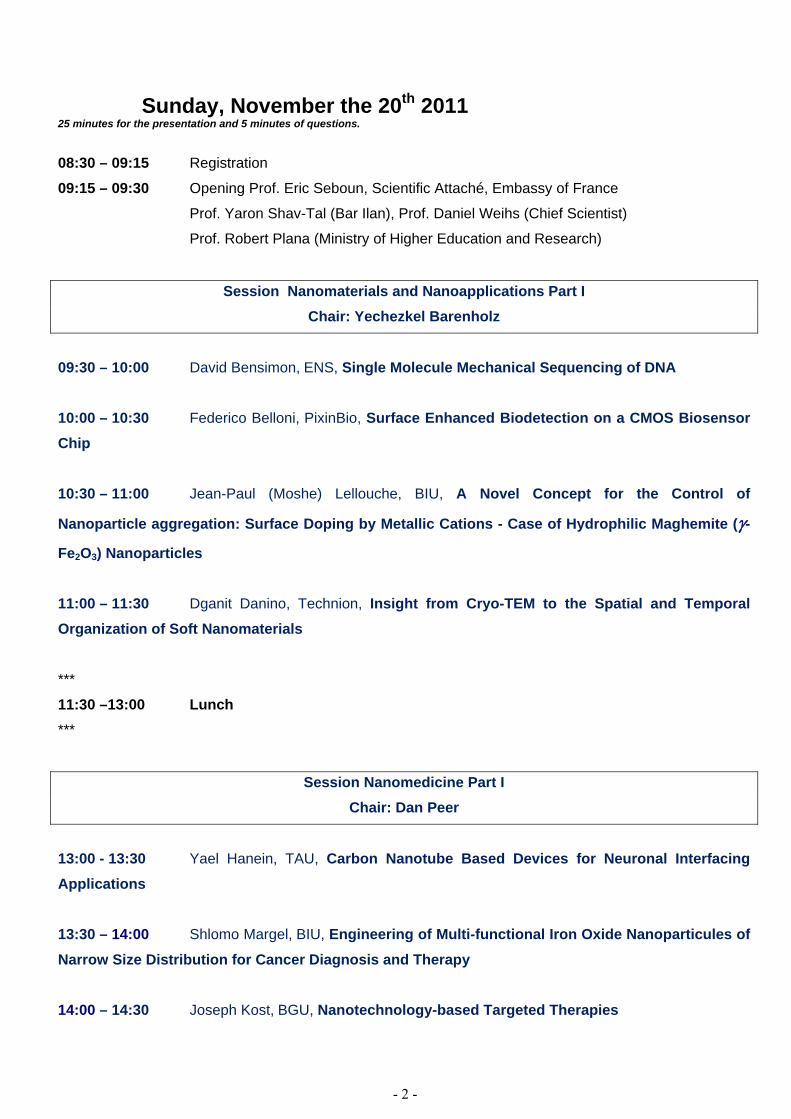
Sunday, November the 20th 2011 25 minutes for the presentation and 5 minutes of questions.
08:30 – 09:15 Registration
09:15 – 09:30 Opening Prof. Eric Seboun, Scientific Attaché, Embassy of France
Prof. Yaron Shav-Tal (Bar Ilan), Prof. Daniel Weihs (Chief Scientist)
Prof. Robert Plana (Ministry of Higher Education and Research)
Session Nanomaterials and Nanoapplications Part I Chair: Yechezkel Barenholz
09:30 – 10:00 David Bensimon, ENS, Single Molecule Mechanical Sequencing of DNA
10:00 – 10:30 Federico Belloni, PixinBio, Surface Enhanced Biodetection on a CMOS Biosensor Chip
10:30 – 11:00 Jean-Paul (Moshe) Lellouche, BIU, A Novel Concept for the Control of
Nanoparticle aggregation: Surface Doping by Metallic Cations - Case of Hydrophilic Maghemite (γ-
Fe2O3) Nanoparticles
11:00 – 11:30 Dganit Danino, Technion, Insight from Cryo-TEM to the Spatial and Temporal Organization of Soft Nanomaterials
***
11:30 –13:00 Lunch ***
Session Nanomedicine Part I Chair: Dan Peer
13:00 - 13:30 Yael Hanein, TAU, Carbon Nanotube Based Devices for Neuronal Interfacing Applications
13:30 – 14:00 Shlomo Margel, BIU, Engineering of Multi-functional Iron Oxide Nanoparticules of
Narrow Size Distribution for Cancer Diagnosis and Therapy
14:00 – 14:30 Joseph Kost, BGU, Nanotechnology-based Targeted Therapies

- 3 -
14:30 – 15:00 Yechezkel Barenholz, HUJI, Liposomes: From Lipid Biophysics and a Membrane Model to Nanotechnology, NanoBio and Approved Nano-drugs
***
15:00 - 15:30 Coffee break ***
Session Nanophotonics Part I
Chair: Shlomo Margel
15:30 – 16:00 Amit Meller, Technion, The art of Nanopore analysis - Threading individual biomolecules through nanoscale pores
16:00 – 16:30 Ron Naaman, Weizmann, Spin Specific Properties of Chiral Molecules
16:30 – 17:00 Laurence Salomé, CNRS, High-throughput Analysis of Single DNA Molecules by Tethered Particle Motion

- 4 -
Monday, November the 21st 2011 25 minutes for the presentation and 5 minutes of questions.
08:30 – 09:00 Registration
Session Nanophotonics Part II Chair: Christian Bergaud
09:00 – 09:30 Xavier Darzacq, INSERM/ENS/CNRS, Multiple Approaches to Study the
Regulation of Nuclear Factors Mobility
09:30 – 10:00 Ulrich Bockelman, CNRS, THRCA: a Versatile New Technique for the Preparation of Periodic DNA
10:00 – 10:30 Christophe Demaille, CNRS, Probing the Distribution and Conformational Dynamics of Surface-Immobilized Redox-Tagged Macromolecules using Electrochemical-Atomic Force Microscopy (AFM-SECM)
10:30 – 11:00 Jérôme Bibette, CNRS, Microorganism Growth Variability in Droplets
***
11:00 – 11:30 Coffee break ***
Session Nanomedicine Part II Chair: Yaron Shav-Tal
11:30 – 12:00 Gershon Golomb, HUJI, Modulation of the Innate Immunity by Nanoparticles for Anti-inflammatory Therapy 12:00 – 12:30 Tal Dvir, TAU, Nanotechnological Strategies for Mending Broken Hearts 12:30 – 13:00 Ehud Gazit, TAU, Molecular Self-Assembly of Short Proteins and Peptides: From Biology and Biochemistry to Nanotechnology and Material Science
***
13:00 – 14:30 Lunch ***

- 5 -
Session Nanomaterials and Nanoapplications Part II Chair: David Bensimon
14:30 – 15:00 Lluis M. Mir, CNRS, Biomedical Applications of Cell Electroporation: towards the Use of Nanosecond Pulsed Electric Fields (nsPEF). Development of a Microfluidic Chamber to Apply nsPEF and to Visualize their Biological Effects. 15:00 – 15:30 Ido Bachelet, BIU, Molecular Robots Emulating a Biological Digital Computer in a Live Insect 15:30 – 16:00 Christian Bergaud, CNRS, Nanowire-based Devices for Chemical and Biological
Applications
***
16:00 – 16:30 Coffee break ***
Session continued
16:30 – 17:00 Loic Auvray, CNRS, Transport of Proteins and Macromolecules through Natural
and Solid-state Nanopores
17:00 – 17:30 Simon Benita, HUJI, A Novel Oral Nanotransporter System for enhanced Intestinal Absorption of P-gp and CYP3A4 Substrate Drugs

- 6 -
Abstracts
1) David Bensimon, Single Molecule Mechanical Sequencing of DNA. D.Fangyuan, JF. Allemand, D. Bensimon and V.Croquette Laboratoire de Physique Statistique, ENS, 24 rue Lhomond, Paris 75005
We have developed a novel method for DNA sequencing based on the mechanical opening and closing of DNA hairpins and the detection of roadblocks to rehybridization due to the presence in solution of complementary fragments. Using a magnetic trap to pull on DNA tethered small magnetic beads, the position of the roadblocks on single molecules can be detected with nanometer precision which is sufficient to sequence DNA by hybridization or by ligation. The technique is simple, PCR-free, single molecule with low error rate, low cost (no fluorescently labeled nucleotides) and potentially high throughput.
2) Federico Belloni, Surface Enhanced Biodetection on a CMOS Biosensor Chip Sylvain Contié, Florence Vicaire, Roisin Owens and Hervé Rigneault PIXinBIO
We present the development of a low-cost, simple, portable device based on CMOS photodiodes technology for the detection and quantification of biological targets through light detection, presenting high sensitivity, multiplex ability, and fast data processing. The key feature of our approach is to perform the analytical test directly on the CMOS sensor surface improving dramatically the optical detection due to optical confinement of the molecule emitted light into the high refractive index semiconductor CMOS material. We compared the developed device with a commercial spectrophotometer and found similar intrinsic sensitivities in the fM range. Based on adequate surface chemistry modifications, we performed proof-of-concept bio-assays directed against typical targets such as inflammatory cytokines (TNF�, IFN�). Due to its simplicity, this promising type of device could be of great interest for remote testing and, through its versatility, may evolve towards a challenging point-of-care device for clinical applications.
3) Jean-Paul Lellouche, A Novel Concept for the Control of Nanoparticle aggregation:Surface Doping by Metallic Cations - Case of Hydrophilic Maghemite(γ-Fe2O3) Nanoparticles
Department of Chemistry, Nanomaterial Research Center, Institute of Nanotechnology & Advanced Materials, Bar-Ilan University, Ramat Gan 52900, Israel
Iron oxide-based nanoparticles (NPs) and/or nanocomposites found quite diverse and numerous applications in magnetism-driven separation of cells, magnetic field-guided drug- and gene-delivery (magnetic drug targeting/gene therapy), detoxification of undesirable toxic species, relaxation and contrast enhancement in non-invasive magnetic resonance imaging (MRI) of tissues, piezoelectric immuno-sensors, and magnetic fluid hyperthermia for cancer therapy. Amongst various well-known technological bottlenecks of nanoparticle/nanocomposite fabrication methods, the detrimental aggregation phenomenon is one of the most difficult to address. Our recent work in the field led to the discovery and successful implementation of a novel method/concept for the aggregation control of hydrophilic magnetically responsive maghemite (γ-Fe2O3) nanoparticles.
In contrast to any process described till now, this novel method does not make use of any passivating organic species such as surface-interacting polymers or ligands. Indeed, we demonstrated that the ultrasound-assisted metal Ce(III) cation doping of the surface of 45/50 nm-sized maghemite nanoparticles strongly modified their surface charge to highly positive values. This doping process enabled (i) a full charge-control of particle aggregation due to charge repulsive effects, as well as (ii) their water-compatibility for biological applications.
Deep characterization investigations (FT-IR, high resolution TEM with compositional EDAX analysis, DLS/�potential, Mössbauer spectroscopy) have been conducted in order to identify the influential parameters of this new fabrication/”inorganic” stabilization method of maghemite

- 7 -
nanoparticles. A plausible mechanism that will explain this innovative key metal cation-based NP surface doping will be proposed and discussed.
In addition, this new nanosizedmagnetic support has been tested for cell toxicity (HeLa, HEK 293, and MEF 3T3 cell lines), and for DNA covalent attachment/hybridization, including DNA detection using a blue-coloured HRP-based enzymatic amplifying system. Moreover, preliminary results that investigated the formation of metal cation-doped maghemite nanoparticle/RNA polycationic/polyanionic complexes for gene silencing will be also presented as a first step towards siRNA/microRNA delivery.
4) Dganit Danino, Insight from Cryo-TEM to the Spatial and Temporal Organization of Soft Nanomaterials
Department of Biotechnology and Food Engineering, Technion, Israel
Cryogenic- EM havelong been recognized as instrumental methods for studying the nanostructure of soft materials(e.g., surfactants, lipids, peptides and proteins) at high resolution. These methodsinvolve ultra-rapid cooling of liquid suspensionsand creation of amorphous, vitrified specimens,whichcaptures the native state of structured liquids thereby allowing to get directly detailed structural information at the nanoscale, as well as to explore self-organization mechanisms and dynamics. Several recent advances in themain cryo-EM techniques,and their application to nanoscience and nanotechnology will be presented, including recent work onpeptide nanotubes [1, 2], DNA/lipid systems for gene therapy [3],and lipid and protein nanovehicles for oral delivery [4,5]. References: Zisermanet al., Unraveling the mechanism of nanotube formation by chiral self-assembly of amphiphiles. J Am Chem Soc133(8), 2511-2517 (2011). Ziserman et al., Curvature instability in chiral amphiphile self-assembly. Phys Rev Lett 106, 238105 (2011). Danino et al., A lamellar to hexagonal phase transition in DNA/lipid complexes induced by osmotic pressure. Biophys J96(7), L43-45 (2009). Efrat et al., Solubilization of hydrophobic guest molecules in the monoolein discontinuous cubic mesophase and its soft nanoparticles. Langmuir25(3), 1316-1326 (2009). Bachar et al., A novel oral drug nanocarrier based on self-assembled beta-casein micelles. (Submitted).
5) Yael Hanein, Carbon Nanotube based Devices for Neuronal Interfacing Applications School of Electrical Engineering, Tel-Aviv University
Electronic devices for retinal and brain implant are currently being developed by several research teams. The feasibility to create such devices rests in the ability to produce proper interfacing between the chip and the biological system. Extensive research, conducted over the last several years, demonstrated that Nanotechnology can help making better bio-materials for effective interfacing between nerve cells and electronic chips. Using carbon nanotubes, we have been able to produce highly effectiveneuro micro-electrodes suited for high efficacy recording and stimulation. Using dissociated retinas we were able to show that carbon nanotube electrodes can record neuronal activity with signal to noise ratio as high as 75 and to achieve stimulation at 1 nC charge injection. Through innovative nano and micro-fabrication methods, we have also developed a flexible, Polydimethylsiloxane based system with carbon nanotube micro-electrodes. This system exhibit excellent electrochemistry and is ideally suited for neuro-prosthetic applications. Recent efforts to realize photo-sensitive electrodes based on carbon nanotubes will be also presented.
6) Shlomo Margel, Engineering of Multi-functional Iron Oxide Nanoparticules of Narrow Size Distribution for Cancer Diagnosis and Therapy
Bar-Ilan University, Dept. of Chemistry,Center for Nanotechnology, Ramat Gan 52900, Israel.
A variety of particles of very narrow size distribution (3 nm up to tens of microns), surface composition and properties (porosity, surface area and surface morphology) have been prepared and characterized during the last decade in our laboratory. These include organic and inorganic solid particles, hollow particles, surface supported particles, biodegradable particles, chiral particles, self-assembled particles, iodinated particles and magnetic particles. These various nanoparticles offer unique solutions to

- 8 -
a variety of basic and applied problems in materials science and biotechnology. The functional groups of the particles have been used for covalent or physical binding of ligands such as drugs, proteins, enzymes, antigens, and antibodies to the particles surfaces. The designed particles were used for applications such as imaging, targeting, therapy, specific cell labeling and cell separation, diagnostics, specific blood filtration by hemoperfusion, heavy metal ions detoxification and various bio-chemical reactions.
The present lecture will focus on the synthesis of magneticmaghemite (γ-Fe2O3) nanoparticles of narrow size distribution , prepared by nucleation and then controlled growth of thin iron oxide films on gelatine/iron oxide nuclei. Fluorescent maghemite nanoparticles were prepared similarly, substituting the gelatin for gelatine covalently bound with the fluorescent dye. The efficient use of these iron oxide nanoparticles for fluorescent and MRI imaging and for brain and colon cancer diagnosis and therapy will be demonstrated.
7) Joseph Kost, Nanotechnology-based Targeted Therapies Department of Chemical Engineering, Ben-Gurion University of the Negev, Beer-Sheva 84105, Israel
Nanotechnology has the potential to revolutionize cancer and other therapies. Advances in protein
engineering and materials science have contributed to novel nanoscale targeting approaches that may bring new hope to patients. Several nanocarriers have been approved for clinical use. However, to date, there are only a few clinically approved nanocarriers with drugs to selectively bind and target cancer cells. Nanoparticles used as drug delivery carriers consist of different biodegradable materials such as natural or synthetic polymers, lipids, or metals. Nanoparticles are taken up by cells more efficiently than larger micromolecules and therefore, could be used as effective delivery systems. For therapeutic applications, drugs can either be integrated in the matrix of the particle or attached to the particle surface. In the presentation the drug delivery aspects of nanomedicine, the molecular mechanisms underlying the interactions of nanoparticles with cell-surface receptors, biological responses and ultrasound as a targeting tool and its effect on cellular transport would be discussed.
8) Yechezkel Barenholz, Liposomes: From Lipid Biophysics and a Membrane Model to Nanotechnology, NanoBio and Approved Nano-drugs
The Laboratory of Membrane and Liposome Research, IMRIC, The Hebrew University – Hadassah Medical School, Jerusalem, Israel
DOXIL™ (doxorubicin in liposomes, also referred to in Europe as CaelyxTM) is an anticancer drug which is the first nano-liposomes based drug and the first nano-drug approved by the US FDA (November 1995), Liposomes were described first by Dr. A.D. Bangham in 1965. Then they served as the main model for biological membrane. Five years later, due to their biocompatibility and versatility, they were proposed as a promising drug delivery system. However, due to major scientific and technical obstacles, it took the liposomes 20 more years to mature into approved drug products. Major research efforts made it obvious that nano-liposomes have unique potential as drug carriers to treat major diseases with inflammatory components (i.e. rheumatoid arthritis and multiple sclerosis) and cancer. The advantages of being at the nano range are related to the microanatomy of blood vessels in most cancers and inflamed tissues which are much more permeable than vascular system in most healthy tissues (EPR effect) and to the unique physico-chemical properties related to the nano scale. However, the nano size also lead to major drawback of low drug loading and therefore low drug to lipid ratio which result in lack of therapeutic efficacy. In order to have viable nano-liposomal drugs these large deficiencies has to be overcome.
In my lecture I will describe: (1) The biological and physico-chemical rationale to use nano-liposomes as drug carriers; (2) The advantage of nanotech principles in improving liposome performance as drug carriers; (3) The relevancy of liposome biophysics, especially the concept of lipid “packing parameter”, lipid phase structure, steric stabilization, and the suitability of drugs to be loaded into, and released of nano-liposomes. The latter are required in order to overcome the major obstacles imposed by the nano scale (their low encapsulation volume and the need for controlled drug release).
The presentation will demonstrate the design and evaluation of a few different drugs based on nano-liposomes (referred to as nano-drugs) and show how their therapeutic performance (that determines

- 9 -
the success and failure of the product) are related to physicochemical and biophysical features and especially to nanotechnology. The systems that will be described include liposomal drugs to treat cancer and inflammation. Physical and chemical means to achieve controlled drug release will also be briefly discussed.
9) Amit Meller, The art of Nanopore analysis - Threading Individual Biomolecules through Nanoscale Pores
Department of Biomedical Engineering, Technion, Israel Institute of Technology, Haifa, Israel
Gel Electrophoresis (GE) is one of the most broadly used methods for nucleic acids and proteins characterization in life sciences. In GE an electrical field is used to mobilize biological molecules through a porous media where molecules are separated by their size and/or charge. Nanopores are the single-molecule analogs of GE, permitting the analysis of individual, unlabeled biopolymers. Two outstanding features make nanopores a prime candidate for the development of next-generation DNA sequencing techniques and related applications: First, due to their nanoscale dimensions nanopores can be used to uncoil long biopolymers, allowing ‘scanning’ their properties. Consequently, the method can be used to probe short as well as extremely long biopolymers, such as genomic DNA with high efficiency. Second, electrostatic focusing of charged biopolymers into the nanopore overcomes thermally driven diffusion, thus facilitating an extremely efficient end threading (or ‘capture’) of DNA. Thus, nanopores can be used to detect minute DNA copy numbers circumventing costly molecular amplifications. Specifically, salt gradients across the pore can be used to further increase the capture rate allowing the detection of an attomole (10-18 mol) of DNA copies.1
In this seminar I will introduce the nanopore technique, discuss the physics of nanopore capture, and two applications: (1) The detection of stacking-unstacking transitions in individual poly-Adenine RNA molecules lodged inside a protein nanopore.2 (2) Optically based DNA sequencing using solid-state nanopore arrays.3
1. Wanunu M, Morrison W, Rabin Y, Grosberg AY, & Meller A (2010) Electrostatic Focusing of Unlabeled DNA into Nanoscale Pores using a Salt Gradient. Nature Nanotechnology 5:160-165.
2. Lin J, Kolomeisky A, & Meller A (2010) Helix-coil kinetics of individual polyadenylic acid molecules in a protein channel. Phys. Rev. Lett. 104:158101-158104.
3. McNally B, et al. (2010) Optical Recognition of Converted DNA Nucleotides for Single-Molecule DNA Sequencing Using Nanopore Arrays. Nano Letters 10(6):2237-2244.
10) Ron Naaman, Spin Specific Properties of Chiral Molecules Dep. of Chemical Physics, Weizmann Institute, Rehovot 76100, Israel
In electron-transfer processes, spin effects normally are seen either in magnetic materials or insystems containing heavy atoms that facilitate spin-orbit coupling. We report spin-selectivetransmission of electrons through self-assembled monolayers of double-stranded DNA. Bydirectly measuring the spin of the transmitted electrons we found spinpolarizations exceeding 60% at room temperature. The observed spinselectivity at room temperature was extremely high as compared with other known spin filters.The spin filtration efficiency depended on the length of the DNA.
In addition, we show that conduction through double stranded DNA oligomers is spin selective, demonstrating a true organic spin filter. The selectivity exceeds that of any known system atroom temperature. The spin dependent resistivity indicates that the effect cannot result solelyfrom the atomic spin-orbit coupling and must relate to a special property resulting from thechirality symmetry. The results may reflect on the importance of spin in determining electrontransfer rates through biological systems.
Double-stranded DNA molecule threaded through a ~5 nm pore fabricated in thin silicone nitride membrane. Side-view of the pore structure obtained using a high-resolution TEM tomography. © A. Meller.

- 10 -
11) Laurence Salomé, High-throughput Analysis of Single DNA Molecules by Tethered Particle Motion
Institut de Pharmacologie et Biologie Structurale- IPBS UPS/CNRS UMR 5089, 205, route de Narbonne 31077 TOULOUSE, France
Tethered Particle Motion (TPM) has the advantage to explore the conformational dynamics of DNA molecules, isolated or interacting with proteins, in the absence of external forces [1]. To take full benefit of this method, we developed a biochip that consists of microarrays of functional sites patterned by a soft lithography technique and thus enables the simultaneous analysis of hundreds of single DNA molecules by TPM[2].
To demonstrate the efficiency of our TPM-on-a-chip method, we investigated the activity of the T7 gene exonuclease. This enzyme leads to the conversion of dsDNA to ssDNA and was classified according to a previous bulk analyses as non processive in contrast to other exonucleases. Our results give evidence for a processivity of the enzyme and revealed subtle characteristics of its mechanism which could not be unveiled by conventional approaches.
This new generation of DNA chips offers promising opportunities for the broader use of single molecule experiments in general and has potential applications in diagnosis and screening. [1] Manghi M., Tardin C. et al. Probing DNA conformational changes with high temporal resolution by Tethered Particle Motion. Phys. Biol. 7: 046003 (2010) [2] Salomé L., T. Plénat, C. Tardin, C. Thibault, E. Trévisiol and C. Vieu, French Patent“Biochip for the Analysis of the dynamics of nucleic acid molecules” FR 1057031.
12) Xavier Darzacq, Multiple Approaches to Study the Regulation of Nuclear Factors Mobility INSERM/ENS/CNRS, Functional Imaging of Transcription, IBENS 46 Rue d'Ulm, Paris, France
Gene expression is the result of a multistep process that needs to be finely orchestrated in order to efficiently potentiate a genetically encoded function. How initiation, elongation, termination, export, and translation regulation are linked is still poorly understood. Over the ~23k genes present in a human cell, only a few thousands are expressed at a given time in order to specify a cellular function integrated in an organism. Transcription factors (TFs) play a key role in the regulation of gene expression controlling the genes that are silenced or activated and the flow of information in genetic networks.
Here we report that the nuclear mobility of a general transcription factor is regulated by a macromolecular complex able to mobilize it within the nucleus. This finding explains how nuclear factors can find specific targets within the nucleus without being present in saturating amounts. We also report new methodologies enabling the tracking of single proteins within the nucleoplasm and the implications on our understanding of polymerase II nuclear distribution and dynamics.
13) Ulrich Bockelman, THRCA: a Versatile New Technique for the Preparation of Periodic DNA
I. Cissé and U. Bockelmann Laboratoire Nanobiophysique,ESPCI Paris
We present tagged hyper-branched rolling circle amplification (THRCA), an enzymatic amplification technique that involves repeated replication of circular DNA using strand-displacing polymerases. The talk will in a first part focus on the amplification mechanism. In a second part two different applications will be considered, (i) the preparation of DNA constructs with periodic base sequences for single molecule force measurements and (ii) THRCA based genotyping of DNA.
14) Christophe Demaille, Probing the Distribution and Conformational Dynamics of Surface-Immobilized Redox-Tagged Macromolecules using Electrochemical-Atomic Force Microscopy (AFM-SECM)
Laboratoire d’Electrochimie Moléculaire, UMR 7591 CNRS, Univ Paris Diderot, Sorbonne Paris Cité, 15 rue Jean-Antoine de Baïf, F-75205 Paris Cedex 13, France.

- 11 -
AFM-SECM is a powerful local probe in-situ technique where an hybrid AFM probe acts both as
a force sensor and as a microelectrode. As a recent development of this combined technique we demonstrated that molecular layers of redox-tagged nanometer-sized chains, either polyethylene glycol chains[1] or DNA strands (oligonucleotides), [2] end-grafted onto electrode surfaces can be directly electrochemically addressed (i.e. contacted) by an incoming AFM-SECM microelectrode-probe. Upon direct Brownian collision with the tip and the surface the redox tag is respectively oxidized and reduced. The intensity of the faradaic current flowing though the tip is a function of the rate at which the redox-tagged free-end of the chain reaches the tip and substrate surfaces, i.e. the current is a direct measure of the motional dynamics of the chains. This original type of electrochemical microscopy, that for convenience we label Mt/AFM-SECM for Molecule touching AFM-SECM, is thus a promising technique for probing the conformational dynamics of surface-attached macromolecules, provided they are suitably redox tagged. Mt/AFM-SECM can also be used for imaging the 2D distribution of redox-tagged macromolecules immobilized on surfaces at the nanometer scale.[3] By combining the resolution of AFM with the selectivity of the electrochemical detection, Mt/AFM-SECM may find useful for resolving the relative positions of various specifically redox- tagged macromolecules on bio-surfaces. [4]
[1] Probing the Structure and Dynamics of End-Grafted Flexible Polymer Chain Layers by Combined Atomic Force - Electrochemical Microscopy. Cyclic Voltammetry within Nanometer-Thick Macromolecular Poly(ethylene glycol) Layers. J. Abbou; A. Anne ; C. Demaille J. Am. Chem. Soc. 2004, 126,10095-10108.
[2] Exploring the Motional Dynamics of End-Grafted DNA Oligonucleotides by in Situ Electrochemical Atomic Force Microscopy. K. Wang ; C. Goyer ; A. Anne ; C. Demaille J. Phys. Chem. B. 2007,111, 6051-6058.
[3] Touching Surface-Attached Molecules with a Microelectrode: Mapping the Distribution of Redox-Labeled Macromolecules by Electrochemical-Atomic Force Microscopy. A. Anne ; E. Cambril ; A. Chovin ; C. Demaille Anal. Chem. 2010, 82, 6353–6362.
[4] High-Resolution Mapping of Redox-Immunomarked Proteins Using Electrochemical-Atomic Force Microscopy in Molecule Touching Mode. A. Anne, A. Chovin, C. Demaille, M. Lafouresse Anal. Chem. 2011, 83, 7924–7932.
15) Jérôme Bibette, Microorganism Growth Variability in Droplets Laboratoire Colloides et Matériaux divisés, ESPCI Paris Tech, 10 Rue Vauquelin 75005 Paris
We have developed new approaches based on water-in-oil droplet manipulation, to explore large libraries of confined microorganisms. We are able to follow the growth of each individual culture (several thousands in parallel) over many hours; each culture is encapsulated in a water droplet and can grow from one individual up to 105. From these data we can explore the diversity of biomass production within monoclonal populations and particularly how this diversity varies with a given selection pressure. Some preliminary conclusions about the inherent existence of a large phenotypic diversity among a monoclonal population may have implications in evolving microorganisms.
16) Gershon Golomb, Modulation of the Innate Immunity by Nanoparticles for Anti-inflammatory Therapy
Institute for Drug Research, School of Pharmacy, Faculty of Medicine, The Hebrew University ofJerusalem, Jerusalem, Israel
Intimal hyperplasia is a universal response of the arterial wall to mechanical injury and it is a major cause of restenosis following angioplasty. Experimental and clinical data indicate that the innate immunity and inflammation are of major importance in the pathophysiology of restenosis. We validated the hypothesis that systemic and transient depletion of circulating monocytes inhibits the inflammatory cascade. Monocytes/macrophage depletion was achieved with a systemic injection of nanoparticles dosage forms (PLGA-based NP and liposomes) containing bisphosphonates (BP), which were formulated

- 12 -
for effective phagocytosis. Following phagocytosis the vesicles discharge their encapsulated drug like a Trojan Horses, inactivating the cell with no effect on non-phagocytic cells. We investigated the effect of different BP, NP type (polymeric or liposomal), and size on the formulation properties, biodistribution, and monocytes sub-populations. Bioactivity and mechanism was examined in tissue cultures, and in animal models of restenosis, MI and endometriosis. Partial and transient depletion of blood monocytes following NP systemic injection correlated with the therapeutic effect. Phase I and phase II clinical studies,in stented patients (one IV injection at the time of angioplasty), confirmed the safety and efficacy of the liposomal delivery system in preventing restenosis.
17) Tal Dvir, Nanotechnological Strategies for Mending Broken Hearts Biotechnology, Tel Aviv University
The heart is a non-regenerating organ. Consequently, the loss of cardiac cells and formation of scar tissue after extensive myocardial infarction frequently leads to congestive heart failure. Given the scarcity of cardiac donors, a potential approach to treat the infarcted heart is to repopulate the ‘dead zone’ with cells capable of spontaneous contraction. In this talk I will present nanoscale technologies for the regeneration of the infarcted heart. The first part will focus on ex-vivo engineering of functional tissues and will include presentation of nanowired 3D cardiac patches, and nanosensored engineered tissues. In the second part of my talk I will describe novel nanoparticulate systems that can target the infarcted heart in vivo and release therapeutic biomolecules in the infarct. This includes a system developed to manipulate the diseased cardiac tissue to recruit stem cells, and light-activated nanoparticles to target diseased organs.
18) Ehud Gazit, Molecular Self-Assembly of Short Proteins and Peptides: From Biology and Biochemistry to Nanotechnology and Material Science
Department Molecular Microbiology and Biotechnology, Tel Aviv University, Tel Aviv 69978, Israel
The formation of ordered amyloid fibrils is the hallmark of several diseases of unrelated origin. In spite of its grave clinical consequence, the mechanism of amyloid formation is not fully understood. We have suggested, based on experimental and bioinformatic analysis, that aromatic interactions may provide energetic contribution as well as order and directionality in the molecular-recognition and self-association processes that lead to the formation of these assemblies. This is in line with the well-known central role of aromatic-stacking interactions in self-assembly processes. Our works on the mechanism of aromatic peptide self-assembly, lead to the discovery that the diphenylalanine recognition motif of the Alzheimer’s beta-amyloid polypeptide self-assembles into ordered peptide nanotubes with a remarkable persistence length. Other aromatic homodipeptides could self-assemble in nano-spheres, nano-plates, nano-fibrils and hydrogels with nano-scale order. We demonstrated that the peptide nanostructures have unique chemical, physical and mechanical properties including ultra-rigidity as aromatic polyamides. We also demonstrated the ability to use these peptide nanostructures as casting mold for the fabrication of metallic nano-wires and coaxial nano-cables. The application of the nanostructures was demonstrated in various fields including electrochemical biosensors, tissue engineering, and molecular imaging. Finally, we had developed ways for depositing of the peptide nanostructures and their organization. We had use inkjet technology as well as vapour deposition methods to coat surface and from the peptide “nano-forests”. We are currently using this notion, as well as a novel β-breakage strategy that was developed in our laboratory, for the development of novel inhibitors of the process of amyloid formation by utilizing hetero-aromatic interactions. Our lead compound is a novel chemical entity that inhibits the formation of β-amyloid oligomers in vitro and protects cultured cell and isolated cortical neurons from cytotoxic effect of β-amyloid aggregates. Chronic administration of the compound was shown safe and significantly effective in preventing memory impairment in this animal model as assayed by Morris Water Maze experiments. Taken together, our hypothesis provides a new approach to understand the self-assembly mechanism that governs amyloid formation and indicates possible ways to control this process.
Relevant publications: Reches, M, & Gazit, E. (2003) Casting Metal Nanowires Within Discrete Self-Assembled Peptide Nanotubes. Science 300, 625-627. Reches, M., & Gazit, E. (2004) Formation of Closed-Cage Nanostructures by Self-Assembly of Aromatic Dipeptides. Nano Lett. 4, 581-585.

- 13 -
Yemini, M., Reches, M., Rishpon, J., & Gazit, E. (2005) Novel Electrochemical Biosensing Platform Using Self-Assembled Peptide Nanotubes. Nano Lett. 5, 183-186.
Kol, N., Abramovich, L., Barlam, D., Shneck, R. Z., Gazit E. , & Rousso, I. (2005) Self-Assembled Peptide Nanotubes are Uniquely Rigid Bioinspired Supramolecular Structures. Nano Lett. 5, 1343 -1346.
Proposed assembly mechanism for the formation of vertically aligned ADNTs [Taken from Adler-Abramovich et al.(2009) Nature Nanotech. 4, 849-854].
Mahler, A., Reches, M., Rechter, M., Cohen, S., and Gazit, E. (2006) Novel Self-Assembled Gel Biomaterial Composed of Modified Aromatic
Dipeptide. Adv. Mater. 18, 1365-1370. Carny, O., Shalev, D., and Gazit, E. (2006) Fabrication of Coaxial Metal Nanowires Using Self-Assembled Peptide Nanotube Scaffold. Nano Lett.
6, 1594-1597. Reches, M., and Gazit, E. (2006) Controlled Patterning of Aligned Self-Assembled Peptide Nanotubes. Nature Nanotechnol.1, 195-200. Gazit, E. (2008) Bioactive nanostructures branch out. Nature Nanotech. 1, 8-9 Frydman-Marom, A. Rechter, M. Shefler, I., Bram, Y., Shalev, D. E., and Gazit, E. (2009) Cognitive Performance Recovery of Alzheimer's Disease
Model Mice by Modulating Early Soluble Amyloidal Assemblies. Angew Chem Int Ed. Engl. 48, 1981-1986. Adler-Abramovich et al., and Gazit E. (2009) Self-assembled arrays of peptide nanotubes by vapour deposition. Nature Nanotech. 4, 849-854. Gazit E. (2010) Bioinspired chemistry: Diversity for self-assembly. Nature Chemistry 2, 1010-1011. Adler-Abramovich, L., Badihi-Mossberg, M., Gazit, E., Rishpon, J. (2010) Charachterization if Peptide Nanostructures Modified Electrodes and
their Application for Ultra- Sensitive Enviromental Monitoring. Small 7, 825-831. Ostrov, N. and Gazit, E. (2010) Genetic engineering of biomolecular scaffolds for the fabrication of organic and metallic nanowires. Angew. Chem.
Int. Ed. Engl. 49, 3018-3021 (Featured as N&V in Nature Chemistry 2, 521-523). Amdursky N, Molotskii M, Gazit E, Rosenman G.(2010) Elementary building blocks of self-assembled peptide nanotubes. J. Am. Chem. Soc. 132,
15632-15636 (Featured as N&V in Nature 468, 526-517). Adler-Abramovich L, Kol N, Yanai I, Barlam D, Shneck RZ, Gazit E, Rousso I. (2010) Self-Assembled Organic Nanostructures with Metallic-Like
Stiffness. Angew Chem Int Ed Engl 49, 9939-9942.
19) Lluis M. Mir, Biomedical Applications of Cell Electroporation: towards the Use of
Nanosecond Pulsed Electric Fields (nsPEF). Development of a Microfluidic Chamber to Apply nsPEF and to Visualize their Biological Effects.
UMR 8203 CNRS, University Paris-Sud and Institute Gustave-Roussy, 114 Rue Edouard Vaillant, 94805 Villejuif Cédex, France.
We previously developed the biomedical applications of cell permeabilisation (“electroporation”).
Short electric pulses (from 100 µs to tens of ms), that cause cell reversible (or irreversible) electroporation are nowadays used in the clinics for cancer treatment (“electrochemotherapy”, already applied in routine in 80 EU cancer centers) or for the development of a non-viral gene therapy approach (clinical trials ongoing). We are now interested in studying the biological effects of the nsPEF (ultra short electric pulses of, e.g., 10 ns duration, also termed “nanopulses”). The technology and the biological effects already described are dramatically different of those related to the use of the micro-or milli-pulses.
We are developing a microfluidic device to apply these nsPEF and to visualize their effects on cells under classical microscopy, fluorescence microscopy or multi photons microscopy. The biochip is designed to show a load adapted to the electrical circuit characteristics and to be placed in a modified microscope support with in home-elaborated solutions for the electrical connections. It also has fluidic

- 14 -
connectors to bring the cells into the microfluidic channels where the cells are pulsed and observed. Preliminary results show cells electropermeabilisation as well as intracellular calcium bursts provoked by, e.g., one pulse of 80 kV/cm and 10 ns in the case of mesenchymal stem cells treated in a calcium-containing medium.
The work with the biochip results from a collaboration between Marie-Amélie de Ménorval, Marie Breton, Aude Silve, Franck André, and Lluis M. Mir at UMR 8203 CNRS, Villejuif, Claire Dalmay, Olivier Français and Bruno Le Pioufle at UMR 8029 CNRS, ENS Cachan, 94235 Cachan, Thomas Schmid, Nelly Dorval and Brigitte Attal-Tretout at ONERA, BP 80100, 91123 Palaiseau Cedex, and Philippe Leveque at UMR 6172 XLIM, 87280 Limoges, in the frame of the project ANR Nanopulsebiochip (ANR-08-pNANO-024).
20) Ido Bachelet, Molecular Robots Emulating a Biological Digital Computer in a Live Insect
Bar Ilan University
A central challenge in DNA computation is the seamless embedding of a functional computing module into a biological system. Here a draft towards this goal is presented. A modular DNA origami chassis is used to fabricate four species of automata that are programmed to interact in response to preset signatures of biological inputs. By means of mixing and matching these four species, different types of logical operators can be generated, for example AND, OR, XOR and NAND, which can then be coupled to an effector output such as a therapeutic or a nano-scale cargo. Here, XOR and AND were coupled to emulate a digital 1-bit adder inside a live cockroach, using the insect's hemocytes as target cells. This system can be scaled up to process multiple bits of biological and physiological information, assembling a true digital computer that reads from and writes to an animal's biochemistry and physiology.
21) Christian BERGAUD, Nanowire-based Devices for Chemical and Biological Applications LAAS-CNRS, 7 avenue du Colonel Roche, 31077 Toulouse Cedex, France
We will present 2 ongoing projects. The first one aims at developing nanowire-based thermal devices to study chemical and biological processes. By optimizing thermal perturbation techniques, it is possible to get insights into the dynamics of fast reactions in gas and liquid phases. Here, metal nanowires, Joule-heated under various current injections in terms of amplitude and frequency modulation, are used as thermal sources to explore a wide range of spatial and temporal windows and to validate different approaches for chemical and biological applications. We will discuss more specifically about the relevance of using such devices for thermophoresis at a reduced scale.
The second project deals with bio-inspired helical nanostructures. They represent a new variety of nano-objects which have recently attracted great attention in nanotechnology and nanoscience. One decisive advantage of helices or twisted ribbons, compared to 1D nanostructures such as nanotubes and nanowires, lies in the fact that 3D morphologies with extremely high surface to volume ratio exhibit unconventional physical properties (confinement, edge effects, nonlinearity, etc.). They can therefore be advantageously used as building blocks in functional nanodevices (NEMS). In this project, self-assembled organic systems based on amphiphilic molecules are used as templates to turn organic and/or inorganic materials into nanoscale helical objects with controlled dimensions. We will present our preliminary AFM results on oxide helical nanostructures to obtain topographical and mechanical information. This work is done in collaboration with R. Oda (IECB, Bordeaux) and M.H. Delville (ICMCB, Bordeaux).
22) Loic Auvray, Transport of Proteins and Macromolecules Through Natural and Solid-state Nanopores
Laboratoire Matière et Systèmes Complexes, UMR 7057 CNRS-Université Paris Diderot, 75025 Paris Cedex 13
Our group studies the transport of single macromolecules through short and narrow channels of
nanometric size by using a simple electrical detection, based on the change of conductance induced by the presence of a macromolecule in a channel. We use two natural passive protein channels (bacterial toxins) reconstituted in lipid bilayers, which differ by their geometry and electrical charge: Hemolysin from

- 15 -
Staphylococcus aureus and Aerolysin from Aeromonas hydrophila. We also drill single nanopores in ultrathin (10-100 nm) Silicon Carbide or Silicon Nitride membranes by Focused Ion Beam (FIB). I will in particular describe how we use nanopores for studying the conformation and the dynamics of partially unfolded proteins, varying the pore size and comparing different mutants differing by their folding properties.
This work results from a collaboration between Loïc Auvray, Juan Pelta, Laurent Bacri, Jérôme Mathé, Manuela Pastoriza, Cécile Merstorf and Benjamin Cressiot at the university of Evry (équipe Matériaux Polymères aux Interfaces, LAMBE UMR 8587), Jacques Gierak, Ali Madouri, Birgitta Schiedt, Ghani Oukhaled at Laboratoire de Photonique et Nanostructures (UPR 20 CNRS, Marcoussis) and Jean-Michel Betton at Institut Pasteur (Paris)
References
Oukhaled G., Mathé J., Biance A-L., Bacri L., Betton J-M., Lairez D., Pelta J., Auvray L., Unfolding of proteins and long transient conformations detected by single nanopore recording, Phys. Rev. Lett., 98, 158101 (2007) B. Schiedt, L. Auvray, L. Bacri, G. Oukhaled, A. Madouri, E. Bourhis, G. Patriarche, J. Pelta, , R. Jede and J. Gierak, Direct FIB fabrication and integration of “single nanopore devices” for the manipulation of macromolecules ,Microelectronic Engineering doi:10.1016/j.mee.2009.12.073 Pastoriza-Gallego M., Rabah L., Gibrat G., Thiebot B., Gisou van der Goot F., Auvray L., Betton J.-M., Pelta J., Dynamics of Unfolded Protein Transport through an Aerolysin PoreJ. J. Am. Chem. Soc. (2011), 2923–2931
23) Simon Benita, A Novel Oral Nanotransporter System for Enhanced Intestinal Absorption of P-gp and CYP3A4 Substrate Drugs
S. Attili-Qadri, A. Rohald, T. Nassar, N. Karra, O. Schwob and S. Benita The Institute for Drug Research of the School of Pharmacy, Faculty of Medicine, The Hebrew University of Jerusalem, Israel
Oral drug delivery is convenient and patient friendly, it is one of the most desirable
administration routes and will most likely continue to dominate the drug delivery market in the future. Docetaxel is a potent anti-microtubule drug widely used for lung cancer treatment. However, its intravenous administration is often associated with acute hypersensitivityreactions owing to the presence of polysorbate 80 in the formulation needed for drug solubilization. Furthermore, docetaxel exhibits low and variable oral bioavailability due to the active efflux by P-gp pump and to CYP3A4 gut metabolism.We have developed a novel polysorbate free oral delivery system of docetaxel Trojan nanocapsules (NCs) embedded in entero-coated microparticles. Recently, we have reported that the oral delivery of docetaxel NCs to rats elicited a higher absolute bioavailability as compared to the intravenous or oral administration of the commercial drug solution. These unexpected results can be explained only if the pharmacokinetics of docetaxelhave been modified. In the present study, the in vivo efficacy of oral docetaxel NCs was investigated in mice using ametastatic lung cancer model. Additionally, to confirm and validate the improved pharmacokinetic profile offered by this formulation, absorption studies of docetaxel NCswere also performed in mini-pigs.
Tumor luminescence's intensity demonstrated that in PBS and Taxotere® treated animals a rapidly progressive disease had developed already at 21 days following tumor inoculation, while docetaxel NCs microparticulate formulation effectively inhibited tumor growth up to 70 days following tumor cells injection; absolute luminescence values measured in Taxotere® and PBS treated animals were around 2x10⁶ at 56 days as opposed to 1.8x10⁴ in NCs treated animals. Notably, animals' survival was prolonged by docetaxel NCs treatment, with 100% survival as opposed to 0% survival at 75 days in PBS and Taxotere® treated animals. It should be emphasized that although the first animal in the docetaxel NC formulation group died only at day 90, none of the animals survived by day 149. These results indicate that even though total eradication of tumor cells did not occur the actual treatment regime did achieve a verysignificant increase in survival time. The absorption studies in mini pigs demonstrated that oral delivery of docetaxel NCs embedded in microparticles elicited a significantly enhanced relative bioavailability as compared to oral administration of the docetaxel commercial solution, resulting in AUC and Cmax values 10 and 8.4 fold higher, respectively. These findings are consistent with the pharmacokinetic parameters previously reported in rats and represent an additional landmark/milestone towards preliminary clinical trials. Similar results were also achieved with an antiretroviral drug, lopinaviralso exhibiting low and variable oral bioavailability due to active efflux by P-gp pump and CYP3A4 gut metabolism. The overall results clearly indicate that the Trojan NCs behave like a

- 16 -
nanotransporter able to overcome the biochemical barriers hampering the intestinalabsorption of potent active drugs. Acknowledgements: This research is supported in part by the Israel Science Foundation (grant576/08).