first workshop on nanomedicine uab-cei · biosensors for diabetes. nanomaterial-based devices are...
TRANSCRIPT

05/06/2012
2012
FIRST WORKSHOP ON NANOMEDICINE UAB-CEI Abstracts compilation
Scientific Comittee: Dr. Antoni Villaverde (Chairman) Dr. Ll. Tort Dr. F. Serra Dr. J. Reverter Dra. N. Barniol

First Workshop on Nanomedicine UAB-CEI
-INDEX-
Abstracts Presented
AUTHOR & TITLE PAGE
Alfredo de la Escosura-Muñiz 1 Nanomaterials based biosensors for rapid and cost effective diagnostic of biomarkers
Ana Belén González-Guerrero 2
High sensitivity detection of human growth hormone using a bimodal waveguide interferometer
Àngels Ruyra 3 Use of liposomes as immunostimulant encapsulation agents in aquaculture
Anna Aviñó 4 Use of modified oligonucleotides for the inhibition of gene expression: branched siRNA
and antisense oligonucleotides carrying cell-penetratingPeptides
César Sánchez-Huertas 5 Rapid and sensitive label-free detection of alternative splicing fas gene RNA isoforms
Cristina Fornaguera 6 Preparation of glucocorticosteroid-loaded nanoparticle dispersions by nano-emulsion templating
as drug delivery systems for pulmonary disease
Cristina Ocaña 7 Label-free impedimetric aptasensor for detection of thrombin
Daphné Duval 8 Integrated bimodal waveguide interferometers for highly sensitive lab-on-a-chip platforms
Delfina Brandão 9 Immunomagnetic separation of pathogenic bacteria for multiplex electrochemical magneto biosensing
Elisa Carenza 10 Magnetic nanoparticles for brain ischemia treatment
Eudald Casals 11 The inorganic nanoparticle biomolecular corona. Formation, evolution and biological impact
Eva Monteagudo 12 Hyperpolarized
13C magnetic resonance metabolic imaging applied to mouse brain gliomas

First Workshop on Nanomedicine UAB-CEI
Evelien Arcay 13 Development of a reusable impedimetric aptasensor for the recognition of cytochrome c
Evelyn Moreno 14 One-step preparation of stable, nanoscopic and unilamellar cholesterol-rich vesicles for application in nanomedicine, using compressed fluids
Eva Samanes 15 Compressed fluids for the micronization of drugs and their formulations as polymeric drug
delivery systems
Fernando Novio 16 Smart metal-organic nanoparticles with application on nanomedicine
Gerard Vales 17 DNA damage induced by silver nanoparticles in three different human cell lines (BEAS-2B, CACO-2
and TK6)
Joan Camuñas-Soler 18
Unraveling the kinetics of aggregation of single peptide-DNA complexes using force spectroscopy
Joan Marc Martínez Láinez 19 Development of a highly efficient purification protocol for the isolation of protein-based
nanoparticles with nanomedical applications
Joaquin Seras 20 Inclusion bodies in biomedical applications: tissue engineering scaffolds
Jordi Piella 21 Gold nanoparticles as drug delivery agents for cancer therapy
Jorge Pérez 22 Raddel: nanocapsules for targeted delivery of radioactivity
Lorena García-Fernández 23 A simple method for the preparation of cationic gold nanoparticle bioconjugates for cell penetration
Nerea Murillo-Cremaes 24 Drug impregnated magnetic nanospheres
Neus Ferrer-Miralles 25 Targeting domains in protain-only gene therapy vehicles trigger cellular responses upon receptor binding
Ngoc Tran 26 Size-controlled synthesis and functionalization of large gold nanoparticles
Ona Illa 27 Synthesis of hybrid cyclobutane-proline γ, γ-peptides as a new family of CPP agents
First Workshop on Nanomedicine UAB-CEI
Olivia Cano 28 Dissection of the supramolecular organization of a new type of protein -based nanoparticles with biomedical applications
Petra Gener 29 New ‘in vitro’ and ‘in vivo’ models for testing the efficacy of cancer stem cell targeted nanomedicines
Piero Ugliengo 30 Drug delivery mediated by silica based support: does dispersion over H-bond interactions?
Rosa M. Ortuño 31 Chiral cyclobutane platforms: applications as new MRI contrast agents
Sandrine Miserere 32 Microfluidics platforms for biosensing applications
Silvia Lope 33 Superparamagnetic iron oxide nanoparticles for tracking amniotic fluid mesenchymal stromal cells in a myelomeningocele ovine fetal model though magnetic resonance imaging
Sofía Rubio 34 Synthesis, surface modification and immunological properties of Peptide-conjugated gold nanoparticles
Soledad Carinelli 35
Magneto immunosensors for the enumeration of CD4+
T lymphocytes in HIV diagnosis
Stephanie Leitner 36 Positively charged polymeric nanoparticles from nano-emulsions appropriate for biomedical applications
Susana Liébana 37 Phagomagnetic separation and electrochemical detection of pathogenic bacteria
Susana Liébana 38 Multiplex electrochemical genosensing of pathogenic bacteria by using silica magnetic particles
Tamara Laube 39 Magneto immunoassays for the detection of ‘plasmodium falciparum’ histidine-rich protein 2 related to Malaria
Ugutz Unzueta 40 Self-assembling protein-only artificial viruses
Witold I. Tatkiewicz 41 Cells growth over surfaces patterned with inclusion bodies: impact on morphology and orientation
revealed by image processing and statistical analysis

First Workshop on Nanomedicine UAB-CEI
1
NANOMATERIALS BASED BIOSENSORS FOR RAPID AND COST EFFECTIVE
DIAGNOSTIC OF BIOMARKERS
Alfredo de la Escosura-Muñiz1, Claudio Parolo
1, Marisa Maltez-da Costa
1, and Arben Merkoçi
1,2,
1 Institut Català de Nanotecnologia, CIN2 (ICN-CSIC), Bellaterra, Barcelona, Spain,
2 ICREA,
Barcelona, Spain.
Biosensors represent an interesting alternative for an efficient, fast, low-cost and user-friendly
clinical analysis, in general, and in diagnostics, particularly. Between different biosensing
alternatives, the nanotechnology oriented biosensors, or nanobiosensors, represent a very
attractive tool for clinical applications. The need for nucleic acid and protein based diagnostic
tests has increased enormously in the last few years, and the design of novel nanostructures
with special optical and electrochemical properties is bringing significant advantages in several
fields, diagnostics being one of the most important.
Protein detection methodologies with interest for rapid and cost-effective detection of
biomarkers and based on several nanostructures, including nanoparticles and nanochannels,
will be described. The developed devices are based on the use of screen-printed technology, a
mass production technology, which allows future application and extension of the developed
devices into many diseases related biomarker diagnostics, in a similar mode to glucose
biosensors for diabetes.
Nanomaterial-based devices are being offered as an excellent screening alternative to
sophisticated and high-cost equipment that requires experts for their use and analyses. These
devices show great promise for clinical diagnostics and treatment.
Acknowledgments
We acknowledge MEC (Madrid) for the projects MAT2008–03079/ NAN, IT2009-0092 and
PIB2010JP-00278 and the E.U.’s support under FP7 contract number 246513 “NADINE”.
References
* M. Perfézou et al., Chem. Soc. Rev. 2012, 41, 2606-2622.
* A. de la Escosura-Muñiz et al., Small 2011, 7(5), 675-682.
* A. Merkoçi, Biosens. Bioelectron. 2010, 26, 1164-1177.
* A. de la Escosura-Muñiz et al., Chem Comm. 2010, 46, 9007-9009.
* A. de la Escosura-Muñiz et al., Biosen. Bioelectron. 2010, 26, 1710-1714.
* A. de la Escosura-Muñiz et al., Anal. Chem. 2009, 81, 10268-10274.

First Workshop on Nanomedicine UAB-CEI
2
HIGH SENSITIVITY DETECTION OF HUMAN GROWTH HORMONE USING A BIMODAL WAVEGUIDE INTERFEROMETER
Ana Belén González-Guerrero a,*, Carlos Domínguez b and Laura M. Lechuga a
a Nanobiosensors and Bioanalytical Application Group, Research Center on Nanoscience and
Nanotechnology (CIN2), CSIC-ICN, Campus UAB, 08193 Bellaterra, Spain. b Chemical Transducers Group. National Microelectronics Center (CNM-IMB), CSIC, Campus
UAB, Bellaterra 08193, Spain.
The demand of society for new detection systems able to achieve extremely high sensitivities and at the same time able to reduce the analysis time required for sample has driven to the scientific community to the quest of a new photonic interferometric transducer totally compatible with its integration in a portable lab-on-a-chip device.
From the previous experience of our group in the development and fabrication of silicon-base rib waveguides emerges a new transducer device called Bimodal Waveguide (BiMW) [1]. The need of a simple design more suitable with the precision and reproducibility that offer actual microfabrication techniques has led us to avoid the Y shape divisor of the two-arms configuration of MZI or Young interferometer and to replace it with a modal splitter by a nanometric step in the core height. The resulting sensing chip (see Figure 1 A), containing 16 different transducers, is characterized in a standard optical set up (Figure 1 B), where a 4-channels PDMS fluidic cell is used. The detection limit obtained for refractive index changes in bulk was calculated as 2.5·10
-7 RIU.
Figure 1. A) BiMW sensing chip picture, B) fluidic cell image, C) sketch of the transducer biofunctionalization and D) competitive calibration curve for hGH detection.
BiMW device has been applied to the detection of human Growth Hormone (hGH) in order to demonstrated the applicability of this high sensitive transducer for biosensing purpose. A competitive assay has been designed in which hGH is covalent immobilizated in the sensor surface (Figure 1 C). The curve for the triplicate detection of hGH in PBS is shown in Figure 1 D. BiMW device has reached an appropriate detection limit for the detection of this protein, overcoming 1000 times the obtained using a Surface Plasmon Resonance (SPR) device [2]. Actually, the detection of hGH in human fluid is in progress.
[1] K. E. Zinoview, Integrated Bimodal Waveguide Interferometric Biosensor for Label-Free Analysis, Journal of Lightwave Technology, (2011), 29, 1926-1930.
[2] J. Treviño, Determination of human growth hormone in human serum samples by surface plasmon resonance immunoassay, Talanta, (2009), 78 (3), 1011-6.

First Workshop on Nanomedicine UAB-CEI
3
USE OF LIPOSOMES AS IMMUNOSTIMULANT
ENCAPSULATION AGENTS IN AQUACULTURE
Àngels Ruyraa,b
, Mary Canob, Simon MacKenzie
a ,Daniel Maspoch
b, Nerea Roher
a
aInstitut de Biotecnologia i Biomedicina (IBB) and
bCIN2(ICN-CSIC), Catalan Institute
of Nanotechnology, Campus de la UAB 08193 Bellaterra (Barcelona) Spain.
E-mail: [email protected] , [email protected]
Intensive aquaculture often involves high pathogenic burdens in farms that can
provoke disease outbreaks accounting for immense economic losses being the
development of protective/vaccination strategies a priority research area for aquaculture
industry. Although there are a number of commercial finfish vaccines the initial
expectations have not been fulfilled because the achieved protection levels are usually
low, particularly viral vaccines. In this particular aspect nano-carriers could help to
increase the fish immunisation levels by improving delivery of vaccines and other
bioactive agents to specific immune actors. It can also be a useful key for a proper
administration of the adequate doses in order not to over stimulate the immune system,
avoiding in this way, the presence of unwanted side effects.
The current project has addressed the following fundamental goals: 1) we have
systematically developed nanocarriers based on biocompatible and environmentally safe
lipid formulations (Nanoliposomes, NLs); 2) we have loaded the NLs with
immunological relevant molecules such as a cocktail of PAMPs (Pathogen-associated
molecular patterns) that will stimulate the innate immune response protecting fish
against a pathogenic challenge; 3) we have studied their in vitro uptake using NL
formulations containing a fluorescent labels (Fluorophores) . This labeled NLs will be
used in the future to evaluate its biodistribution and portals of entry, that would allow
for the design of rational immunisation protocols and the comparision of three different
immunisation routes: injection, inmersion and oral in three different aquacultured fishes
(trout, seabream and seabass).

4
First Workshop on Nanomedicine UAB-CEI
C/ Jordi Girona, 18-26 08034 Barcelona
Tel: +34 93 400 61 00 Fax: + 34 93 204 59 04
Title: Use of Modified Oligonucleotides for the Inhibition of Gene Expression:
Branched siRNA and Antisense Oligonucleotides Carrying Cell-
PenetratingPeptides Authors: Anna Aviñó, Santiago Grijalvo, Sandra M. Ocampo,ד José C. Perales ד and Ramon
Eritja*
Group: Networking Center on Bioengineering, Biomaterials and Biomedicine (CIBER-BBN);
Institute for Research in Biomedicine (IRB Barcelona); Institute for Advanced Chemistry of Catalonia (IQAC-CSIC); Baldiri Reixac 10, E-08028 Barcelona (Spain) Biophysics Unit, Department of Physiological Sciences II, Universitat de Barcelona; Feixa Llargaד
s/n, E-08907 L’Hospitalet del Llobregat, Barcelona (Spain) *[email protected]
The discovery that nucleic acids could be used in inhibiting a specific gene by blocking
translation or transcription or by stimulating the degradation of a particular
messenger RNA have generated a tremendous interest in therapeutics. Two strategies
can be followed: 1. In RNA interference (RNAi)-based therapies, small RNA duplexes
complementary to messenger RNA bind to a protein complex named RISC. The
complex formed by the antisense or guide RNA strand and RISC catalyzes the efficient
degradation of a specific messenger RNA, thereby lowering the amount of target
protein; 2. In the antisense strategy, synthetic oligonucleotides (ASO) complementary
to the messenger RNA of a given gene are used to inhibit the translation of messenger
RNA to protein. Herein, we reported the synthesis of novel branched RNAs with two
and four strands. Branched RNAs are considered novel structures for siRNA
technology, and they provide an innovative tool for specific gene inhibition. The
branched siRNA duplexes had similar inhibitory capacity as those of unmodified siRNA
duplexes, as deduced from gene silencing experiments of the TNF-α protein.
On the other hand, antisense strategy has been used to synthesize ASO
phosphorothioate derivatives designed to inhibit Renilla luciferase gene in SH-SY5Y
cells. In particular, we have studied the gene silencing properties of an ASO
phosphorothioate carrying a cell-penetrating peptide (SAP peptide) at 3’-termini. SAP
peptide and ASO phosphorothioate were anchored with two spacers of different
length. The presence of the peptide sequence did not interfere with the inhibitory
activity of the antisense oligonucleotide in mammalian cells.
REFERENCES
Aviñó, S. M. Ocampo, J. C. Perales and R. Eritja J. Nucleic Acids 2011,
doi:10.4061/2011/586935
S. Grijalvo, R. Eritja 2012, Mol. Div. DOI: 10.1007/s1130-012-9365-2

5
SH-56 Receptor
SH-57 Receptor
SH-57 Receptor
n
(R
IU)
n
(R
IU)
n
(R
IU)
n
(R
IU)
First Workshop on Nanomedicine UAB-CEI
RAPID AND SENSITIVE LABEL-FREE DETECTION OF ALTERNATIVE SPLICING FAS GENE RNA ISOFORMS
L. G. Carrascosa *a, C. Sánchez-Huertas a, A. B. González a, S. Bonnal b, J. Valcárcel b and
L. M. Lechuga a
a Nanobiosensors and Bioanalytical Applications Group – CIBER-BBN & Research Center on
Nanoscience and Nanotechnology (CIN2) CSIC, Barcelona, Spain
b Centre de Regulació Genòmica (CRG), Barcelona, Spain.
Alternative splicing is a biological process by which a cell can generate different proteins from a single RNA transcript. In this process, the exons of the RNA produced by gene transcription are edited and recombined in multiple ways resulting in different mRNAs that may be translated into different protein isoforms. Different factors in the environment surrounding the cell can influence the splicing pathway of the transcription and, therefore, the production of the final protein. Splicing is a key pathway of gene regulation and protein expression and the alterations on the splicing process may have dramatic effects in the organism, with a crucial role in the development and progression of some types of cancer.
FAS gene produces a pre-mRNA which is alternatively edited in either anti- or pro-apoptotic isoforms depending on how this pre-RNA is spliced. This alternative splicing pattern leads FAS gene to be an important target for diagnosis and therapy of diseases in which FAS is involved (i.e. Alzheimer, autoimmune lympho-proliferative syndrome and other types of lymphomas and tumours). However, the detection of FAS splicing isoforms entails several difficulties due to the similarities in their sequences as both isoforms differ only in one exon. It is crucial to effectively capture each isoform without any cross-talk reaction from the other.
Using SPR biosensing we have detected and label-free quantified the specific level of the main isoforms generated by FAS alternative splicing: (i) the one including exon 6 (FAS-567), which encodes CD95 receptor and (ii) the one excluding it (FAS-57), the soluble FAS form (Figure 1). In addition, the optimized splicing SPR methodology was transferred to a novel highly sensitive nanophotonic platform based on bimodal waveguides (BIMW) largely improving the limit of detection. Results inferred by both sensing platforms were used to deliver a suitable quantification method which was employed in the evaluation of alternative splicing variants in real samples from cultured HELA cells, leading to a fast and sensitive label-free method for routine analysis of splicing variants.
CHANNEL 1
3,0x10
-4
T-57
T-567
Control
SH-57 Receptor T-57 5nM
T-567 5nM 2,0x10
-4 Linear Fit of Mean 6,0x10
-5
1,0x10
-4 3,0x10
-5
SH-57 SH-57
0,0
0 25 50
Concentration (nM)
0,0
0 500 1000
Time (s)
CHANNEL 2 1,5x10
-4
T-567
T-57
Control
SH-56 Receptor
6,0x10⁻⁵
T-57 5nM
T-567 5nM
1,0x10⁻⁴ Linear Fit of Mean
SH-56 SH-56
5,0x10⁻⁵
0,0
0 25 50
Concentration (nM)
3,0x10⁻⁵
0,0
0 500 1000
Time (s)
Figure 1. Label-free detection of the FAS gene splicing isoforms by SPR

6
First Workshop on Nanomedicine UAB-CEI
Preparation of glucocorticosteroid-loaded nanoparticle
dispersions by nano-emulsion templating as drug delivery
systems for pulmonary disease
C. Fornaguera, G. Calderó, M. Llinàs, C. Solnas Institut de Química Avançada de Catalunya (IQAC-CSIC), C/Jordi Girona 18-26, 08034, Barcelona, Spain. CIBER de
Bioingeniería, Biomateriales y Nanomedicina (CIBER-BBN), Barcelona, Spain
Polymeric nanoparticle dispersions are colloidal materials of great interest for biomedical
applications. In this context, the inhalatory administration of glucocorticosteroid-loaded
nanoparticles is promising for the treatment of inflammatory pulmonary diseases, since they are
suitable drug carriers for inhalatory administration of glucocorticosteroids such as
dexamethasone. These drugs have been used for the treatment of chronic pulmonary diseases
like asthma or COPD. The main objectives of this study were: 1) the formation and
characterization of O/W glucocorticosteroid-loaded polymeric nano-emulsions; 2) the
preparation of drug-loaded nanoparticles from nano-emulsions and 3) the assessment of drug
encapsulation and release from nanoparticle dispersions. Polymeric O/W nano-emulsions have
been obtained in a water/polysorbate surfactant/ [poly(lactic-co-glycolic) acid in ethyl acetate]
system, with or without drug in the oil component, by the phase inversion composition me thod
at constant temperature (25ºC). In the absence of glucocorticosteroid, nano-emulsions were
formed at oil to surfactant ratios between 40/60 and 70/30 and water contents above 85wt%,
while in the presence of drug, the region of formation of nano-emulsions is broader. Nano-
emulsions showed droplet sizes below 200nm, as determined by dynamic light scattering.
Nano-emulsion stability tests, assessed by light backscattering experiments, indicated that they
were stable for the required period of time. Nanoparticle dispersions were prepared from nano-
emulsions by solvent evaporation. TEM image analysis revealed that nanoparticles had
spherical shape (Figure 1) and mean sizes below 150nm, appropriate for inhalatory
administration. The encapsulation efficiency assessed by filtration/centrifugation was above
74wt%. Release studies performed on drug-loaded nanoparticle dispersions and an aqueous
solution showed that the diffusion of the drug from the nanoparticle dispersions was about one
or der of magnitude slower than from the aqueous solution. Therefore, these nanoparticle
dispersions could be suitable candidates for the sustained release of glucocorticosteroids to the
lungs.
Figures
Figure 1: TEM micrograph of negatively stained PLGA nanoparticles showing the characteristic
spherical shape and cracked surface.
.

First Workshop on Nanomedicine UAB-CEI
7
Label-free impedimetric aptasensor for deteccion of thrombin
Cristina Ocaña, Mercè Pacios, Manel del Valle
Sensors and Biosensors Group, Universitat Autònoma de Barcelona, Edifici CN, 08193, Bellaterra, Barcelona, SPAIN
*E-mail: [email protected]
We reported a label-free electrochemical aptasensor for the detection of
thrombin based on a graphite-epoxy composite. Aptamers are artificial oligonuceotides
selected in vitro which have ability to bind to proteins, small molecules or even whole
cells, recognizing their target with affinities and specificities often matching or
exceeding those of antibodies. In this work, aptamers were immobilized onto the
electrodes surface using wet physical adsorption. The detection principle is based on
changes of interfacial properties of the electrode these were probed in the presence of
the reversible redox couple [Fe(CN)6]3-
/ [Fe(CN)6]4-
, using impedance measurements.
The electrode surface was partially blocked due to formation of aptamer-thrombin
complex, resulting in an increase of the interfacial electron-transfer resistance detected
by Electrochemical Impedance Spectroscopy. The aptasensor showed a linear response
for thrombin in the range 7.5·10-12
M to 1.0·10-10
M and a detection limit of 4.5.0·10-
12M. Moreover, sensor was shown to be regenerable by breaking the complex formed
between the aptamer and thrombin using 2.0M NaCl solution and increasing
temperature. Finally, the interference response caused by main proteins in serum has
been characterized.

First Workshop on Nanomedicine UAB-CEI
8
Integrated bimodal waveguide interferometers for highly sensitive lab-on-a-chip platforms
D. Duval 1, S. Dante 1, A. B. González-Guerrero 1, J. Osmond 2, L. J. Fernández 3, A. Garcia Castaño 4, C. Domínguez 4, L. M. Lechuga 1,*
1. Nanobiosensors and Bioanalytical Applications Group, CIN2 (CSIC-ICN) and CIBER-BBN, Bellaterra,
Barcelona, Spain 2. Institute of Photonic Sciences, ICFO, Castelldefels, Barcelona, Spain 3. Group of Structural Mechanics and Material Modelling (GEMM-I3A), University of Zaragoza and
CIBER-BBN, Zaragoza, Spain 4. National Microelectronics Center, CNM (CSIC), Bellaterra, Barcelona, Spain
Silicon photonic biosensors based on evanescent wave detection are very attractive for the development of user-friendly point-of-care platforms, avoiding the inconvenient of time consuming and expensive laboratory tests. Advantages such as miniaturization, extreme sensitivity, robustness, reliability, potential for multiplexing and mass production at low cost can be offered. They also offer the possibility to integrate several analytical steps, from sample preparation to detection, into a single miniaturized device, the so-called lab-on-a-chip (LOC) platform. Such devices could allow the identification of any disease at the earliest stage possible in a fast, direct, simple and cost-effective way. Among the different configurations, the interferometric transducers are the most promising as they show extreme sensitivity for label-free and real-time detection at the picomolar level with detection limit close to 10
-7-10
-8 in bulk refractive index [1]. In this context, we present our last results
towards the assembly of a LOC platform based on Bimodal Waveguide (BiMW) interferometers [2,3].
The envisioned LOC is represented in Fig. 1a) and includes: the BiMW interferometers in a multiplexed configuration, the flow cells and the flow delivery system, a phase modulation system, the surface functionalization, immobilization and regeneration protocols for the receptor, the light sources, the photodetectors and the processing electronics.
Fig. 1. a) Scheme of the envisioned LOC platform; b) photograph of a 30x10 mm2
containing 16 BiMW sensors; c) BiMW excited via a grating coupler (period: 450 nm) at 633 nm, inset: SEM image of the grating.
In particular, the following items will be highlighted: (i) the sensor chip (Fig. 1b); (ii) the integration of gratings couplers for efficient light incoupling (Fig. 1c); (iii) the implementation of an all-optical wavelength modulation system to provide a direct phase read-out with high signal to noise ratio; (iv) the integration of a 3D microfluidic network in SU-8 polymer, monolithically integrated at wafer level and (v) the demonstration of the specificity and reproducibility of the wavelength modulated BiMW sensor through the label-free immunodetection of BSA/anti-BSA.
This work has been sponsored by M. Botín Foundation. S. Dante acknowledges the “Programa de Formación de Profesorado Universitario (FPU)” of the “Ministerio de Educación” of Spain.
References [1] M. C. Estevez, M. Alvarez and L. M. Lechuga, “Integrated optical devices for lab-on-a-chip biosensing applications”,
Laser Photonics Rev, DOI: 10.1002/lpor.201100025, 2011. [2] K. E. Zinoviev, A. B. González-Guerrero, C. Domínguez and L. M. Lechuga, “Integrated bimodal waveguide
interferometric biosensor for label-free analysis”, J. Lightwave Technol., 29, 1926-1930, 2011. [3] D. Duval, A. B. González-Guerrero, S. Dante, J. Osmond, R. Monge, L. J. Fernández, K. E. Zinoviev, C. Domínguez, L.
M. Lechuga, “Nanophotonic lab-on-a-chip platforms including novel bimodal interferometers, microfluidics and grating couplers”, Lab Chip, DOI: 10.1039/C2LC40054E, 2012.
First Workshop on Nanomedicine UAB-CEI
9
Immunomagnetic Separation of Pathogenic Bacteria for Multiplex
Electrochemical Magneto Biosensing
D. Brandão1, S. Liébana
1, S. Campoy
2, M. I. Pividori
1
1 Grup de Sensors i Biosensors, Departament de Química, Universitat Autònoma de
Barcelona, Bellaterra, Spain 2
Unitat de Microbiologia, Departament de Genètica i Microbiologia, Universitat Autònoma
de Barcelona, Bellaterra, Spain
The increasing incidence of infectious disease pathogens is a significant public health
concern for consumers worldwide. Among all food pathogens, Escherichia coli O157:H7,
Salmonella enterica and Listeria monocytogenes are considered examples of important
pathogens causing the most food-related human illnesses. [1]
In recent years, many improvements have been made in order to replace time-consuming
conventional culture detection methods by rapid methodologies, such as polymerase chain
reaction, immunological assays and biosensors. Moreover, the integration of magnetic particles
into immunoassays provides improved analytical performances, allowing miniaturization,
development of integrated systems and also the reduction of reagent and sample consumption.
[2]
In the present work, a simple methodology for the simultaneous immunomagnetic
separation (IMS) of different bacteria using magnetic particles modified with specific antibodies
is reported. Salmonella, E. coli and Listeria were selected as a model.
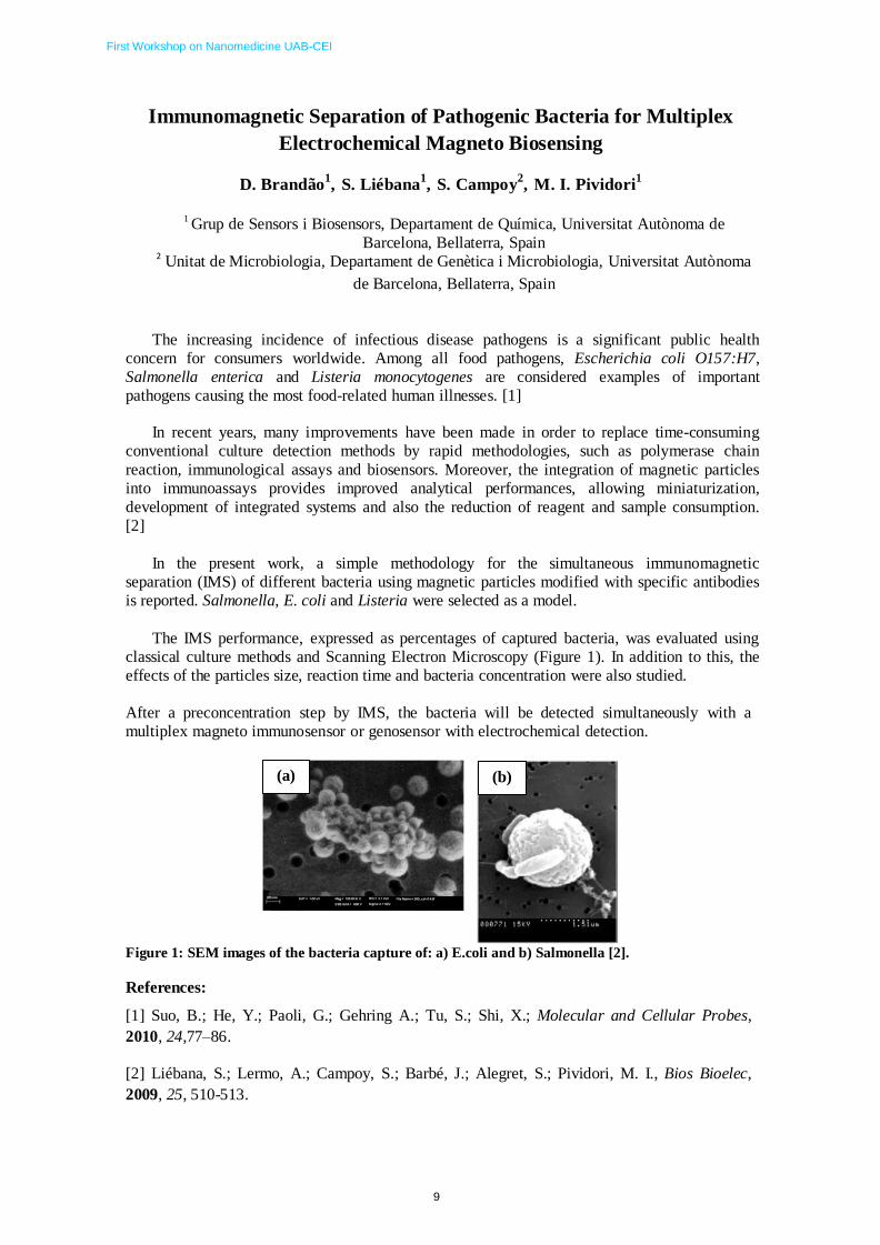
The IMS performance, expressed as percentages of captured bacteria, was evaluated using
classical culture methods and Scanning Electron Microscopy (Figure 1). In addition to this, the
effects of the particles size, reaction time and bacteria concentration were also studied.
After a preconcentration step by IMS, the bacteria will be detected simultaneously with a
multiplex magneto immunosensor or genosensor with electrochemical detection.
(a) (b)
Figure 1: SEM images of the bacteria capture of: a) E.coli and b) Salmonella [2].
References:
[1] Suo, B.; He, Y.; Paoli, G.; Gehring A.; Tu, S.; Shi, X.; Molecular and Cellular Probes,
2010, 24,77–86.
[2] Liébana, S.; Lermo, A.; Campoy, S.; Barbé, J.; Alegret, S.; Pividori, M. I., Bios Bioelec,
2009, 25, 510-513.

First Workshop on Nanomedicine UAB-CEI
10
Magnetic Nanoparticles for Brain Ischemia Treatment
Elisa Carenzaa, Verónica Barceló
b, Anna Roig
a, Joan Montaner
b, Anna Rosell
b
aInstitut de Ciència de Materials de Barcelona, Consejo Superior de Investigaciones Científicas (ICMAB-
CSIC), Campus de la UAB, 08193 Bellaterra, Catalunya, Spain.
bNeurovascular Research Laboratory and Neurovascular Unit. Vall d'Hebron Institut de Recerca,
Hospital Universitari Vall d'Hebron, Universitat Autònoma de Barcelona, Passeig Vall d’Hebron 119-
129, 08035, Barcelona, Catalunya, Spain.
Endothelial progenitor cells (EPCs) show stemness characteristics with the ability of differentiating into
endothelial cells (1). These cells constitute a new model for angiogenesis, endothelial regeneration and
vessels repair (2). In recent years stem cell labeling with superparamagnetic iron oxide nanoparticles
(SPIONPs) has been used as strategy for cellular therapy and tissue repair, as in central nervous system
diseases (3).
Our project aims to develop highly magnetized functional EPCs which can be accumulated in damaged
brain areas by using an external magnetic field to induce angiogenesis and tissue repair.
Citrate coated SPIONPs were synthesized through thermal decomposition route (4) with a γ-Fe 2 O3 core
of 6 ±1 nm in diameter and subsequent transfer in water with anionic surfactants. Stable aqueous
dispersion at pH= 7.5 showed nanoparticles aggregates with hydrodynamic size of 50 nm and 30% of
polydispersity. Magnetic measurement at room temperature showed absence of remnant magnetization,
and a high saturation magnetization value (54 emu/g Fe2 O3 ).
Early EPCs from mouse were successfully labeled with aqueous dispersions of citrate coated SPIONPs
after 24h of incubation at iron concentration of 50 µg/ml, showing uptake of around 24 pg Fe/ cell. TEM
images proved cellular uptake and storing of SPIONPs into endosomal compartments.
Our results show that magnetized outgrowth EPCs were fully functional since they shaped vessel-like
structures as non-magnetized cells. Furthermore we have found that magnetized human and mouse EPCs
secrete more VEGF and FGF than control cells. Finally a preliminary in vivo cell tracking demonstrates
that magnetized EPCs can be guided to cortical areas of the brain by an external magnetic field as
confirmed by MRI images.
1) Urbich C, Dimmeler S (2004) Endothelial progenitor cells functional characterization. Trends
Cardiovasc Med Nov 14:318–322.
2) Rafii S, Lyden D (2003) Therapeutic stem and progenitor cell transplantation for organ
vascularization and regeneration. Nat Med 9:702–712.
3) Syková E, Jendelová P (2007) Migration , fate and in vivo imaging of adult stem cells in the
CNS. Cell Death and Differ 14:1336-1342.
4) Sun S, et al. (2004) Monodisperse MFe2O4 (M = Fe, Co, Mn) nanoparticles. J Am Chem Soc
126:273-279.

First Workshop on Nanomedicine UAB-CEI
11
The Inorganic Nanoparticle Biomolecular Corona.
Formation, Evolution and Biological Impact Eudald Casals1, Tobias Pfaller2, Albert Duschl2, Gertie J. Oostingh2, Víctor F. Puntes1,3
1. Catalan Institute of Nanotechnology, Campus UAB, Q-building, 08193 Bellaterra (Barcelona), Spain
2. Department of Molecular Biology, University of Salzburg, Salzburg, Austria
3. Institut Català de Recerca i Estudis Avançats (ICREA), Barcelona, Spain. Email: [email protected]
Physicochemical changes of inorganic nanoparticles (NPs) in biological environments determine their
effects. Blood, lymph, mucus, complete cell culture media, and other biological fluids contain a large
amount and variety of different molecules. Nanoparticles dispersed in these fluids are sensitive to such
environment [1]. One of the most significant alterations is the formation of the NP Protein Corona (PC) as
a result of the adsorption of proteins onto the inorganic surface. Currently, there is an increasing
awareness of the importance of the NP-PC in the field of inorganic NPs, which is reflected in the
increasing number of recent publications that cover different aspects of this topic [2-4]. Largely, this is
because this spontaneous coating provides the biological identity to the composite NP-PC and determines
the interactions between the NPs and the host in living systems. As a result, the proper understanding of
the NP-PC formation has emerged as a crucial aspect to study the evolution, biodistribution and reactivity
of NPs in organisms and, therefore, for the safe design of engineered NPs [2].
Our studies aim to understand PC formation on model NPs, comprising metal (Au, Ag) and metal oxide
(Fe3O4, CeO2 and CoO), with sizes ranging from 7 to 20 nm and dispersed in commonly used cell culture
medium supplemented with serum. As a result, we have observed that all tested NPs adsorb proteins onto
their surface, thereby forming a PC through a dynamic process. Remarkably, an evolution from a loosely
attached PC (soft PC) towards an irreversible PC (hard PC) have been observed over time. Despite
studied NPs have similar characteristics (i.e., hydrophobicity and surface charge), different temporal
patterns of PC formation have been observed, which can be considered as a fingerprint for NP
classification and identification [5, 6]. Moreover, different PC formation processes have been observed
which depend on the NPs composition, size and surface state. All this aspects are of special relevance
since interactions and interference of inorganic NPs with cells and tissues take place at different time
scales. Similarly, biodistribution and residence times in different biological environments depends on the
NP surface characteristics. Importantly, some fundamental questions are still unclear such as the format
of presentation of the proteins in the PC, the role of the ubiquitin proteasome system (which identify and
"tag" proteins the body no longer need, as aged proteins, denaturated and aggregated) and the metabolic
degradation of the corona after extended period of time. All of this aspects need to be analyzed and
resolved aiming to design nanomaterials to be applied safely.
NP-protein interactions. The process of conjugation of the NP when inserted in biological media takes few minutes in
the working conditions (I), which evolves to a NP coated with protein in equilibrium with the proteins in the medium
(II), then later evolves towards an irreversible protein corona with proteins that are no longer in equilibrium with
their in-solution counterparts (III) [6].
--------------------------------- [1] Rivera-Gil, P. et al. Acs Nano 2010, 4 (10), 5527-5531.
[2] Faunce, T.A. et al., Nanomedicine, 2008, 3, 859-866.
[3] Rocker, C. et al. Nature Nanotechnology, 2009, 4, 577-580.
[4] Monopoli, M.P. et al. Journal of the American Chemical Society, 2011, 133, 2525-2534.
[5] Casals, E. et al. Acs Nano, 2010, 4, 3623-3632.
[6] Casals E., et al. Small, 2011 7(24), 3479-3486.

First Workshop on Nanomedicine UAB-CEI
12
Hyperpolarized 13C Magnetic Resonance Metabolic Imaging Applied to Mouse Brain Gliomas
Eva Monteagudo,a Teresa Delgado‐Goñi,b,c Teodor Parella,a Silvia Lope‐Piedrafitaa,c, Carles Arúsb,c,d
aServei de Ressonància Magnètica Nuclear, Universitat Autònoma de Barcelona (UAB), Cerdanyola del Vallés, Spain;
bDept. Bioquímica i Biologia
Molecular, UAB, Cerdanyola del Vallés, Spain; cCentro de Investigación Biomédica en Red‐Bioingeniería, Biomateriales y Nanomedicina (CIBER‐BBN),
Cerdanyola del Vallés, Spain; dInstitut de Biotecnologia i de Biomedicina, UAB, Cerdanyola del Vallés, Spain.
[email protected]; [email protected]
Magnetic resonance spectroscopic imaging (MRSI) of hyperpolarized 1‐13C‐pyruvate is a promising non‐invasive
technique to monitor metabolic changes in‐vivo. This method uses dynamic nuclear polarization1 (DNP) technique to
obtain tens of thousands fold enhancement in the polarization of 1‐13C‐pyruvate (Fig. 1) and its metabolic products, like
lactate, providing sufficient MR signal for high spatial and temporal resolution spectroscopic imaging of these
metabolites.2 The technique is based on cooling down the sample into a strong magnetic field in presence of a trityl
radical. Under such conditions, the radical unpaired electrons become hyperpolarized and this strong polarization can be
transferred to nearby atomic nuclei using microwave irradiation at the appropriate frequency. The hyperpolarized
sample is dissolved in a hot buffer and quickly injected at body temperature into a mouse allowing the study of in‐vivo 1‐ 13C‐pyruvate metabolic pathways. This technology is especially promising in oncology, where lactate apparent labeling
intensity have been shown to correlate with disease progression and response to therapy.3 Hence, the injection of 13C‐
pyruvate and assessment of 13C‐lactate can be used to distinguish, and best characterize, cancerous tissue. In recent
experiments, we were able to optimize experimental conditions to detect pyruvate and lactate 13C labeling following
intravenous injection of hyperpolarized 1‐13C‐pyruvate into mice with implanted GL261 mouse glioma cells. Metabolic
images showed significant labeling of pyruvate and lactate within the tumor region but comparatively low levels in
surrounding brain (Fig. 2). Active investigation is currently being carried out to hyperpolarize other compounds that may
help in the understanding of tumor metabolism. Substrates hyperpolarization was performed with a Hypersense® DNP
polarizer (Oxford Instruments) and magnetic resonance data were acquired using a 7 Tesla Biospec MRI spectrometer
(Bruker Biospin). Both equipments are located at the Servei de Ressonància Magnètica Nuclear of the UAB.
A)
B)
ppm
Fig. 1. 13
C MR spectra of 80 mM 1‐13
C‐pyruvate
at thermal equilibrium (A) and hyperpolarized (B)
acquired at 7T in a single scan.
References
Fig. 2. 13
C MRSI of a mouse brain bearing a high grade GL261 glioma tumor
after intravenous injection of hyperpolarized 1‐13
C‐pyruvate.
1 Ardenkjær‐Larsen, et al., PNAS, 2003, 100: 10158; 2 Golman, et al., PNAS, 2003, 100: 10435; 3 Day et al., Magn. Reson.
Med., 2011, 65: 557.
Acknowledgement
The purchase of the HyperSense® equipment has been funded by the Biomedical Research Networking Center in
Bioengineering, Biomaterials and Nanomedicine (CIBER‐BBN), the UAB Campus of International Excellence (UABCEI), and
the European Regional Development Fund (ERDF).

First Workshop on Nanomedicine UAB-CEI
13
Development of a reusable impedimetric aptasensor for the
recognition of cytochrome c
Evelien Arcay, Cristina Ocaña, Manel del Valle
Sensors and Biosensors Group, Universitat Autònoma de Barcelona, Edifici CN, 08193, Bellaterra, Barcelona, SPAIN
*E-mail: [email protected]
The application of a reusable impedimetric aptamer-based biosensor employing
a graphite-epoxy composite electrode has been increasing the last few years. The
method employed is electrochemical impedance spectroscopy because of the simplicity
and high sensitivity of the technique as well as its capacity for low concentration
detection and ability for label-free detection. In this work the technique has been used
for the detection of the protein cytochrome c. Detection occurs when the protein
interacts with the immobilized aptamer on the aptasensor. An aptamer can bind with
high specificity and affinity to small target ligands such as molecules, proteins or cells.
The recognition technique is based on the physical adsorption of the aptamer on the
electrode. The first step is the optimization of the graphite-epoxy composite electrode,
followed by the label-free detection of cytochrome c by the aptamer on the sensor. The
result of the interaction between cytochrome c and aptamer is quantified by the
observed increase of the electron-transfer resistance that can then be analyzed with
electrochemical impedance spectroscopy. The detection method for the resistance
involves a [Fe(CN)6]3-,4-
redox marker solution in a potentiostated electrochemical cell.
From the results, it can be concluded that the produced graphite-epoxy composite
electrode has a good detection range for cytochrome c between 5.10-11
M and 5.10-8
M,
as well as a high sensitivity of 5,24.108
M-1
and a low detection limit of 6,3.10-11
M.

First Workshop on Nanomedicine UAB-CEI
14
ONE-STEP PREPARATION OF STABLE, NANOSCOPIC AND
UNILAMELLAR CHOLESTEROL-RICH VESICLES FOR
APPLICATION IN NANOMEDICINE, USING COMPRESSED
FLUIDS I.Cabrera, E.Elizondo, E.Moreno, L.Ferrer, S.Sala, N.Ventosa, J.Veciana
Department of Molecular Nanoscience and Organic Materials. Institut de Ciència de Materials de Barcelona (ICMAB-CSIC),
Networking Research Center on Bioengineering, Biomaterials and Nanomedicine, CIBER-BBN, Campus de la Universitat
Autònoma de Barcelona (UAB), 08193-Bellaterra (Spain)
e-mail: [email protected]; [email protected]
Vesicles constitute one of the most studied drug delivery systems (DDS) since their discovery in the mid 60s. However, a high grade of structural homogeneity, not only in size and morphology, but also in their membrane composition and supramolecular organization is required for an
optimal performance of these self-assembled structures as functional material [1]
. Attending to this, methods for the precise synthesis of homogeneous vesicular systems are required for fully exploiting the potential of these self-assembled structures in the development of new nanomedicines.
In the early 90’s, compressed fluid (CF)-based processes emerged as an alternative to conventional methods using liquid solvents, attracting enormous interest for the production of
micro- and nanoparticulate materials [2]
. Our research group has experience in using these novel technologies for the controlled nanostructuration of molecular materials to be used in drug
delivery [3]
. In this poster we will present a CF-based method for the production and integration of actives in vesicular systems. This one-step process allows the preparation of stable,
nanoscopic and unilamellar protein and peptide loaded cholesterol-rich vesicles [4]
, which present higher structural homogeneity regarding size and morphology tan those, with the same composition, produced by a conventional multi-step hydration method.
Figure 1. Cryo-TEM image of peptide functionalized cholesterol rich SUVs. Remarkably, by
analyzing the membrane composition and supramolecular organization of
vesicles prepared by both methodologies, we have observed that apart from size and morphology, the superior homogeneity observed for vesicular systems produced by CFs is also present in the molecular assembly of the lipidic constituents forming the vesicular membrane, which is crucial for an optimum performance of these supramolecular structures as
pharmaceutical carriers [5]
.
References [1] R. Sawant, V. Torchilin, Soft Matter 2010, 6, 4026. [2] J.D. Holmes, K.P. Johnston, R.C. Doty, B.A. Korgel, Science 2000, 287, 1471.
[3] E. Elizondo, S. Sala, E. Imbuluzqueta, D. González, M. J. Blanco-Prieto, C. Gamazo, N. Ventosa,
J. Veciana, Pharmaceutical Research 2011, 28, 309.
[4] M. Cano-Sarabia, N. Ventosa, S.Sala, C. Patino, R. Arranz, J. Veciana, Langmuir 2008, 24, 2433.
[5] E. Elizondo, J. Larsen, N. S. Hatzakis, I. Cabrera, T. Bjørnholm, J. Veciana, D. Stamou, N. Ventosa,
J.Am.Chem. Soc. 2012, 134, 1918.

First Workshop on Nanomedicine UAB-CEI
15
Compressed Fluids for the Micronization of Drugs and their Formulation as Polymeric
Drug Delivery Systems
E. Elizondo1,2, E. Moreno1,2, E. Samanes1,2, S.Sala2,1, E. Rojas1,2, N. Ventosa1,2,*, J. Veciana1,2,*
1Departament de Nanociència Molecular i Materials Orgànics, Institut de Ciència de Materials de
Barcelona (ICMAB-CSIC), Campus UAB, Bellaterra, 08193, Spain 2CIBER de Bioingeniería, Biomateriales y Nanomedicina (CIBER-BBN), Bellaterra, 08193, Spain
Corresponding authors: [email protected]; [email protected]
Structuring of synthetic and biological therapeutic actives as micro and
nanoparticulate materials is a widely accepted formulation strategy to improve efficacy and
reduce toxicity of drugs. However, the development of efficient production platforms that
enable the obtaining of these nanomedicines at industrial scale and with the quality
requirements imposed by the regulatory agencies remains a challenge. In this framework,
compressed fluid-based methods, which offer advantages like reduction of organic solvent use,
low working temperatures, few operational steps and easy scale-up, are promising
technologies for the controlled and reproducible preparation of uniform micro- and
nanoparticulate nanomedicines at large scale.
In our group, compressed fluid-based methods have been successfully applied for the
one-step micronization of pharmaceutical compounds like ibuprofen, naproxen, aspirin or
acetaminophen [1]. This type of processes have also been used for the formulation of
particulate drug delivery systems, focusing special attention to the processing of
biodegradable polymers such as poloxamer [2] and poly (methyl vinyl ether-co-maleic
anhydride) [3], and the preparation of drug/polymer composites [4].
References
[1] Sala S, Cordoba A, Moreno-Calvo E et al. Cryst. Growth Des. 12(4), 1717-1726 (2012).
[2] Munto M, Ventosa N, Veciana J. Synergistic. J. Supercrit. Fluids 47(2), 290-295 (2008).
[3] Elizondo E, Cordoba A, Sala S et al. J. Supercrit. Fluids 53(1-3), 108-114 (2010).
[4] Elizondo E, Sala S, Imbuluzqueta E, et al. Pharm. Res. 28(2), 309-321 (2011).
First Workshop on Nanomedicine UAB-CEI
16
Smart Metal-Organic Nanoparticles with Application on Nanomedicine
F. Novio,a
A. Raman,a
D. Ruiz-Molinaa
aCentre d’Investigació en Nanociència i Nanotecnologia CIN2, CSIC-ICN (Spain).
E-mail: [email protected].
The successful design of the metal-organic frameworks and the good control on the
release of a wide variety o drugs open an interesting research field in which metal-
organic nanostructures can afford novel and revolutionary drug delivery systems. With
this aim, the development of nanoscale drug delivery metal-organic systems follows
three different approximations: I) drugs are adsorbed in porous metal-organic
frameworks (NMOF´s), II) drugs are the constitutive building blocks of the metal-
organic nanostructures and III) a novel approach described in our group by which drugs
are encapsulated inside an amorphous metal-organic nanoparticles.
Here we show how our research group has developed the synthesis of polymeric
metal-organic nanoparticles able to encapsulate a wide variety of substances [1] and
materials with interesting applications on nanomedicine.[2] These systems exhibit good
rates of drug release and notable in vitro citotoxicity effects. Preliminary results show
that the drug or active species can be encapsulated on smart nanoparticles that respond
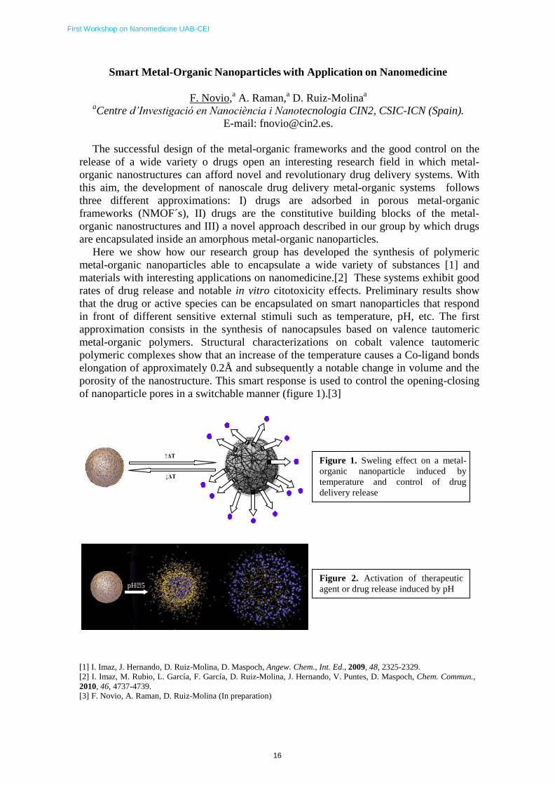
in front of different sensitive external stimuli such as temperature, pH, etc. The first
approximation consists in the synthesis of nanocapsules based on valence tautomeric
metal-organic polymers. Structural characterizations on cobalt valence tautomeric
polymeric complexes show that an increase of the temperature causes a Co-ligand bonds
elongation of approximately 0.2Å and subsequently a notable change in volume and the
porosity of the nanostructure. This smart response is used to control the opening-closing
of nanoparticle pores in a switchable manner (figure 1).[3]
↑ΔT
↓ΔT
Figure 1. Sweling effect on a metal-
organic nanoparticle induced by
temperature and control of drug
delivery release
pH5 Figure 2. Activation of therapeutic
agent or drug release induced by pH
[1] I. Imaz, J. Hernando, D. Ruiz-Molina, D. Maspoch, Angew. Chem., Int. Ed., 2009, 48, 2325-2329.
[2] I. Imaz, M. Rubio, L. García, F. García, D. Ruiz-Molina, J. Hernando, V. Puntes, D. Maspoch, Chem. Commun.,
2010, 46, 4737-4739. [3] F. Novio, A. Raman, D. Ruiz-Molina (In preparation)
17
First Workshop on Nanomedicine UAB-CEI
DNA DAMAGE INDUCED BY SILVER NANOPARTICLES IN THREE
DIFFERENT HUMAN CELL LINES (BEAS-2B, CACO-2 AND TK6)
Vales, Gerard1; Rubio, Laura
1; Vela, Lourdes
1; Creus, Amadeu
1,2; Marcos, Ricard
1,2
1Grupo de Mutagénesis, Departamento de Genética y de Microbiología, Facultad de Biociencias,
Universidad Autónoma de Barcelona; 2CIBER de Epidemiología y Salud Pública, Instituto de Salud
Carlos III.
Nanotechnology is an emergent field and many products commercially available have
engineered nanomaterials in their composition. Besides the increasing presence of these
compounds, the same novel properties that make them interesting for industrial purposes had
also raised some concerns about their toxicity. Therefore, the analysis of the genotoxic risk
associated to nanomaterials exposure has become an expansive field.
Many different materials are used as additives, being the silver-based nanoparticles the most
common material found in product description among the nanotechnology-based products. In
this work we have carried out the genotoxic evaluation of silver nanoparticles in three different
human cell lines (BEAS-2B, Caco-2 and TK6). Exposure treatments for the three cell lines lasted for 3 hours and, in addition, TK6 cells were
also treated for 24 hours. The dose range was up to 100 μg/mL, and the genetic damage was
measured by means of the comet assay. The standard comet assay was complemented by
using the formamidopyrimidine-DNA glycosylase (FPG) enzyme, to determine DNA oxidation as
a possible mechanism for the genotoxic action of silver nanoparticles. In parallel, the apoptosis
rate and the effect on the cell cycle was analyzed in the BEAS-2B and Caco-2 cell lines by flow
cytometry.
The results showed that, although no significant increases in the levels of DNA damage were
observed in the standard version of the comet assay, significant increases in the percentage of
DNA in the comet tail were observed when FPG was used. Also, no effect on the apoptosis rate
was seen neither in BEAS-2B nor in Caco-2 cells, although cell cycle arrest in Caco-2 was
observed in 50 and 100 μg/mL. With respect to the sensitivity of the cell lines to the oxidative effects of silver nanoparticles it
was, Caco-2 > BEAS-2B > TK6. The results indicate that the selection of the cell line is an
important factor to avoid positive/negative false results, when testing the toxicity of
nanomaterials.

First Workshop on Nanomedicine UAB-CEI
18
Unraveling the kinetics of aggregation of single peptide-DNA
complexes using force spectroscopy
J. Camunas-Soler,1, 2 S. Frutos,1, 2 C.V. Bizarro,1, 2 S. de Lorenzo,1, 2 M.E. Fuentes-Perez,3 R. Ramsch,4, 2 S. Vilchez,4, 2 C. Solans,4, 2 F. Moreno-Herrero,3
F. Albericio,2, 5 R. Eritja,2, 4, 5 E. Giralt,2, 5 S.B. Dev,5 and F. Ritort1, 2
1Small Biosystems Lab, Departament de Física Fonamental, Facultat de Física, Universitat de Barcelona, Barcelona, Spain 2CIBER de Bioingeniería, Biomateriales y Nanomedicina, Instituto de Salud Carlos III, Madrid, Spain 3Centro Nacional de Biotecnologia, CSIC, Cantoblanco, Madrid, Spain 4Institut de Química Avançada de Catalunya, Consejo Superior de Investigaciones
Científicas (IQAC-CSIC), Barcelona, Spain 5Institute for Research in Biomedicine(IRB Barcelona), Barcelona Science Park, Baldiri Reixac 10-12,08028, Barcelona, Spain
Correspondence: [email protected]
Abstract
The knowledge of the mechanisms of interaction between hydrophobic molecules and essential cellular components is key to our understanding of many aggregation processes underlying several human diseases. Kahalalide F (KF) is an hydrophobic marine-derived peptide with a strong anticancer activity which contains a positively charged residue (L-Orn). KF is an ideal model to elucidate the mechanisms by which self-aggregation competes with binding to a strongly charged polyelectrolite such as DNA. Here we carry out mechanical stretching and unzipping experiments of single DNA molecules (in double and single stranded form) complexed with KF using optical tweezers. We show that KF and DNA interact forming large aggregate complexes promoted by the recruitment and wrapping of DNA around the aggregate which are further stabilized by hydrophobic interactions within the KF-DNA complex. These experiments reveal unique features of the aggregation process, and the proposed methodology might be useful to quantitatively characterize other compounds or proteins in which the formation of aggregates is of relevance.
First Workshop on Nanomedicine UAB-CEI
19
Development of a highly efficient purification protocol for the isolation of
protein‐based nanoparticles with nanomedical applications
Martínez‐Láinez, J.M. 1,2,3, Seras‐Franzoso, J. 1,2,3, Peebo, K. 1,4 Corchero, J.L. 2,1,3, Vázquez, E.1,2,3,
Villaverde, A. 1,2,3, García‐Fruitós, E. 2,1,3
1Institut de Biotecnologia i de Biomedicina, Universitat Autònoma de Barcelona, Bellaterra, Barcelona, Spain
2CIBER de Bioingeniería, Biomateriales y Nanomedicina (CIBER‐BBN), Bellaterra, Barcelona, Spain
3Department of Genetics and Microbiology, Universitat Autònoma de Barcelona, Bellaterra, Barcelona, Spain
4Competence Centre of Food and Fermentation Technologies, Akadeemia tee 15b, 12618 Tallinn, Estonia
Inclusion bodies (IBs) are small protein aggregates produced in recombinant bacteria under stress
conditions1
with a size ranging from 50 nm to 500 nm2. Interestingly, in the last years, and after the
characterization of IBs as biologically active aggregates2,3
, many applications using these aggregates
have been described 2,3,4,5
. In this context, recent studies have shown their potential in the biomedical
applications as vehicles to deliver therapeutic proteins (nanopills), since they have the capacity to cross
the eukaryotic cell membrane and deliver their compounds into the target cell6.
One of the main bottlenecks of the IBs isolation is the purification process, since the purity and the
quality of the final product are crucial. With the aim to optimize the purification method, considering
both the conformational quality and the purity of the final nanoparticles, we have developed a new
protocol, in which we have obtained higher amounts of IBs. This data is really interesting, especially if
we consider that the specific activity remains constant, when compared with the results obtained with
the previous procedure7. On the other hand, it is important to stress that, with this new protocol, the
purity of the final product increases significantly. In this context, and with the aim to specifically
optimize bacterial lysis, being the most limiting step in the purification process8, we have carried out a
comparative study using different concentrations of lysozyme, an enzyme with bactericide properties
able to hydrolyze peptidoglycans present in the bacterial wall9. The results obtained show that there is
no positive correlation between the concentration of lysozyme used and the degree of cellular lysis.
However, when using low concentration of lysozyme we observe a loss of inclusion bodies. Therefore, if
we consider the need to have an optimal cellular lysis and at the same time achieve a good yield, we
suggest an intermediate concentration, 1 μg lysozyme/mL culture media, as the most effective lysozyme
concentration for the new protocol. Moreover, we have also observed that the number of freeze/thaw
cycles is important regarding the protocol efficiency. To carry out this study we used different strains of
Escherichia coli deficient in the main proteases and chaperones, and different proteins, in order to
evaluate the new protocol in a large number of cases.
References:
1. A. Villaverde, M.M Carrio, Biotechnol. 22, 1385 (2003)
2. E. Garcia‐Fruitos et al., Advanced Materials 21, 4249 (2009)
3. E. Garcia‐Fruitos et al., Microb Cell Fact. 4, 27 (2005)
4. M. J. Dalby, Nanomedicine. (Lond) 4, 247 (2009)
5. Diez‐Gil C al. Biomaterials. 12, 5805 (2010).
6. E. Vazquez et al, Adv Mater. 24(13): 1742‐7 (2012)
7. E. Rodriguez‐Carmona et al, Microbial Cell Fact. 9; 71 (2010)
8. E. Peternel, N Biotechnol, [Epub ahead of print] (2011)
9. R. W. Burley, D.V. Vadehra, The Avian Egg: Chemestry and Biology (New York, John Wiley) (1989)
First Workshop on Nanomedicine UAB-CEI
20
Inclusion bodies in Biomedical Applications: Tissue engineering scaffolds
Joaquin Seras‐ Franzoso1,2,3
, César Díez‐ Gil3,4
, Esther Vazquez1,2,3
, Elena García–Fruitós3,1
, Rafael
Cubarsi3,5
, Imma Ratera3,4
, Jaume Veciana3,4
& Antonio Villaverde1,2,3
1 Institute for Biotechnology and Biomedicine, Universitat Autònoma de Barcelona, Bellaterra, 08193
Barcelona, Spain
2 Department of Genetics and Microbiology, Universitat Autònoma de Barcelona, Bellaterra, 08193
Barcelona, Spain
3 CIBER de Bioingeniería, Biomateriales y Nanomedicina (CIBER‐BBN), Bellaterra, 08193 Barcelona, Spain
4 Institut de Ciencia de Materials de Barcelona (ICMAB‐CSIC) Bellaterra, 08193 Barcelona, Spain
5 Departament de Matemàtica Aplicada IV. Universitat Politècnica de Catalunya. Jordi Girona 1‐3, 08034
Barcelona, Spain
Bacterial Inclusion Bodies (IBs) are protein aggregates commonly formed during recombinant
protein production by the deposition of polypeptide chains in different conformational states. Most of
them show a sphere‐like morphology with amyloid fibrils acting as a net in which folded or partially
folded protein is trapped. Despite being regarded during years as useless by products of the protein
production process the discovery of several appealing features has dramatically reverted this
perception.
These aggregates can be easily purified resulting in stable protein particles ranging in size
between 50nm to 500nm. Moreover it has been observed that IBs can retain a certain grade of
biological activity. These properties make IBs promising cost effective biocatalysts. In this regard many
processes using IBs as immobilized biocatalysts have been successfully carried out in the last decade.1‐3
However, our laboratory has focused its recent research in new applications for bacterial IBs
directing this technology to biomedical fields such as regenerative medicine and tissue engineering. It
has been shown how these protein particles are suitable to generate scaffolds for cell culture being a
biocompatible material and enhancing cell adhesion and proliferation, both events crucial for tissue
engineering applications. More precisely here we show evidence of how the mechanical features of
VP1GFP IBs can be recognized by the cell sensing machinery and induce cell division through a
mechanotransduction cascade in 1BR3.G fibroblast‐like cells. In addition IB‐based scaffold adhesion was
assayed in four different cell types 1BR3.G, HepG2, PC12 and BHK observing and increment of retained
cells when comparing to nude polystyrene surfaces after several washing steps in PBS. These data prove
a dual effect of IB‐based scaffolds by increasing cell adhesion and depending on the cell line also
stimulating cell proliferation.4
In addition, IBs have been shown to be an easily tunable material
modulated by the producing genetic background5, 6
. Thus it is possible to produce IB‐based scaffolds
with different mechanical properties in order to achieve the desired response. All these data reinforce
the potential vested in IBs as suitable material for surface coating in tissue engineering applications.
Reference List
1. Sans,C. et al. Inclusion bodies of fuculose‐1‐phosphate aldolase as stable and reusable biocatalysts. Biotechnol. Prog.
28, 421‐427 (2012). 2. Nahalka,J., Gemeiner,P., Bucko,M., & Wang,P.G. Bioenergy beads: a tool for regeneration of ATP/NTP in
biocatalytic synthesis. Artif. Cells Blood Substit. Immobil. Biotechnol. 34, 515‐521 (2006). 3. Nahalka,J. Physiological aggregation of maltodextrin phosphorylase from Pyrococcus furiosus and its application in a
process of batch starch degradation to alpha‐D‐glucose‐1‐ phosphate. J. Ind. Microbiol. Biotechnol. 35, 219‐223 (2008). 4. Seras‐Franzoso,J. et al. Bioadhesiveness and efficient mechanotransduction stimuli synergistically provided by
bacterial inclusion bodies as scaffolds for tissue engineering. Nanomedicine. (Lond) 7, 79‐93 (2012).
5. Diez‐Gil,C. et al. The nanoscale properties of bacterial inclusion bodies and their effect on mammalian cell proliferation. Biomaterials 31, 5805‐5812 (2010).
6. Garcia‐Fruitos,E., Seras‐Franzoso,J., Vazquez,E., & Villaverde,A. Tunable geometry of bacterial inclusion bodies as substrate materials for tissue engineering. Nanotechnology. 21, 205101 (2010).

First Workshop on Nanomedicine UAB-CEI
21
Gold Nanoparticles as Drug Delivery Agents for Cancer Therapy.
Gold nanoparticles present unique properties as drug delivery scaffolds due to their size
and surface tunability. Cisplatin is the most used chemotherapeutic agent in many types
of cancers. Here we show that toxicity, which is the main limiting factor for
chemotherapy, is clearly reduced without affecting the therapeutic benefits of the drug
by attaching a cisplatin derivative to AuNPs via a pH-sensitive coordination bond. This
is related to the change on the biodistribution as well as the different processing of the
drug when it is attached to gold nanoparticles. Nanoparticles not only act as a delivery
agent, but protect the drug from being deactivated by plasma proteins until they are
internalized via endocytosis and cisplatin is released. The possibility of tracking the
drug and the vehicle separately enables a better understanding on how nanocarriers are
processed by the organism.
Jordi Piella (1), Joan Comenge (1) (2), Carmen Sotelo (3), Francisco Romero (4), Óscar
Gallego (5), Agustí Bernadas (5), Fernando Domínguez (3), Víctor Puntes (1) (6)
(1) CIN2 (ICN-CSIC), Catalan Institute of Nanotechnology, Spain
(2) INL, International Iberian Nanotechnology Laboratory, Portugal
(3) USC, Univerity of Santiago de Compostela, Spain
(4) UV, University of Valencia, Spain
(5) Sant Pau Hospital, Spain
(6) ICREA, Institució Catalana de Recerca i Estudis Avançats

First Workshop on Nanomedicine UAB-CEI
22
RADDEL: Nanocapsules for Targeted Delivery of Radioactivity
Jorge Pérez, Gerard Tobias
Institut de Ciència de Materials de Barcelona, Campus de la UAB, 08193 Bellaterra, Barcelona. E-mail: [email protected]
Abstract
Tailored functionalization of nanomaterials for biomedical applications is an emerging trend in
nanotechnology. Carbon nanotubes offer an attractive platform for the developement of “smart”
systems for drug delivery, diagnosis and therapy. Multifunctional carrier systems based on
carbon nanotubes can be designed in which their internal cavity encapsulates a chosen payload
whilst the outer surface is chemically modified to match specific needs. However, despite their
potential, these filled and functionalized nanotubes (carbon nanocapsules) had not been
previously studied. We have recently reported on the covalent functionalization of radionuclide-
filled single-walled carbon nanotubes and their use as radioprobes [1]. These nanocapsules
allow the delivery of an unprecedented radiodosage and ultrasensitive imaging. They remain
stable for extended periods thus guaranteeing essentially zero leakage of the radionuclides.
Surface functionalisation of these nanocapsules offers versatility towards modulation of tissue
biodistribution of the radioemitting crystals in a manner determined by the nanocapsule that
delivers them. The delivery of radioactivity takes place through the walls of the nanocapsules
and release of the encapsulated radionuclides is therefore not needed and certainly not desired.
Further studies on these systems are now being performed within the frame of the RADDEL
(RADioactive DELivery) project, an Initial Training Network funded by the European
Comission under the FP7-PEOPLE program (2012-2016). The aim of the project is to train
young researchers in a multidisciplinary research environment on the development of novel
nanomaterials for biomedical applications, always taking industrial aspects into account
References
[1] S. Y. Hong, G. Tobias, K. T. Al-Jamal, B. Ballesteros, H. Ali-Boucetta, S. Lozano-Pérez, P.
D. Nellist, R. B. Sim, C. Finucane, S. J. Mather, M. L. H. Green, K. Kostarelos and B. G. Davis;
Nature Materials, 9, 485-490 (2010).
RADDEL project is funded by the European Comission under the FP7 People Program - Marie Curie Actions (Grant
agreement number: 290023).

First Workshop on Nanomedicine UAB-CEI
23
A simple method for the preparation of Cationic Gold Nanoparticle Bioconjugates
for Cell Penetration
L. García-Fernández1, I. Ojea
1, J. Lorenzo
2, V. F. Puntes
1,3
1
Institut Català de Nanotecnologia, Campus UAB, 08193 Bellaterra-Barcelona, Spain. 2
Institut de
Biotecnologia i Biomedicina, Campus UAB, 08193 Bellaterra-Barcelona, Spain. 3
Institut Català de
Recerca i Estudis Avançats (ICREA), Barcelona, Spain.
The surface charge of NPs plays a critical role in determining their molecular
interactions with target cells. These interactions could determine intracellular uptake,
localization of the NPs and their biological functions, which is of a broad interest for the
use of these NPs in advanced biomedical applications. For example, cationic Au NPs
have attracted a great interest over the recent years for transfecting molecules into cells
and for drug delivery applications. However, there are major concerns regarding the
toxicity of these conjugates and there is a limited number of reports describing their
synthesis. To date, only a few reports can be found of cationic lipids, synthetic cationic
polymers such as poly(ethyleneimine) (PEI) or poly(allylamine), aminoalkanethiols and
quaternary ammonium salts, decorating the surface of Au NPs of small size (e.g. 2 nm
Au core). NP size and concentration, among others, are limiting factors in these
syntheses. In this work, we present a fast, easy and effective method for the preparation
of cationic Au NPs of several sizes (from ~8 to 23 nm) in high concentrations (i.e. up to
3-4 mM of 13 nm Au NPs). This approach is based on a phase-transfer methodology
from organic to aqueous solutions with a simultaneous ligand exchange and formation
of a dense, positively charged monolayer. Importantly, this method has been
successfully applied for the covalent functionalization of bioactive peptides with an
identical sequence but positive (-NH2) and negative (-COOH) terminal charged groups.
In vitro studies demonstrated that the cationic bioconjugates were extremely penetrating
in human dermal fibroblasts at short incubation times (3 h) as opposite to their negative
counterparts, and remarkably, no toxic effects were found after 24 h incubation. The
phase-transfer methodology shows a great potential and feasibility for promising
applications such as gene delivery.

First Workshop on Nanomedicine UAB-CEI
24
OH
Drug impregnated magnetic nanospheres
Nerea Murillo-Cremaes
1, Javier Saurina
2, Concepción Domingo,
1* Anna Roig
1*
1 Institut de Ciència de Materials de Barcelona, ICMAB-CSIC, Campus de la UAB, 08193 Bellaterra, Spain. 2
Department of Analytical Chemistry. University of Barcelona, 08028 Barcelona, Spain.
The use of supercritical carbon dioxide (scCO2) as a synthesis medium as well as a solvent to perform
adsorption and impregnation processes and materials functionalization has received considerable attention as a viable and sustainable alternative to conventional liquid solvents. We will present the use of supercritical
fluid assisted sol-gel method for the production of a multi-core magnetic silica carriers as well as the use of
supercritical carbon dioxide to impregnate a therapeutic agent (triflusal) in the nanospheres. Trifusal is an antithrombotic therapeutic agent used here as a model of a hydrophobic and moisture sensitive active agent
with poor solubility in water.
Fabrication of the magnetic silica nanospheres was done in a straight forward one-pot method combining sol- gel chemistry and supercritical fluids technology [1]. Resulting nanoparticles present a narrow particle size
distribution with sizes of the order of 100 nm. Each nanosphere consists of a magnetic multi-core of non- contacting Fe3O4 nanoparticles surrounded by a microporous silica shell. Nanospheres are superparamagnetic
at room temperature. Some advantages of the method are short reaction times, purity of the product and potentiality of the process to be scaled up. Cytotoxicity studies of the composites will be presented.
We have previously reported on the potential use of the nanospheres as enhanced T 2 contrast agent for MRI
[1,2]. In addition, the designed material may find applications as a target drug delivery system having the greatest therapeutic potential in those clinical scenarios that require the delivery of active agents at a specific
point of the body while avoiding systemic effects of toxicity. The silica–based matrix is found to prevent the hydrolization of the active ingredient more efficiently than a polymeric matrix PMMA used for comparison,
the drug vehicle serving in this way as a moisture protection barrier. Moreover, the trifusal is dispersed in a
molecular form inside the material and fast release kinetics has been assessed, both features being of great interest to enhance the bioavailability of low solubility drugs.
A
G
N
E
T
O
O O
O
IMPREGNATION PROCESS O O
OH
F3C
Drug dissolution and diffusion in
SCCO2
O
O
F3C
O
OH O
F3C
F3C
O O O
O O
OH
OH
F3C
ϕ = 65 nm (19%) 100 nm
[1] Taboada et al., Advanced Functional Material, 19, 14 (2009) 2319.
[2] Taboada et al., Mater. Res. Soc. Symp. Proc. 1257, (2010), 1257-O05-06

First Workshop on Nanomedicine UAB-CEI
25
Targeting domains in protein-only gene therapy vehicles trigger
cellular responses upon receptor binding
Joan Domingo-Espín 1, 2, 3
, Valérie Petegnief 4, Núria de Vera
4, Oscar Conchillo
1,
Paolo Saccardo 1, 2, 3
, Ugutz Unzueta 1, 2, 3
, Esther Vazquez 1, 2, 3
, Juan Cedano 5, Luciana
Negro 6, Xu Zhikun
1, 2, 3 , Xavier Daura
1,6, Hugo Peluffo
7,8, Anna M. Planas
4, Antonio
Villaverde 1, 2, 3
, Neus Ferrer-Miralles 1, 2, 3
1 Institut de Biotecnologia i de Biomedicina, Universitat Autònoma de Barcelona, Bellaterra,
08193 Barcelona, Spain 2 Department of Genetics and Microbiology, Universitat Autònoma de Barcelona, Bellaterra,
08193 Barcelona, Spain
3 CIBER de Bioingeniería, Biomateriales y Nanomedicina (CIBER-BBN), Bellaterra,
08193 Barcelona, Spain 4 Departament d’Isquèmia Cerebral i Neurodegeneració, Institut d’Investigacions Biomèdiques
de Barcelona (IIBB), Consejo Superior de Investigaciones Científicas (CSIC)-Institut
d’Investigacions Biomèdiques August Pi i Sunyer (IDIBAPS), Barcelona, Spain
5 Laboratory of Immunology, Regional Norte, Universidad de la Republica, Gral. Rivera 1350;
Salto, 50.000, Uruguay 6 Institució Catalana de Recerca i Estudis Avançats (ICREA), Barcelona, Spain
7 Neurodegeneration Laboratory, Institut Pasteur de Montevideo, CP 11400, Montevideo,
Uruguay 4
8 Department of Histology & Embryology, Faculty of Medicine, UDELAR, CP 11800,
Montevideo, Uruguay
Novel protein-only nanoparticles can be designed to display crucial functions to get
access to the target cells, to get internalized, to escape from endosomes and finally
deliver nucleic acids to the desired cell compartment. This type of biological
nanomaterial are aimed to be used in therapy, diagnosis and imaging. [1-3]. Among
those activities, targeting moieties are usually added to improve the specificity of the
protein vehicle to the target cell, through the interaction of the recombinant protein and
a targeted cell receptor. However, the effect caused on the targeted cell upon receptor-
ligand interaction has not been described in detail so far. In that sense, we have analysed
the effect of a RGD integrin binding domain in a model modular protein designed to
transfect integrin displaying cells. The results show that the RGD-containing protein
acts as an agonist dependent on integrin receptor interaction triggering in PC12 cell line
a proliferative effect. In fact, we have demonstrated that the proliferative effect depends
on the ERK1/2 stimulation cascade and we also observe a partial differentiation
phenotype in this neuron-like PC12 cell line.
Therefore, we hypothesize that the presence of targeting domains such as RGD-
containing motifs in recombinant proteins or functionalized nanoparticles might have an
impact on the targeted cells that need to be deeply studied.
References
[1] J. Domingo-Espin, E. Vazquez, J. Ganz, O. Conchillo-Sole, E. García-Fruitós, J. Cedano, U. Unzueta,
V. Petegnief, N. Gonzalez-Montalban, A. M. Planas, X. Daura, H. Peluffo, N. Ferrer-Miralles, and A.
Villaverde, Nanomedicine 6 (2011) 1047-1061.
[2] Vazquez E, Ferrer-Miralles N, Mangues R, Corchero JL, Schwartz S Jr, Villaverde A.
Curr.Pharm.Des 2009;15:893-916.
[3] Vazquez E, Ferrer-Miralles N, Villaverde A. Drug Discov.Today 2008;13:1067-1074.

First Workshop on Nanomedicine UAB-CEI
26
Size-controlled Synthesis and Functionalization of Large Gold Nanoparticles
Ngoc Tran1,2, Neus Bastús1, Joan Comenge1,2,3, Víctor Puntes 1,2,4,
1Institut Català de Nanotecnologia, Barcelona, Spain 2 Universitat Autònoma de Barcelona, Barcelona, Spain
3 International Iberian Nanotechnology Laboratory, Braga, Portugal 4Institut Català de Recerca i Estudis Avançats, Barcelona, Spain
Email: [email protected]
Highly monodispersed spherical gold nanoparticles with controlled sizes were obtained by seeded growth method in which sodium citrate played the role of both reducing and stabilizing agent. The achieved uniformity was attributed to the kinetic control of the homogeneous growth process against secondary nucleation by adjusting the reaction conditions: temperature, gold precursor to seed particle ratio, and pH [1]. This well- developed method allows the reproducible synthesis of large particles up to ~200 nm with higher concentration and narrower size distribution compared to the traditional Frens method [1, 2]. Moreover, gold nanoparticles prepared using this method, are ready to be further functionalized with a wide variety of molecules [3]. This conjugation process leads to the possibility to tune the surface properties of resultant particles and therefore test its suitability for medical and biological applications [4].
Figure 1: TEM images of Au seed particles and those obtained after different growth steps
[1] Bastus, N et al. 2011. Kinetically controlled seeded growth synthesis of citrate-stabilized gold nanoparticles of up to 200 nm: Size focusing versus Ostwald ripening. Langmuir 27: 11098-11105 [2] Frens, G et al. 1973. Controlled nucleation for the regulation of the particle size in monodisperse gold suspensions. Nature Physical Science 241: 20-22 [3] Bastus, N et al. 2009. Homogeneous conjugation of peptides onto gold nanoparticles enhances macrophage response. ACS Nano 3: 1335-1344 [4] Casals, E et al. 2010. Time evolution of the nanoparticle protein corona. ACS Nano 4: 3623-3632

First Workshop on Nanomedicine UAB-CEI
27
Synthesis of hybrid cyclobutane-proline γ,γ-peptides as a new family of CPP agents
E. Gorrea,a
R. Gutiérrez-Abad ,a
D. Carbajo,b
O. Illa,a
M. Royob
and R.M. Ortuñoa
aDepartament de Química, Universitat Autònoma de Barcelona,08193-Cerdanyola del Vallès, Spain;
bParc
Científic de Barcelona, Universitat de Barcelona,08028- Barcelona, Spain
ona. [email protected] [email protected]
In recent works, two generations of hybrid -peptides containing cyclobutane amino acids1
and cis-γ-amino-L-
proline joined in alternation were synthesized and their capacity to cross the eukaryotic cell membrane was
evaluated.
The first generation consists of di-, tetra- and hexapeptides, and their ability to penetrate cells was analyzed as
well as the influence of peptide length and absolute configuration of the cyclobutane residues. Results showed
that hexapeptides have the best cell-uptake properties and that the absolute configuration of the cyclobutane
amino acid does not have a relevant influence.2,3
The second generation consists of hybrid -hexapeptides with a common backbone and distinct side chains
introduced with different linkage types through the -amino group (N) of the proline monomers (Figure 1).
These peptides have shown to be non toxic towards HeLa cells and to enter into them satisfactorily, the best
results being obtained for the peptides with a spacer of five carbons between N
atom and a guanidinium group.
The introduction of cyclobutane residues affords a good balance between charge and hydrophobicity, reducing
the number of positive charges. This results in lower toxicity and similar cell-uptake properties when compared
to previously described peptide agents.3
Figure 1. Hybrid cyclobutane-proline γ,γ-hexapeptides
References 1. J. Aguilera, A.G. Moglioni, G.Y. Moltrasio, R.M. Ortuño, Tetrahedron: Asymmetry 2008, 19, 302.
2. R. Gutiérrez-Abad, D. Carbajo, P. Nolis, C. Acosta-Silva, J.A. Cobos, O. Illa, M. Royo, R.M. Ortuño, Amino Acids
2011, 41, 673.
3. E. Gorrea, D. Carbajo, R. Gutiérrez-Abad, O. Illa, V. Branchadell, M. Royo, R.M. Ortuño, Org. Biomol. Chem. 2012,
10, 4050.

First Workshop on Nanomedicine UAB-CEI
28
Dissection of the supramolecular organization of a new type of protein-based
nanoparticles with biomedical applications
Cano-Garrido, O. 1,2,3
, Rodríguez-Carmona, E. 3,1,2
, Vázquez, E.1,2,3
, Díez-Gil, C. 4,2
, Elizondo, E. 4,2
,
Seras-Franzoso, J 1,2,3
., Cubarsí, R. 5,3
, Ratera, I. 4,2
, Ventosa, N. 4,2
, Veciana J. 4,2
, Villaverde, A. 1,2,3
,
García-Fruitós, E. 2,1, 3
1Institut de Biotecnologia i de Biomedicina, Universitat Autònoma de Barcelona, Bellaterra, Barcelona, Spain
2CIBER de Bioingeniería, Biomateriales y Nanomedicina (CIBER-BBN),Bellaterra, Barcelona, Spain
3Department of Genetics and Microbiology, Universitat Autònoma de Barcelona, Bellaterra, Barcelona, Spain
4Department of Molecular Nanoscience and Organic Materials, Institut de Ciència de Materials de Barcelona
(CSIC), Bellaterra
5Departament de Matemàtica Aplicada i Telemàtica, Universitat Politècnica de Catalunya, 08034, Barcelona, Spain
Inclusion body (IB) formation is a common phenomenon in recombinant protein production processes in
bacteria, particularly in E.coli. In the last years, it has been described that these protein nanoparticles are
formed, at least partially, by biologically active protein. In this context, IBs have been recently explored
as nanostructured, protein-based materials with broad biomedical applications, such as tissue engineering
and nanomedicine. Due to their broad applicability, it would be of extreme interest to finely dissect the IB
material organization. Therefore, we have approached here the dissection of the supramolecular
organization of IB protein by the combined use of diverse analytical approaches.
In this work, we have determined the proteinase K resistance of VP1GFP IBs produced in different E.coli
mutants’ strains lacking the main chaperones and proteases involved in the protein quality control
network. Proteolysis kinetics obtained show that the disintegration ratio of IBs is clearly influenced by the
cell genetic background, suggesting that protease sensibility could be related to stronger or lighter
embeddition of the native-like protein, depending on the producer strain.
In order to test the architecture of the proteinase K-sensitive core, the size, the activity (fluorescence) and
the appearance of the remaining protein were monitored during protein digestion kinetics. Data show that
IB size remains constant after protease incubation; however, fluorescence progressively declines during
digestion. In this context, confocal and cryo-TEM microscopy images confirm that the digestion indeed
ablated the protein activity but there are not effects in the IB size. Interestingly, the cryo-TEM
microscopy images revealed a notable loss of IB density after being treated with proteinase K. Finally, to
check if IB skeleton was responsible for the mechanical stability in the whole particle, we have also tested
as well the partially digested IB as scaffolds to improve the mammalian cell proliferation. Experiments
have evidenced that IBs treated with proteinase K none treated IBs ameliorate identically mammalian cell
proliferation; confirming that IB integrity is fundamentally because of the proteinase K-resistant core.
To sum up, the study proposed a structural model for bacteria IBs formed by a proteinase-K sensitive core
recovered by native-protein, in cotton-like contractile matrix.

First Workshop on Nanomedicine UAB-CEI
29
New in vitro and in vivo models for testing the efficacy of cancer stem cell targeted nanomedicines.
Petra Gener, Rafael Mifiana, Nuria Bergada Fort, Ma Eugenia Lopez, Ibane Abasolo, Simo Schwartz Jr.
CIBBIM-Nanomedicine & CIBER-BBN, Vall d'Hebron Institut de Recerca, Hospital Universitari Vall d'Hebron,
Universitat Autonoma de Barcelona, Barcelona, Spain
In many tumors, resistance to therapy and metastatic disease seem to be sustained by the presence
of cancer stem cells (CSC) within the tumors. According to the "cancer stem cell hypothesis", cancers
originate from the malignant transformation of an adult stem cell, through the deregulation of the normally
tightly controlled self-renewal program. This leads to the clonal expansion of stem/progenitor cells that
undergo further genetic or epigenetic alterations to become fully transformed (1, 2). As a consequence of
this, tumors contain a cellular compartment of CSC that retains the capacity of repopulating the tumor,
while being insensitive to conventional anticancer therapies, antimitotic agents or radiation (3, 4).
Therefore, in order to eradicate cancer, nanotechnology based drug delivery systems (DDS) under
development should specifically target the CSC. Precise delivery of chemotherapeutic compounds to solid
tumors and metastatic foci by DDS has great potential to reduce the toxicity and adverse effects related to
most anticancer drugs used today, by reducing the exposure of normal cells and tissues to cytotoxic drugs.
Based on fluorescent reporter gene tdTomato, expressed under the control of a CSC specific
promoter, we were able to separate and to track CSC in breast (MCF7, MDA-MB-231) and colon
(HCT116) cancer cell lines. To ensure specific CSC enrichment, isolated cells were tested for
pluripotency, the ability to self-renew and for expression of specific stem cell markers (ALDH1A1,
OCT4, ABCG2, CXCR1, CD44, CD24). In addition, MCF7 and MDA-MB-231 CSC showed higher
resistance to paclitaxel compared to parental cell lines. The in vivo tumor-forming capacity of the isolated
cells is currently under investigation.
New in vitro and in vivo CSC models will serve to test PTX-loaded, CD44-targeted lipidots and
PLGA polymers specifically designed to target CSC. Active CSC-targeting, together with passive
Enhanced Permeability Retention (EPR) to tumor areas, will improve accumulation of PTX specifically
within tumors and will yield better therapeutic responses.
References:
1. Dick JE. Proc Natl Acad Sci U S A. (2003) Apr 1;100(7):3547-9
2. Clevers H. Med. (2011) Mar;17(3):313-9
3. Dean, M., et al. (2005) Nat Rev Cancer 5, 275-284 4. Lagadec C., et al. (2010) Breast Cancer Res. 1,R13
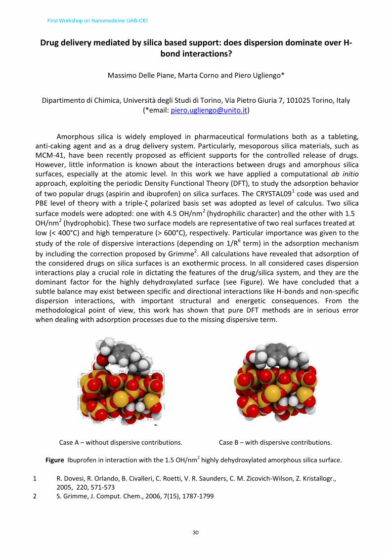
First Workshop on Nanomedicine UAB-CEI
30
Drug delivery mediated by silica based support: does dispersion dominate over H- bond interactions?
Massimo Delle Piane, Marta Corno and Piero Ugliengo*
Dipartimento di Chimica, Università degli Studi di Torino, Via Pietro Giuria 7, 101025 Torino, Italy
(*email: [email protected])
Amorphous silica is widely employed in pharmaceutical formulations both as a tableting, anti-caking agent and as a drug delivery system. Particularly, mesoporous silica materials, such as MCM-41, have been recently proposed as efficient supports for the controlled release of drugs. However, little information is known about the interactions between drugs and amorphous silica surfaces, especially at the atomic level. In this work we have applied a computational ab initio approach, exploiting the periodic Density Functional Theory (DFT), to study the adsorption behavior
of two popular drugs (aspirin and ibuprofen) on silica surfaces. The CRYSTAL091 code was used and PBE level of theory with a triple-ζ polarized basis set was adopted as level of calculus. Two silica
surface models were adopted: one with 4.5 OH/nm2 (hydrophilic character) and the other with 1.5 OH/nm2 (hydrophobic). These two surface models are representative of two real surfaces treated at low (< 400°C) and high temperature (> 600°C), respectively. Particular importance was given to the
study of the role of dispersive interactions (depending on 1/R6 term) in the adsorption mechanism
by including the correction proposed by Grimme2. All calculations have revealed that adsorption of the considered drugs on silica surfaces is an exothermic process. In all considered cases dispersion interactions play a crucial role in dictating the features of the drug/silica system, and they are the dominant factor for the highly dehydroxylated surface (see Figure). We have concluded that a subtle balance may exist between specific and directional interactions like H-bonds and non-specific dispersion interactions, with important structural and energetic consequences. From the methodological point of view, this work has shown that pure DFT methods are in serious error when dealing with adsorption processes due to the missing dispersive term.
Case A – without dispersive contributions. Case B – with dispersive contributions.
Figure Ibuprofen in interaction with the 1.5 OH/nm2 highly dehydroxylated amorphous silica surface.
1 R. Dovesi, R. Orlando, B. Civalleri, C. Roetti, V. R. Saunders, C. M. Zicovich-Wilson, Z. Kristallogr.,
2005, 220, 571-573 2 S. Grimme, J. Comput. Chem., 2006, 7(15), 1787-1799
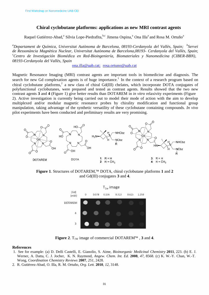
First Workshop on Nanomedicine UAB-CEI
31
Chiral cyclobutane platforms: applications as new MRI contrast agents
Raquel Gutiérrez-Abad,a
Sílvia Lope-Piedrafita,b,c
Jimena Ospina,a
Ona Illaa and Rosa M. Ortuño
a
aDepartament de Química, Universitat Autònoma de Barcelona, 08193-Cerdanyola del Vallès, Spain;
bServei
de Ressonància Magnètica Nuclear, Universitat Autònoma de Barcelona,08193- Cerdanyola del Vallès, Spain; cCentro de Investigación Biomédica en Red-Bioingeniería, Biomateriales y Nanomedicina (CIBER-BBN),
08193-Cerdanyola del Vallès, Spain
[email protected]; [email protected] Magnetic Resonance Imaging (MRI) contrast agents are important tools in biomedicine and diagnosis. The
search for new Gd complexation agents is of huge importance.1
In the context of a research program based on
chiral cyclobutane platforms,2
a new class of chiral Gd(III) chelates, which incorporate DOTA conjugates of polyfunctional cyclobutanes, were prepared and tested as contrast agents. Results showed that the two new contrast agents 3 and 4 (Figure 1) give better results than DOTAREM in in vitro relaxivity experiments (Figure
2). Active investigation is currently being carried out to model their mode of action with the aim to develop
multiplexed and/or modular magnetic resonance probes by chirality modification and functional group
manipulation, taking advantage of the synthetic versatility of these cyclobutane containing compounds. In vivo
pilot experiments have been conducted and preliminary results are very promising.
Figure 1. Structures of DOTAREM, DOTA, chiral cyclobutane platforms 1 and 2
and Gd(III) conjugates 3 and 4.
Figure 2. T1W image of commercial DOTAREM , 3 and 4.
References 1. See for example: (a) D. Delli Castelli, E. Gianolio, S. Aime, Bioinorganic Medicinal Chemistry 2011, 223. (b) E. J.
Werner, A. Datta, C. J. Jocher, K. N. Raymond, Angew. Chem. Int. Ed. 2008, 47, 8568. (c) K. W.-Y. Chan, W.-T.
Wong, Coordination Chemistry Reviews 2007, 251, 2428.
2. R. Gutiérrez-Abad, O. Illa, R. M. Ortuño, Org. Lett. 2010, 12, 3148.

First Workshop on Nanomedicine UAB-CEI
32
MICROFLUIDICS PLATFORMS FOR BIOSENSING APPLICATIONS
Mariana Medina-Sanchez
1, Maria Guix
1, Sandrine Miserere
1, and Arben Merkoçi
1,2
1Nanobioelectronics and Biosensors Group, Catalan Institute of Nanotechnology (ICN), SPAIN
2Catalan Institute of Research and Advanced Studies (ICREA), SPAIN
Electrochemical detection exhibits great advantages such as low power requirements, low limits of
detection, high sensitivity. The last years, the use of microfluidic platforms for electroanalysis has
increased in a remarkable way. This trend can be explained by the fact that most of microdevices
work under certain hydrodynamic conditions which enhance mass transport toward the detector
surface (working electrode) resulting in an increase of the obtained current and sensitivity compared
to the classical static measurement modes. Simple and miniaturized micro/nanofluidic platforms are
especially interesting due to their advantages like the reduction of sample and reagent volumes, the
decrease of the time of analysis, the possibility of portability and the integration of conventional
analytical techniques. Furthermore it’s important to point out the role that nanomaterials can play in
terms of enhancing optical and electrochemical properties after being integrated into the microfluidic
platform or even in the electrode, where the detection event will be performed. Combined together,
nanotechnology, electrochemistry and microfluidics could provide a really powerful biosensor
platform.
Fluids can be pumped in microfluidic systems by means of physical systems, such as syringe or
peristaltic pumps. However, there is also the possibility of using applied electric fields in microfluidic
chips based on materials which can be charged, like glass. Analytes will be moved by electroosmosis
and they will have different retention times depending on the charge that they present. Electrophoretic
and magnetic manipulation can provide us very versatile microfluidic platform, where different
biosensing applications, like proteins, DNA analysis, cell counting or environmental and food control
can be held. Phenol was detected as electroactive alkaline phosphatase product, using rabbit IgG as
model protein. Magnetic beads were manipulated by using permanent magnets, and were used as a
solid support for immunoassay for the preparation, separation and detection of the model protein.
To find their way into routine clinical analysis, such devices must be inexpensive, disposable and amenable to mass production. Therefore, polymers and ”soft lithography” are increasingly replacing traditional materials and microfabrication technologies. Polydimethylsiloxane (PDMS) is the most popular substrate for fast prototyping. In our laboratory we are developing a flexible hybrid PDMS/polycarbonate (PC) microfluidic chip with integrated screen-printed electrodes in order to achieve electrochemical detection of QDs as labeling alternative. The detection of CdS QDs in the
range of 50 to 8000 ng mL−1
with a sensitivity of 0.0009 μA/(ng mL−1
) has been achieved. In addition
to the single in-chip flow through measurements, the design of a recirculation system with the aim of achieving lower detection limits using reduced volumes of sample (25 μL) was proposed as a proof- of-concept.
But sometimes PDMS is not the best candidate, in particular it is poorly adapted to mass production
requirements. That is why thermoplastics are preferred, as for example Cyclo-Olefin-Copolymer or
COC, which is a thermoplastic polymer that exhibits really high chemical resistance to acids, bases
and most polar solvents in comparison to other thermoplastics, such as PMMA or PC. They also have
good optical and mechanical properties and are FDA approved. Thus we are also working on the
integration of screen-printed electrodes (SPE) in a COC microfluidic platform. This microdevice is
still in development at our laboratory so as to integrate analytical steps such as preconcentration and
amplification with the objective to obtain a complete lab-on-a chip with interest for diagnostic
applications.

First Workshop on Nanomedicine UAB-CEI
33
Superparamagnetic Iron Oxide Nanoparticles for Tracking Amniotic Fluid Mesenchymal Stromal Cells in a
Myelomeningocele Ovine Fetal Model Though Magnetic Resonance Imaging
Margarita Codinach,a,b,c César G. García Fontecha,b,c Luciano Rodríguez,d,b , Cleofé Romagosae, Teresa Molinée and Silvia Lope‐Piedrafitafg
aBanc de Sang I Teixits, Divisió de Teràpies avançades (Xcelia), Barcelona;
bBioengineering, orthopedics and surgery in pediatrics, Vall d’Hebron
Institut de Recerca (VHIR), Barcelona; cUniversitat Autònoma de Barcelona (UAB);
dBanc de Sang i Teixits, Divisió de Teixits, Barcelona; ePathology
Department, Vall d’Hebron Hospital, Barcelona; fServei de Ressonància Magnètica Nuclear, UAB;
fgCentro de Investigación Biomédica en
Red‐Bioingeniería, Biomateriales y Nanomedicina (CIBER‐BBN), Spain.
[email protected], [email protected]
Myelomeningocele (MMC) is the most severe and common form of spina bifida. MMC is a congenital malformation
characterized by a closure defect of the spinal column that can result in considerable disability at birth [1]. Experimental
animal studies have shown that prenatal repair of the neural tube closure defect prevents additional neural injury [2],
however, simple closure seems to fail to reverse the neurologic injury incurred before surgery [3]. Different types of
biodegradable scaffolds to promote tissue remodeling and neuronal pathfinding are being evaluated in animal models of
MCC. These tissue engineering‐based implantation therapies would greatly benefit of imaging techniques for
noninvasive tracking of the implanted tissues. Superparamagnetic iron oxide nanoparticles (SPIONs) are used as cellular
contrast agents for the noninvasive detection of labeled cells on high‐resolution magnetic resonance images (MRI) [4]. In
this study, a protocol for ovine amniotic fluid‐derived mesenchymal stromal cell (oAF‐MSC) labeling with SPIONs has
been developed and optimized for MRI detection in a MMC sheep fetal model. oAF‐MSC cells cultivated with SPIONs at
different concentrations were prepared to evaluate MRI sensitivity in detecting labeled cells (Fig.1A). Using high
resolution MRI, it was possible to detect single labeled cells (Fig.1B). Biocompatible and biodegradable scaffolds seeded
with unlabeled and labeled oAF‐MSC cells were used in prenatal repair of MMC in lambs. Postnatal vertebras were later
examined by MRI for cell tracking (Fig. 2).
Regenerative medicine offers great promise for many diseases, especially those without current effective treatments.
This type of non‐invasive imaging techniques, which offer the ability to track the localization of labeled cells after
transplantation, will expedite progress in this field and help to achieve maximized therapeutic effect.
A) B) A) B)
f e
d
g a
b c
1 cm 1 mm
1 cm 1 cm
Fig. 1. Cell culture MRI studies. A) Phantoms containing labeled cells incubated at different SPION concentrations: a, media only;
b, unlabeled cells; c‐g, labeled cells at 1, 5, 10, 25, and 50
SPIONs/cell, respectively. B) High resolution MRI of the phantom with cells incubated at 1 SPION/cell.
References
Fig. 2. MRI of resected postnatal lamb vertebras repaired with scaffolds seeded with oAF‐MSC cells: A) Unlabeled cells and B) SPION labeled
cells (see arrow).
[1] Hirose et al., World J Surg. 2003, 27: 87; [2] Fontecha et al., Eur J Obstet Gynecol Reprod Biol. 2011, 156:171; [3] Spinner et al. J Obstet Gynecol Neonatal Nurs 2012, doi: 10.1111/j.1552‐6909.2012.01357.x.; [4] Slotkin et al., Neurotherapeutics 2007, 4:428.
Acknowledgement The present study was supported by a grant from the Mútua Madrileña Foundation (project number 08/2749). We also thank the Servei de Ressonància Magnètica Nuclear of the UAB for allocating time to this project in the 7T Bruker Biospec MRI spectrometer.

First Workshop on Nanomedicine UAB-CEI
34
Synthesis, Surface Modification and Immunological properties of
Peptide-conjugated Gold Nanoparticles
S. Rubio1, N. G. Bastús1 and V. F. Puntes1, 2, 3
1. Institut Català de Nanotecnologia (ICN), Campus UAB, 08193 Bellaterra, Barcelona, Spain. 2. Universitat Autònoma de Barcelona (AUB), Campus UAB, 08193 Bellaterra, Barcelona, Spain. 3. Institut Català de Recerca i Estudis Avançats (ICREA), 08010 Barcelona, Spain.
Although the remarkable rapid development of synthetic and functionalization protocols for
the use of inorganic nanoparticles (NPs) in biomedical applications, relatively little is known
about NP's behavior in complex biological systems. Thereby it is particularly true for the
immune system, which is responsible for maintaining body integrity, detecting and
categorizing self and non-self molecules in order to protect the host from succumbing to
infections.
Herein we explore the use of engineered inorganic nanoparticles as substrates to carry
multifunctional ligands to manipulate the immune system in a controlled manner via its
interaction with specific cell-membrane receptors1-3. For this purpose, we synthesized highly
monodispersed citrate-stabilized gold NPs following a kinetically-controlled seeded growth
strategy4. As-synthesized NPs were further functionalized with peptides by following a ligand
exchange via the thiol group of the cysteine moiety. Obtained samples were characterized
by UV-Vis spectroscopy, Dynamic Light Scattering and Z-Potential in order to determine the
success of the conjugation process as well as the stability and aggregation state of final
colloidal solutions. Special efforts were devoted to study the conformation structure of the
peptide layer onto the gold NP's surface by using monitoring the etch resistance to sodium
cyanide digestion5. Obtained results allowed us not only to determine the robustness of
functionalized NPs but also to study the degree of order of the peptide shell, a key factor
when correlating final structure of conjugated NP with biological effects.
References
1. N. G. Bastús, E. Sánchez-Tilló, S. Pujals, C. Farrera, C. López, E. Giralt, A. Celada, J.
Lloberas and V. Puntes, ACSNano, 2009, 3(6), 1335-1344.
2. I. Hamad, O. Al-Hanbali, A. C. Hunter, K. J. Rutt, T. L. Andresen and S. M. Moghimi, ACSNano 2010, 4(11), 6629-6638.
3. S. M. Moghimi, D. Peer and R. Langer, ACSNano, 2011, 5(11), 8454-8458.
4. N. G. Bastús, J. Comenge, V. F. Puntes, Langmuir, 2011, 27, 11098-11105.
5. B. C. Mei, E. Oh, K. Susumu, D. Farrell, T. J. Mountziaris, and H. Mattoussi, Langmuir 2009, 25(18), 10604–10611.

First Workshop on Nanomedicine UAB-CEI
35
Magneto immunosensor for the enumeration of CD4+
T lymphocytes in
HIV diagnosis
S. Carinelli1, C. Xufré Ballesteros2, M. Martí2, S. Alegret1, M.I. Pividori1
1 Grup de Sensors I Biosensors, Universitat Autònoma de Barcelona, Spain
2 Institute of Biotechnology and Biomedicine, Spain
The incidence of human immunodeficiency virus (HIV) infection and clinical disease continues to
increase rapidly in underdeveloped and developing countries.
In a patient with HIV infection, CD4 counts help determine the stage of infection, guide drug
choices and indicate the response of the patients to the treatment as well as disease progression.
Moreover, this indicator is also recommended for immune disorders, after an organ transplant or a
graft. In developed countries, CD4 counts for patient with HIV infection are usually determined every
3–6 months. Flow cytometry is the standard method for CD4+
cells counts, but the high investment of
the instrument and costly reagents make it unaffordable to most of the centres in a developing
country. To solve the urgent need for improved diagnostic tools of HIV, a magneto immunosensor with
electrochemical detection is presented, as a simple, rapid and inexpensive strategy for CD4+
T cells counting.
In this strategy, CD4+
T cells were successfully separated from the sample and preconcentrated
using one-step immunomagnetic separation based on the CD3 receptor and using magnetic particles
modified with antiCD3 antibody. The optimization of the immunomagnetic separation was performed
using optical microscopy as well as flow cytometry. After the immunomagnetic separation, the captured cells were then labelled by a biotinylated
antiCD4 antibody, followed by the reaction with the streptavidin-peroxidase conjugate. Finally, the
electrochemical detection was performed using a magneto electrode based on graphite epoxy
composite and compared with the optical detection in a magneto-ELISA procedure.
Preliminary results indicated that the LOD was as low as 20 CD4+
T cells per L of human serum
being t L, involving the whole medical interest range for counts for HIV-1-infected patients. Future work will be focused on the evaluation of this new strategy
in both healthy volunteers as well as HIV infected patients. Moreover, the immunomagnetic
separation will be also evaluated coupled with a genosensing strategy for detection of virus transcripts
for HIV confirmatory diagnosis.
Figure 1. Schematic representation of the bioassay for the rapid
CD4+
cell counting in peripheral blood by using magnetic particles.

First Workshop on Nanomedicine UAB-CEI
36
Positively charged polymeric nanoparticles from nano-emulsions
appropriate for biomedical applications
S. Leitner1, G. Calderó1, M. J. García-Celma2, C. Solans1
1 Institut de Química Avançada de Catalunya (IQAC)
Consejo Superior de Investigaciones Cientificas (CSIC) Centro de Investigación Biomédica en Red en Bioingeniería, Biomateriales y
Nanomedicina (CIBER-BBN) Barcelona, Spain
2 Dept. de Farmàcia i Tecnologia Farmacèutica. Univ. de Barcelona. Unitat Associada
d’I+D al CSIC. Barcelona, Spain
The use of nanoscience in medicine and, more specifically, in drug delivery is expected to spread rapidly. Nano-emulsions as non-viral vectors have become an important tool to give rise to this application. Especially cationic nano- emulsions have awaken enormous interest as aside from their small size cellular uptake could be enhanced due to the positive surface charges. Electrostatic interactions with negatively charged DNA are warranted resulting in nano-sized complexes appropriate for biomedical applications [1-2]. The aims of this work are the preparation and characterization of oil-in-water cationic nano-emulsions containing a biocompatible preformed polymer and their use for nanoparticle preparation. The Polymer-in-water (P/W) cationic nano-emulsions were obtained by the low-energy Phase Inversion Composition (PIC) emulsification method [3]. A mixture of a cationic and a nonionic surfactant was used to stabilize the nano-emulsions and a hydrophobically modified polysaccharide (HMPS), dissolved in a non-toxic volatile solvent, constituted the oil component. Nano-emulsions were obtained at O/S ratios between 60/40 and 80/20 and at water contents above 83 wt%. They showed an average droplet size of (typically) about 100 nm and positive Zeta potential values (about 30 mV). Visual stability assessment of nano-emulsions at 25ºC revealed no macroscopic changes during 11 days. Further stability studies performed by light backscattering at 25º C showed that no destabilisation phenomena like creaming or sedimentation took place during at least 24 hours after preparation. Nano-particles were prepared from the (P/W) nano-emulsions by using the solvent evaporation method, showing smaller sizes (about 90 nm) and Zeta potential values similar to those of the template nano-emulsion. The positive Zeta potential values obtained suggest that these polymeric nanoparticles are promising candidates for transfection purposes.
References:
[1] Torchilin V. P. (2006) Adv. Drug Delivery Rev. 58, 1532-1555. [2] Liu C.-H., Yu S.-Y. (2010) Colloid Surface B 79: 509-515. [3] Calderó C. et al (2011) J Colloid Interf Sci 353: 406-411.

First Workshop on Nanomedicine UAB-CEI
37
Phagomagnetic Separation and Electrochemical Detection of
Pathogenic Bacteria
S.Liébana1, D.Spricigo
2, P.Cortés
2, M. Llagostera
2, S.Alegret
1, M.I. Pividori
1*
1 Grup de Sensors i Biosensors, Departament de Química, Universitat Autònoma de Barcelona,
Bellaterra, Spain. 2
Unitat de Microbiologia, Departament de Genètica i Microbiologia, Universitat Autònoma de Barcelona, Bellaterra, Spain
* Tel: +34 93 581 4937, E-mail address: [email protected]
Phage-based diagnostic is attracting much interest due to bacteriophages possess features such
as specificity and rapid growth, which make them ideal agents for the rapid detection of bacteria. As a
new bioselective material, phages combine unique characteristics of affinity reagents and self-
assembling proteins. As a biorrecognition interface in biosensors devices, they may have improved
properties as the antibodies, since they are inexpensive, highly specific and strong binders, resistant
to high temperatures and environmental stresses [1].
In this work, phages are used as biorrecognition element for the magnetic separation of
pathogenic bacteria for the first time. The phage capabilities as biorrecognition element are explored
by using the model phage nanoparticle P22 towards Salmonella. P22 nanoparticles are immobilized
in an oriented way on activated magnetic beads. Further evaluation of this immobilization was done
by Coomassie Bradford Protein Assay, SEM microscopy and classical phage plating technique. The
bacteria are then captured and preconcentrated by the phage-modified magnetic beads throughout
the phage-host interaction. To confirm the identity of the bacteria, further double-tagging PCR
amplification of the captured bacteria DNA and electrochemical magneto genosensing of the amplicon
are performed. The method is able to detect as low as 1 cfu·mL-1
showing highly specificity in a
considerable reduced assay time of 3 hours including PCR amplification time. The features of this
approach are discussed and compared with classical culture methods and PCR-based assay.
PHAGOMAGNETIC
SEPARATION AND
BACTERIA LYSIS
PCR
AMPLIFICATION
ELECTROCHEMICAL
GENOSENSING
H2O H2O2
DIG primer
BIO primer
HRP
ox HRP
red
red HQ ox
Double-tagged amplicon
magneto sensor m-GEC
Schematic representation of the phagomagnetic separation and electrochemical detection approach.
References
[1] A.E. Smartt, S. Ripp. Anal Bioanal Chem., 400, 991-1007, 2011.

First Workshop on Nanomedicine UAB-CEI
38
Multiplex Electrochemical Genosensing of Pathogenic Bacteria by
using Silica Magnetic Particles
S.Liébana1, S. Campoy
2, P.Cortés
2, S.Alegret
1, M.I. Pividori
1*
1 Grup de Sensors i Biosensors, Departament de Química, Universitat Autònoma de Barcelona,
Bellaterra, Spain. 2
Unitat de Microbiologia, Departament de Genètica i Microbiologia, Universitat Autònoma de Barcelona, Bellaterra, Spain
* Tel: +34 93 581 4937, E-mail address: [email protected]
The control of food quality has become of growing interest for both consumer and food industry
since the increasing incidence of food poisoning is a significant public health concern for customers
worldwide. Among food pathogens, Escherichia coli, Listeria monocytogenes and Salmonella
typhimurium have been the source –in the last decade– of many outbreaks [1].
Several strategies of detection using Salmonella as a model have been developed previously in
our group [2], [3]. In this work, a rapid and sensitive method for the multiplex detection of food
pathogenic bacteria is reported. After the lysis of the bacteria, further amplification of the genetic
material by multiplex PCR with a labelled set of specific primers for each pathogen is performed. The
amplicon, tagged with fluorescein, digoxigenin and biotin for Salmonella, E.coli and Listeria
respectively, was then immobilized on silica magnetic beads. The method is based on the lysing and
nuclease-inactivating properties of the chaotropic agent guanidinium thiocyanate together with the
nucleic acid-binding properties of silica particles in the presence of this agent, previously described by
Boom et al. [4], [5]. To confirm the identity of the three bacteria a magneto genosensing strategy is
performed using three different electrochemical tags, antiFluorescein-HRP, antiDigoxigenin-HRP and
Streptavidin-HRP conjugates for Salmonella, E. coli and Listeria amplicons, respectively.
This method is able to clearly distinguish among pathogenic bacteria and can be considered as
rapid alternative to the time consuming classical methodology by replacing the selective enrichment
and biochemical/serological tests.
Schematic representation of the multiplex electrochemical genosensing approach.
References
[1] M. Upmann, C. Bonaparte. “Rapid methods for food hygiene inspection. In Encyclopedia of Food Microbiology”; Academic Press, New York, p 1887, 2004. [2] S.Liébana, A.Lermo, S.Campoy, J.Barbé, S.Alegret, MI. Pividori. Anal.Chem., 81, 5812-5820, 2009. [3] S.Liébana, A.Lermo, S.Campoy, MP.Cortés, S.Alegret, MI. Pividori, Biosensors and Bioelectronics, 25, 510-513, 2009. [4] R. Boom, C. J. A. Sol, M. M. M. Salimans, C. L. Jansen, P. M. E. Wertheim-Van Dillen, J. Van der Noordaa. Journal of Clinical Microbiology, 495-503, 1990. [5] R. Boom, C. Sol, M. Beld, J. Weel, J. Goudsmit, P. Wertheim-Van Dillen. Journal of Clinical Microbiology, 615-619, 1999.

First Workshop on Nanomedicine UAB-CEI
39
Magneto Immunoassays for the detection of Plasmodium falciparum
Histidine-Rich Protein 2 Related to Malaria
Tamara Laube†, Michelle De Souza
‡, Hideko Yamanaka
‡, Salvador Alegret
†, Isabel Pividori
†.
†Grup de Sensors i Biosensors, Universitat Autònoma de Barcelona. Edifici Cn, 08193, Bellaterra. ‡UNESP, Laboratorio de Eletroanalítica,Instituto de Química, Universidade Estadual Paulista.
Campus de Araraquara, Sao Paulo, Brazil.
About 40% of the world’s population living in the poorest countries is at risk of malaria
infection. A recent study emphasizes the difficulty in making a presumptive diagnosis of malaria and
highlights the urgent need for improved diagnostic tools that can be used at the community and
primary care level, especially in poorer populations. Rapid diagnostic tests based on the detection of
Plasmodium-specific proteins as parasite biomarkers detectable in whole blood, serum, and plasma
seem to be good candidates for the development of novel rapid and decentralized methods. Some
examples are the histidine-rich protein 2 (HRP2) (only found in Plasmodium falciparum), as well as
the glycolytic lactate dehydrogenase (LDH) and the Plasmodium aldolase (both found in all
Plasmodium species). However, better sensitivity was reported for P. falciparum HRP2-based assays
compared with LDH and aldolase tests for the detection of P. falciparum. In this work, two magneto immunoassay-based strategies for the detection of HRP2 related to
malaria are described for the first time by using magnetic micro- and nanoparticles. The covalent immobilization of a commercial monoclonal antibody toward the HRP2 protein in magnetic beads and
nanoparticles was evaluated and compared. The immunological reaction for the protein HRP2 was successfully performed in a sandwich assay on both kinds of particles by using a second monoclonal antibody labeled with the horseradish peroxidase enzyme (HRP). Then, the modified magnetic particles were easily captured by a magneto sensor made of graphite-epoxy composite (m-GEC) which was also used as the transducer for the electrochemical detection. The performance of the immunoassay-based strategy with the electrochemical magneto immunosensors was successfully evaluated and compared with a novel magneto-ELISA based on optical detection using spiked serum samples. Improved sensitivity was obtained when using 300 nm magnetic nanoparticles in both cases. The electrochemical magneto immunosensor coupled with magnetic nanoparticles have shown better
analytical performance in terms of limit of detection (0.36 ng mL-1
), which is much lower than the
LOD reported by other methods. Moreover, at a low level of HRP2 concentration of 31.0 ng mL-1
, a
signal of 15.30 μA was reached with a cutoff value of 0.34 μA, giving a clear positive result with a nonspecific adsorption ratio of 51. Due to the high sensitivity, this novel strategy offers great promise for rapid, simple, cost-effective, and on-site detection of falciparum malaria disease in patients, but also to screen out at-risk blood samples for prevention of transfusion-transmitted malaria.
Reference:
De Souza Castilho, M. ; Laube, T. ; Yamanaka, H. ; Alegret, S. ; Pividori, M.I. Anal. Chem.. 2011, 83, 5570–
5577

First Workshop on Nanomedicine UAB-CEI
40
Self‐assembling protein‐only artificial viruses
U. Unzueta 1,2,3, J. Domingo‐Espín 1,2,3, P. Saccardo 1,2,3, X. Zhikun 1,2,3 J. Cedano 4,5, O. Conchillo 5, E. García‐Fruitós 1,2,3, X. Daura 4,5, N. Ferrer‐Miralles 1,2,3 A.Villaverde 1,2,3, E. Vázquez 1,2,3.
1. Applied Microbiology Unit, Institut de Biotecnologia i de Biomedicina, Universitat Autònoma de Barcelona, Bellaterra (Spain)
2. Department of Genetics and Microbiology, Universitat Autònoma de Barcelona, Bellaterra (Spain) 3. CIBER de Bioingeniería, Biomateriales y Nanomedicina (CIBER‐BBN) Bellaterra, Barcelona (Spain) 4. Departament de Bioquímica i Biologia Molecular, Universitat Autònoma de Barcelona. Bellaterra (Spain) 5. Computational Biology and Proteomics, Institut de Biotecnologia i de Biomedicina, Universitat
Autònoma de Barcelona, Bellaterra (Spain)
Different studies have described the ability of arginine‐rich peptides to condensate DNA, to cross cell membranes and to localize in cell nucleus. Since these properties make them very useful functional elements of artificial viruses for gene therapy and drug delivery, they have become very well known peptides in molecular medicine. Very recently, we have shown that the homogeneous peptide R9 acts, in addition, as an unexpected architectonic agent at the nanoscale, promoting the self‐assembling of a multifunctional protein that contains the R9 domain in it, as protein‐only, planar nanoparticles of 20 nm in diameter [1]. These particles that efficiently accumulate in the cell nucleus in less than two hours after exposition [2], are able to bind, condensate and deliver expressible DNA [1]. Conventional self‐assembling amyloidogenic peptides usually render fibers or amorphous aggregates. However, the self‐organizing properties of R9 seemed to result in much more regular nanoparticles. Thus, in this work we explored if cationic peptides other than R9 could also promote the self‐assembling of holding building blocks. A series of diverse amino acid sequences and structures containing unrelated peptides were tested as architectonic tags by using an EGFP as convenient building block. Interestingly, all these peptides were able, at different extents, to promote the spontaneous formation of protein nanoparticles of different sizes, ranging from 20 to 100 nm, in a process in which the arginine residues seems to be critical for the final geometry of the resulting particles. On the contrary, lysine‐rich peptides, which are also strongly positively charged and are very useful as DNA condensers, do not show any architectonic ability when incorporated to artificial viruses [3]. The use of arginine‐rich peptides as structural agents of protein‐only nanoparticles opens interesting possibilities to the tailoring particle geometry through classical protein engineering, a possibility so far unprecedented in bionanomedicine
References:
[1] Vazquez, E., Roldán, M, Diez‐Gil, C, Unzueta, U., Domingo‐Espin, J., Cedano, J, Conchillo‐Sole, O.,
Ratera, I, Veciana, J, Ferrer‐Miralles, N, and Villaverde, A. Nanomedicine 5 (2010).259‐268. [2] E. Vazquez, R. Cubarsi, U. Unzueta, M. Roldan, J. Domingo‐Espin, N. Ferrer‐Miralles, and A. Villaverde, Biomaterials, 31 (2010) 9333‐9339. [3] J. Domingo‐Espin, E. Vazquez, J. Ganz, O. Conchillo‐Sole, E. García‐Fruitós, J. Cedano, U. Unzueta, V. Petegnief, N. Gonzalez‐Montalban, A. M. Planas, X. Daura, H. Peluffo, N. Ferrer‐Miralles, and A. Villaverde, Nanomedicine 6 (2011) 1047‐1061.

First Workshop on Nanomedicine UAB-CEI
41
Cells growth over surfaces patterned with inclusion bodies: impact on morphology and orientation revealed by image
processing and statistical analysis
Witold I. Tatkiewicz1,2
, Joaquin Seras-Franzoso1,3
, Elena García-Fruitós1,3
, Esther Vazquez1,3,4
, Imma
Ratera1,2
, Nora Ventosa1,2
, Antonio Villaverde1,3,4
, Jaume Veciana1,2
1
CIBER de Bioingeniería, Biomateriales y Nanomedicina (CIBER-BBN), Bellaterra, Barcelona (Spain) 2
Department of Molecular Nanoscience and Organic Materials, Institut de Ciencia de Materials de
Barcelona (CSIC), Bellaterra, Barcelona (Spain) 3 Institute for Biotechnology and Biomedicine (IBB), Universitat Autonoma de Barcelona, Bellaterra,
Barcelona (Spain) 4 Department of Genetics and Microbiology, Universitat Autònoma de Barcelona, Bellaterra, Barcelona
(Spain)
A B
C D
Figure. 1 Confocal microscopy images of fibroblasts cultivated over IB´s-decorated supports with various patterns: A) stripes 5µm, B) stripes 20µm, C) stripes 50µm, D) dots 20µm. Green – IB´s (GFP), red – membrane (CellMask), blue – nuclei (Hoestch). Bar length indicates 100 µm.
Inclusion Bodies (IB´s) are highly pure protein deposits in the size range of a few hundred nanometers produced by recombinant bacteria. Although IBs were supposed to be undesirable side products on protein transcription processes it has been recently probed that those materials retain part of their original functionality
[ 1] and
that it is possible to tailor its properties during production
[ 2]. We already presented experiments with IB
assisted cell growth[ 3]
and proved that IB´s are biocompatible and have positive impact on cell proliferation
[ 4] and bioadhesivenes
[ 5]. In this study, we
have stepped forward studying the impact of cell growth over microengineered surfaces with the presence of green fluorescence protein (GFP) IB´s.
We have used the microcontact printing (µCP)[ 6]
to decorate silicon supports with 2D patterns of the IBs. This technique is recognized as a cheap, fast and versatile tool to control the surface chemistry and address surface properties at the micro-level scale. However, unlike other research on this topic we have not changed the local wettability of the supports, letting cells
adhere freely all over the surface[ 7]
. Six different pattern geometries to print the IBs were selected: stripes of 5, 20 and 50 μm width (and the same distance between stripes), dots of 20 μm diameter (spaced correspondingly) and two control surfaces with randomly distributed IBs and without IB´s. We cultured 1BR3.G human skin fibroblasts during 24, 48 and 72 hours.
Fluorescence images of dyed cells (membrane and nuclei) were used as a tool to study the cell´s position, orientation and morphology in respect to the patterned IBs which gives a green fluorescence. The results obtained from the image analysis were treated in a novel, statistical way. Gathered data clearly proves that cells a) adhere to IB rich areas, b) align and elongate according to the IB’s pattern, c) choose the shortest way between IBs to reach new adhesion (IBs) points.
This work fills the gap between the protocols that use local changes of surface chemistry and the ones using modification of topography at the submicron level for cell proliferation studies. It also proves that IB´s is an interesting and useful material in cell proliferation studies making it especially appealing for regenerative medicine.
[1] E. García-Fruitós et.al.; Trends in Biotechnology, 2012, 30, 2, 65-70 [2] E. García-Fruitós et.al. Nanotechnology ,2010, 21, 205101 (8pp) [3] E. García-Fruitós et.al. Adv. Mater. 2009, 21, 4249–4253 [4] C. Díez-Gil et.al. Biomaterials 2010, 31, 5805-5812 [5] J. Seras-Franzoso et.al. Nanomedicine, 2012,, 7(1), 79–93 [6] A. C. Philipsborn et.al. Nature Protocols, 2006, 1 (3), 1322-1328 [7] J. Zhang et.al. Adv. Funct. Mater. 2011, XX, 1–9