fitrah mujahidah abdul hamid - core.ac.uk · hela human cervical adenocarcinoma cell line kpa ......
TRANSCRIPT
THE EFFECT OF OTUB1 OVEREXPRESSION ON
HUMAN CERVICAL CANCER HeLa
By
FITRAH MUJAHIDAH ABDUL HAMID
Dissertation Submitted in Partial Fulfillment Of The Requirement For The
Degree Of Master Of Science
UNIVERSITI SAINS MALAYSIA
2015

ii
ACKNOWLEDGEMENT
Alhamdulillah, praise to the Almighty Allah for the successful completion of this
dissertation.
I would like to express my appreciation to my supervisor Dr. Nurulisa Zulkifle for
introducing me to the methodology of work, and for her passion, support and guidance.
I would like to thank to my co-supervisor Dr. Nor Hazwani Ahmad for the guidance and
encouragement in carrying out this project work.
I also wish to express my gratitude to my fellow friends in M.Sc Medical Research
(2014/15) and all teaching staff as well as non-teaching staff in IPPT who rendered their
help during the period of my project work. This thesis is dedicated to my husband and
child who have always stood by me and give me continuous support and strength.
iii
TABLE OF CONTENTS
ACKNOWLEDGEMENT ii
TABLE OF CONTENTS iii
LIST OF FIGURES vi
LIST OF TABLES vii
LIST OF SYMBOLS AND ABBREVIATIONS viii
ABSTRAK x
ABSTRACT xii
CHAPTER I - INTRODUCTION
1.1 Research background 1
1.2 Hypothesis of study 3
1.3 Objectives of study 3
CHAPTER II - LITERATURE REVIEW
2.1 Cancer statistic 4
2.2 Protein ubiquitination 5
2.3 DUB and cancer pathway 8
2.4 OTUB1 11
2.5 Therapeutic application of ubiquitination 13
2.6 Targeting p53 as anticancer therapy 14
2.7 Regulations of p53 by OTUB1 15
CHAPTER III - MATERIALS AND METHODS
3.1 Introduction 16
3.2 Preparation of cells 16
3.2.1 Cells lines 16
3.2.2 Thawing of HeLa cells and L929 17
3.2.2 Subculturing of cell lines 17

iv
3.3 pEGFP-N1-OTUB1 Vector Information 18
3.4 Transfection using Lipofectamine ™ 3000 20
3.5 Viability assays 21
3.5.1 Reagent preparation 21
3.6 Statistical analysis 22
3.7 Flow chart of the study 23
CHAPTER IV - RESULT
4.1 Transfection of pEGFP-N-OTUB1 24
4.1.1 Transfection of pEGFP-N-OTUB1 on HeLa cells 24
4.1.2 Transfection of pEGFP-N-OTUB1 on L929 cells 28
4.2 Effect of OTUB1 on the proliferation of HeLa cells 32
4.3 Effect of OTUB1 on the proliferation of L929 cells 34
CHAPTER V - DISCUSSION
5.1 Transfection of pEGFP-N-OTUB1 36
5.1.1 Transfection of pEGFP-N-OTUB1 on HeLa cells and L929 cells 36
5.2 Effect of OTUB1 on the proliferation of HeLa cells and L929 cells 39
CHAPTER VI - CONCLUSION
6.1 Summary 43
6.2 Recommendation 44
REFERENCES 45
v
LIST OF FIGURES
Page
Figure 2.1 Ubiquitination of proteins (D’Arcy et al ., 2015).. 8
Figure 2.2
Deubiquitinases are important regulators of oncogenes and tumour
suppressors (Sacco et al ., 2010).
11
Figure 3.1 Vector information of pEGFP-N1-OTUB1 19
Figure 4.1 Image of transfected OTUB1 on HeLa cells after 24 hour of
transfection.
25
Figure 4.2 Image of transfected OTUB1 on HeLa cells after 48 hour of
transfection.
26
Figure 4.3 Image of transfected OTUB1 on HeLa cells after 72 hour of
transfection.
27
Figure 4.4 Image of transfected OTUB1 on L929 cells after 24 hour of
transfection.
29
Figure 4.5 Image of transfected OTUB1 on L929 cells after 48 hour of
transfection.
30
Figure 4.6 Image of transfected OTUB1 on L929 cells after 72 hour of
transfection.
31
Figure 4.7 The effect of OTUB1 on HeLa cancer cell proliferation. 33
Figure 4.8 The effect of OTUB1 on L929 cancer cell proliferation. 35

vi
LIST OF TABLES
Page
Table 1.1 List of OTU DUBs that have mutation in cancer cell lines (Sacco
et al., 2010)
13

vii
LIST OF SYMBOLS AND ABBREVIATIONS
% Percentage
°C Degree celcius
µl Microliter
ANOVA Analysis of variance
CO2 Carbon dioxide
DMEM Dulbecco’s Modified Eagle Medium
DMSO Dimethyl Sulfide
DNA Deoxyribonucleic acid
DUBs Deubiquitinating enzyme
E1 Ubiquitin-activating enzyme
E2 Ubiquitin-conjugating enzymes
E3 Ubiquitin-protein ligases
FBS Fetal Bovine Serum
g Gram
GFP Green Fluorescent Protein
HCL Hydrogen chloride
HeLa Human cervical adenocarcinoma cell line
kPA Kilopascal
ml Milliliter
viii
mM Milimolar
mm Milimeter
MTT 3-(4,5-dimethylthiazol-2-yl)-2,5-diphenyltetrazolium
nm Nanometer
OTUB1 Ovarian tumour domain-containing Ub aldehyde-binding protein 1
PBS Phosphate buffer saline
RT-PCR Real-time polymerase chain reaction
SEM Standard error of the mean
SDS Sodium dodecyl sulfate
SPSS Statistical Package for the Social Sciences
USA United States of America
USP Ub-specific protease
UPS Ubiquitin–proteasome system
ix
KAJIAN KESAN EKSPRESI BERLEBIHAN OTUB1 PADA BARAH SERVIKAL
MANUSIA HeLa
ABSTRAK
Protein ubiquitinasi adalah proses regulasi yang mengawal kebanyakan mekanisma
fisiologi dan patologi dan yang melibatkan perkembangan tumor. Enzim deubiquitinasi
(DUBs) semakin diterima bahawa ia bermutasi pada kanser manusia dan mempunyai
peranan sebagai onkogen dan penahan tumor. Kajian terdahulu mendapati DUBs
mengawal proses yang berkaitan dengan proliferasi sel dan apoptosis, menjadikan ia
sasaran untuk terapi kanser. Dalam kajian ini, kami ingin mengkaji sama ada tumor
ovari protease (OTU) iaitu dalam keluarga DUBs mempengaruhi sel proliferasi sel
HeLa. Kami melakukan kajian kesan ekspresi berlebihan pada kanser servikal HeLa dan
sel L929. Tranfeksi dilakukan menggunakan Lipofectamine™3000 pada kedua-dua sel.
Kecekapan transfeksi diukur menggunakan mikroskop fluorescence. Ujian MTT
dijalankan untuk melihat proliferasi sel HeLa dan L929. Analisis statistic dijalankan
menggunakan ujian ANOVA satu hala. OTUB1 secara signifikan membantut
pertumbuhan sel HeLa pada semua masa inkubasi jika dibandingkan dengan sel
kawalan. Kesimpulannya, ekspresi berlebihan OTUB1 membantut proliferasi sel HeLa.
x
THE EFFECT OF OTUB1 OVEREXPRESSION ON HUMAN CERVICAL
CANCER HeLa
ABSTRACT
Protein ubiquitination is a highly regulated process that controls multiple physiologically
and pathologically relevant mechanisms involved in tumor development. There is
growing recognition that mutated deubiquitinating enzyme (DUBs) in human cancers
suggesting their roles as oncogenes and tumor suppressors. Previous studies have
identified the DUBs regulate processes associated with cell proliferation and apoptosis,
and as such represent candidate targets for cancer therapeutics. Here we investigated
whether members of the ovarian tumor proteases (OTU) family of DUBs influence the
proliferation in HeLa cells. We intended to study the effect of OTUB1 overexpression in
cervical cancer HeLa and L929 cells lines. Transfection with Lipofectamine™ 3000 was
performed in both cell lines. The transfection efficiency was measured by fluorescence
microscopy. MTT assay was performed to evaluate cell proliferation on HeLa and L929
cells. Statistical analysis was performed by using one way ANOVA test. OTUB1 has
significantly inhibit the proliferation of Hela cells at all incubation times in comparison
to control. In conclusion, overexpression of OTUB1 inhibit the cell proliferation of
HeLa.

1
CHAPTER 1
INTRODUCTION
1.1 Research background
Cervical cancer is a cancer in tissues of the cervix. It is usually a slow growing
cancer that may not have symptoms but can be found with regular pap tests. Most cervical
cancer cases (83%) occur in developing countries in which they account for 15% of female
cancers, compared to 3.6% in developed regions (Gakidou et al., 2008). Cervical cancer is
the third most frequent cancer in women after breast and colorectal cancers and is one of
the leading causes of cancer death among women in the world (Ferlay et al., 2010).
According to Globocan statistics (2012) in Malaysia, about 2,145 new cervical cancer cases
are diagnosed annually. Cervical cancer ranks as the second cause of female cancer in
Malaysia and the second most common female cancer in women aged 15 to 44 years in
Malaysia. Compared among the major races, Chinese women had the highest incidence for
cervical cancer followed by the Indians and Malays (National Cancer Registry, 2006).
Therefore, there is critical need for better targeted therapies for cervical cancer.
OTUB1 is a member of OTU family which are part of deubiquitinating enzymes
(DUBs) family. It is establish for its deubiquitinating properties (Messick et al., 2008).
They play an important role in many physiological and pathological process such as
interferon signaling (Huang et al., 1995 ;Sass et al., 1995 ; Li et al., 2014). DUBs has been
reported regarding an association with cancer as it found mutated in human cancer
2
suggesting their roles as oncogenes and tumor suppressor genes. DUBs also found to play
crucial aspect in regulating cell proliferation (Hussain et al., 2009).
There are approximately 100 identified OTU family members of proteins from
eukaryotes, viruses and pathogenic bacteria (Balakirev et al., 2003). It is include OTUB1,
OTUB2, YOD1 and OTULIN. Previous study has shown that OTUB1 mediates certain
types of cancer such as prostate cancer cell invasion through RhoA activation and promotes
tumorigenesis in vivo (Iglesias-Gato et al., 2015). OTUB1 also interact with ERα which is
key factor involved in the development of breast and endometrial cancers in cells and in
vitro (Stanišić et al., 2009). Previous literature shows that OTUB1 from ovarian tumor
(OTU) family has been identified as a novel p53 regulator. There are an evidence show that
overexpression of OTUB1 cause apoptosis and inhibition of cell proliferations in a p53
dependant manner (Sun et al., 2011).
As a result of these finding, OTU family members rise as a promising regulator in
cancer associated pathway, human DUBs are increasingly regarded as a potential drug
target including cancer and neurodegeneration disease (Pfoh et al., 2015). However, most
of the research activities in OTU family are still limited. It could be interesting to see
overexpression of OTUB1 in cervical cancer using HeLa cell lines as the role of OTUB1 in
this disease has still not been fully elucidated.

3
1.2 Objective of the study
The aim of present study is to study overexpression of OTUB1 could significantly
enhance or inhibit proliferation of cancer cells in vitro. The specific study were :
1) To transfect and express OTUB1 clones in human cancer cell lines HeLa and
mouse fibroblast L929.
2) To assess the effect on proliferation of human cancer cell lines upon
overexpression of OTUB1 protein by MTT assay.
1.3 Hypothesis of the study
The expected outcomes of the study were :
1) Overexpression of OTUB1 could significantly promote proliferation of cancer
cells in vitro or inhibit proliferation of cancer cells in vitro.

4
CHAPTER II
LITERATURE REVIEW
2.1 Cancer Statistic
An estimated 14.1 million new cancer cases and 8.2 million cancer-related deaths
occurred in 2012, compared with 12.7 million and 7.6 million, respectively, in 2008. In
year 2012, there were 32.6 million people (over the age of 15 years) alive who had a cancer
diagnosed in the previous five years (World Health Organization, 2013).
The most commonly diagnosed cancers worldwide were those of the lung (1.8
million, 13.0% of the total), breast (1.7 million, 11.9%), and colorectum (1.4 million,
9.7%). The most common causes of cancer death were cancers of the lung (1.6 million,
19.4% of the total), liver (0.8 million, 9.1%), and stomach (0.7 million, 8.8%) (World
Health Organization, 2013).
Worldwide, cervical cancer is second only to breast cancer as the most common
female malignancy in both incidence and mortality, and results in approximately 275000
deaths annually (Parkin et al., 2005). Some 83% of the cases occur in developing
countries, where cervical cancer accounts for 15% of female cancers, with a risk before age
65 of 1.5%, while in developed countries it accounts for only 3.6% of new cancers, with a
cumulative risk (ages 0–64) of 0.8% ( Ferlay et al., 2004).
5
2.2 Protein Ubiquitination
Protein ubiquitination is a highly regulated process that controls multiple
physiologically and pathologically relevant mechanisms involved in tumor development.
The degree of ubiquitination of specific proteins is controlled by the concerted actions of
E3 ubiquitin ligases, deubiquitinating enzymes (DUBs) and the proteasome (Deshaies RJ &
Joazeiro CA., 2009; Komander D et al., 2009).
The attachment of ubiquitin to target proteins is mediated by an enzymatic cascade
consisting of E1 (ubiquitin-activating enzyme), E2 (ubiquitin-conjugating enzymes), and
E3 (ubiquitin-protein ligases) proteins (Fang & Weissman 2004). The existence of a large
number (N500) of E3 ligases makes them the main specificity factor in the UPS. Target
proteins may be monoubiquitinated or, as in this example, polyubiquitinated. A target
protein must be tagged with at least four ubiquitin monomers (forming a polyubiquitin
chain) to be recognized by the proteasome. DUBs, are components of the UPS that catalyze
the removal of ubiquitin moieties from target proteins or polyubiquitin chains, resulting in
altered signaling or changes in protein stability (D’Arcy et al., 2015).
Human genome encodes approximately 95 putative DUBs, grouped into five
families: Ub-specific protease (USP), Ub C-terminal hydrolase (UCH), ovarian tumour
(OTU) domain-containing protease, Machado–Joseph disease (MJD) protease, and
JAB1/MPN/Mov34 metalloenzyme (JAMM) (Nijman et al., 2005).

6
The ubiquitin–proteasome system (UPS) has many critical regulatory roles in
eukaryotic cellular processes including cell cycle progression, stress response, signal
transduction, transcriptional activation, and DNA repair (Ciechanover et al., 2000;
Ciechanover 2006).

7
Figure 2.1: Ubiquitination of proteins. Proteins are targeted for degradation by the addition
of ubiquitin chains to lysine residues by a process that involves three enzymes, E1,E2 and
E3. DUBs catalyze the removal of ubiquitin moieties from target proteins or polyubiquitin
chains, resulting in altered signaling or changes in protein stability. (D’Arcy et al., 2015).

8
2.3 DUB and cancer pathway
Oncogenes and tumor suppressors act at various points along the signal transduction
pathway between the plasma membrane and the nucleus affecting mitogenic processes in
ways that either enhance or slow cell growth. Given the pervasive role of ubiquitin DUBs
and cancer mediated signaling or targeted proteolysis in these pathways, it is not surprising
to find that DUBs play critical roles in regulating cell proliferation (Hussain et al., 2009).
Ubiquitination of oncoproteins and tumour suppressors can promote their
destabilization by targeting them for degradation (e.g., K48-linked poly-ubiquitination
specifies proteasomal degradation), or regulate their activity (activation or inactivation).
Activation here may refer to a variety of processes like translocation to the nucleus (e.g
PTEN and FOXO), or engagement in signalling protein interaction networks (TRAF6,
RIP1). Specific DUBs implicated in tumourigenesis. Previous study by Iglesias-Gato et al.,
2015 demonstrated that OTUB1 is overexpressed in prostate cancer suggests a role for
OTUB1 in tumorigenesis and invites additional exploration of its mechanisms of action.
The ubiquitin proteasome pathway is intricately involved in nearly all aspects of
cell biology. Ubiquitination is covalent attachment of the small protein modifier ubiquitin
to a substrate protein is involved in virtually all cellular processes by mediating the
regulated degradation of proteins. Deubiquitination is have been shown to play a role in the
cleavage of ubiquitin from translational precursors and in the maintenance of free ubiquitin
levels within the cell by the deubiquitinating enzymes (or DUBs). However, DUBs can also
remove both monoubiquitin and polyubiquitin chains from proteins, or can trim the distal

9
ubiquitin from polyubiquitin chains. Consequently, these activities can potentially
antagonize the functions of ubiquitination within the cell (Komander et al, 2009).
Deubiquitination plays an equally important regulatory role as well as ubiquitination. There
is a growing list of human cancers in which direct mutational alterations in DUBs has been
observed. Much more work is required in order to fully appreciate the role of
deubiquitination in malignancy (Hussain et al., 2009).

10
Figure 2.2. Deubiquitinases are important regulators of oncogenes and tumour suppressors.
Both overexpression and loss of function of DUBs can promote cancer (Sacco et al., 2010).
11
2.4 OTUB1
Ovarian-tumor-domain-containing proteases (OTUs) are part of the deubiquitinating
enzymes (DUBs) family (Makarova et al., 2000 ; Edelmann M, 2009). One of the most
recently recognized DUBs is the OTUs. This family mainly comprises a group of putative
cysteine proteases including OTUB1, OTUB2, A20 and yeast OTU1 (Edelmann M, 2009).
OTUB1 was the first member of OTU family to be confirmed for its
deubiquitinating properties. It is located at chromosomal position 11q13.1, and is
ubiquitously expressed in human tissues such as in kidney tissue (Messick et al., 2008 ;
Zhang et al., 2012).
OTUB1 is thought to play an important role in many physiological and pathological
processes of human being. The OTUB1 gene product is identified to be involved in the
control of cell division and differentiation of the cystoblast into an oocyte and nurse cells
(Huang YZ et al. 1995; Sass et al., 1995).
Although widely expressed, OTUB1 was specifically implicated in mediating
lymphocyte antigen responsiveness through affecting the stability of the lymphocyte-
specific E3 ligase GRAIL (gene related to anergy in lymphocytes) in CD4+ T-lymphocytes
(Soares L et al., 2004). Moreover, OTUB1 was also found in Lewy bodies of the brain on
mass spectrometry, and may be involved in the pathogenesis of neurodegenerative
disorders (Xia Q et al., 2008).

12
OUT
DUB
name
Mutation/translocation
Published
abnormalities/protein
level
Oncomine
Upregulation Downregu-
lation
OTUB1
None reported
None reported
Bladder/lung,pr
ostate, HNSCC,
breast
Brain,
HNSCC,
testis,
cervical,
sarcoma
A20 Chromosomal deletions and
inactivating mutations
found in several lymphoma
subtype. 2 out 11 lung
cancer
Study showing
overexpression in
Hodgkins and anaplastic
B-cell lymphomas with
downregulation in other
lymphoma types
HNSCC,
luekemia, lung,
brain, cervical.
Bladder,
ovary, lung,
lymphoma,
sarcoma
Cezanne None reported None reported Liver, myeloma Ovarian
TRABID 1/202 kidney cancer None reported Brain,
testiscular,
leukemia
Oesophagus,
liver
Brain,
leukemia,
liver,
testicular,
bladder
Table 1.1: Example of OTU DUBs including OTUB1 that have mutation and altered
expression in cancer cell lines, listed from COSMIC (Catalogue of Somatic Mutations in
Cancer) and Oncomine database. (Sacco et al., 2010)
13
2.5 Therapeutic application of ubiquitination
Key cancer-associated proteins whose levels are tightly controlled by the ubiquitin
proteasome include p53, p27, cyclins and BCL2 family members. The enzymes involved in
conjugation and deconjugation of ubiquitin to protein substrates include an activating ATP
dependent ubiquitin enzyme (E1), an ubiquitin-conjugating enzyme (E2), ubiquitin-protein
ligases (E3s) that often form multi-component complexes key for substrate recognition, and
deubiquitinases (DUBs) that cleave ubiquitin from protein substrates. In humans, there are
just a few E1 enzymes, around 40 E2 enzymes, over 500 E3 ligases (most commonly RING
and HECT domain E3s) and around 100 DUBs, the majority belonging to the ubiquitin-
specific protease (USP) sub-family (Lipkowitz & Weissman 2011, Budhidarmo et al.,
2012, Jacq et al., 2013).
These enzymes have major regulatory roles in normal cellular processes, both
within and independently of the ubiquitin-proteasome, including DNA repair, maintaining
genomic stability and transcription. Aberrant expression of a number of DUBs and E3s has
been linked to cancer (Lipkowitz & Weissman 2011 ;Clague et al., 2013). As a
consequence, many of these enzymes are generating extensive interest as targets for the
treatment of cancer (Marsh 2015).
Several DUBs have been implicated in various diseases, including neurological
disorders, infectious diseases and cancer. A genome-wide RNAi (RNA interference) screen
of the catalytically active human USPs in cancer-relevant cellular models and phenotypic
assays was performed to identify potential USP targets in cancer (Colland, F. 2006).
14
2.6 Targeting p53 as anticancer therapy
p53 plays a critical role in tumor suppression mainly by inducing growth arrest,
apoptosis, and senescence, as well as by blocking angiogenesis. In addition, p53 generally
confers the cancer cell sensitivity to chemoradiation. Thus, p53 becomes the most
appealing target for mechanism-driven anti-cancer drug discovery. The approaches
currently undertaken to target p53 and its regulators with an overall goal either to activate
p53 in cancer cells for killing or to inactivate p53 temporarily in normal cells for
chemoradiation protection. The compounds that activate wild type (wt) p53 would have an
application for the treat- ment of wt p53-containing human cancer. Likewise, the
compounds that change p53 conformation frommutant to wt p53 (p53 reactivation) or that
kill the cancer cells with mutant p53 using a synthetic lethal mechanismcan be used to
selectively treat human cancer harboring a mutant p53. The inhibitors of wt p53 can be
used on a temporary basis to reduce the normal cell toxicity derived from p53 activation.
Thus, successful development of these three classes of p53 modulators, to be used alone or
in combination with chemoradiation, will revolutionize current anticancer therapies and
benefit cancer patients (Wang & Sun 2010).
15
2.7 Regulations of p53 by OTUB1
The ubiquitin (Ub) proteasome system plays a pivotal role in the regulation of p53
protein stability and activity. p53 is ubiquitinated and destabilized by MDM2 and several
other Ub E3s, whereas it is deubiquitinated and stabilized by Ub-specific protease (USP)7
and USP10.
According to study by Sun et al., (2011) OTUB1 is a novel p53 regulator. OTUB1
directly suppresses MDM2-mediated p53 ubiquitination in cells and in vitro.
Overexpression of OTUB1 drastically stabilizes and activates p53, leading to apoptosis and
marked inhibition of cell proliferation in a p53-dependent manner.
Under physiological conditions, p53 is maintained at low levels primarily by the
oncoprotein MDM2. MDM2 also promotes p53 ubiquitination and degradation through the
proteasome system (Haupt et al, 1997; Kubbutat et al, 1997). Together, their results suggest
that OTUB1 have a novel function in regulating p53 stability and activity.
16
CHAPTER III
MATERIALS AND METHODS
3.1 Introduction
The major requirement is to maintain an aseptic work area that is restricted to cell culture
work. The procedure must be performing in a designated cell culture including sterile
handling, incubation, and storage of cell cultures, reagents, and media. The simplest and
most economical way to provide aseptic conditions is to use a cell culture hood (biosafety
cabinet). All material need in this study including glassware and plasticware were sterelize.
Autoclavable materials were autoclaved at 121 °C for 30 minutes at the pressure of 100
kPA prior to use.
3.2 Preparation of Cells
3.2.1 Cell line
Two cell lines were chosen in this study which is HeLa cell lines and L929 mouse
fibroblast. L929 mouse fibroblast was selected as it represent normal cell. Both cell lines
were maintain in the same medium. Dulbecco’s Modified Eagle Medium (DMEM) (Gibco,
USA) medium was used to culture HeLa (American Type Culture Collection ATCC, USA)
and L929 ( Life technology, USA) in suspension and supplemented with 10% (v/v) Fetal
Bovine Serum (FBS) (Gibco, USA), 1% (v/v) Penicillin-Streptomycin (Gibco, USA).
17
Centrifugation was done at 500 × g for 10 minutes after the collection of cells in log phase
of growth.
3.2.2 Thawing of HeLa Cells
The cryopreserved HeLa and L929 cells were taken out from a liquid nitrogen tank and
immediately soaked in a water bath at 37°C until cells become semi fluid. The cells were
transferred into a 15ml tube (BD Biosciences, USA) containing 5 ml of prewarmed
complete DMEM (Gibco, USA) and mixed gently. The cells suspension was centrifuged at
500 x g for 10 minutes. The supernatant of DMSO then discarded and the pellet was
resuspended with pre-warmed complete DMEM growth medium. The cells suspension was
transferred into 25 cm2 tissue culture flask. Then the cells suspension was incubated at
37°C in 5% CO2. Cells were routinely checked under inverted microscope to determine the
confluence of cells growth. The cells were subculture when the cells reached 70 to 80%
confluent of cell growth.
3.2.3 Subculturing of Cell lines
HeLa and L929 cells were detached from the flask by incubating 1 ml of trypsin express
(Gibco, USA) for 5 to 10 minutes in 5% CO2 air at 37 °C. Then the flask was gently tapped
to detached the cells from the wall of flask. The floating cells were transferred into the 15
ml tube and centrifuged for 10 minutes at 500 × g. The supernatant was discarded and the
pallet was resuspended with complete DMEM growth medium. The cells were transferred
into flask and incubated in an incubator at 37 °C in 5% CO2.
18
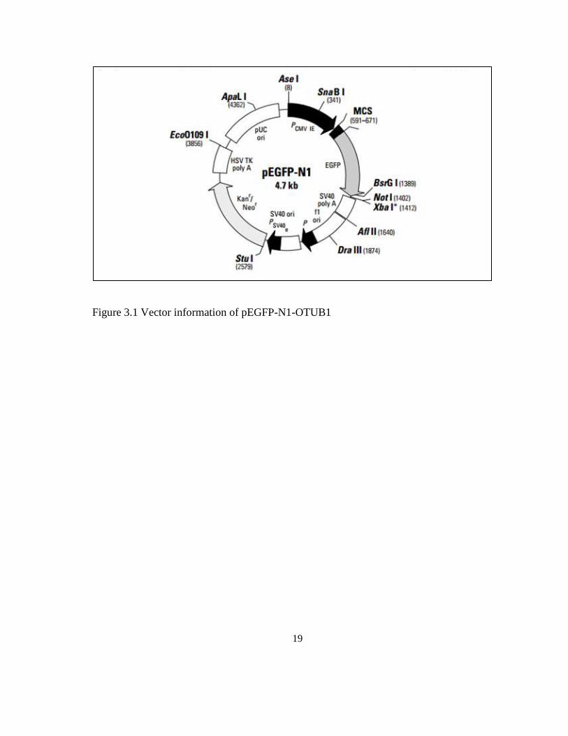
3.3 pEGFP-N1-OTUB1 Vector Information
pEGFP-N1 is used to insert genes of interest, OTUB1. It encodes a red-shifted variant of
wild-type GFP which has been optimized for brighter fluorescence and higher expression
in mammalian cells. pEGFP-N1 encodes the GFPmut1 variant which contains the double-
amino-acid substitution of Phe-64 to Leu and Ser-65 to Thr. The coding sequence of the
EGFP gene contains more than 190 silent base changes which correspond to human codon-
usage preferences. The MCS in pEGFP-N1 is between the immediate early promoter of
CMV (P CMV IE) and the EGFP coding sequences. Genes cloned into the MCS will be
expressed as fusions to the N-terminus of EGFP if they are in the same reading frame as
EGFP and there are no intervening stop codons. SV40 polyadenylation signals downstream
of the EGFP gene direct proper processing of the 3' end of the EGFP mRNA. The vector
backbone also contains an SV40 origin for replication in mammalian cells expressing the
SV40 T antigen. A neomycin-resistance cassette (Neor), consisting of the SV40 early
promoter, the neomycin/kanamycin resistance gene of Tn5, and polyadenylation signals
from the Herpes simplex virus thymidine kinase (HSV TK) gene, allows stably transfected
eukaryotic cells to be selected using G418. A bacterial promoter upstream of this cassette
expresses kanamycin resistance in E. coli. The pEGFP-N1 backbone also provides a pUC
origin of replication for propagation in E. coli and an f1 origin for single-stranded DNA
production.
19
Figure 3.1 Vector information of pEGFP-N1-OTUB1

20
3.4 Transfection using Lipofectamine ™ 3000
Cells were transfected at high cell density. One day before transfection, Cells were plated
0.5-2 x 10x 5 cells in 500 µl of growth medium without antibiotics so that cells will be 90
to 95% confluent at the time of transfection for high efficiency, high expression levels, and
to minimize cytotoxicity. Complexes were prepared using a DNA (µg) to Lipofectamine™
3000 (µl). Opti-MEM medium were used to dilute Lipofectamine™ 3000 and DNA before
complexing. Add diluted DNA to diluted Lipofectamine™ 3000 Reagent (1:1 ratio). DNA
concentration should contain 0.1 ng/ml in each well. But DNA stock available For HeLa
cells is 495 ng/ml. So each well of treated cell contain 0.2 µl of DNA. In L929 cells, 683.3
ng/ml were used and each well of treated cell were contain 0.15 µl of DNA. Incubate for 5
minutes at room temperature. After 5 minute incubation, combined diluted DNA with
diluted Lipofectamine™ 3000. Mix gently and incubate at 37°C for 4 days and continue
with viability assays.
3.5 Transfection efficiency analysis
Proliferation of HeLa cells and L929 cells in response to the OTUB1 overexpression were
were identified by fluorescence microscopy for expression of the OTUB1. Transfections
were identified by using Green Fluorescent Protein (GFP) as reporter gene.
21
3.4 Viability assays
3.4.1 Reagent Preparation
Proliferation of HeLa cells and L929 cells in response to the OTUB1 overexpression was
assessed by using MTT assays and the cell number determined by using standard
microplate absorbance readers. The protocol of MTT assay was performed according to the
method of developed by Mossman T (1983). 12 mM MTT stock solution were prepared by
adding 1 mL of sterile PBS to one 5 mg vial of MTT (Component A). The solution were
mix by vortexing or sonication until dissolved. Some particulate material that will not
dissolve is removed by filtration or centrifugation. Component B were prepared by add 10
mL of 0.01 M HCl to one tube containing 1 gm of SDS. The solution gently dissolves by
inversion or sonication. Once prepared, the solution should be used promptly. Cell were
treated with complex Opti-MEM medium, Lipofectamine ™ 2000, Lipofectamine ® 2000
Reagent. Cell were analysed in 0 hour, 12 hour, 24 hour and 48 hour. Cells seeded at
densities between 5000 to 10,000 cells per well. The medium were removed and replace
with 100 µL of fresh culture medium. 10 µL of the 12 mM MTT stock solution were added
to each well. 10 µL of the MTT stock solution also were added to 100 µL of medium of
negative control. Microplate were incubate for 4 hours. After that 100 µL of the SDS HCl
solution were added to each well and mix thoroughly using the pipette. Microplate were
incubate again at 37°C for 4 to 18 hours . Samples were mix again using a pipette and
absorbance were read at 570 nm.

22
3.5 Statistical Analysis
The representative data were presented as mean ± SEM. Statistical analysis was performed
using IBM SPSS Statistic Version 2.0. The comparison between control and treated was
tested for significance using one way ANOVA. Differences at p <0.05 were considered to
be statistically significant.
23
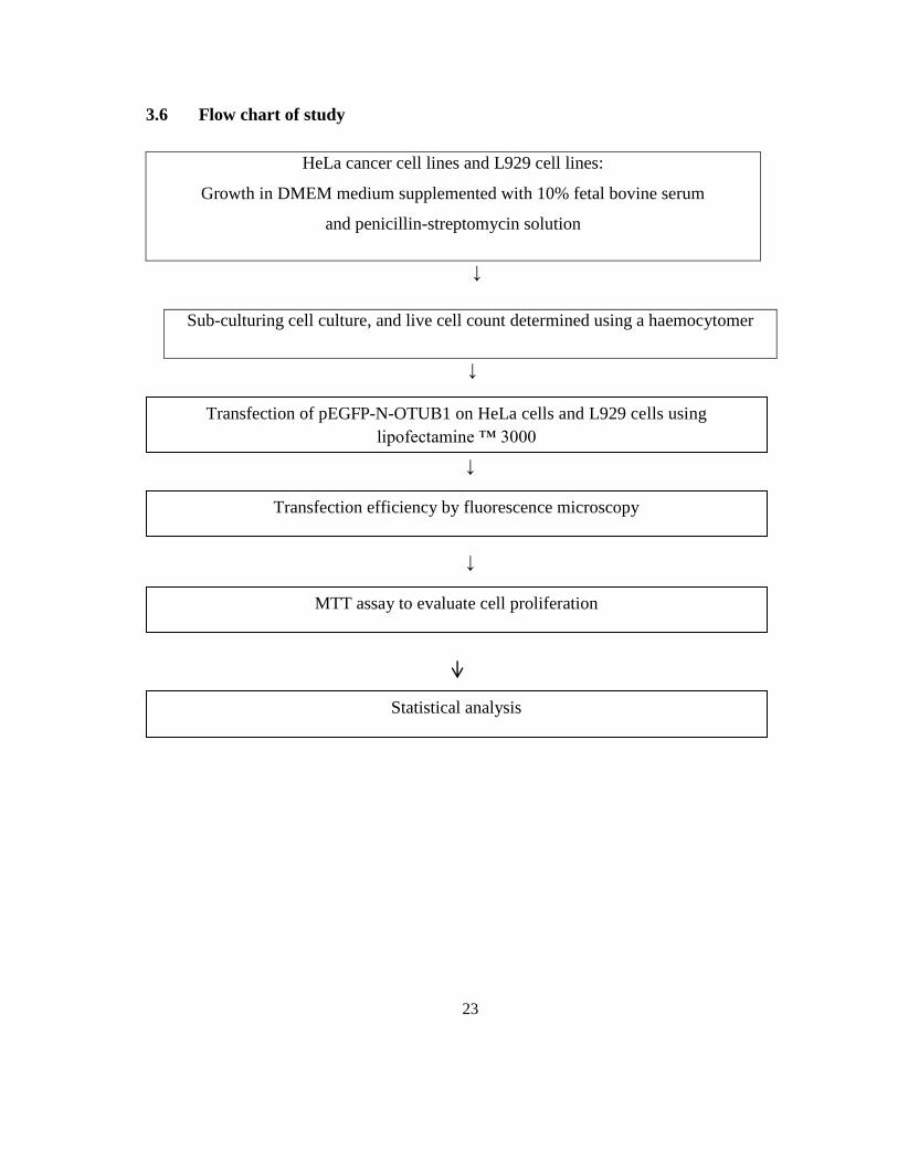
3.6 Flow chart of study
HeLa cancer cell lines and L929 cell lines:
Growth in DMEM medium supplemented with 10% fetal bovine serum
and penicillin-streptomycin solution
↓
Sub-culturing cell culture, and live cell count determined using a haemocytomer
↓
↓
↓
↓
Transfection of pEGFP-N-OTUB1 on HeLa cells and L929 cells using
lipofectamine ™ 3000
Transfection efficiency by fluorescence microscopy
MTT assay to evaluate cell proliferation
Statistical analysis

24
CHAPTER IV
RESULT
4.1 Transfection of pEGFP-N-OTUB1
4.1.1 Transfection of pEGFP-N-OTUB1 on HeLa cells
HeLa cancer cell lines were transfected with plasmid containing pEGFP-N1-OTUB1 in 96
well plates at 70% confluent. Cells were transfected by vectors with Lipofectamine™
3000 following the manufacturer’s protocol. Transfection were performed using an
appropriate concentration of DNA and 5 μL of Lipofectamine™ 3000 Reagent. Clones
were identified by fluorescence microscopy for expression of the OTUB1. Transfections
were identified by using Green Fluorescent Protein (GFP) as reporter gene.