flatworms, roundworms, & rotifers chapter 34. phylum platyhelminthes section 34.1
TRANSCRIPT

Flatworms, Roundworms, & Rotifers
Chapter 34

Phylum Platyhelminthes
Section 34.1

General Structure:
3 germ layers – ectoderm, mesoderm, and endoderm acoelomates
Bilateral symmetry Anterior and posterior ends
Dorsal and ventral surfaces only Flat body plan Flatworms!
General Functions:
Exchange oxygen and carbon dioxide directly with the environment to cells by diffusion No circulatory system or respiratory
system needed
Only one opening where food and wastes pass through!
Cephalization
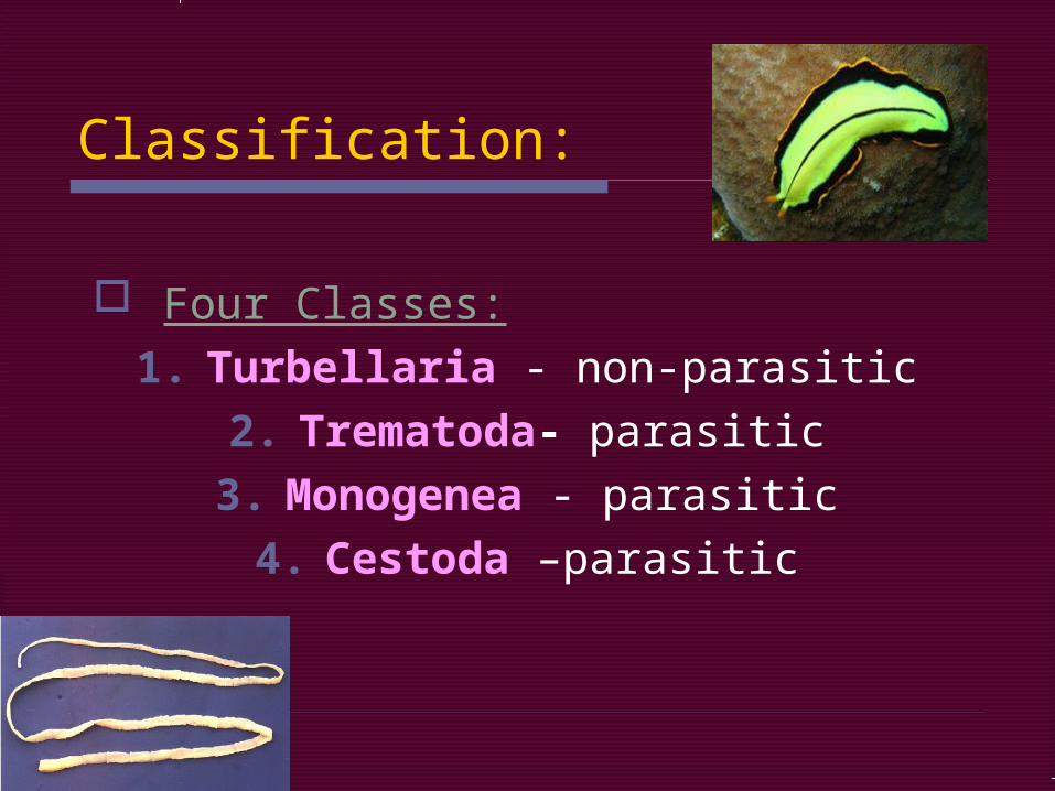
Classification:
Four Classes:1. Turbellaria - non-parasitic
2. Trematoda- parasitic3. Monogenea - parasitic
4. Cestoda –parasitic

1. Class Turbellaria: 4,500 species Mostly marine Swim in wavelike
motion Glide over solid surfaces
on layer of mucus Example: Planarian
Dugesia freshwater
Video

Planarian Body Plan:

Planarian Organ Systems:
Digestive System: Scavengers & predators
Decaying plants & animal matter Prey on smaller organism
Pharynx – throat that extends to the middle of body video
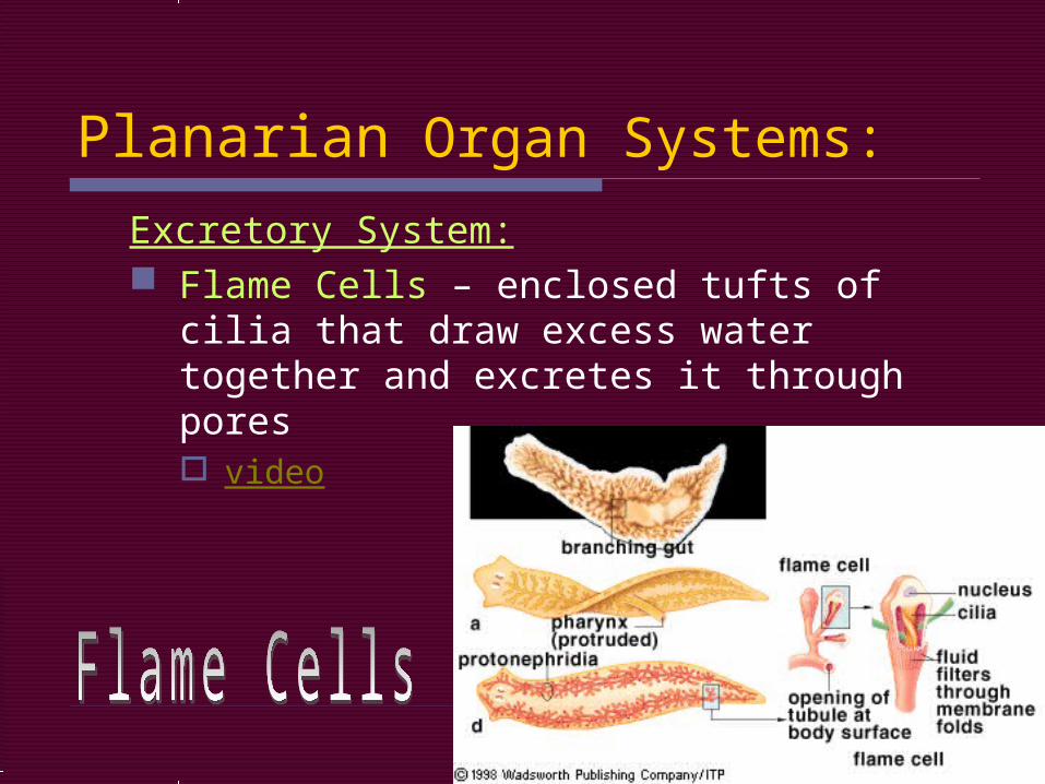
Planarian Organ Systems:
Excretory System: Flame Cells – enclosed tufts of cilia that
draw excess water together and excretes it through pores video

Planarian Organ Systems:
Nervous System: Cerebral ganglia: two
clusters of nerve cells at anterior “Brain” Can learn
Eyespots: sense direction and intensity of light
Other senses: touch, water currents, chemicals

Planarian Organ Systems:
Reproductive System: Sexual:
Hermaphrodites Eggs laid in protective capsule
Hatch in 2-3 weeks
Asexual: Regeneration

2. Class Trematoda & 3. Class Monogenea:
Both are parasitic flukes Leaf-shaped flatworms
Endoparasites: Live in blood, intestines, lungs, liver, etc.
Ectoparasites: Live on external surfaces of aquatic
hosts

Structure of Flukes
Anterior & ventral suckers for attachment to host
Nervous system like planarian Except NO eyespots
Tegument – outer layer that protects from host’s immune and digestive system

Liver fluke

Reproduction of flukes: Most are hermaphroditic May release 10,000+ eggs at a time! Complicated life cycle (p. 692)
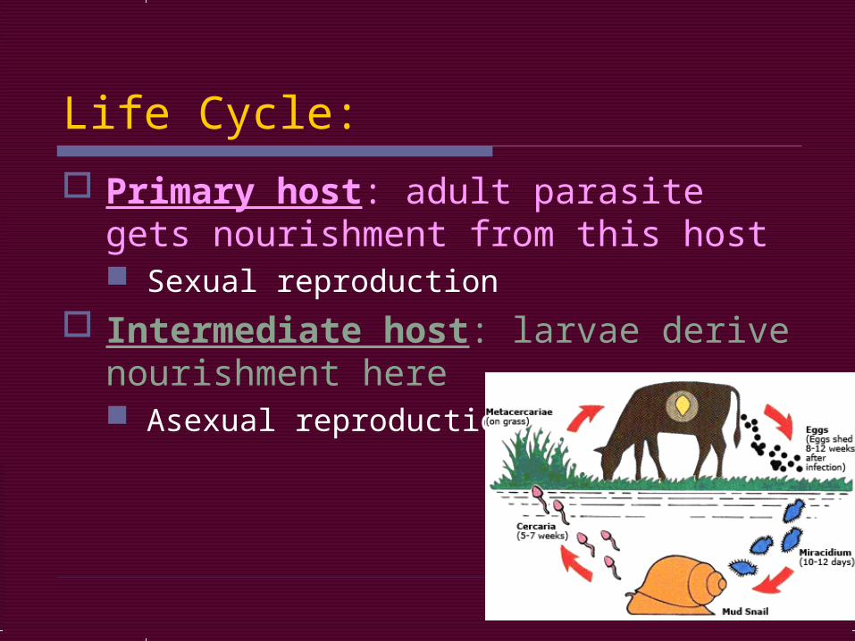
Life Cycle:
Primary host: adult parasite gets nourishment from this host Sexual reproduction
Intermediate host: larvae derive nourishment here Asexual reproduction

Fluke Diseases in Humans
Swimmer’s itch: minor skin irritation and swelling Small brown fluke in lakes (in Ohio) Dies within skin because humans are not
ideal hosts

Swimmer’s itch
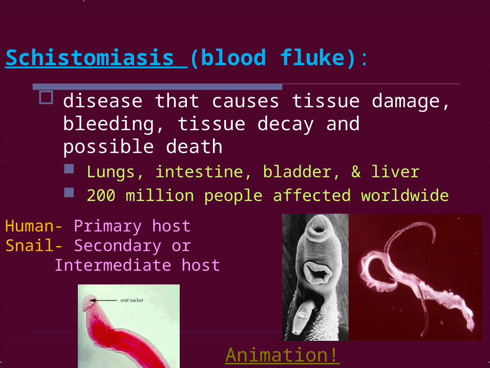
disease that causes tissue damage, bleeding, tissue decay and possible death Lungs, intestine, bladder, & liver 200 million people affected worldwide
Human- Primary hostSnail- Secondary or
Intermediate host
Animation!
Schistomiasis (blood fluke):

WHO Info.
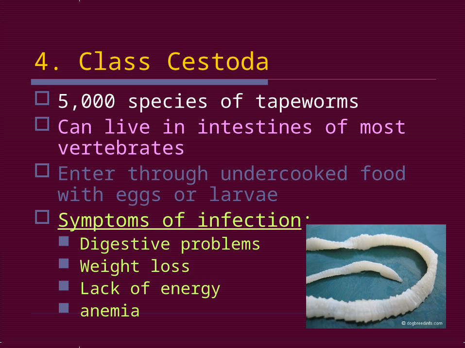
4. Class Cestoda 5,000 species of tapeworms Can live in intestines of most
vertebrates Enter through undercooked food with
eggs or larvae Symptoms of infection:
Digestive problems Weight loss Lack of energy anemia

Structure:
Tegument to protect from host Also absorbs nutrients from host
Scolex: knob-shaped organ with hooks and suckers to attach to host
Proglottids: body sections after a short neck Up to 2,000 per tapeworm!

Reproduction:
Hermaphrodites Each proglottid has ovaries and testes
Filled with 100,000+ eggs each! Eggs fertilized by sperm of different
proglottid

Life Cycle:
Cysts: dormant larvae surrounded by protective covering in animal muscle
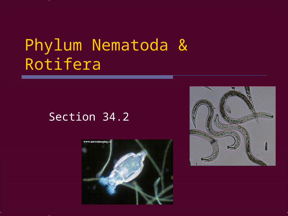
Phylum Nematoda & Rotifera
Section 34.2
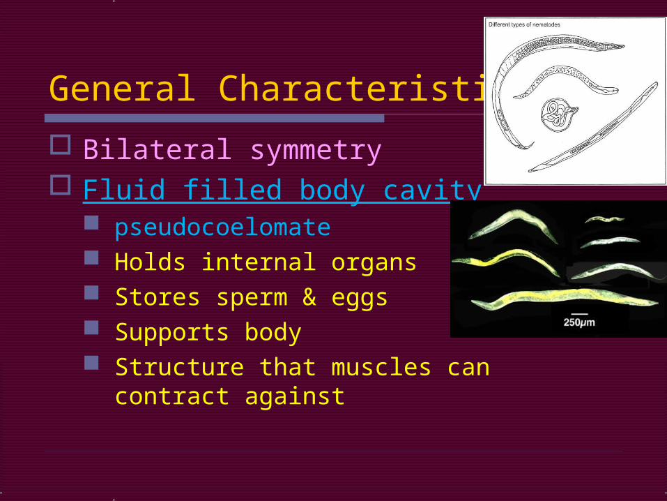
General Characteristics:
Bilateral symmetry Fluid filled body cavity
pseudocoelomate Holds internal organs Stores sperm & eggs Supports body Structure that muscles can contract
against

Phylum Nematoda Roundworms
Long, slender bodies that taper at both ends
1mm to 4ft Digestive tract with 2 openings
Anterior – mouth Posterior – anus One directional movement

Continued…
Most have separate sexes Cuticle – protective covering Free-living on land, salt and
freshwater 15,000 species known
150 species parasitic to plants and animals
Humans are host to 50 species!
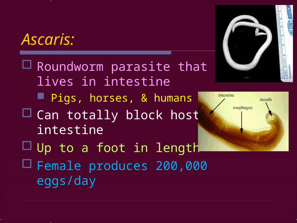
Ascaris:
Roundworm parasite that lives in intestine Pigs, horses, & humans
Can totally block host’s intestine
Up to a foot in length Female produces 200,000
eggs/day


Eggs leave with feces and enter soil Enter humans with contaminated food and water
Larvae enter intestines and move to blood stream, then lungs, coughed up and swallowed back to intestines where they mate and reproduce
Life Cycle:

Hookworms:
Another intestinal parasite Mouth has cutting plates that clamp
onto intestine wall Feed on host’s blood which may lead to
anemia May cause slow mental and physical
development in children Affects 1 billion people in tropical and
subtropical regions

Hookworm
Enter host by boring through the feet

Eggs leave with feces Larvae develop in soil Enter host’s feet Hitch a ride with blood to the lungs Coughed up and swallowed to
intestines where adult develop
Life Cycle:

Trichinella: Infect humans and pigs
Adults embed in walls of intestine Larvae travel via blood to muscles Form cysts
Humans get it from eating undercooked pork
Causes disease trichinosis Muscle pain & stiffness Can cause death

Pinworm – most common in U.S. Live and mate in lower intestine Female crawls out at night and lays eggs
around anus Person scratches during sleep and
spreads eggs to everything touched Eggs ingested and hatch

Pinworm

Filarial worms – 250 million people infected in tropics Found in lymphatic system (collects
excess fluid from blood vessels) Can cause elephantiasis
Swollen limbs, skin hardens & thickens Can cause heartworm in dogs and cats Spread by mosquitoes

Phylum Rotifera Most are transparent (see-through)
Free-living in freshwater 100 to 50 micrometers
No water = dry up and look like grains of sand; when water is present again they go back to normal Cool adaptation!

Rotifer Structures: Cilia – sweep food into mouth Mastax – breaks down food Stomach Intestine – absorbs nutrients Cloaca – digestive, reproductive, and
excretory systems empty here Universal hole
Flame cells – pull excess water together
Anus – hole to the outside

Body Parts: video

Have cerebral ganglia and eyespots Reproduction by:
Parthenogenesis – unfertilized eggs become adult females