fluid transfer from the peritoneal to the pleural cavities in rodents
TRANSCRIPT

62
FLUID TRANSFER FROM THE PERITONEAL TO THEPLEURAL CAVITIES IN RODENTS
R. C. NAIRNFrom the Department of Pathology. University of Aberdeen^
Received for publication October 15, 1956
INFORMATION about the transfer of fluid between the peritoneal and pleuralcavities is scanty, although most authorities accept the view that there is a directchannel of communication across the diaphragm. The existence of such a channelis commonly postulated to explain the concurrence of ascites and hydrothoraxin Meigs' syndrome and in many cases of hepatic cirrhosis. In theory, it mighttake the form of pores in the serous membranes (the evidence is reviewed byDrinker and Yoffey (1941)), or there might be a true diaphragmatic hiatus as issometimes obvious in congenital diaphragmatic malformations (Sweet, 1950;Deacon, 1954), or the diaphragmatic lymphatics might provide the channel oftransport (Meigs, Armstrong and Hamilton, 1943; Meigs, 1954; Cowan, Cron,Burgess and Karioris, 1954).
The present experiments were designed to determine whether artificiallyinduced ascites causes hydrothorax in rodents and to study the means wherebysuch fluid transfer inay occur.
METHODSRabbits weighing 2-3 kg. and rats weighing 250-350 g. were given mammalian Ringer
solution (in some experiments mixed with methylene blue, India ink or colloidal graphite)intraperitoneally, either by continuous drip over a 4-5 hr. period or by rapid infusioncompleted in a few minutes. The drips were set up or the infusions administered with sterileprecautions under ether anaesthesia, preceded in the rabbits by intravenous nembutalinduction. The volume of fluid injected was 350-500 ml. in the rabbits and 35-60 ml. in therats, and the duration of the experiments was usually 5-7 hr. though an occasional animalwas observed for a shorter or longer period (2-24 hr.). At the end of the observation period theanimals were killed, their serous cavities examined immediately, and any fluid present wasmeasured. The diaphragms of the animals which had the India ink or colloidal graphiteinjections were fixed in 10 per cent formol-acetate for histological examination. To assessthe influence of simple hydration in the preceding experiments, two groups of control animalswere given subcutaneous fluid by continuous drip in like or larger volume. Urine outputwas measured in all the drip experiments. These animals required partial immobilizationin their cages to prevent kinking of the drip tubes and this was accomplished by the applicationof plaster-of-Paris jackets which were loosely tethered to the cage-sides.
RESULTS
As shown in the accompanying Table, hydrothorax developed in all the rabbitsand rats in which artificial ascites had been induced by the intraperitonealinjections, while no serous effusions occurred in those receiving subcutaneousfluid. The animals which received intraperitoneal injections showed considerablevariation in the rate of absorption of fluid from the peritoneal cavity, but whetheradministered by slow drip or by rapid infusion, 25-80 per cent of the fluid had
FLUID TRANSFER THROUGH THE DIAPHRAGM
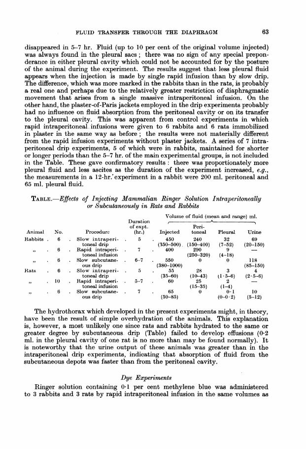
disappeared in 5-7 hr. Fluid (up to 10 per cent of the original volume injected)was always found in the pleural sacs; there was no sign of any special prepon-derance in either pleural cavity which could not be accounted for by the postureof the animal during the experiment. The results suggest that less pleural fluidappears when the injection is made by single rapid infusion than by slow drip.The difference, which was more marked in the rabbits than in the rats, is probablya real one and perhaps due to the relatively greater restriction of diaphragmaticmovement that arises from a single massive intraperitoneal infusion. On theother hand, the plaster-of-Paris jackets employed in the drip experiments probablyhad no influence on fluid absorption from the peritoneal cavity or on its transferto the pleural cavity. This was apparent from control experiments in whichrapid intraperitoneal infusions were given to 6 rabbits and 6 rats immobilizedin plaster in the same way as before; the results were not materially differentfrom the rapid infusion experiments without plaster jackets. A series of 7 intra-peritoneal drip experiments, 5 of which were in rabbits, maintained for shorteror longer periods than the 5-7 hr. of the main experimental groups, is not includedin the Table. These gave confirmatory results: there was proportionately morepleural fluid and less ascites as the duration of the experiment increased, e.g.,the measurements in a 12-hr.'experiment in a rabbit were 200 ml. peritoneal and65 ml. pleural fluid.
TABLE.-Effects of Injecting Mammalian Ringer Solution Intraperitoneallyor Subcutaneously in Rats and Rabbits
Volume of fluid (mean and range) ml.Duration r
Aof expt. Peri-
Animal No. Procedure (hr.) Injected toneal Pleural UrineRabbits . 6 . Slow intraperi- 5 . 450 240 32 69
toneal drip (350-500). (150-400) (7-52) (20-150)6 Rapid intraperi- 7 . 400 290 9
toneal infusion (250-320) (4-18)6 . Slow subcutane- . 6-7 . 550 0 0 118
ous drip (380-1000) (85-150)Rats 6 . Slow intraperi- . 5 . 55 28 3 4
toneal drip (35-60) (10-43) (1-5-6) (2 5-6)10 Rapid intraperi- 5-7 60 25 2
toneal infusion (15-35) (1-4)6 . Slow subcutane- . 7 . 65 0 0.1 10
ous drip (50-85) (0-0-2) (5-12)
The hydrothorax which developed in the present experiments might, in theory,have been the result of simple overhydration of the animals. This explanationis, however, a most unlikely one since rats and rabbits hydrated to the same orgreater degree by subcutaneous. drip (Table) failed to develop effusions (0.2ml. in the pleural cavity of one rat is no more than may be found normally). Itis noteworthy that the urine output of these animals was greater than in theintraperitoneal drip experiments, indicating that absorption of fluid from thesubcutaneous depots was faster than from the peritoneal cavity.
Dye ExperimentsRinger solution containing 0-1 per cent methylene blue was administered
to 3 rabbits and 3 rats by rapid intraperitoneal infusion in the same volumes as
63
R. C. NAIRN
before. The addition of the dye had no demonstrable effect on the distributionof fluid: the rabbits had a mean volume of 260 ml. peritoneal fluid and 11 ml.pleural fluid; the rats had 26 ml. and 3 ml. The fluid in the chest was, however,less deeply stained than in the abdomen, indicating that the fluid channel betweenthe two cavities was not a simple opening or collection of openings.
This observation was elaborated in 6 similar experiments in rabbits and 6 inrats using injection solutions of Ringer containing 1 per cent India ink. Themean values obtained in the rabbits were 290 ml. peritoneal fluid and 7 ml.pleural fluid, and in the rats, 41 ml. and 3 ml. Again, the dye was present inlower concentration in the chest and in one rat the pleural fluid looked quiteunstained. No special precaution, beyond ordinary care, was taken in openingthe pleural cavities in these animals and it is possible that some of the relativelylightly staining fluid here was contaminated by India ink from incised pleurallymphatics. Subsequent histological examination demonstrated that India inkwas in fact present in these lymphatics.
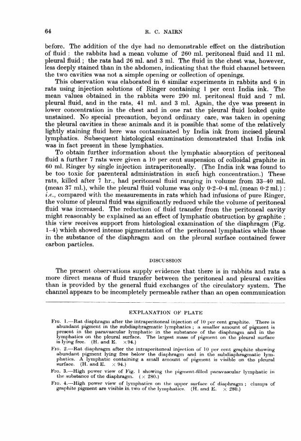
To obtain further information about the lymphatic absorption of peritonealfluid a further 7 rats were given a 10 per cent suspension of colloidal graphite in60 ml. Ringer by single injection intraperitoneally. (The India ink was found tobe too toxic for parenteral administration in suchi high concentration.) Theserats, killed after 7 hr., had peritoneal fluid ranging in volume from 33-40 ml.(mean 37 ml.), while the pleural fluid volume was only 0 2-0 4 ml. (mean 0-2 ml.):i.e., compared with the measurements in rats which had infusions of pure Ringer,the volume of pleural fluid was significantly reduced while the volume of peritonealfluid was increased. The reduction of fluid transfer from the peritoneal cavitymight reasonably be explained as an effect of lymphatic obstruction by graphite;this view receives support from histological examination of the diaphragm (Fig.1-4) which showed intense pigmentation of the peritoneal lymphatics while thosein the substance of the diaphragm and on the pleural surface contained fewercarbon particles.
DISCUSSION
The present observations supply evidence that there is in rabbits and rats amore direct means of fluid transfer between the peritoneal and pleural cavitiesthan is provided by the general fluid exchanges of the circulatory system. Thechannel appears to be incompletely permeable rather than an open communication
EXPLANATION OF PLATE
FIG. 1. Rat diaphragml after the intraperitoneal injection of 10 per cent graphite. There isabundant pigment in the subdiaphragmatic lymphatics; a smaller amount of pigment ispresent in the paravascular lymphatic in the substance of the diaphragm and in thelymphatics on the pleural surface. The largest mass of pigment on the pleural surfaceis lying free. (H. and E. x 94.)
FiG. 2.-Rat diaphragm after the intraperitoneal injectioin of 10 per cent graphite showirngabundant pigment lying free below the diaphragm and in the subdiaphragmatic lym-phatics. A lymphatic containing a small amount of pigment is visible on the pleuralsurface. (H. and E. x 94.)
FIG. 3.-High power view of Fig. 1 showinig the pigment-filled paravasculai lyinphatic inthe substance of the diaphragm. ( x 280.)
FIG. 4.-High power view of lymphatics oni the upper surface of diaphragmi; clunps ofgraphite pigment are visible in two of the lymiphatics. (H. and E. x 280.)
64

BRITISH JOURNAL OF EXPERIMENTAL PATHOLOGY. Vol. XXXVIII, No. 1.
Ii'A
Pt: '. e"
:.~,t , AP op a
A-., tf* - ,Vo0 ...
b.#' 4
'.hZ
v .. ':ALI-
., A,
.F. ,A
I ,W. B
/1.VA4
Nairn.
r4ri
J~I

FLUID TRANSFER THROUGH THE DIAPHRAGM
since colloidal particles traversed the diaphragm less readily than the crystalloidsolution. When the local lymphatics were blocked by a heavy concentration ofgraphite, fluid transfer from the abdomen to the chest was diminished, suggestingthat the diaphragmatic lymphatic system is the important link between the serouscavities. The alternative explanation, that the cavities communicate by way ofmicroscopic pores in the diaphragm, is an unlikely one for several reasons. Ifthere were open pores in the diaphragm, it should be possible to transfer air aswell as fluid from the abdomen to the chest or in the opposite direction. Directexperiment in the present study showed that this could not be done in rats orrabbits. It is also everyday clinical experience that pneumothorax does notcause pneumoperitoneum and vice versa, which is in accord with the experimentsquoted by Meigs et al., on cases of Meigs' syndrome, showing that air introducedinto the chest or abdomen failed to traverse the diaphragm. There is, moreover.no satisfactory morphological evidence that diaphragmatic pores exist, and inthe histological preparations of the diaphragm from the present dye experiments,extra-lymphatic pigment was not seen. If it may be concluded that the lymphaticsare the main fluid channel across the diaphragm, their unusual role in dischargingfluid into the chest instead of absorbing it is likely to be an accidental one, dueperhaps to leakage from over-filling. Respiratory movements doubtless have someinfluence here both in increasing the rate of lymphatic absorption below thediaphragm and in exposing lymphatic channels in the chest to transient inter-mittent reductions in external pressure.
It is common experience that ascites, in experimental animals and in man, isfrequently associated with hydrothorax. Marshall (1949) states that all hisautopsies of cases of ascites showed some pleural effusion, and although this is noteveryone's experience, it emphasizes the frequency with which the two conditionsare associated. One of the questions which prompted the present investigationwas: can hydrothorax, in the presence of ascites, be ascribed solely to the localpermeation of fluid or must some general cause of the serous effusions be postulated?The results indicate that substantial hydrothorax can rapidly develop in experi-mental animals from the transdiaphragmatic passage of ascitic fluid. The possi-bility that human ascites behaves similarly is supported by the tracer experimentsof Cowan et al. (1954) in a clinical case of ascites and hydrothorax in which theydemonstrated a more rapid transport of fluid across the diaphragm than couild beaccounted for by general circulatory fluid exchanges.
SUMMARY
The induction of artificial ascites in rabbits and rats by the intraperitonealinjection of mammalian Ringer solution led to the rapid development of substantialpleural effusions. This was not simply an effect of over-hydrating the animalssince serous effusions did not develop when the solution was administered in likeor greater volume subcutaneously.
When the fluid injected into the peritoneal cavity was mixed with methyleneblue or India ink it was found that the colloidal particles passed less readily intothe chest than the crystalloid solution. Moreover, when a more concentratedcolloidal suspension was used (10 per cent graphite) the transfer of fluid from theabdomen was reduced, probably because the lymphatics were obstructed by thepigment. Histological examination confirmed that the subdiaphragmatic lym-
65

66 R. C. NAIRN
phatics were in fact filled with graphite which was also readily demonstrable,although in smaller amount, in the lymphatics writhin and above the diaphragm.
The findings indicate that there is a direct transdiaphragmatic fluid pathwaybetween the peritoneal and pleural cavities which is probably provided by thelocal lymphatics.
I wish to thank Professor J. S. Young for his interest in this paper.
REF1ERENCESC'OWAN, T. I., CRON, R. S., BURGESS, (G. F. AND KARIORIS, F. G.-(1954) SurY. Gvynec.
Obstet., 98, 721.DEACON, A. L. (1954) Brit. med. J., i, 317.DRINKER, C. K. AND YOFFEY, J. M. (1941) 'Lymphatics, LyImiph and Lymnplhoid
Tissue'. Cambridge, Mass. (Harvard University Press), p. 88.MARSHALL, A. G. (1949) J. Obstet. Gynaec., Brit. Emp., 56, 86.MEIGS, J. V.-(1954) Amer. J. Obstet. Gynec., 67, 962.Idem, ARMSTRONG, S. H. AND HAMILTON, H. H.-(1943) Ibid.. 46, 19.SWEET, R. H.-(1950) 'Thoracic Surgery'. Philadelphia and London (Saunders),
p. 316.