fluorescence resonance energy transfer (fret)-based biosensors: visualizing cellular dynamics and...
TRANSCRIPT
MINI-REVIEW
Fluorescence resonance energy transfer (FRET)-basedbiosensors: visualizing cellular dynamics and bioenergetics
Sohila Zadran & Steve Standley & Kaylee Wong &
Erick Otiniano & Arash Amighi & Michel Baudry
Received: 3 August 2012 /Revised: 17 September 2012 /Accepted: 17 September 2012 /Published online: 6 October 2012# Springer-Verlag Berlin Heidelberg 2012
Abstract Förster (or fluorescence) resonance energytransfer (FRET) is a process involving the radiation-lesstransfer of energy from a “donor” fluorophore to an“acceptor” fluorophore. FRET technology enables thequantitative analysis of molecular dynamics in biophysicsand in molecular biology, such as the monitoring ofprotein–protein interactions, protein–DNA interactions,and protein conformational changes. FRET-based biosen-sors have been utilized to monitor cellular dynamics notonly in heterogeneous cellular populations, but also at thesingle-cell level in real time. Lately, applications ofFRET-based biosensors range from basic biological tobiomedical disciplines. Despite the diverse applicationsof FRET, FRET-based sensors still face many challenges.There is an increasing need for higher fluorescence res-olution and improved specificity of FRET biosensors.Additionally, as more FRET-based technologies extendto medical diagnostics, the affordability of FRET reagentsbecomes a significant concern. Here, we will reviewcurrent advances and limitations of FRET-based biosensortechnology and discuss future FRET applications.
Keywords Förster resonance energy transfer (FRET) .
Biosensors . Imaging . Fluorescence . Medical diagnostics
Introduction
Since the emergence of the fluorescence protein in 1992,fluorescence-based biosensors have become critical playersin monitoring and identifying cellular molecular dynamics,cellular physiology, and cell–cell interactions, or, more sim-ply, what occurs inside and outside of the cell (Periasamy2001). Förster or fluorescent resonance energy transfer(FRET) is a process involving the radiation-less transfer ofenergy from a “donor” fluorophore to an “acceptor” fluo-rophore. FRET is commonly utilized to demonstrate near-field communication or interaction between two molecules(Day et al. 2001). FRET-based assays are able to transduce anear-field interaction into a far-field signal, a unique opticaltool to assess biological phenomena well below the resolu-tion of standard optical microscopy (Roy et al. 2008;Moerner and Fromm 2003). In essence, FRET magnifiesmicroscopic conformational changes by emitting light,which can be captured by a sensor (Ha et al. 1996). Gov-erned by the physics of molecular proximity, FRET can onlyallow energy transfer to occur when the distance betweenthe donor and the acceptor fluorophores are within 1–10nanometers (nm) of each other. An excited fluorophoreemits an essentially virtual photon, which is then absorbedby a receiving fluorophore. Due to the conservation ofenergy and momentum, FRET is a mostly radiation-lessevent where the transfer is largely undetectable (Merchantet al. 2007; Kapanidis and Weiss 2002).
The donor fluorophore and the acceptor fluorophore canbe brought together in several ways. For example, onefluorophore can be attached to a substrate while the othercould be attached to its binding site, and once bound the
S. Zadran :K. Wong : E. Otiniano :A. AmighiDavid Geffen School of Medicine, University of California,Los Angeles, CA, USA
S. Standley :M. BaudryGraduate College of Biomedical Sciences,Western University of Health Sciences,Pomona, CA, USA
S. Zadran (*)Department of Pathology and Lab Medicine,University of California, Los Angeles,10833 LeConte Ave, CHS 13-375,Los Angeles, CA 90089, USAe-mail: [email protected]
Appl Microbiol Biotechnol (2012) 96:895–902DOI 10.1007/s00253-012-4449-6
fluorophores would be in close enough proximity to interactvia FRET (Joo et al. 2007). Alternatively, two fluorophorescan be attached to a single protein and, due to environmentalconstraints, force the protein to alter its conformation andinitiate FRET (Kajihara et al. 2006). Another means throughwhich FRET is stimulated and utilized to monitor cellulardynamics occurs when the FRET fluorophores are simulta-neously attached to a molecule at close positions to contin-ually engage in FRET; this interaction will cease when acellular process causes a cleavage somewhere on the bind-ing site of one of the fluorophores, separating it from theother fluorophores and decreasing the ability of FRET tooccur (Kohl et al. 2002).
The ability of FRET to occur is heavily dependent on theFRET efficiency, E. E is essentially the measurement of everyincident of energy transfer during every incident of donorexcitation. The efficiency depends on the overlap of the donorand acceptor spectra and the orientation of the dipolemomentsof the donor and acceptor (Ansbacher et al. 2012). FRETefficiency is also dependent on the distance R0 between thedonor and acceptor. The relationship between FRET efficien-cy and R0 is elucidated by the following equation:
E ¼ 1 1þ R R0=ð Þ6h i.
where R0 is the Förster’s radius, or the distance between thefluorophores at which E050 %, and where
R0 ¼ 8:79� 10�5 � n�4 � Q� k2� J lð Þ� �
R0 is in Angstrom, n is the refractive index of medium inthe range of overlap, Q is the quantum yield of the donor inthe absence of acceptor, and J(λ) is the spectral overlap. κ2is the orientation factor of the two dipoles. κ20[cos θT−3cos θA cos θD], where θT is the angle between donor (D)and acceptor (A) moments. The dynamic value of κ2 iscommonly 2/3 for freely rotating donor and acceptor fluo-rophores (Jares-Erijman and Jovin 2003; Hoppe et al. 2002).
Usually, the donor and acceptor fluorophores are charac-teristically different, particularly in emission. In this case,FRET can be detected either by monitoring the fluorescencereadout of the acceptor or by acceptor quenching. Both ofthese cases result in a decrease in the amount of donorfluorescence. FRET donor fluorophores are always fluores-cent. Upon excitation, the electrons of the donor fluorophorejump from their ground state to a higher energy level. Asthey return to the ground state, a particle of light known as aphoton is emitted. During FRET, the photon is not emittedfrom the donor; rather, the energy is transferred from thedonor molecule to the acceptor molecule, exciting theacceptor’s electrons and causing the acceptor to emit aphoton instead (Meyer and Teruel 2003). FRET quenchershave been developed to reduce fluorescence bleed-throughbetween donor and acceptor. Much like traditional FRET-
based fluorophores, FRET quenchers act as FRET accept-ors; they are capable of similar energy acceptance, butinstead of returning to the ground state from the excitedstate with the emission of light, quenchers return to theground state via non-radiative decay pathways. Quenchershave a wide absorption spectrum and a high extinctioncoefficient (Hangauer and Bertozzi 2008; Leriche et al.2012; Ogawa et al. 2009).
FRET has been a key tool in analyzing biological sys-tems for decades, but it continues to be faced with chal-lenges. In the remainder of this review, we will discuss thecurrent applications of FRET ranging from basic science tobiomedicine. We will also address current limitations andpossible routes to overcome them.
Design and limitations of FRET probes
Although it is an incredibly useful tool, FRET is not withoutshortcomings. The first problem that researchers must addressis that the donor fluorophore emission range (how muchenergy is emitted from an excited donor) and the acceptorfluorophore excitation range (how much energy is necessaryto excite an acceptor) have to overlap by at least 30 % (Lovellet al. 2009). This means that there are a limited number offlorescent tag proteins that can be used in combination witheach other. This can hinder the ability to use this technologyon certain sensitive cellular mechanisms. Pairs of organicfluorescent tags are often classified as “FRET pairs” (Ai etal. 2008). The combination of cyan fluorescent protein (CFP)-and yellow fluorescent protein (YFP)-labeled fusion proteinshas been widely used for FRET measurements in living cells.Other donor and acceptor fluorophore pairs that have beenused for FRET include CFP and dsRED, BFP and GFP, GFP(or YFP) and dsRED, Cy3 and Cy5, Alexa488 andAlexa555, Alexa488 and Cy3, and FITC and rhodamine.These pairs are only some of the many probes commonlyused in FRET-based assays (Wiedenmann et al. 2009).
There is also a phenomenon called auto-fluorescence thatcan interfere with the FRET data. When mitochondria andlysosomes are excited by light photons, they release energy inthe form of photons, causing auto-fluorescence (Sturmey et al.2006). This radiation will be measured by sensors in additionto the radiation produced by the cellular mechanisms of inter-est and can produce significant error when trying to monitorcellular activity. The additional light may cause researchersand clinicians to believe a certain metabolic pathway is chang-ing or not changing when the opposite is actually occurring.
Another disadvantage to the use of fluorophores is thatthey have a short fluorescence lifetime. This means thatonce they are excited, they stay excited for only a very shortperiod of time. Because of the brevity of the opportunity tomeasure fluorophore emissions, auto-fluorescence can take
896 Appl Microbiol Biotechnol (2012) 96:895–902
up a significant percentage of the recorded emissions insteadof the emissions produced by the cellular process of interest(Davydov et al. 2008; Fernando et al. 2006). To help alle-viate this problem, FRET quenchers were developed toreplace certain acceptor fluorophores. These quenchers donot have emission spectra themselves and can transfer theenergy they absorb without producing any visual “noise”that can make the data harder to interpret. This also allowsfor multiple acceptor fluorophores to be used for one assay,greatly increasing efficiency. One further benefit is thatthese quenchers are more resilient and are therefore easierto synthesize (Linder et al. 2011).
There are many different types of quenchers, just as thereare many different types of fluorophores. One of the firstreported quenchers was the azobenzene dye, Dabcyl. With abroad absorbance centered around 478 nm, Dabcyl is ideal forquenching dyes by FRET that fluoresce in the blue to greenregion, such as EDANS or fluorescein (Marras et al. 2002).Recently, the development of quenchers that fall into the redand near-infrared dyes, known as Black Hole Quenchers(BHQs), has further enhanced FRET. BHQ dyes are robustdark quenchers. They exhibit excellent coupling efficiencies,have large extinction coefficients and, like other quenchers,are non-fluorescent. The absorbance capability of BHQ dyesenables BHQs to quench a variety of popular fluorophores,including Cy3 and Cy5 (Johansson 2006).
It is important to note that FRET fluorophores must haveproper excitation and emission spectra as well as no effecton the three-dimensional structure of the molecule(s) towhich they are attached. For large macromolecules such aslarge proteins, the aforementioned stipulations are not sig-nificant problems, but it is not uncommon for small changesin the conformation of a macromolecule to alter both itsstructure and behavior (Bujalowski and Jezewska 2012).
Another important concern with regards to the detection ofFRET involves analyte concentration (Long et al. 2012). Onlymolecules that interact with one another will result in FRET. Iflarge amounts of donor and acceptor molecules are present,but do not interact, the amount of FRET taking place would bequite low. In this example, while the donor and the acceptormolecules may be very easy to detect separately, the actualamount of FRETactivity may not be sufficient to detect. Thereare also limitations in the development of more sensitivefluorogenic probes capable of greater solubility, greater ab-sorption coefficients, and greater pairing compatibilities(Kwak et al. 2010). One approach to overcome these currentprobe limitations is the use of quantum dots, the excitation ofwhich in the ultra violet range does not produce any excitationof its coupled fluorescent protein acceptor (mOrange). Thusquantum dots prolong the functional life and thus the visibilityof the fluorescent protein of interest (Liu et al. 2011).
Designing FRET experiments has limitations. The mostobvious limitation of current FRET models is the necessity
of a close physical proximity between donor and acceptorprobes for FRET to even occur. Depending on the assaydesign, close proximity will either be established or re-moved during the assay, resulting in a change in signalingthat can be measured. Additionally, appropriate donor andacceptor pairs need to have enough spectral overlap forefficient energy transfer to take place or have enough of adifference in spectrums to be visually distinguishable fromone another. The choice of filters for fluorescent wavelengthselection is also critical, as the excitation filter for the donorhas to be able to selectively excite the donor while mini-mizing excitation of the acceptor. Furthermore, the insertionof biosensors into live cells has had an efficiency rating ofaround 20–30 % (Komatsua et al. 2011).
FRET-based assays: from the (lab) benchto the (hospital) bedside
The FRET mechanism can be used to monitor cellular activityin a variety of ways. There are many different types of changesoccurring in a cell that involve different classes of molecules.FRET donor and acceptor fluorophores have been conjugatedto a variety of biomolecules creating functional assays such asprotein–protein binding, antigen–antibody binding, ligand–re-ceptor binding, DNA or RNA hybridization and DNA orRNA-protein binding (Okamura and Watanabe 2006; Howell2006; Li et al. 2011). A recently developed FRET-based bio-sensor assay is capable of detecting in vivo Bcr-Abl andepidermal growth factor receptor activity and its inhibition bytyrosine kinase inhibitors in real time (Waterhouse et al. 2011).This assay utilizes a specific SH2-phosphotyrosine moduleseparated by a flexible linker, and flanked at each termini byCFP and YFP. When the biosensor is phosphorylated, FREToccurs, and when the biosensor is dephosphorylated, the FRETdiminishes. These FRET-biosensor probes can also detectchanges in tyrosine kinase activity. A FRET-based assay hasalso been developed to monitor ligand-dependent interactionsof estrogen receptor isoform, ERα and Sp1 in MCF-7 breastcancer cells. Chimeric ERα and Sp1 proteins fused to CFP andYFP have been transfected into MCF-7 cells, and a FRETsignal was induced after treatment with 17β-estradiol, 4′-hydroxytamoxifen, or ICI 182,780 (Kim et al. 2005).
Oligonucleotides (short nucleic acid polymers) withDNA or RNA backbones, labeled with one or several FRETprobes, can also form FRET systems. FRET can occur whenthe oligonucleotide duplex is formed, bringing the donorand acceptor dyes in close proximity. Alternatively, thehairpin configuration of the oligonucleotide labeled duallywith both donor and acceptor fluorophores can result inFRET (Tsuji et al. 2001). DNA or RNA-based FRET probesare used in vitro and in vivo in a plethora of applications thatmonitor various types of DNA and RNA reactions including
Appl Microbiol Biotechnol (2012) 96:895–902 897
PCR, hybridization, ligation, cleavage, recombination, andsynthesis (Cardullo et al. 1988; Li et al. 2011; Dietrich et al.2002). This DNA or RNA FRET-based assay essentiallyutilizes two oligonucleotide probes hybridized to adjacentDNA or RNA sequences. The probes are end-labeled withFRET probes so that one has a 3′ donor and the other has a5′ acceptor, or vice versa. Conformation-wise, the probescan be linear, hairpin-shaped, or have more complex con-figurations. The FRET system is formed after the hybridiza-tion of the oligonucleotide probes, which signals thecompletion of the reaction by quenching the donor and thesensitization of the acceptor fluorescence (Lassaunière et al.2010). The introduction of FRET-based hybridizationprobes greatly improves DNA and RNA hybridization bio-technology. FRET-based hybridization assays are speedyand simple assays that enable the signal from the groundstate probes to be quenched, permitting observations ofhybridization reactions in real time.
FRET-based assays have also been developed to monitorDNA methylation status (Bellau-Pujol et al. 2005; Bailey etal. 2009). A study used genomic DNA from cancer cells and
pretreated it with a methylation-sensitive restriction endo-nuclease, an optically amplifying cationic conjugated poly-mer, followed by polymerase chain reaction (PCR)amplification, in the presence of fluorescein-labeled dNTPand Taq polymerase. The PCR only occurred for methylatedDNA. DNA methylation of the gene sequence of interest isdetected as a resulting FRET.
Oligonucleotides with a DNA backbone and one or severalchromophore tags havemultiple applications as FRET probes.They are especially advantageous for the real-time monitoringof biochemical reactions and in vivo studies (Kehl and Kumar2009; Hangauer and Bertozzi 2008). FRET has also beenutilized to detect DNA hybridization in a microfluidic channel(Crivat et al. 2010; Long et al. 2012). Antibiotics targeting thebacterial rRNA A-site have also been developed by incorpo-rating a new emissive surrogate into the RNA and labeling theaminoglycosides with an appropriate fluorescence acceptor(Means et al. 2005). This allowed for real-time detection ofRNA-small molecule binding.
Recently, Marshfield Clinic has developed a FRET-basedsensor to detect food-borne illness. This technology utilizes
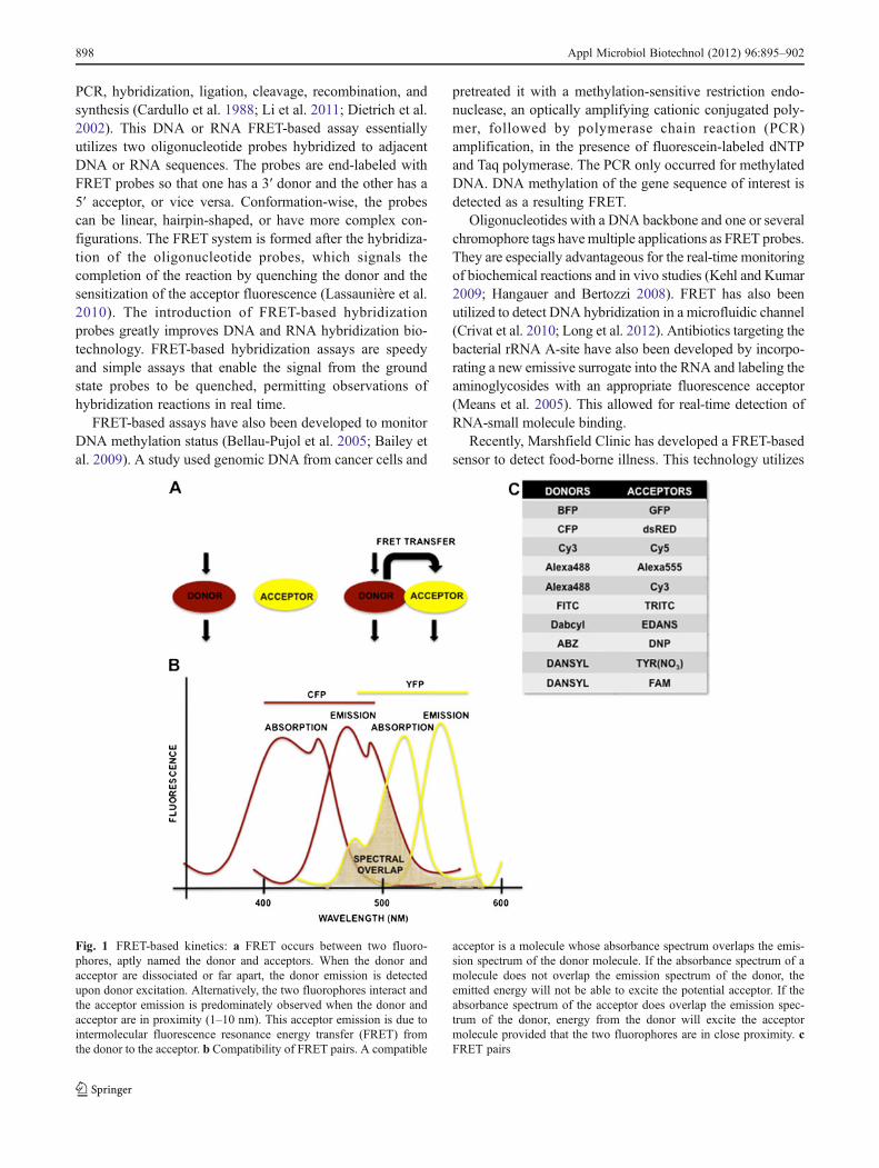
Fig. 1 FRET-based kinetics: a FRET occurs between two fluoro-phores, aptly named the donor and acceptors. When the donor andacceptor are dissociated or far apart, the donor emission is detectedupon donor excitation. Alternatively, the two fluorophores interact andthe acceptor emission is predominately observed when the donor andacceptor are in proximity (1–10 nm). This acceptor emission is due tointermolecular fluorescence resonance energy transfer (FRET) fromthe donor to the acceptor. b Compatibility of FRET pairs. A compatible
acceptor is a molecule whose absorbance spectrum overlaps the emis-sion spectrum of the donor molecule. If the absorbance spectrum of amolecule does not overlap the emission spectrum of the donor, theemitted energy will not be able to excite the potential acceptor. If theabsorbance spectrum of the acceptor does overlap the emission spec-trum of the donor, energy from the donor will excite the acceptormolecule provided that the two fluorophores are in close proximity. cFRET pairs
898 Appl Microbiol Biotechnol (2012) 96:895–902
FRET hybridization probe technology to quickly detectmarkers for enterohemmorhagic Escherichia coli and Sal-monella (Ellingson et al. 2005). Also, a cell-based high-throughput screening method utilizing FRET kinetics canselectively detect apoptosis and is highly effective in iden-tifying anti-cancer compounds. This FRET-based assay candetect apoptosis, but not necrosis, and is thus more specificthan MTT-based cell viability assays (Tian et al. 2007; Zhuet al. 2012) (Fig. 1).
Regulation of cellular bioenergetics has become the sub-ject of increased interest, particularly in cancer biology,biomedical engineering, and medicine. Genetically encodedFRET-glucose nanosensors have been used for imagingglucose flux in single cells. These FRET-based sensors arehighly sensitive and thus capable of detecting glucose at a
dynamic range of 0.05–11 mM in vivo, permitting analysesin a physiologically relevant range (Deuschle et al. 2006).Single-cell FRET-based biosensors have been developed tomonitor cellular energy flux as well (Willemsel et al. 2007).
FRET is very much a quantitative technique that canassist in developing and bringing to fruition previouslytheoretical models as well. Already FRET has been appliedto the observation of transition paths in protein-folding todetermine the average transition-path time for a fast- andslow-folding protein from a photon-by-photon analysis ofthe results of a kinetic model (Wang et al. 2010). In thismodel, the FRET efficiency trajectory was used to describethe transition path from the unfolded (U) to folded (F) statesof a protein exhibiting two-state kinetics and thermodynam-ics (Wigelsworth et al. 2004).
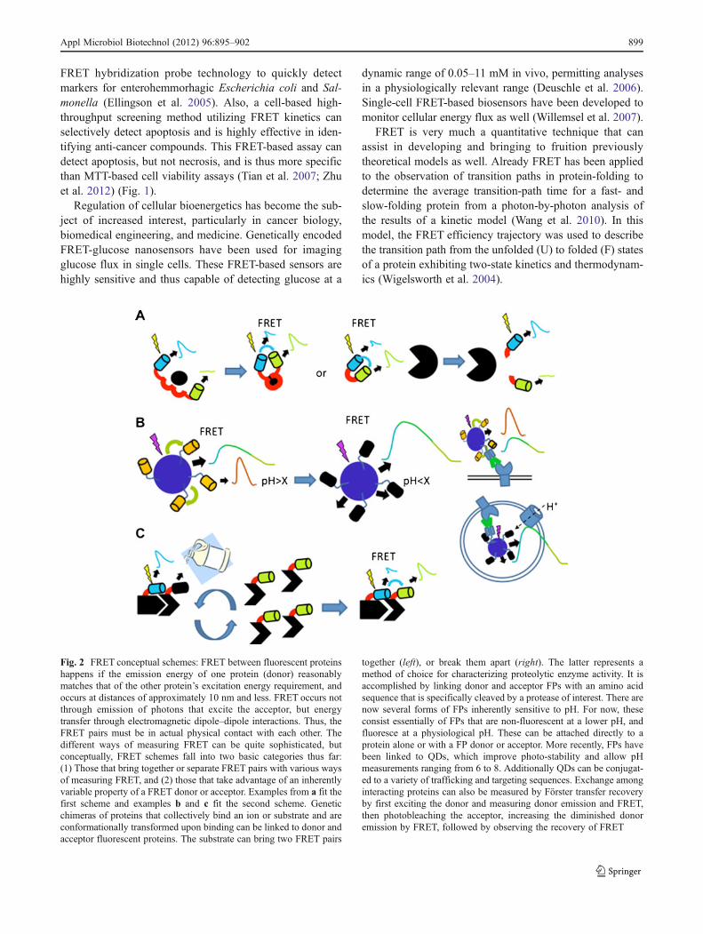
Fig. 2 FRET conceptual schemes: FRET between fluorescent proteinshappens if the emission energy of one protein (donor) reasonablymatches that of the other protein’s excitation energy requirement, andoccurs at distances of approximately 10 nm and less. FRET occurs notthrough emission of photons that excite the acceptor, but energytransfer through electromagnetic dipole–dipole interactions. Thus, theFRET pairs must be in actual physical contact with each other. Thedifferent ways of measuring FRET can be quite sophisticated, butconceptually, FRET schemes fall into two basic categories thus far:(1) Those that bring together or separate FRET pairs with various waysof measuring FRET, and (2) those that take advantage of an inherentlyvariable property of a FRET donor or acceptor. Examples from a fit thefirst scheme and examples b and c fit the second scheme. Geneticchimeras of proteins that collectively bind an ion or substrate and areconformationally transformed upon binding can be linked to donor andacceptor fluorescent proteins. The substrate can bring two FRET pairs
together (left), or break them apart (right). The latter represents amethod of choice for characterizing proteolytic enzyme activity. It isaccomplished by linking donor and acceptor FPs with an amino acidsequence that is specifically cleaved by a protease of interest. There arenow several forms of FPs inherently sensitive to pH. For now, theseconsist essentially of FPs that are non-fluorescent at a lower pH, andfluoresce at a physiological pH. These can be attached directly to aprotein alone or with a FP donor or acceptor. More recently, FPs havebeen linked to QDs, which improve photo-stability and allow pHmeasurements ranging from 6 to 8. Additionally QDs can be conjugat-ed to a variety of trafficking and targeting sequences. Exchange amonginteracting proteins can also be measured by Förster transfer recoveryby first exciting the donor and measuring donor emission and FRET,then photobleaching the acceptor, increasing the diminished donoremission by FRET, followed by observing the recovery of FRET
Appl Microbiol Biotechnol (2012) 96:895–902 899
The coupling of more efficient FRET donor and acceptorfluorescent proteins, the optimization of linker-length, themutation of fluorescent protein amino acid residues thatmediate multi-dimerization, and the subsequent couplingto two zinc binding proteins, Atox1 and WD4, that togethereach contribute two cysteines to form a single tetrahedralzinc binding pocket, now make a sensitive measurement ofintracellular zinc concentrations across a wide dynamicrange feasible (Lee et al. 2010). Further manipulation ofthis construct has made it amenable to measuring cadmiumas well (Halivni et al. 2012; Gill et al. 2005). One can easilyimagine this kind of construct being further coupled totrafficking or subcellular targeting signals to allow discretecharacterization of ionic concentrations in specific cellularcompartments. A particularly intriguing possibility wouldbe the prospect of measuring zinc-induced FRET in realtime with a chimeric construct enabling specific extracellu-lar synaptic targeting in neurons, as zinc concentrationaffects not only glutamatergic receptors like NMDA receptors,but also synaptic function. Specific glutamatergic agonists,such as α-amino-3-hydroxy5-methyl-4-isoxazolepropionicacid and kainic acid, both increase extracellular zinc concen-trations in tissue and these fluctuations affect cellular function.FRET-based assessment and sensitive quantification of prote-ase activity has emerged as probably the most refined meansof obtaining real time and single-cell functional measure-ments, since coupling a donor and acceptor fluorophore be-tween a specific proteolytic consensus sequence for aparticular protease provides the opportunity to measure notonly the induction or change in protease activation, but alsothe subcellular loci wherein the protease activation takesplace. Such FRET-based constructs have been used to assessCaspase-3 activation (Paulsson et al. 2008), matrix metallo-proteinase (MMP) activation (Yang et al. 2007) andm-calpainactivation (Zadran et al. 2009). Moreover, these can be adap-ted to high-throughput types of assays for identification ofmolecules, for instance, that treat cancer (Caspase-3, MMP),or possibly stroke (m-calpain) (Fig. 2).
Summary
FRET is an imaging technique widely used to monitorcellular dynamics in living cells in real time. Despite thevalue of this ability, the accuracy of FRET is still dependenton the FRET efficiency. Despite recent advances in the useof FRET, this technology still requires further investigationand development. The improvement of FRET technologymay lead to the development of alternative, non-invasive,and live-cell resonance energy transfer (RET) techniques.Fluorescent RET (FRET) and its variation, bioluminescentRET, can be used to assess the real-time responses withoutaltering cellular protein network, compartmentalization, and
spatial arrangement. This allows medical diagnostics toproceed through less invasive procedures, enhancing patientcare. Such techniques will undoubtedly be of great impor-tance in the future of fluorescence-based assays for theanalysis and real-time monitoring of cellular events in livecells, and thus allow for a more exact and thorough under-standing of such cellular events. With the increased under-standing that it will produce, the adaptation of FRET willprovide a key biological tool in learning critical lessonsabout cellular processes, disease progression, and drugtherapy.
Acknowledgments We are thankful guidance and suggestions pro-vided by Dr. Raphael Levine for this review. We would also like tothank the UCLA Department of Pathology and Laboratory Medicineand the David Geffen School of Medicine for providing Early CareerAward and support to SZ. This mini-review is dedicated to the lateProfessor Paola S. Timiras.
Competing interest Authors declare no competing financialinterests.
References
Ai H, Hazelwood K, Davidson M, Campbell R (2008) Fluorescentprotein FRET pairs for ratiometric imaging of dual biosensors.Nat Methods 5:401–403. doi:10.1038/nmeth.1207
Ansbacher T, Srivastava HK, Stein T, Baer R, Merkx M, Shurki A(2012) Calculation of transition dipole moment in fluorescentproteins—towards efficient energy transfer. Phys Chem ChemPhys 14(12):4109–4117
Bailey VJ, Easwaran H, Zhang Y, Griffiths E, Belinsky SA, HermanJG, Baylin SB, Carraway HE, Wang TH (2009) MS-qFRET: aquantum dot-based method for analysis of DNA methylation.Genome Res 19(8):1455–1461
Bellau-Pujol S, Vabret A, Legrand L, Dina J, Gouarin S, Petitjean-Lecherbonnier J, Pozzetto B, Ginevra C, Freymuth FJ (2005)Development of three multiplex RT-PCR assays for the detectionof 12 respiratory RNA viruses. Virol Methods 126(1–2):53–63
Bujalowski WM, Jezewska MJ (2012) Using structure–function con-straints in FRET studies of large macromolecular complexes.Methods Mol Biol 875:135–164
Cardullo RA, Agrawal S, Flores C, Zamecnik PC, Wolf DE (1988)Detection of nucleic acid hybridization by nonradiative fluores-cence resonance energy transfer. Proc Natl Acad Sci U S A 85(23):8790–8794
Crivat G, Da Silva SM, Reyes DR, Locascio LE, Gaitan M, RosenzweigN, Rosenzweig Z (2010) Quantum dot FRET-based probes in thinfilms grown in microfluidic channels. J Am Chem Soc 132(5):1460–1461
Davydov DR, Davydova NY, Halpert JR (2008) Allosteric transitionsin cytochrome P450eryF explored with pressure-perturbationspectroscopy, lifetime FRET, and a novel fluorescent substrate,Fluorol-7GA. Biochemistry 47(43):11348–11359
Day RN, Periasamy A, Schaufele F (2001) Fluorescence resonanceenergy transfer microscopy of localized protein interactions in theliving cell nucleus. Methods 25(1):4–18
Deuschle K, Chaudhuri B, Okumoto S, Lager I, Lalonde S, FrommerWB (2006) Rapid metabolism of glucose detected with FRETglucose nanosensors in epidermal cells and intact roots of
900 Appl Microbiol Biotechnol (2012) 96:895–902
Arabidopsis RNA-silencing mutants[W][OA]. Plant Cell 18(9):2314–2325. doi:10.1105/tpc.106.044073
Dietrich A, Buschmann V, Müller C, Sauer M (2002) Fluorescenceresonance energy transfer (FRET) and competing processes indonor-acceptor substituted DNA strands: a comparative study ofensemble and single-molecule data. J Biotechnol 82(3):211–231
Ellingson JLE, Koziczkowski JJ, Anderson JL, Carlson SA, SharmaVK (2005) Rapid PCR detection of enterohemorrhagic Escher-ichia coli (EHEC) in bovine food products and feces. Mol CellProbes 213–217
Fernando H, Halpert JR, Davydov DR (2006) Resolution of multiplesubstrate binding sites in cytochrome P450 3A4: the stoichiome-try of the enzyme-substrate complexes probed by FRET and Job’stitration. Biochemistry 45:4199–4209
Gill R, Willner I, Shweky I, Banin U (2005) Fluorescence resonanceenergy transfer in CdSe/ZnS-DNA conjugates: probing hybridiza-tion and DNA cleavage. J Phys Chem B 109(49):23715–23719
Ha T, Enderle T, Ogletree DF, Chemla DS, Selvin PR, Weiss S (1996)Probing the interaction between two single molecules: fluores-cence resonance energy transfer between a single donor and asingle acceptor. Proc Natl Acad Sci U S A 93:6264–6268
Halivni S, Sitt A, Hadar I, Banin U (2012) Effect of nanoparticledimensionality on fluorescence resonance energy transfer innanoparticle-dye conjugated systems. ACS Nano 6(3):2758–2765
Hangauer MJ, Bertozzi CR (2008) A FRET-based fluorogenic phosphinefor live cell imaging with the Staudinger ligation. Angew Chem IntEd Engl 47(13):2394–2397. doi:10.1002/anie.200704847
Hoppe A, Christensen K, Swanson JA (2002) Fluorescence resonanceenergy transfer-based stoichiometry in living cells. Biophys J83:3652–3664
Howell WM (2006) Detection of DNA hybridization using inducedfluorescence resonance energy transfer. Methods Mol Biol335:33–41
Jares-Erijman EA, Jovin TM (2003) FRET imaging. Nat Biotechnol21:1387–1395. doi:10.1038/nbt896
Johansson MK (2006) Choosing reporter-quencher pairs for efficientquenching through formation of intramolecular dimers. MethodsMol Biol 335:17–29
Joo C, Ha T, Selvin P (2007) Single-molecule FRETwith total internalreflection microscopy. Single Mol Tech Lab Man 3–36
Kajihara D, Abe R, Iijima I, Komiyama C, Sisido M, Hohsaka T(2006) FRET analysis of protein conformational change throughposition-specific incorporation of fluorescent amino acids. NatMethods 3(11):923–929
Kapanidis AN, Weiss S (2002) Fluorescent probes and bioconjugationchemistries for single-molecule fluorescence analysis of biomole-cules. J Chem Phys 117:10953–10964
Kehl SC, Kumar S (2009) Utilization of nucleic acid amplificationassays for the detection of respiratory viruses. Clin Lab Med 29(4):661–671
Kim K, Barhoumi R, Burghardt R, Safe S (2005) Analysis of estrogenreceptor alpha-Sp1 interactions in breast cancer cells by fluores-cence resonance energy transfer. Mol Endocrinol 19(4):843–854
Kohl T, Heinze KG, Kuhlemann R, Koltermann A, Schwille P (2002)A protease assay for two-photon crosscorrelation and FRET anal-ysis based solely on fluorescent proteins. Proc Natl Acad Sci U SA 17; 99(19): 12161–12166. doi:10.1073/pnas.192433499
Komatsua N, Aokia K, Yamadac M, Yukinagac H, Fujitac Y, KamiokacY, Matsudaa M (2011) Development of an optimized backbone ofFRET biosensors for kinases and GTPases. Mol Biol Cell 22(23)
Kwak CK, Kim DM, Lee CS, Lee M, Lee TS (2010) Aldehyde-functionalized, water-soluble poly(para-phenylene): synthesisand streptavidin assay using FRET. J Nanosci Nanotechnol 10(10):6920–6924
Lassaunière R, Kresfelder T, Venter M (2010) A novel multiplex real-time RT-PCR assay with FRET hybridization probes for the
detection and quantitation of 13 respiratory viruses. J Virol Meth-ods 165(2):254–260
Lee AJ, Ensign AA, Krauss TD, Bren KL (2010) Zinc porphyrin as adonor for FRET in Zn(II)cytochrome c. J Am Chem Soc 132(6):1752–1753
Leriche G, Budin G, Darwich Z, Weltin D, Mély Y, Klymchenko AS,Wagner A (2012) A FRET-based probe with a chemically deacti-vatable quencher. Chem Commun 48:3224–3226
Li H, Luo Y, Sun X (2011) Fluorescence resonance energy transferdye-labeled probe for fluorescence-enhanced DNA detection: aneffective strategy to greatly improve discrimination ability towardsingle-base mismatch. Biosens Bioelectron 27(1):167–171
Linder KE, Metcalfe E, Nanjappan P, Arunachalam T, Ramos K,Skedzielewski TM,Marinelli ER, TweedleMF, NunnAD, SwensonRE (2011) Synthesis, in vitro evaluation, and in vivo metab-olism of fluor/quencher compounds containing IRDye 800CWand Black Hole Quencher-3 (BHQ-3). Bioconjug Chem 22(7):1287–1297
Liu H, Liang G, Abdel-Halim ES, Zhu J (2011) A sensitive andselective quantum dots-based FRET biosensor for the detectionof cancer marker type IV collagenase. Anal Methods 3:1797–1801
Long F, Gu C, Gu§ AZ, Shi H (2012) Quantum dot/carrier–protein/haptens conjugate as a detection nanobioprobe for FRET-basedimmunoassay of small analytes with all-fiber microfluidic bio-sensing platform. Anal Chem 84(8):3646–3653
Lovell JF, Chen J, Jarvi MT, Cao WG, Allen AD, Liu Y, Tidwell TT,Wilson BC, Zheng G (2009) FRET quenching of photosensitizersinglet oxygen generation. J Phys Chem B 113(10):3203–3211
Marras SAE, Kramer FR, Tyagi S (2002) Efficiencies of fluorescenceresonance energy transfer and contact-mediated quenching inoligonucleotide probes. Nucleic Acids Res 30(21):e122
Means JA, Hines JV, Merchant KA, Best RB, Louis JM, Gopich IV,Eaton WA (2005) Fluorescence resonance energy transfer studiesof aminoglycoside binding to a T box antiterminator RNA. Bio-org Med Chem Lett 15(8):2169–2172
Merchant KA, Best RB, Louis JM, Gopich IV, Eaton WA (2007)Characterizing the unfolded states of proteins using single-molecule FRET spectroscopy and molecular simulations. ProcNatl Acad Sci U S A 104:1528–1533
Meyer T, Teruel MN (2003) Fluorescence imaging of signaling net-works. Trends Cell Biol 13:101–106
Moerner WE, Fromm DP (2003) Methods of single-molecule fluores-cence spectroscopy and microscopy. Rev Sci Instrum 74:3597–3619
Ogawa M, Kosaka N, Longmire M, Urano Y, Choyke PL, KobayashiH (2009) Fluorophore-quencher based activatable targeted opticalprobes for detecting in vivo cancer metastases. Mol Pharm 6(2):386–395. doi:10.1021/mp800115t
Okamura Y, Watanabe Y (2006) Detecting RNA/DNA hybridizationusing double-labeled donor probes with enhanced fluorescenceresonance energy transfer signals. Methods Mol Biol 335:43–56
Paulsson JF, Schultz SW, Köhler M, Leibiger I, Berggren P, WestermarkGT (2008) Real-time monitoring of apoptosis by caspase-3-like protease induced FRET reduction triggered by amyloidaggregation. Exp Diabetes Res Vol 2008. doi:10.1155/2008/865850
Periasamy A (2001) Fluorescence resonance energy transfer microsco-py: a mini review. J Biomed Opt 6(3):287–291
Roy R, Hohng S, Ha T (2008) A practical guide to single-moleculeFRET. Nat Methods 5:507–516. doi:10.1038/nmeth.1208
Sturmey RG, O’Toole PJ, Leese HJ (2006) Fluorescence resonanceenergy transfer analysis of mitochondrial:lipid association in theporcine oocyte. Reproduction 132(6):829–837
Tian H, Ip L, Luo H, Chang DC, Lu KQ (2007) A high throughputdrug screen based on fluorescence resonance energy transfer(FRET) for anticancer activity of compounds from herbal
Appl Microbiol Biotechnol (2012) 96:895–902 901
medicine. Br J Pharmacol 150(3):321–334. doi:10.1038/sj.bjp.0706988
Tsuji A, Sato Y, Hirano M, Suga T, Koshimoto H, Taguchi T, Ohsuka S(2001) Development of a time-resolved fluorometric method forobserving hybridization in living cells using fluorescence reso-nance energy transfer. Biophys J 81(1):501–515
Wang Z, Elbaz J, Remacle F, Levine RD, Willner I (2010) All-DNAfinite-state automata with finite memory. Proc Natl Acad Sci U SA 107(51):21996–22001. doi:10.1073/pnas.1015858107
Waterhouse BR, Gijsen M, Barber PR, Tullis ID, Vojnovic B, Kong A(2011) Assessment of EGFR/HER2 dimerization by FRET-FLIMutilizing Alexa-conjugated secondary antibodies in relation totargeted therapies in cancers. Oncotarget 2(9):728–736
Wiedenmann J, Oswald F, Nienhaus GU (2009) Fluorescent proteinsfor live cell imaging: opportunities, limitations, and challenges.IUBMB Life 61(11):1029–1042
Wigelsworth DJ, Krantz BA, Christensen KA, Lacy DB, Juris SJ,Collier RJ (2004) Binding stoichiometry and kinetics of the
interaction of a human anthrax toxin receptor, CMG2, with pro-tective antigen. J Biol Chem 279:23349–23356
Willemsel M, Janssen E, de Lange F, Wieringa B, Fransen J (2007)ATP and FRET—a cautionary note. Nat Biotechnol 25:170–172.doi:10.1038/nbt0207-170
Yang J, Zhang Z, Lin J, Lu J, Liu BF, Zeng S, Luo Q (2007)Detection of MMP activity in living cells by a geneticallyencoded surface-displayed FRET sensor. Biochem BiophysActa 1773(3):400–407
Zadran S, Qin Q, Bi X, Zadran H, Kim Y, Foy MR, Thompson R,Baudry M (2009) 17-Beta-estradiol increases neuronal excitabil-ity through MAP kinase-induced calpain activation. Proc NatlAcad Sci U S A 106(51):21936–21941
Zhu X, Fu A, Luo KQ (2012) A high-throughput fluorescenceresonance energy transfer (FRET)-based endothelial cell apo-ptosis assay and its application for screening vascular dis-rupting agents. Biochem Biophys Res Commun 418(4):641–646
902 Appl Microbiol Biotechnol (2012) 96:895–902