fluorescent peptides and amino acids
TRANSCRIPT
FLUORESCENTPEPTIDES
Peptides and amino acids labeled with Tide FluorTM and Tide QuencherTM We offer peptides and amino acids tagged with Tide FluorTM fluorescent dyes. They meet highest demands in fluorescence intensity and photo-stability, and outper-form most conventional and proprietary dyes for these properties. For optimum results in FRET, Tide FluorTM dyes should be combined with Tide QuencherTM acceptors. The donor and acceptor spectra of the resulting FRET pairs overlap ideally, leading to an efficient quenching. Tide FluorTM and Tide QuencherTM labels are available as diverse derivatives and can be used for labeling of the majority of relevant peptides and amino acids.
Outstanding Performance and Wide Application Range
Intensive fluorescent donor emission
• Efficient excitation with light from common sources
• Strong fluorescence
• High photo-stability
• Tolerant against pH and different buffer conditions
Excellent quenching
• Optimum spectral overlap of Tide FluorTM donor and Tide QuencherTM acceptor
• Efficient donor energy absorption
Broad selection of chemical derivatives
• Active succinimidyl esters (NHS)
• Alkynes
• Amines
• Azides
• Carboxylic acids
• Maleimides
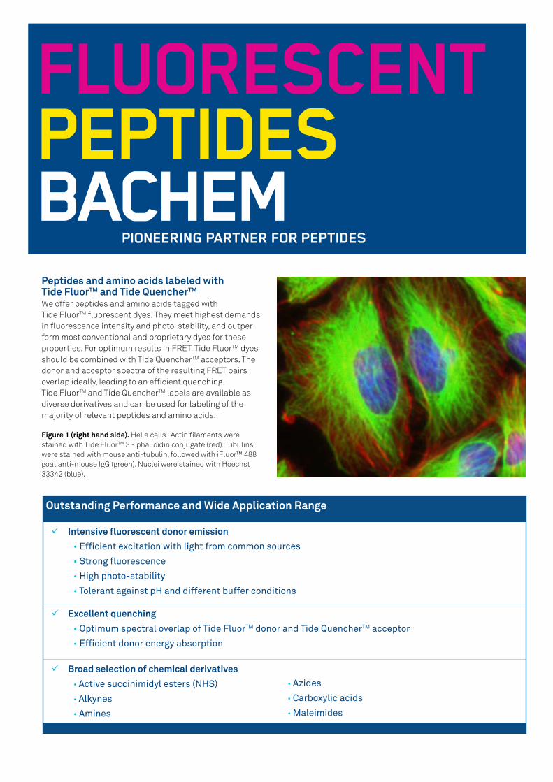
Figure 1 (right hand side). HeLa cells. Actin filaments were stained with Tide FluorTM 3 - phalloidin conjugate (red). Tubulins were stained with mouse anti-tubulin, followed with iFluor™ 488 goat anti-mouse IgG (green). Nuclei were stained with Hoechst 33342 (blue).
Tide FluorTM Dye Absorption (TF) Emission ε (M -1cm-1) * QY** Optimum Tide QuencherTM Absorption (TQ)
Tide FluorTM 1 345 nm 442 nm 20,000 0.95 Tide Quencher™ 1 ~490 nm
Tide FluorTM 2
Tide FluorTM 2 WS
500 nm
495 nm
527 nm
518 nm
75,000
75,000
0.90
0.90
Tide Quencher™ 2
Tide Quencher™ 2 WS~520 nm
Tide FluorTM 3
Tide FluorTM 3 WS
555 nm
555 nm
584 nm
565 nm
78,000
150,000
0.85
0.10***
Tide Quencher™ 3
Tide Quencher™ 3 WS~570 nm
Tide FluorTM 4 590 nm 618 nm 90,000 0.91 Tide Quencher™ 4 WS ~610 nm
Tide FluorTM 5 WS 649 nm 664 nm 250,000 0.25 Tide Quencher™ 5 WS ~670 nm
Tide FluorTM 6 WS 676 nm 695 nm 220,000 0.18 Tide Quencher™ 6 WS ~704 nm
Tide FluorTM 7 WS 749 nm 775 nm 275,000 0.12 Tide Quencher™ 7 WS ~763 nm
Tide FluorTM 8 WS 775 nm 807 nm 250,000 0.08
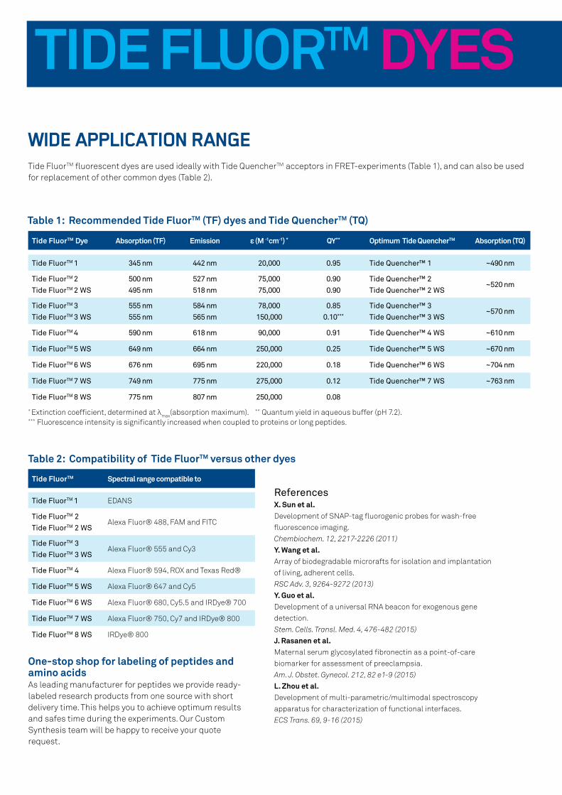
Table 1: Recommended Tide FluorTM (TF) dyes and Tide QuencherTM (TQ)
* Extinction coefficient, determined at λmax(absorption maximum). ** Quantum yield in aqueous buffer (pH 7.2).*** Fluorescence intensity is significantly increased when coupled to proteins or long peptides.
TIDE FLUORTM DYESWIDE APPLICATION RANGETide FluorTM fluorescent dyes are used ideally with Tide QuencherTM acceptors in FRET-experiments (Table 1), and can also be used for replacement of other common dyes (Table 2).
Tide FluorTM Spectral range compatible to
Tide FluorTM 1 EDANS
Tide FluorTM 2
Tide FluorTM 2 WSAlexa Fluor® 488, FAM and FITC
Tide FluorTM 3
Tide FluorTM 3 WSAlexa Fluor® 555 and Cy3
Tide FluorTM 4 Alexa Fluor® 594, ROX and Texas Red®
Tide FluorTM 5 WS Alexa Fluor® 647 and Cy5
Tide FluorTM 6 WS Alexa Fluor® 680, Cy5.5 and IRDye® 700
Tide FluorTM 7 WS Alexa Fluor® 750, Cy7 and IRDye® 800
Tide FluorTM 8 WS IRDye® 800
Table 2: Compatibility of Tide FluorTM versus other dyes
One-stop shop for labeling of peptides and amino acidsAs leading manufacturer for peptides we provide ready-labeled research products from one source with short delivery time. This helps you to achieve optimum results and safes time during the experiments. Our Custom Synthesis team will be happy to receive your quote request.
ReferencesX. Sun et al. Development of SNAP-tag fluorogenic probes for wash-free
fluorescence imaging.
Chembiochem. 12, 2217-2226 (2011)
Y. Wang et al. Array of biodegradable microrafts for isolation and implantation
of living, adherent cells.
RSC Adv. 3, 9264-9272 (2013)
Y. Guo et al. Development of a universal RNA beacon for exogenous gene
detection.
Stem. Cells. Transl. Med. 4, 476-482 (2015)
J. Rasanen et al. Maternal serum glycosylated fibronectin as a point-of-care
biomarker for assessment of preeclampsia.
Am. J. Obstet. Gynecol. 212, 82 e1-9 (2015)
L. Zhou et al. Development of multi-parametric/multimodal spectroscopy
apparatus for characterization of functional interfaces.
ECS Trans. 69, 9-16 (2015)
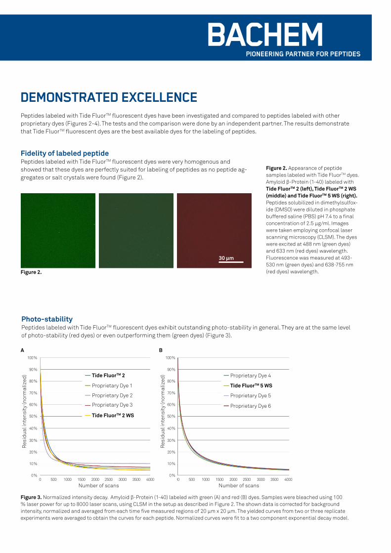
TIDE FLUORTM DYESDEMONSTRATED EXCELLENCEPeptides labeled with Tide FluorTM fluorescent dyes have been investigated and compared to peptides labeled with other proprietary dyes (Figures 2-4). The tests and the comparison were done by an independent partner. The results demonstrate that Tide FluorTM fluorescent dyes are the best available dyes for the labeling of peptides.
Figure 3. Normalized intensity decay. Amyloid β-Protein (1-40) labeled with green (A) and red (B) dyes. Samples were bleached using 100 % laser power for up to 8000 laser scans, using CLSM in the setup as described in Figure 2. The shown data is corrected for background intensity, normalized and averaged from each time five measured regions of 20 µm x 20 µm. The yielded curves from two or three replicate experiments were averaged to obtain the curves for each peptide. Normalized curves were fit to a two component exponential decay model.
100 %
90 %
80 %
70 %
60 %
50 %
40 %
30 %
20 %
10 %
0 %
100 %
90 %
80 %
70 %
60 %
50 %
40 %
30 %
20 %
10 %
0 %0 500 1000 1500 2000 2500 3000 3500 4000 0 500 1000 1500 2000 2500 3000 3500 4000
A B
Tide FluorTM 2
Tide FluorTM 2 WS
Tide FluorTM 5 WSProprietary Dye 1
Proprietary Dye 2
Proprietary Dye 3
Proprietary Dye 4
Proprietary Dye 5
Proprietary Dye 6
Number of scansNumber of scans
Res
idua
l int
ensi
ty (n
orm
aliz
ed)
Res
idua
l int
ensi
ty (n
orm
aliz
ed)
Photo-stability Peptides labeled with Tide FluorTM fluorescent dyes exhibit outstanding photo-stability in general. They are at the same level of photo-stability (red dyes) or even outperforming them (green dyes) (Figure 3).
Figure 2. Appearance of peptide samples labeled with Tide FluorTM dyes. Amyloid β-Protein (1-40) labeled with Tide FluorTM 2 (left), Tide FluorTM 2 WS (middle) and Tide FluorTM 5 WS (right). Peptides solubilized in dimethylsulfox-ide (DMSO) were diluted in phosphate buffered saline (PBS) pH 7.4 to a final concentration of 2.5 µg/ml. Images were taken employing confocal laser scanning microscopy (CLSM). The dyes were excited at 488 nm (green dyes) and 633 nm (red dyes) wavelength. Fluorescence was measured at 493-530 nm (green dyes) and 638-755 nm (red dyes) wavelength.
Fidelity of labeled peptide Peptides labeled with Tide FluorTM fluorescent dyes were very homogenous and showed that these dyes are perfectly suited for labeling of peptides as no peptide ag-gregates or salt crystals were found (Figure 2).
Figure 2.
30 µm
TIDE FLUORTM DYES
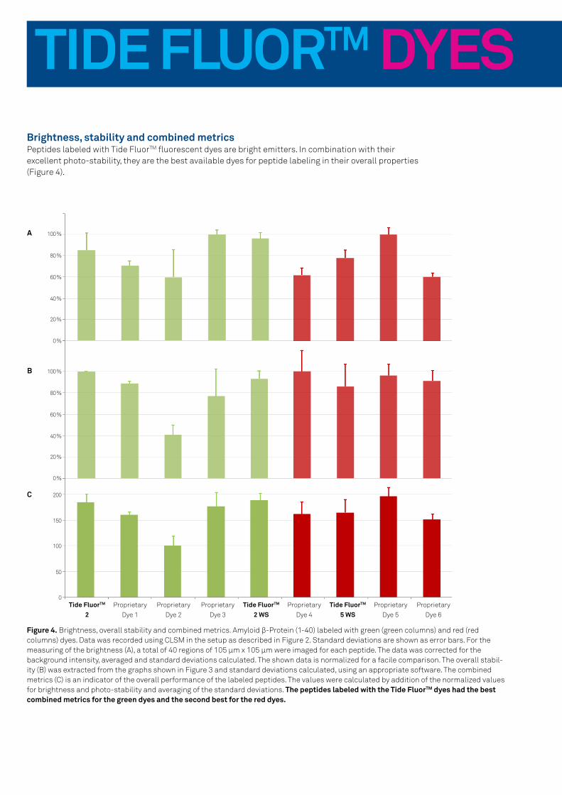
Figure 4. Brightness, overall stability and combined metrics. Amyloid β-Protein (1-40) labeled with green (green columns) and red (red columns) dyes. Data was recorded using CLSM in the setup as described in Figure 2. Standard deviations are shown as error bars. For the measuring of the brightness (A), a total of 40 regions of 105 µm x 105 µm were imaged for each peptide. The data was corrected for the background intensity, averaged and standard deviations calculated. The shown data is normalized for a facile comparison. The overall stabil-ity (B) was extracted from the graphs shown in Figure 3 and standard deviations calculated, using an appropriate software. The combined metrics (C) is an indicator of the overall performance of the labeled peptides. The values were calculated by addition of the normalized values for brightness and photo-stability and averaging of the standard deviations. The peptides labeled with the Tide FluorTM dyes had the best combined metrics for the green dyes and the second best for the red dyes.
100 %
80 %
60 %
40 %
20 %
0 %
100 %
80 %
60 %
40 %
20 %
0 %
200
150
100
50
0 Tide FluorTM
2Proprietary
Dye 1
Tide FluorTM 2 WS
Proprietary
Dye 2
Proprietary
Dye 3
Proprietary
Dye 4
Tide FluorTM 5 WS
Proprietary
Dye 5
Proprietary
Dye 6
A
B
C
Brightness, stability and combined metricsPeptides labeled with Tide FluorTM fluorescent dyes are bright emitters. In combination with their excellent photo-stability, they are the best available dyes for peptide labeling in their overall properties (Figure 4).

TIDE FLUORTM DYES

Pub
lishe
d b
y G
lob
al M
arke
ting
, Bac
hem
Gro
up, J
anua
ry 2
018
All information is compiled to the best of our knowledge. We cannot be made liable for any possible errors or misprints. Some products may be restricted in certain countries. www.bachem.com shop.bachem.com
Marketing & Sales Contact
Europe, Africa, Middle East and Asia Pacific
Bachem AGTel. +41 58 595 [email protected]
Americas
Bachem Americas, Inc.Tel. +1 888 422 2436 (toll-free in USA & Canada) +1 310 539 [email protected]
Visit our website www.bachem.com or shop online shop.bachem.com
CUSTOM PEPTIDE SYNTHESIS AT BACHEMPEPTIDE LABELING AND CONJUGATION
Our highly experienced custom synthesis teams with extensive know-how in sequence design and modifications will deliver every peptide in the quality you need.
• Biotinylated Peptides
• Conjugation to Imaging Agents
• Conjugation to Oligonucleotides
• Conjugation to Proteins:
BSA and KLH
• FRET or TR-FRET Peptides
• Heavy Isotope Labeling
• Labeling with standard
dyes and patented dyes
• Labeling with Tide FluorTM
and Tide QuencherTM
• Pegylated Peptides