food and chemical toxicology - ippt.pan.pl · dominika malinskaa,jędrzej szymańskia, paulina...
TRANSCRIPT
Contents lists available at ScienceDirect
Food and Chemical Toxicology
journal homepage: www.elsevier.com/locate/foodchemtox
Assessment of mitochondrial function following short- and long-termexposure of human bronchial epithelial cells to total particulate matter froma candidate modified-risk tobacco product and reference cigarettes
Dominika Malinskaa, Jędrzej Szymańskia, Paulina Patalas-Krawczyka, Bernadeta Michalskaa,Aleksandra Wojtalaa, Monika Prilla, Małgorzata Partykaa, Karolina Drabika, Jarosław Walczaka,Alain Sewerb, Stephanie Johneb, Karsta Luettichb, Manuel C. Peitschb, Julia Hoengb,Jerzy Duszyńskia, Joanna Szczepanowskaa,1, Marco van der Toornb,∗,1,Mariusz R. Wieckowskia,∗∗,1
aNencki Institute of Experimental Biology, Polish Academy of Sciences, 3 Pasteur Street, 02-093 Warsaw, Polandb PMI R&D, Philip Morris Products S.A., Quai Jeanrenaud 5, 2000 Neuchâtel, Switzerland
A R T I C L E I N F O
Keywords:MitochondriaMitochondrial respiratory chainOxidative stressBEAS-2B cellsCigaretteTobacco heating system
A B S T R A C T
Mitochondrial dysfunction caused by cigarette smoke is involved in the oxidative stress-induced pathology ofairway diseases. Reducing the levels of harmful and potentially harmful constituents by heating rather thancombusting tobacco may reduce mitochondrial changes that contribute to oxidative stress and cell damage. Weevaluated mitochondrial function and oxidative stress in human bronchial epithelial cells (BEAS 2B) following 1-and 12-week exposures to total particulate matter (TPM) from the aerosol of a candidate modified-risk tobaccoproduct, the Tobacco Heating System 2.2 (THS2.2), in comparison with TPM from the 3R4F reference cigarette.After 1-week exposure, 3R4F TPM had a strong inhibitory effect on mitochondrial basal and maximal oxygenconsumption rates compared to TPM from THS2.2. Alterations in oxidative phosphorylation were accompaniedby increased mitochondrial superoxide levels and increased levels of oxidatively damaged proteins in cells ex-posed to 7.5 μg/mL of 3R4F TPM or 150 μg/mL of THS2.2 TPM, while cytosolic levels of reactive oxygen specieswere not affected. In contrast, the 12-week exposure indicated adaptation of BEAS-2B cells to long-term stress.Together, the findings indicate that 3R4F TPM had a stronger effect on oxidative phosphorylation, gene ex-pression and proteins involved in oxidative stress than TPM from the candidate modified-risk tobacco productTHS2.2.
1. Introduction
Oxidative stress caused by tobacco smoke can result in inflamma-tion, epithelial injury, and cell death, and is a major causative factor inairway diseases such as chronic obstructive pulmonary disease and lungcancer (Durham and Adcock, 2015; Rahman and MacNee, 1996). Be-cause tobacco smoke contains thousands of harmful and potentiallyharmful constituents (HPHCs) such as radicals, carcinogens, aromatichydrocarbons, and α, β-unsaturated aldehydes (Baker, 1974; Rodgmanand Perfetti, 2013), it is likely that the toxicity associated with oxida-tive stress is the result of a complex interaction of a variety of con-stituents, reacting either directly or indirectly with the extracellular and
intracellular components of the respiratory tract.Mitochondria are among the first responders to various stress factors
that challenge cell and tissue homeostasis. Therefore, mitochondrialmalfunction is increasingly recognized as a key component in acute andchronic cellular stress. In vitro and in vivo studies have shown thatHPHCs in tobacco smoke can disturb the complexes of the mitochon-drial respiratory chain (Anbarasi et al., 2005; van der Toorn et al.,2007). When this happens, a proton motive force across the inner mi-tochondrial membrane cannot be generated, leading to mitochondrialrespiratory chain inhibition, and electrons leak from the electrontransfer chain and react with oxygen to form reactive oxygen species(ROS) (Lebiedzinska et al., 2013; van der Toorn et al., 2009; Wojewoda
https://doi.org/10.1016/j.fct.2018.02.013Received 19 December 2017; Accepted 7 February 2018
∗ Corresponding author. Systems Toxicology, Biological Systems Research, Philip Morris International, R&D, Switzerland.∗∗ Corresponding author. Laboratory of Bioenergetics and Biomembranes, Department of Biochemistry, Nencki Institute of Experimental Biology, Polish Academy of Sciences, Poland.
1 These three authors share senior co-authorship.E-mail addresses: [email protected] (M. van der Toorn), [email protected] (M.R. Wieckowski).
Food and Chemical Toxicology 115 (2018) 1–12
Available online 13 February 20180278-6915/ © 2018 The Authors. Published by Elsevier Ltd. This is an open access article under the CC BY license (http://creativecommons.org/licenses/BY/4.0/).
T
et al., 2010, 2011). Apart from increased generation, elevated ROS le-vels also depend on the levels of antioxidant enzymes, which form thefirst line of defense against ROS. Furthermore, impaired mitochondrialfunctions not only affect energy production and ROS levels, but alsoexert a significant impact on cellular physiology. The consequences ofmitochondrial malfunction in lung epithelium are reflected in sig-nificant changes in epithelial barrier function, inflammation, and lunginjury, which are seen in patients with tobacco smoke-induced lungdiseases (CDC, 2010).
Lowering HPHC levels by heating rather than combusting tobacco isa promising approach in reducing mitochondrial dysfunction and cel-lular stress associated with smoking combustible tobacco products.Heating rather than combusting tobacco has been shown to sub-stantially reduce HPHC levels in the resulting aerosol (Patskan andReininghaus, 2003). In a 90-day nose-only inhalation study in Spra-gue–Dawley rats, it was demonstrated that exposure to aerosol from anew candidate modified-risk tobacco product (cMRTP), the TobaccoHeating System 2.2 (THS2.2), significantly reduced the degree of cel-lular stress and lung inflammation compared with exposure to tobaccosmoke from the 3R4F reference cigarette (Wong et al., 2016). In thepresent study, we evaluated the mitochondrial function of culturedhuman bronchial epithelial cells following short- and long-term ex-posure to total particulate matter (TPM) from THS2.2 aerosol and 3R4Fcigarette smoke (CS).
THS2.2 is a battery-operated heating system into which a tobaccostick is inserted and heated at a maximum temperature of 350 °C; thisgenerates an aerosol primarily composed of water, glycerol, and nico-tine (Smith et al., 2016). In contrast, during the combustion of con-ventional cigarettes, the temperature of the tobacco increases up to900 °C, resulting in more than 6000 combustion and pyrolysis products(Baker, 1974; Rodgman and Perfetti, 2013). TPM, which represents theparticulate phase of mainstream tobacco smoke, comprises approxi-mately 80% of the total weight of tobacco smoke (Barsanti et al., 2007).We used TPM as the test item to investigate the effects of HPHCs, asmultiple HPHCs have been found in TPM (Rodgman and Perfetti, 2013).
To assess mitochondrial physiology, a human immortalized bron-chial epithelial cell line, BEAS-2B, was continuously exposed to TPMfrom 3R4F CS and three different concentrations of TPM from THS2.2aerosol for 1 or 12 weeks. Multiple cellular parameters were measured,including mitochondrial oxygen consumption, ROS levels, the effi-ciency of the antioxidant defense system, and markers of oxidativestress. This enabled us to assess the impact of the THS2.2 TPM on mi-tochondrial functions in bronchial epithelial cells in comparison withthose seen following exposure to CS TPM.
2. Materials and methods
2.1. Chemicals
MitoSOXTM Red (mitochondrial superoxide indicator), CM-H2DCFDA (general oxidative stress indicator), and JC-1 (mitochondrialmembrane potential probe) were obtained from Invitrogen (Carlsbad,CA, USA). Total OXPHOS rodent antibody cocktail and anti-SOD2 an-tibody were obtained from Abcam (Cambridge, UK). Antibody against
GPx1 was obtained from Cell Signaling Technology (Danvers, MA,USA). Antibody against GAPDH was obtained from Merck Millipore(Billerica, MA, USA). Antibody against SOD1 was obtained from SantaCruz Biotechnology (Dallas, TX, USA). CellTiter-Glo® 2.0 was obtainedfrom Promega (Madison, WI, USA). Anti-rabbit IRDye 800CW antibodywere obtained from LI-COR Biosciences (Lincoln, NE, USA). Antibodyagainst anti-β-actin, DMSO, ATP, pyruvate, oligomycin, carbonyl cya-nide-4-(trifluoromethoxy)phenylhydrazone (FCCP), mercaptoethanol,glycerol, HCl, Tween® 20, glucose, H2O2, Tris and antimycin A wereobtained from Sigma-Aldrich (St. Louis, MO, USA). Glutamine wasobtained from GE Healthcare Bio-Sciences (Pittsburgh, USA).
2.2. Cell culture
The human bronchial epithelial cell line BEAS-2B was obtainedfrom the American Type Culture Collection (ATCC, #CRL-9609;Manassa, VA, USA). Cells were cultured in bronchial epithelial cellgrowth medium consisting of bronchial epithelial cell basal mediumsupplemented with a SingleQuots™ kit (Lonza Walkersville, MD, USA),following the vendor's recommendations. Cells were grown in 75-cm2
collagen I-coated culture flasks (VWR, Radnor, PA, USA) at 37 °C in anatmosphere of 5% CO2 until reaching 75%–80% confluence. A TC20Automated Cell Counter and Muse Count & Viability Assay (Bio-RadLaboratories, Hercules, CA, USA) were used to count the cells and assesscell viability according to the manufacturer's instructions. Substanceswere added to cells 4 h after seeding on culture dishes.
2.3. Generation of 3R4F and THS2.2 TPM
3R4F reference cigarettes were purchased from the University ofKentucky (Lexington, KY, USA; http://www.ca.uky.edu/refcig/).THS2.2 sticks and stick holders were provided by Philip MorrisProducts S.A. (Neuchâtel, Switzerland); the holder heats the tobaccoplug to generate an aerosol containing water, glycerin, nicotine, andtobacco flavors (Smith et al., 2016). The tobacco stick holder includes abattery, electronics for control, a heating element, and a stick extractor.Prior to use, 3R4F cigarettes and THS2.2 sticks were conditioned ac-cording to ISO Standard 3402 (International Organization forStandardization, 2010) for at least 48 h at a temperature of 22 ± 1 °Cand a relative humidity of 60 ± 3% before being used for smoke oraerosol generation. Smoke from 3R4F cigarettes was generated on a 20-port Borgwaldt smoking machine (Hamburg, Germany), and aerosolfrom THS2.2 was generated on a 30-port SM2000/P1 smoking machine(Philip Morris International, Neuchâtel, Switzerland) according to theHealth Canada Intense protocol (55mL puff volume; 2-s puff duration;2min−1 puff frequency; 100% blocking of 3R4F filter ventilation holes)(Health Canada, 1999). TPM from the mainstream smoke of 3R4F re-ference cigarettes or aerosol from THS2.2 sticks was trapped on glassfiber filters, from which it was then extracted with an appropriate vo-lume of DMSO to give a final concentration of 100mg TPM/mL.
2.4. TPM exposure
The assessment of mitochondrial function was performed using
Abbreviations
A.U. Arbitrary unitsCS Cigarette smokeCOPD Chronic obstructive pulmonary diseasecyt.ROS Cytosolic reactive oxygen speciesDMSO Dimethyl sulfoxideECAR Extracellular acidification rateEDTA Ethylenediaminetetraacetic acid
FCCP Carbonyl cyanide-4-(trifluoromethoxy)phenylhydrazoneHPHCs Harmful and potentially harmful constituentsmtO2
●− Mitochondrial superoxideOCR Oxygen consumption rateRpm Rounds per minuteROS Reactive oxygen speciesTHS Tobacco heating systemTPM Total particulate matter
D. Malinska et al. Food and Chemical Toxicology 115 (2018) 1–12
2
BEAS-2B cells collected during the long-term exposure study describedby van der Toorn et al. (van der Toorn et al., 2018). In brief, BEAS-2Bcells were exposed to TPM from 3R4F (7.5 μg/mL) or low, medium, andhigh concentrations of TPM from THS2.2 aerosol (7.5, 37.5, or 150 μg/mL, respectively) for either 1 or 12 weeks in cell culture flasks. Cells inmedium with 0.1% (v/v) DMSO were included as a vehicle control.
2.5. Measurement of ATP levels
BEAS-2B cells were evenly seeded on 96-well collagen I-coatedwhite plates (Corning BioCoat, Corning, NY, USA). ATP levels weremeasured using a CellTiter-Glo® 2.0 assay in an Infinity microplatereader (Tecan, Männedorf, Switzerland), according to the manufac-turer's instructions. A standard curve was prepared to calculate cellularATP content, according to the kit manual. ATP measurements werenormalized to the mean cell count in each particular exposure group.
2.6. Measurement of oxygen consumption
The mitochondrial oxygen consumption rate (OCR) in intact cellswas measured with a Clark electrode (5300A biological oxygenmonitor; YSI, Rye Brook, NY, USA). All measurements were performedat 37 °C. Cells growing on 75-cm2 culture flasks were harvested withAccutase (Gibco, Waltham, MA, USA), rinsed with ice-cold phosphate-buffered saline (PBS), and sedimented by centrifugation. Cells werethen resuspended in PBS containing Ca2+ and Mg2+ ions; the totalnumber of cells and the number of living cells were estimated with anautomated cell counter and cell viability assay, respectively. The cellsuspension was transferred to a measurement chamber, where the basalrespiration rate was recorded in the presence of 5mM pyruvate. Next,1 μM oligomycin was added to inhibit oxygen consumption related toATP synthesis and to measure proton leak. To measure uncoupled(maximal) respiration rate, 0.5 μM FCCP was added. Respirationdriving ATP production under basal conditions (basal respiration rateminus proton leak), the coupling efficiency (basal respiration rate di-vided by basal respiration), and the spare respiratory capacity (maximalrespiration rate minus basal respiration rate) were calculated.
2.7. Seahorse phenotype test
The extracellular acidification rate (ECAR) and OCR of cells atstarting assay conditions and under energy demand were measuredusing the Seahorse XF96 Extracellular Flux Analyzer (SeahorseBioscience, North Billerica, MA, USA) according to the manufacturer'sinstructions. Briefly, Seahorse XFp DMEM media (Seahorse) supple-mented with 1mM pyruvate, 2 mM glutamine, and 10mM glucose wasused. Oligomycin (final concentration: 1 μM) and FCCP (final con-centration: 1.5 μM) were simultaneously applied to measure the twomajor energy-producing pathways in the cells.
2.8. Whole-cell extracts and immunoblotting
Cells growing on 75-cm2 culture flasks were harvested withAccutase® (Gibco), rinsed with ice-cold PBS, sedimented by cen-trifugation, resuspended in RIPA lysis buffer (Sigma-Aldrich) withprotease and phosphatase inhibitor cocktails (Sigma-Aldrich), and in-cubated for 20min on ice. Next, cell lysates were centrifuged at16,000× g for 20min at 4 °C to remove insoluble cellular material. Thesupernatants were collected and protein concentration was determinedaccording to the Bradford method (Bradford, 1976). The samples weresupplemented with reducing Laemmli loading buffer (0.5M Tris-HCl,2.3% SDS, 5% mercaptoethanol [v/v], 12.5% glycerol [v/v]; pH 6.8)and denatured at 95 °C or 45 °C (in the case of OXPHOS subunits de-tection) for 5min. Lysates containing equal amounts of protein(10–50 μg) were separated by SDS-PAGE, transferred to nitrocelluloseor PVDF membranes, and blocked in Odyssey Blocking Buffer (LI-COR
Biosciences) diluted 1:1 in Tris-buffered saline for 1 h. Blots were thenincubated overnight with primary antibodies at appropriate con-centrations (as described in the following section), rinsed with PBS-0.1% Tween 20, and incubated with fluorescently labeled secondaryantibodies for 1 h in a 1:10,000 dilution.
To evaluate the levels of proteins of interest, the following primaryantibodies were used: Total OXPHOS rodent antibodies cocktail(1:1000), anti-GPx1 (1:1000); anti-GAPDH (1:20,000), anti-SOD2(1:1000), anti-SOD1 antibody (1:1000) and anti-β-actin antibody(1:150,000).
The relative levels of the proteins on the membranes were visualizedusing an Odyssey Infrared Imaging System (LI-COR Biosciences). Thefluorescence intensity of the bands was analyzed using Image Studio™software for the Odyssey® 3021. Protein level was expressed relativelyto β-actin or GAPDH as a reference protein.
2.9. Measurement of cytosolic ROS levels
Three days before the end of the TPM exposure, cells were seeded in24-well plates at a density of 3500–7000 cells per well. Seeding densitywas individually adjusted for each exposure group to account for dif-ferences in proliferation intensities, so that on the day of the mea-surement, all cells had similar confluence (about 80%). On the day ofmeasurement, cells were treated for 30min at 37 °C with 1 μM CM-H2DCFDA ROS indicator in PBS containing 5.56mM glucose. Next, cellswere rinsed twice with fresh PBS, and 0.5mL of fresh glucose-supple-mented PBS were added to each well. Cytosolic ROS levels were mea-sured with an iCys laser scanning cytometer (20×objective; Thorlabs,Newton, NJ, USA). From each well, 9–12 fields of view were collectedand analyzed. A single field of view corresponded to an image size of500× 384. The fluorescence intensity of the oxidized probe was mea-sured at λEx= 488 nm and λEm= 530 nm, using a 530/30 nm filtercube. As a positive control, cells were loaded in the presence of 20 μMH2O2. For probe-loading, washing, and measurements, PBS containing0.9 mM CaCl2 and 0.49mM MgCl2 was used. Images were analyzedwith the iCys software (Thorlabs). Each microscopy image was dividedinto smaller regions (phantom contours) in which the fluorescent signalwas quantified. Based on the histogram of the obtained phantom in-tensities, different areas of the image corresponding to the signal andbackground were identified. The radius of the applied phantom con-tours was in the range of 1–2 μm. The mean number of detectedphantom contours per well was in the order of 104–105, correspondingto several hundred (102–103) cells. The signal originating from the cellsat the edge of the image was discarded, and only the signal from singlyilluminated 2′, 7′-dichlorofluorescein (DCF) was integrated over thewhole cell. The cells situated at the edge of the field of view were il-luminated several times during image acquisition of neighboring areas,resulting in a strong signal increase in those areas owing to the photo-activation of the DCF probe.
2.10. Measurement of mitochondrial superoxide (mt.O2•−) levels
Cells were exposed and seeded in 24-well plates in the same manneras for cytosolic ROS measurements (Section 2.9). On the day of mea-surement, cells were treated for 30min at 37 °C with 5 μM mitochon-drial superoxide indicator MitoSOXTM Red in PBS containing 5.56mMglucose. Next, cells were rinsed twice with fresh PBS, and 0.5 mL offresh glucose-supplemented PBS was added to each well. Levels ofmitochondrial superoxide were measured with an iCys laser scanningcytometer (20× objective). The fluorescence intensity of the oxidizedMitoSOXTM Red was measured at λEx= 488 nm and λEm=580 nm. Asa positive control, 1 μg/μL antimycin A was added to the selected wells.For probe-loading, washing, and measurements, PBS containing 0.9 mMCaCl2 and 0.49mM MgCl2 was used. As in the case of cytosolic ROSmeasurement, collected images were analyzed using the iCys softwarewhile applying the phantom contour method. Only the cytosolic signal
D. Malinska et al. Food and Chemical Toxicology 115 (2018) 1–12
3
of oxidized MitoSOXTM Red was analyzed.
2.11. Detection of oxidatively modified proteins
The levels of carbonylated proteins were estimated using an OxyBlotProtein Oxidation Detection Kit (Merck Millipore). Protein aliquots(7.5 μg) were prepared according to the manufacturer's protocol andseparated on 10% SDS-PAGE. The dinitrophenyl (DNP)-derivatizedproteins were detected by incubation with rabbit anti-DNP antibody(1:150) followed by incubation with anti-rabbit IRDye® 800CW anti-body (1:5000; LI-COR Biosciences), both in Odyssey Blocking Buffer(LI-COR Biosciences). Levels of oxidatively modified proteins werenormalized to GAPDH levels.
2.12. Generation and analysis of transcriptomics data
The material used in this study corresponds to a subset of the BEAS-2B cells collected during the long-term exposure study described by vander Toorn et al. (van der Toorn et al., 2018). Therefore, the tran-scriptomics data were readily obtained by extracting the 1- and 12-week values from the corresponding ArrayExpress submission (E-MTAB-5697), disregarding the 2-, 4-, and 8-week exposure groups. Wefocused on a subset of genes related to the oxidative stress response,which was obtained by intersecting the microarray gene annotation and
the 84 genes contained in the biologically relevant “Human OxidativeStress QIAseq Targeted RNA Virtual” panel (https://www.qiagen.com/ch/shop/sequencing/qiaseq-targeted-rna-virtual-panels/?catno=CRHS-00065Z-100#geneglobe). The corresponding gene response tothe exposures was quantified by applying the moderated statistics-based approach implemented in the Limma R package to eight pairwisecomparisons between the four TPM-exposed groups (3R4F TPM, andlow-, medium-, and high-dose THS2.2 TPM) and the matching DMSO-exposed control groups (Smyth, 2005). Linear models containing thebiological replicate number as a covariate were used to estimate thedifferential expression, and raw p-values were calculated for each gene.The Benjamini-Hochberg false discovery rate (FDR) method was thenapplied to adjust the raw p-values for multiple testing effects(Benjamini and Hochberg, 1995). FDR values below 0.05 were con-sidered statistically significant.
2.13. Statistical analysis
Statistical analysis of the data was performed in GraphPad Prism 6.0(GraphPad Software, Inc., La Jolla, CA, USA). Principal componentanalysis (PCA) was performed in R.
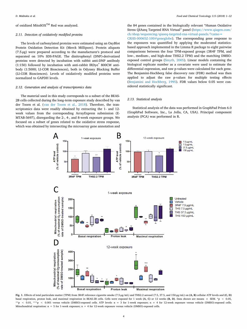
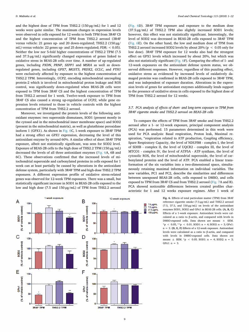
Fig. 1. Effects of total particulate matter (TPM) from 3R4F reference cigarette smoke (7.5 μg/mL) and THS2.2 aerosol (7.5, 37.5, and 150 μg/mL) on (A, B) cellular ATP levels and (C, D)basal respiration, proton leak, and maximal respiration in BEAS-2B cells. Cells were exposed for 1 week (A, C) or 12 weeks (B, D). Data shown are means ± SEM. *p < 0.05,**p < 0.01, ***p < 0.001 versus vehicle (DMSO)-exposed cells. ATP levels: n = 3 for 1-week exposure; n = 4 for 12-week exposure versus vehicle (DMSO)-exposed cells.Mitochondrial respiration: n = 5 for 1-week exposure; n = 4 for 12-week exposure versus vehicle (DMSO)-exposed cells.
D. Malinska et al. Food and Chemical Toxicology 115 (2018) 1–12
4
3. Results
3.1. TPM from 3R4F cigarette smoke and from THS2.2 aerosol decreasesATP levels in the BEAS-2B cells dependent on exposure time and dose
To elucidate the effect of short and chronic exposure to TPM from3R4F CS and TPM from THS2.2 aerosol on cellular bioenergetics ofhuman bronchial epithelial BEAS-2B cells, we examined the ATP levelsin response to exposure. Changes in the ATP level can suggest mi-tochondrial defect and can highlight alterations in cellular energyproduction processes. One-week exposure to 7.5 μg/mL of 3R4F TPMsignificantly decreased the level of ATP in BEAS-2B cells versus vehicle
(DMSO)-exposed cells (Fig. 1A). However, 3R4F TPM had no effect onATP levels in cells exposed for 12 weeks (Fig. 1B). Similarly, the lowand high concentrations of TPM from THS2.2 aerosol (7.5 and 150 μg/mL) had no impact on BEAS-2B ATP levels, both after 1 and 12 weeks ofexposure (Fig. 1A and B). Only the medium concentration of THS2.2TPM (37.5 μg/mL) significantly decreased ATP levels after 1 week aswell as after 12 weeks of exposure. These results clearly indicate me-tabolic alterations in cells exposed for 1 week to 3R4F TPM and in cellsexposed to a medium concentration of THS2.2 TPM for 1 and 12 weeks.The effect of DMSO (DMSO vs. non-exposed cells) on ATP levels inBEAS-2B cells is shown in Supplementary Fig. 1. Interestingly, at bothtime points, DMSO-exposed cells had higher ATP levels than non-
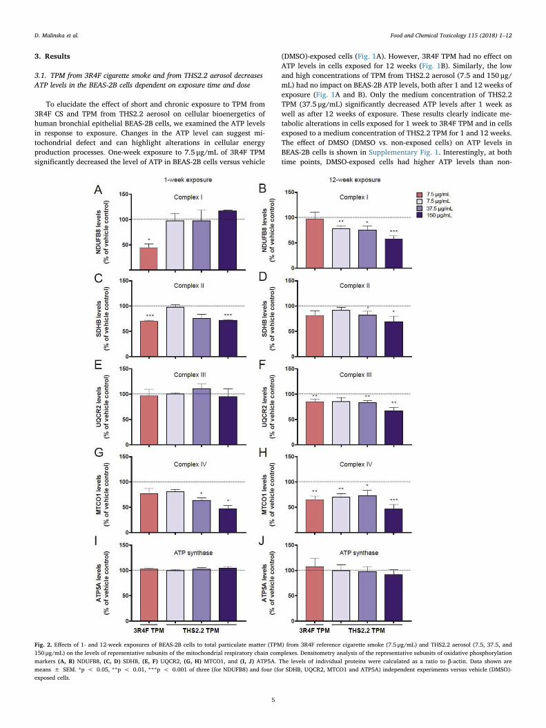
Fig. 2. Effects of 1- and 12-week exposures of BEAS-2B cells to total particulate matter (TPM) from 3R4F reference cigarette smoke (7.5 μg/mL) and THS2.2 aerosol (7.5, 37.5, and150 μg/mL) on the levels of representative subunits of the mitochondrial respiratory chain complexes. Densitometry analysis of the representative subunits of oxidative phosphorylationmarkers (A, B) NDUFB8, (C, D) SDHB, (E, F) UQCR2, (G, H) MTCO1, and (I, J) ATP5A. The levels of individual proteins were calculated as a ratio to β-actin. Data shown aremeans ± SEM. *p < 0.05, **p < 0.01, ***p < 0.001 of three (for NDUFB8) and four (for SDHB, UQCR2, MTCO1 and ATP5A) independent experiments versus vehicle (DMSO)-exposed cells.
D. Malinska et al. Food and Chemical Toxicology 115 (2018) 1–12
5
exposed BEAS-2B cells.
3.2. The effect of short- and long-term exposure to TPM from 3R4Fcigarette smoke and from THS2.2 aerosol on mitochondrial respiration
To explore the possible mechanism by which TPM exposure affectsATP levels, we measured cellular oxygen consumption, which is relatedto mitochondrial respiratory chain function. The rate of respirationrecorded at the initial phase of measurement corresponding to “Basalrespiration” gives insights into the resting energetics of the cells.Addition of oligomycin inhibits mitochondrial ATP synthesis, whichresults in slowing the rate of oxygen consumption by the mitochondrialrespiratory chain. The remaining oxygen consumption rate is used tocompensate mitochondrial “Proton leak”, but can also have a non-mi-tochondrial origin (other cellular processes that use oxygen). In ourstudies, examination of non-mitochondrial respiration was omitted dueto its negligible value. Next, addition of FCCP stimulates oxygen con-sumption to its maximum value (“Maximal respiration”) and shows themaximum capacity of the mitochondrial respiratory chain. Based on“Basal respiration”, “Proton leak” and “Maximal respiration”, it ispossible to calculate parameters describing: a) the respiration rate beingused to drive “ATP synthesis” under basal conditions, b) “Couplingefficiency” describing which fraction of “Basal respiration” is used todrive ATP production versus “Proton leak”, and c) “Spare RespiratoryCapacity” indicating how close to maximal efficiency of the respiratorychain the cells are operating under basal conditions. As shown inFig. 1C, 1-week exposure to 3R4F TPM decreased, while 1-week ex-posure to the highest dose of THS2.2 TPM (150 μg/mL) increased “Basalrespiration” of BEAS-2B cells in comparison with vehicle-exposedcontrols. There were no significant changes in the “Proton leak” -coupled respiration in BEAS-2B cells exposed for 1 week to either 3R4FTPM or the lowest dose of TPM from THS2.2 aerosol. Only in cells in-cubated in medium or exposed to the highest dose of THS2.2 TPM (37.5and 150 μg/mL) a higher proton leak was observed, suggesting thatthese exposures had a negative effect on the inner mitochondrialmembrane. Additionally, there was a clear inhibitory effect of TPMfrom 3R4F CS on mitochondrial respiratory chain as evidenced by a ca.25% decrease in “Maximal respiration” when compared with vehicle-exposed controls (Fig. 1C).
A slightly opposite effect is observed when TPM exposures are ex-tended to 12 weeks (Fig. 1D). “Basal respiration” of cells exposed toTPM from 3R4F reference CS increased compared with vehicle controls.Moreover, increased “Proton leak” suggests a harmful effect of pro-longed exposure to 3R4F TPM on the permeability of the inner mi-tochondrial membrane in BEAS-2B cells. In contrast, exposure of BEAS-
2B cells to medium or high doses of TPM (37.5 and 150 μg/mL) fromTHS2.2 aerosol decreased “Basal respiration”, suggesting these ex-posures had a negative effect on the mitochondrial respiratory chain(Fig. 1D). Interestingly, “Maximal respiration” following addition ofFCCP to cells exposed to vehicle (DMSO) for 12 weeks was not higherthan that seen in DMSO controls following 1 week of exposure, butsimilar to the value of “Basal respiration”. This indicates that long-termexposure to DMSO causes dysfunction of the mitochondrial respiratorychain. Moreover, comparison of the “Basal respiration” and “Maximalrespiration” rates in vehicle (DMSO)-exposed cells and non-exposedcells clearly showed that 12-week exposure to DMSO has a strong in-hibitory effect on the mitochondrial respiratory chain.
As mentioned above, based on the Basal and Maximal respirationrates and “proton leak”, we calculated additional parameters indicativeof OXPHOS function. Results presenting oxygen consumption coupledto “ATP synthesis”, “Spare Respiratory Capacity” and “Coupling effi-ciency” calculated for individual exposures after 1 and 12 weeks areshown in Supplementary Fig. 2 A, C, E and Supplementary Fig. 2 B, D,F, respectively. The differences in the effects on respiration of BEAS-2Bcells between 1- and 12-week exposure are clearly visible in oxygenconsumption coupled to “ATP synthesis” and coupling efficiency.
3.3. The effect of short- and long-term exposure to TPM from 3R4Fcigarette smoke and from THS2.2 aerosol on the expression levels ofindividual subunits of the mitochondrial respiratory chain and ATP synthase
To understand the effect of TPM exposure on oxygen consumptionand to determine whether alterations in the oxygen consumption can berelated to the changes in OXPHOS composition, we evaluated the ex-pression level of individual subunits of the mitochondrial respiratorychain. With the use of antibodies against representative OXPHOS sub-units (NDUFB8 - complex I, SDHB - complex II, UQCR2 - complex III,MTCO1 - complex IV and ATP5A - ATP synthase) we evaluated the levelof mitochondrial respiratory chain complexes I, II, III, IV and ATPsynthase in BEAS-2B cells exposed to TPM from 3R4F CS and fromTHS2.2 aerosol for 1 or 12 weeks (Fig. 2). One-week exposure of BEAS-2B cells to 3R4F TPM caused a significant decrease in Complex I and II(Fig. 2A and C). The level of Complex IV also seemed to be decreased;however, the effect was not statistically significant (Fig. 2G). WhenBEAS-2B cells were exposed to THS2.2 TPM, there was a dose-depen-dent decrease in Complex II and IV levels. Prolonged exposure to 3R4FTPM caused a statistically significant decrease in the level of complexesIII and IV (Fig. 2F and H). A dose-dependent effect on the level of allfour respiratory chain complexes (I, II, III and IV) was observed forTHS2.2 TPM exposures (Fig. 2B, D, F, and H). However, there was no
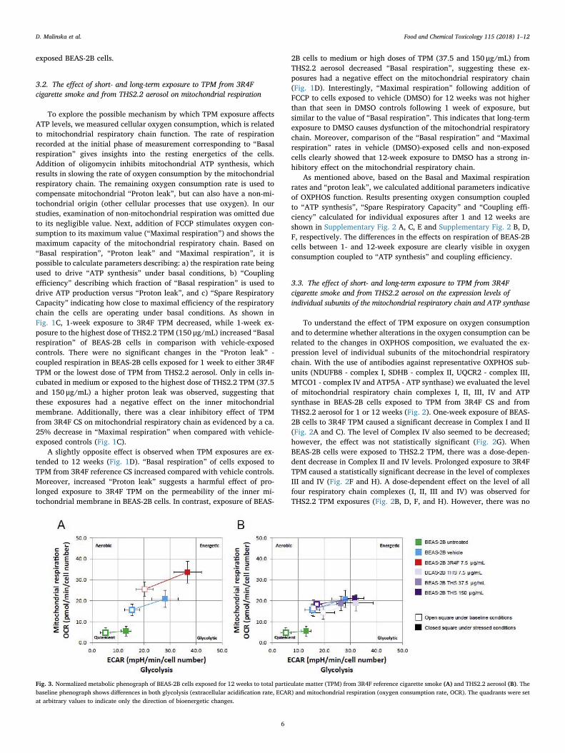
Fig. 3. Normalized metabolic phenograph of BEAS-2B cells exposed for 12 weeks to total particulate matter (TPM) from 3R4F reference cigarette smoke (A) and THS2.2 aerosol (B). Thebaseline phenograph shows differences in both glycolysis (extracellular acidification rate, ECAR) and mitochondrial respiration (oxygen consumption rate, OCR). The quadrants were setat arbitrary values to indicate only the direction of bioenergetic changes.
D. Malinska et al. Food and Chemical Toxicology 115 (2018) 1–12
6
TPM exposure effect on the level of ATP synthase (Fig. 2I and J). In-terestingly, 1-week exposure of BEAS-2B cells to vehicle (DMSO) in-creased the level of complex II, while 12-week exposure induced sig-nificant higher levels of complexes I, II, III and IV in comparison withthe non-exposed BEAS-2B cells (data not shown).
3.4. Long-term exposure to TPM from 3R4F cigarette smoke and THS2.2aerosol alters the metabolic phenotype of BEAS-2B cells
In order to examine if long term exposure-dependent differences inthe profile of OXPHOS function parameters are connected with meta-bolic reprogramming, we measured the metabolic potential of BEAS-2Bcells after 12 weeks of exposure to TPM (Fig. 3). The extracellularacidification rate (ECAR), which estimates glycolytic activity undercertain conditions, and the mitochondrial oxygen consumption rate(OCR), which is a performance indicator of mitochondrial function,where both determined simultaneously within the BEAS-2B cell popu-lation. Twelve week exposure to TPM from 3R4F CS elevated bothECAR and OCR, indicating that the cells increased their metabolism dueto increased glycolysis and mitochondrial function. This shifted thebaseline phenotype of the original BEAS-2B cells from a quiescent to amore energetic phenotype compared with non-exposed or vehicle-ex-posed cells. This shift is known in oncology as the Warburg effect, and isthought to be an adaptation mechanism for supporting the biosyntheticrequirements of uncontrolled proliferation (Liberti and Locasale, 2016).Long-term exposure of BEAS-2B cells to low, medium, and high con-centrations (7.5, 37.5 and 150 μg/mL) of TPM from THS2.2 aerosol didnot alter the metabolic phenotype compared with effects in the vehiclecontrol (Fig. 3A and B).
3.5. The effect of short- and long-term exposure to TPM from 3R4Fcigarette smoke and from THS2.2 aerosol on the oxidative stressmanifestation in BEAS-2B cells
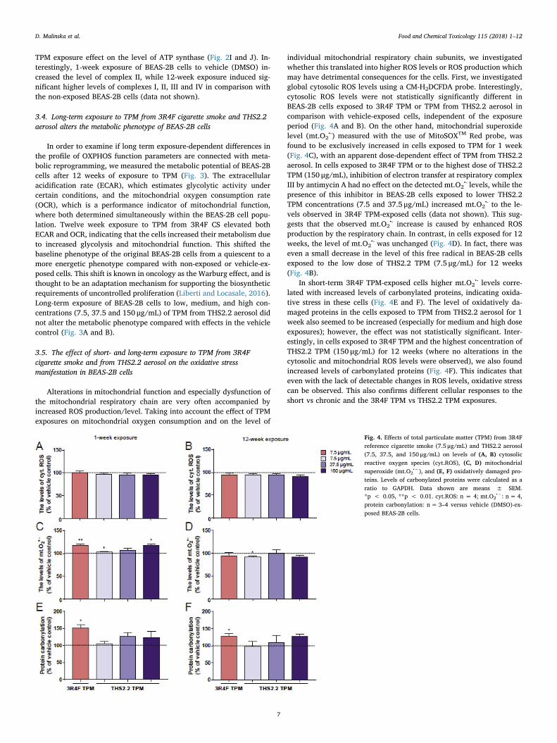
Alterations in mitochondrial function and especially dysfunction ofthe mitochondrial respiratory chain are very often accompanied byincreased ROS production/level. Taking into account the effect of TPMexposures on mitochondrial oxygen consumption and on the level of
individual mitochondrial respiratory chain subunits, we investigatedwhether this translated into higher ROS levels or ROS production whichmay have detrimental consequences for the cells. First, we investigatedglobal cytosolic ROS levels using a CM-H2DCFDA probe. Interestingly,cytosolic ROS levels were not statistically significantly different inBEAS-2B cells exposed to 3R4F TPM or TPM from THS2.2 aerosol incomparison with vehicle-exposed cells, independent of the exposureperiod (Fig. 4A and B). On the other hand, mitochondrial superoxidelevel (mt.O2
•-) measured with the use of MitoSOXTM Red probe, wasfound to be exclusively increased in cells exposed to TPM for 1 week(Fig. 4C), with an apparent dose-dependent effect of TPM from THS2.2aerosol. In cells exposed to 3R4F TPM or to the highest dose of THS2.2TPM (150 μg/mL), inhibition of electron transfer at respiratory complexIII by antimycin A had no effect on the detected mt.O2
•- levels, while thepresence of this inhibitor in BEAS-2B cells exposed to lower THS2.2TPM concentrations (7.5 and 37.5 μg/mL) increased mt.O2
•- to the le-vels observed in 3R4F TPM-exposed cells (data not shown). This sug-gests that the observed mt.O2
•- increase is caused by enhanced ROSproduction by the respiratory chain. In contrast, in cells exposed for 12weeks, the level of mt.O2
•- was unchanged (Fig. 4D). In fact, there waseven a small decrease in the level of this free radical in BEAS-2B cellsexposed to the low dose of THS2.2 TPM (7.5 μg/mL) for 12 weeks(Fig. 4B).
In short-term 3R4F TPM-exposed cells higher mt.O2•- levels corre-
lated with increased levels of carbonylated proteins, indicating oxida-tive stress in these cells (Fig. 4E and F). The level of oxidatively da-maged proteins in the cells exposed to TPM from THS2.2 aerosol for 1week also seemed to be increased (especially for medium and high doseexposures); however, the effect was not statistically significant. Inter-estingly, in cells exposed to 3R4F TPM and the highest concentration ofTHS2.2 TPM (150 μg/mL) for 12 weeks (where no alterations in thecytosolic and mitochondrial ROS levels were observed), we also foundincreased levels of carbonylated proteins (Fig. 4F). This indicates thateven with the lack of detectable changes in ROS levels, oxidative stresscan be observed. This also confirms different cellular responses to theshort vs chronic and the 3R4F TPM vs THS2.2 TPM exposures.
Fig. 4. Effects of total particulate matter (TPM) from 3R4Freference cigarette smoke (7.5 μg/mL) and THS2.2 aerosol(7.5, 37.5, and 150 μg/mL) on levels of (A, B) cytosolicreactive oxygen species (cyt.ROS), (C, D) mitochondrialsuperoxide (mt.O2
•−), and (E, F) oxidatively damaged pro-teins. Levels of carbonylated proteins were calculated as aratio to GAPDH. Data shown are means ± SEM.*p < 0.05, **p < 0.01. cyt.ROS: n = 4; mt.O2
•−: n= 4,protein carbonylation: n= 3–4 versus vehicle (DMSO)-ex-posed BEAS-2B cells.
D. Malinska et al. Food and Chemical Toxicology 115 (2018) 1–12
7
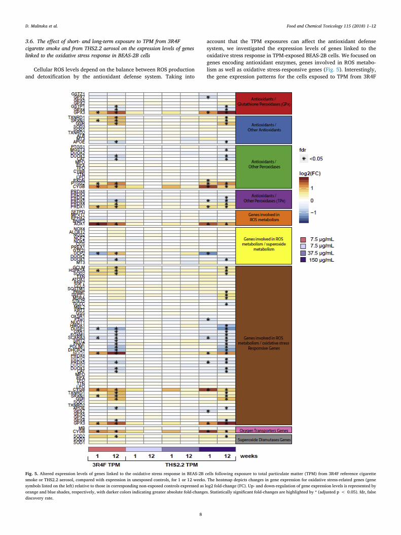
3.6. The effect of short- and long-term exposure to TPM from 3R4Fcigarette smoke and from THS2.2 aerosol on the expression levels of geneslinked to the oxidative stress response in BEAS-2B cells
Cellular ROS levels depend on the balance between ROS productionand detoxification by the antioxidant defense system. Taking into
account that the TPM exposures can affect the antioxidant defensesystem, we investigated the expression levels of genes linked to theoxidative stress response in TPM-exposed BEAS-2B cells. We focused ongenes encoding antioxidant enzymes, genes involved in ROS metabo-lism as well as oxidative stress-responsive genes (Fig. 5). Interestingly,the gene expression patterns for the cells exposed to TPM from 3R4F
Fig. 5. Altered expression levels of genes linked to the oxidative stress response in BEAS-2B cells following exposure to total particulate matter (TPM) from 3R4F reference cigarettesmoke or THS2.2 aerosol, compared with expression in unexposed controls, for 1 or 12 weeks. The heatmap depicts changes in gene expression for oxidative stress-related genes (genesymbols listed on the left) relative to those in corresponding non-exposed controls expressed as log2 fold-change (FC). Up- and down-regulation of gene expression levels is represented byorange and blue shades, respectively, with darker colors indicating greater absolute fold-changes. Statistically significant fold-changes are highlighted by * (adjusted p < 0.05). fdr, falsediscovery rate.
D. Malinska et al. Food and Chemical Toxicology 115 (2018) 1–12
8
and the highest dose of TPM from THS2.2 (150 μg/mL) for 1 and 12weeks were quite similar. The maximum changes in expression levelswere observed in cells exposed for 12 weeks to both TPM from 3R4F CSand the highest concentration of TPM from THS2.2 aerosol (3R4Fversus vehicle: 21 genes up- and 20 down-regulated; THS2.2 (150 μg/mL) versus vehicle: 22 genes up- and 25 down-regulated; FDR:< 0.05).Neither the low nor 5-fold higher concentrations of THS2.2 TPM (7.5and 37.5 μg/mL) significantly changed expression of genes linked tooxidative stress in BEAS-2B cells over time. A number of up-regulatedgenes, including PXDN, PRNP, SEPP1 and MSRA as well as down-regulated genes, including GPX7, MGST3, PRDX2, GCLC, and FTH1were exclusively affected by exposure to the highest concentration ofTHS2.2 TPM. Interestingly, UCP2, encoding mitochondrial uncouplingprotein-2 which is involved in mitochondrial proton leakage and ROScontrol, was significantly down-regulated when BEAS-2B cells wereexposed to TPM from 3R4F CS and the highest concentration of TPMfrom THS2.2 aerosol for 1 week. Twelve-week exposure to TPM from3R4F CS also caused a strong up-regulation of UCP2, while gene ex-pression levels returned to those in vehicle controls with the highestconcentration of TPM from THS2.2 aerosol.
Moreover, we investigated the protein levels of the following anti-oxidant enzymes: two superoxide dismutases, SOD1 (present mostly inthe cytosol and in the mitochondrial inner membrane space) and SOD2(present in the mitochondrial matrix), as well as glutathione peroxidaseisoform 1 (GPX1). As shown in Fig. 6C, 1-week exposure to 3R4F TPMhad a strong effect on GPX1 expression, decreasing the level of thisantioxidant enzyme by around 60%. A similar effect of short 3R4F TPMexposure, albeit not statistically significant, was seen for SOD2 level.Exposure of BEAS-2B cells to the high dose of THS2.2 TPM (150 μg/mL)decreased the levels of all three antioxidant enzymes (Fig. 6A, 6B and6C). These observations confirmed that the increased levels of mi-tochondrial superoxide and carbonylated proteins in cells exposed for 1week can at least partially be caused by alterations in the antioxidantdefense system, particularly with 3R4F TPM and high-dose THS2.2 TPMexposures. A different expression profile of oxidative stress-relatedgenes was observed for 12-week TPM exposures. There was a small, butstatistically significant increase in SOD1 in BEAS-2B cells exposed to thelow and high dose (7.5 and 150 μg/mL) of TPM from THS2.2 aerosol
(Fig. 6D). 3R4F TPM exposure and exposure to the medium dose(37.5 μg/mL) of THS2.2 TPM also slightly increased SOD1 levels;however, this effect was not statistically significant. Interestingly, thelevel of SOD2 was decreased in BEAS-2B cells exposed to 3R4F TPM(Fig. 6E). On the other hand, the low and medium dose of TPM fromTHS2.2 aerosol increased SOD2 levels by about 20% (p < 0.05 only forlow dose). 3R4F TPM exposure for 12 weeks also had the strongesteffect on GPX1 levels which increased by about 20%, but which wasalso not statistically significant (Fig. 6F). Comparing the effect of 1- and12-week exposures on the antioxidant defense system status, we ob-served different responses. However, regardless of this puzzling effect,oxidative stress as evidenced by increased levels of oxidatively da-maged proteins was confirmed in BEAS-2B cells exposed to 3R4F TPM,and was independent of exposure duration. Alterations in the expres-sion levels of genes for antioxidant enzymes additionally lends supportto the presence of oxidative stress in cells exposed to the highest dose ofTPM from THS2.2 aerosol for 12 weeks.
3.7. PCA analysis of effects of short- and long-term exposure to TPM from3R4F cigarette smoke and THS2.2 aerosol on BEAS-2B cells
To compare the effects of TPM from 3R4F smoke and from THS2.2aerosol after a 1- or 12-week exposure, principal component analysis(PCA) was performed. 15 parameters determined in this work wereused for PCA analysis: Basal respiration, Proton leak, Maximal re-spiration, “respiration related to ATP production, Coupling efficiency,Spare Respiratory Capacity, the level of NDUFB8 - complex I, the levelof SDHB - complex II, the level of UQCR2 - complex III, the level ofMTCO1 - complex IV, the level of ATP5A - ATP synthase, the level ofcytosolic ROS, the level of mitochondrial superoxide, the level of car-bonylated proteins and the level of ATP. PCA enabled a linear trans-formation of the six variables into a two-dimensional space, simulta-neously retaining maximal information on individual variables. Thenew variables, PC1 and PC2, describe the similarities and differencesbetween unexposed BEAS-2B cells, cells exposed to DMSO, and cellsexposed to TPM from 3R4F CS and from THS2.2 aerosol (Fig. 7A and B).PCA showed noticeable differences between created profiles char-acteristic for 1 and 12 weeks exposure regimes. After 1 week of
Fig. 6. Effects of total particulate matter (TPM) from 3R4Freference cigarette smoke (7.5 μg/mL) and THS2.2 aerosol(7.5, 37.5, and 150 μg/mL) on levels of the antioxidantenzymes SOD1, SOD2 and GPx1 in BEAS-2B cells. (A, B, C)Effects of a 1-week exposure. Antioxidant levels were cal-culated as a ratio to β-actin, and compared with levels inDMSO-exposed cells. Data shown are means ± SEM.*p < 0.05, **p < 0.01. SOD1: n = 4; SOD2: n = 3; GPx1:n = 3. (D, E, F) Effects of a 12-week exposure. Antioxidantlevels were calculated as a ratio to β-actin, and comparedwith levels in DMSO-exposed cells. Data shown aremeans ± SEM. *p < 0.05. SOD1: n = 4; SOD2: n = 3;GPx1: n = 3.
D. Malinska et al. Food and Chemical Toxicology 115 (2018) 1–12
9
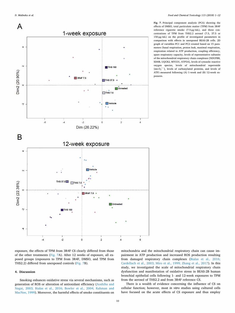
exposure, the effects of TPM from 3R4F CS clearly differed from thoseof the other treatments (Fig. 7A). After 12 weeks of exposure, all ex-posed groups (exposures to TPM from 3R4F, DMSO, and TPM fromTHS2.2) differed from unexposed controls (Fig. 7B).
4. Discussion
Smoking enhances oxidative stress via several mechanisms, such asgeneration of ROS or alteration of antioxidant efficiency (Aoshiba andNagai, 2003; Bialas et al., 2016; Bowler et al., 2004; Rahman andMacNee, 1999). Moreover, the harmful effects of smoke constituents on
mitochondria and the mitochondrial respiratory chain can cause im-pairment in ATP production and increased ROS production resultingfrom damaged respiratory chain complexes (Bialas et al., 2016;Cardellach et al., 2003; Miro et al., 1999; Zhang et al., 2017). In thisstudy, we investigated the scale of mitochondrial respiratory chaindysfunction and manifestation of oxidative stress in BEAS-2B humanbronchial epithelial cells following 1- and 12-week exposures to TPMfrom the aerosol of THS2.2 and from 3R4F reference CS.
There is a wealth of evidence concerning the influence of CS oncellular function; however, most in vitro studies using cultured cellshave focused on the acute effects of CS exposure and thus employ
Fig. 7. Principal component analysis (PCA) showing theeffects of DMSO, total particulate matter (TPM) from 3R4Freference cigarette smoke (7.5 μg/mL), and three con-centrations of TPM from THS2.2 aerosol (7.5, 37.5 or150 μg/mL) on the profile of investigated parameters incomparison with effects in unexposed BEAS-2B cells. 2Dgraph of variables PC1 and PC2 created based on 15 para-meters (basal respiration, proton leak, maximal respiration,respiration related to ATP production, coupling efficiency,spare respiratory capacity, levels of representative subunitsof the mitochondrial respiratory chain complexes [NDUFB8,SDHB, UQCR2, MTCO1, ATP5A], levels of cytosolic reactiveoxygen species, levels of mitochondrial superoxide[mt.O2
•−], levels of carbonylated proteins, and levels ofATP) measured following (A) 1-week and (B) 12-week ex-posures.
D. Malinska et al. Food and Chemical Toxicology 115 (2018) 1–12
10
relatively short incubation times (usually up to 24 h) (Aug et al., 2014;Guan et al., 2013; van der Toorn et al., 2007; Zhang et al., 2017). Dataon the effects of long-term exposures, which would resemble the in vivosituation more closely, are limited (Hoffmann et al., 2013; Veljkovicet al., 2011). Therefore, we were interested in highlighting the potentialdifferences between short- and long-term exposures to TPM from 3R4Freference CS compared to aerosol from THS2.2.
The adverse effects of CS on mitochondrial respiratory chain func-tion have been reported in isolated mitochondria (van der Toorn et al.,2007) as well as in cells and tissues of animals (Agarwal et al., 2014;Gvozdjakova et al., 1999) and humans (Bouhours-Nouet et al., 2005;Cardellach et al., 2003; Miro et al., 1999). Similarly, in our study model(BEAS-2B cells), we observed decreased mitochondrial respiration(both basal and maximal rates) in cells exposed for 1 week to TPM fromthe 3R4F reference cigarette. This was accompanied by a decrease inthe content of selected subunits of respiratory chain complexes I and II.This finding contradicts that of Hoffmann et al., who observed in-creased levels of the tested respiratory chain subunits in BEAS-2B cellsexposed to CS extracts (Hoffmann et al., 2013). In that study, however,a much longer exposure duration of 6 months was applied. In addition,the dose-dependence of this effect was much stronger: above a certainCSE dose, the increase in the levels of respiratory chain proteins wassmaller. Generally, there is discrepancy in the literature concerning theinfluence of CS exposure on the amount and activities of respiratorychain complexes: both increases (Agarwal et al., 2012; Hoffmann et al.,2013) and decreases (Cardellach et al., 2003; Miro et al., 1999) werereported, depending on the experimental model, investigated tissue,incubation time, and applied concentrations. Contrary to the 1-weekexposure, the 12-week exposure of BEAS-2B cells to 3R4F TPM in-creased basal and maximal respiration rates as well as proton leak. Thiscoincided with higher expression of UCP2, but the available data do notallow for a conclusion about the causal relationship between these twoobservations.
Among other aspects of mitochondrial function, we observed in-creased mitochondrial ROS levels in cells exposed for 1 week. Similarobservations were also reported in human airway smooth muscle cells(Aravamudan et al., 2014) exposed to CSE for 24 h. The effect of anti-mycin A suggests that increased mt.O2
•− levels was the result of ele-vated superoxide production by the respiratory chain, rather than ofimpaired ROS scavenging. Surprisingly, no changes in cytosolic ROSlevels were detected in our study, contrary to findings in multiple stu-dies using CS-exposed cultured cells, where elevation of ROS levels wasobserved (Lin et al., 2014; van der Toorn et al., 2009; Wylam et al.,2015). In these studies, however, shorter exposure times had been ap-plied (up to 24 h) and typically higher concentrations, resulting in de-creased cell viability. Despite the unaltered levels of cytosolic ROS inour study, we observed hallmarks of oxidative stress following both 1and 12 weeks of exposure to TPM from CS, such as increased proteincarbonylation and up- as well as down-regulated expression of oxida-tive stress response genes. Additionally, the levels of the antioxidantenzyme GPx1 decreased. In line with these findings, Zhang et al. re-ported multiple symptoms of oxidative stress and redox balance dis-ruption after short exposure of BEAS-2B cells to CS, including a de-creased ratio of reduced glutathione to oxidized glutathione andoxidative damage of lipids and DNA (Zhang et al., 2017).
Compared to 3R4F TPM, THS2.2 TPM needed to be applied at 20-fold higher concentration to cause a similar extent of oxidative stressand changes in cellular bioenergetics. 7.5 μg/mL 3R4F TPM and150 μg/mL THS2.2 TPM exerted similar effects on mitochondrial su-peroxide levels, protein carbonylation and levels of GPx1. Gene ex-pression analysis showed similar changes in expression patterns ofoxidative stress response genes in cells exposed to 7.5 μg/mL of TPMfrom reference CS and to 150 μg/mL of TPM from THS2.2 aerosol. Bothtreatments decreased the levels of selected respiratory chain proteins,but a 1-week exposure to TPM from the reference cigarette induced astrong decrease in the levels of complex I subunit NDUFB8, while TPM
from THS2.2 had a more pronounced effect on the levels of the complexIV subunit. The difference between the mechanisms of toxicity of TPMfrom 3R4F and THS2.2 was seen in their varied effects on respirationrates and glycolysis, where the reference cigarette caused strongerdisturbances than even the highest concentration of TPM from THS2.2.These observations would suggest that different compounds in CS couldbe responsible for respiratory disturbances and for oxidative stress, andthat these alterations may be caused, at least partially, by independentmechanisms.
The presence of oxidative stress was directly confirmed in BEAS-2Bcells exposed to TPM from 3R4F CS for both 1 and 12 weeks by theincreased levels of oxidatively damaged proteins. Moreover, based onthe expression pattern of genes involved in ROS metabolism, thehighest concentration of TPM from THS2.2 aerosol also caused oxida-tive stress even though the levels of cytosolic ROS and mitochondrialsuperoxide were unchanged. The greatest effect on expression levels ofgenes involved in ROS metabolism was observed in cells exposed for 12weeks to TPM from 3R4F CS and the highest concentration of TPM fromTHS2.2 aerosol. Interestingly, when comparing results for the two ex-posure periods, the effects are usually stronger after shorter exposure.For some parameters, an opposite direction of changes are observed, asin the case of basal and maximal respiration rates, which decreasedafter 1-week exposure, but increased after 12-week exposure. Some ofthese differences may reflect adaptive mechanisms, allowing cells tosurvive under chronic stress conditions. It is, however, also known thatlong-term exposure of bronchial epithelial cells to CS results in persis-tent epigenetic changes characteristic of malignant transformation(Veljkovic et al., 2011), and we cannot exclude that some of the effectsof long-term exposure are already reflecting this hallmark of cancer.
5. Conclusion
This study showed that alterations in mitochondrial respiratorychain function are accompanied by oxidative stress in BEAS-2B cellsexposed to TPM from 3R4F CS and THS2.2 aerosol, and that the effectsvaried by exposure duration. A concentration of TPM from THS2.2aerosol 20-fold higher than the concentration of TPM from 3R4F wasrequired to disturb cellular function to a similar extent. This indicatesthat reducing levels of HPHCs by heating rather than combusting to-bacco could reduce mitochondrial dysfunction and oxidative stress-re-lated diseases associated with smoking combustible tobacco products.
Interestingly, the effect of TPM exposures on the mitochondrial re-spiratory chain is different between the two exposure durations. Incontrast to the effects of short-term stress, a chronic exposure appearsto result in cellular adaptation to the stressors. Future investigationsshould focus on elucidating the mechanism underlying this adaptation.
Conflict-of-interest statement
Alain Sewer, Stephanie Johne, Karsta Luettich, Manuel C Peitsch,Julia Hoeng, Marco van der Toorn are employees of and paid by PhilipMorris International.
Philip Morris International was the sole source of funding andsponsor of this project.
Acknowledgments
This work was funded by Philip Morris International R&D.The authors are grateful to V. Biernat for specialist technical assis-
tance.
Transparency document
Transparency document related to this article can be found online athttp://dx.doi.org/10.1016/j.fct.2018.02.013.
D. Malinska et al. Food and Chemical Toxicology 115 (2018) 1–12
11
Appendix A. Supplementary data
Supplementary data related to this article can be found at http://dx.doi.org/10.1016/j.fct.2018.02.013.
References
Agarwal, A.R., Zhao, L., Sancheti, H., Sundar, I.K., Rahman, I., Cadenas, E., 2012. Short-term cigarette smoke exposure induces reversible changes in energy metabolism andcellular redox status independent of inflammatory responses in mouse lungs.American journal of physiology. Lung cellular and molecular physiology 303,L889–L898.
Agarwal, A.R., Yin, F., Cadenas, E., 2014. Short-term cigarette smoke exposure leads tometabolic alterations in lung alveolar cells. Am. J. Respir. Cell Mol. Biol. 51,284–293.
Anbarasi, K., Vani, G., Devi, C.S., 2005. Protective effect of bacoside A on cigarettesmoking-induced brain mitochondrial dysfunction in rats. J. Environ. Pathol. Toxicol.Oncol. 24, 225–234.
Aoshiba, K., Nagai, A., 2003. Oxidative stress, cell death, and other damage to alveolarepithelial cells induced by cigarette smoke. Tob. Induc. Dis. 1, 219–226.
Aravamudan, B., Kiel, A., Freeman, M., Delmotte, P., Thompson, M., Vassallo, R., Sieck,G.C., Pabelick, C.M., Prakash, Y.S., 2014. Cigarette smoke-induced mitochondrialfragmentation and dysfunction in human airway smooth muscle. American journal ofphysiology. Lung cellular and molecular physiology 306, L840–L854.
Aug, A., Altraja, A., Altraja, S., Laaniste, L., Mahlapuu, R., Soomets, U., Kilk, K., 2014.Alterations of bronchial epithelial metabolome by cigarette smoke are reversible byan antioxidant, O-methyl-L-tyrosinyl-gamma-L-glutamyl-L-cysteinylglycine. Am. J.Respir. Cell Mol. Biol. 51, 586–594.
Baker, R.R., 1974. Temperature distribution inside a burning cigarette. Nature 247,405–406.
Barsanti, K.C., Luo, W., Isabelle, L.M., Pankow, J.F., Peyton, D.H., 2007. Tobacco smokeparticulate matter chemistry by NMR. Magn. Reson. Chem. 45, 167–170.
Benjamini, Y., Hochberg, Y., 1995. Controlling the false discovery rate: a practical andpowerful approach to multiple testing. J. Roy. Stat. Soc. B 289–300.
Bialas, A.J., Sitarek, P., Milkowska-Dymanowska, J., Piotrowski, W.J., Gorski, P., 2016.The role of mitochondria and oxidative/antioxidative imbalance in pathobiology ofchronic obstructive pulmonary disease. Oxid Med Cell Longev 2016 7808576.
Bouhours-Nouet, N., May-Panloup, P., Coutant, R., de Casson, F.B., Descamps, P., Douay,O., Reynier, P., Ritz, P., Malthiery, Y., Simard, G., 2005. Maternal smoking is asso-ciated with mitochondrial DNA depletion and respiratory chain complex III defi-ciency in placenta. Am. J. Physiol. Endocrinol. Metab. 288, E171–E177.
Bowler, R.P., Barnes, P.J., Crapo, J.D., 2004. The role of oxidative stress in chronic ob-structive pulmonary disease. COPD 1, 255–277.
Bradford, M.M., 1976. A rapid and sensitive method for the quantitation of microgramquantities of protein utilizing the principle of protein-dye binding. Anal. Biochem. 72,248–254.
Cardellach, F., Alonso, J.R., Lopez, S., Casademont, J., Miro, O., 2003. Effect of smokingcessation on mitochondrial respiratory chain function. J. Toxicol. Clin. Toxicol. 41,223–228.
Durham, A.L., Adcock, I.M., 2015. The relationship between COPD and lung cancer. LungCanc. 90, 121–127.
Guan, S.P., Tee, W., Ng, D.S., Chan, T.K., Peh, H.Y., Ho, W.E., Cheng, C., Mak, J.C., Wong,W.S., 2013. Andrographolide protects against cigarette smoke-induced oxidative lunginjury via augmentation of Nrf2 activity. Br. J. Pharmacol. 168, 1707–1718.
Gvozdjakova, A., Simko, F., Kucharska, J., Braunova, Z., Psenek, P., Kyselovic, J., 1999.Captopril increased mitochondrial coenzyme Q10 level, improved respiratory chainfunction and energy production in the left ventricle in rabbits with smoke mi-tochondrial cardiomyopathy. Biofactors 10, 61–65.
Health Canada, 1999. Health Canada Test Method T-115, Determination of ‘‘Tar’’ andNicotine in Sidestream Tobacco Smoke.
Hoffmann, R.F., Zarrintan, S., Brandenburg, S.M., Kol, A., de Bruin, H.G., Jafari, S., Dijk,F., Kalicharan, D., Kelders, M., Gosker, H.R., Ten Hacken, N.H., van der Want, J.J.,van Oosterhout, A.J., Heijink, I.H., 2013. Prolonged cigarette smoke exposure altersmitochondrial structure and function in airway epithelial cells. Respir. Res. 14, 97.
CDC, 2010. Tobacco Smoke Causes Disease: The Biology and Behavioral Basis forSmoking-attributable Disease: a Report of the Surgeon General, Atlanta (GA).
International Organization for Standardization, 2010. ISO 3402: 1999–Tobacco andTobacco Products – Atmosphere for Conditioning and Testing, fourth ed. .
Lebiedzinska, M., Karkucinska-Wieckowska, A., Wojtala, A., Suski, J.M., Szabadkai, G.,Wilczynski, G., Wlodarczyk, J., Diogo, C.V., Oliveira, P.J., Tauber, J., Jezek, P.,Pronicki, M., Duszynski, J., Pinton, P., Wieckowski, M.R., 2013. Disrupted ATPsynthase activity and mitochondrial hyperpolarisation-dependent oxidative stress isassociated with p66Shc phosphorylation in fibroblasts of NARP patients. Int. J.Biochem. Cell Biol. 45, 141–150.
Liberti, M.V., Locasale, J.W., 2016. The Warburg effect: how does it benefit cancer cells?Trends Biochem. Sci. 41, 211–218.
Lin, X.X., Yang, X.F., Jiang, J.X., Zhang, S.J., Guan, Y., Liu, Y.N., Sun, Y.H., Xie, Q.M.,2014. Cigarette smoke extract-induced BEAS-2B cell apoptosis and anti-oxidative Nrf-2 up-regulation are mediated by ROS-stimulated p38 activation. Toxicol. Mech.Meth. 24, 575–583.
Miro, O., Alonso, J.R., Jarreta, D., Casademont, J., Urbano-Marquez, A., Cardellach, F.,1999. Smoking disturbs mitochondrial respiratory chain function and enhances lipidperoxidation on human circulating lymphocytes. Carcinogenesis 20, 1331–1336.
Patskan, G., Reininghaus, W., 2003. Toxicological evaluation of an electrically heatedcigarette. Part 1: overview of technical concepts and summary of findings. J. Appl.Toxicol. 23, 323–328.
Rahman, I., MacNee, W., 1996. Role of oxidants/antioxidants in smoking-induced lungdiseases. Free Radical Biol. Med. 21, 669–681.
Rahman, I., MacNee, W., 1999. Lung glutathione and oxidative stress: implications incigarette smoke-induced airway disease. Am. J. Physiol. 277, L1067–L1088.
Rodgman, A., Perfetti, T., 2013. The Chemical Components of Tobacco and TobaccoSmoke, second ed. CRC Press, pp. 2238.
Smith, M.R., Clark, B., Luedicke, F., Schaller, J.P., Vanscheeuwijck, P., Hoeng, J., Peitsch,M.C., 2016. Evaluation of the tobacco heating system 2.2. Part 1: description of thesystem and the scientific assessment program. Regul. Toxicol. Pharmacol. 81 (Suppl.2), S17–S26.
Smyth, G.K., 2005. Limma: Linear Models for Microarray Data. Bioinformatics andComputational Biology Solutions Using R and Bioconductor. Springer, pp. 397–420.
van der Toorn, M., Slebos, D.J., de Bruin, H.G., Leuvenink, H.G., Bakker, S.J., Gans, R.O.,Koeter, G.H., van Oosterhout, A.J., Kauffman, H.F., 2007. Cigarette smoke-inducedblockade of the mitochondrial respiratory chain switches lung epithelial cell apop-tosis into necrosis. American journal of physiology. Lung cellular and molecularphysiology 292, L1211–L1218.
van der Toorn, M., Rezayat, D., Kauffman, H.F., Bakker, S.J., Gans, R.O., Koeter, G.H.,Choi, A.M., van Oosterhout, A.J., Slebos, D.J., 2009. Lipid-soluble components incigarette smoke induce mitochondrial production of reactive oxygen species in lungepithelial cells. American journal of physiology. Lung cellular and molecular phy-siology 297, L109–L114.
van der Toorn, M., Sewer, A., Marescotti, D., Johne, S., Baumer, K., Bornand, D., Dulize,R., Merg, C., Corciulo, M., Scotti, E., Pak, C., Leroy, P., Guedj, E., Ivanov, N., Martin,F., Peitsch, M., Hoeng, J., Luettich, K., 2018. The biological effects of long-term ex-posure of human bronchial epithelial cells to total particulate matter from a candi-date modified-risk tobacco product. Toxicol. Vitro (in press).
Veljkovic, E., Jiricny, J., Menigatti, M., Rehrauer, H., Han, W., 2011. Chronic exposure tocigarette smoke condensate in vitro induces epithelial to mesenchymal transition-likechanges in human bronchial epithelial cells, BEAS-2B. Toxicol. Vitro 25, 446–453.
Wojewoda, M., Duszynski, J., Szczepanowska, J., 2010. Antioxidant defence systems andgeneration of reactive oxygen species in osteosarcoma cells with defective mi-tochondria: effect of selenium. Biochim. Biophys. Acta 1797, 890–896.
Wojewoda, M., Duszynski, J., Szczepanowska, J., 2011. NARP mutation and mtDNAdepletion trigger mitochondrial biogenesis which can be modulated by selenitesupplementation. Int. J. Biochem. Cell Biol. 43, 1178–1186.
Wong, E.T., Kogel, U., Veljkovic, E., Martin, F., Xiang, Y., Boue, S., Vuillaume, G., Leroy,P., Guedj, E., Rodrigo, G., Ivanov, N.V., Hoeng, J., Peitsch, M.C., Vanscheeuwijck, P.,2016. Evaluation of the Tobacco Heating System 2.2. Part 4: 90-day OECD 413 ratinhalation study with systems toxicology endpoints demonstrates reduced exposureeffects compared with cigarette smoke. Regul. Toxicol. Pharmacol. 81 (Suppl. 2),S59–S81.
Wylam, M.E., Sathish, V., VanOosten, S.K., Freeman, M., Burkholder, D., Thompson,M.A., Pabelick, C.M., Prakash, Y.S., 2015. Mechanisms of cigarette smoke effects onhuman airway smooth muscle. PLoS One 10 e0128778.
Zhang, S., Li, X., Xie, F., Liu, K., Liu, H., Xie, J., 2017. Evaluation of whole cigarette smokeinduced oxidative stress in A549 and BEAS-2B cells. Environ. Toxicol. Pharmacol. 54,40–47.
D. Malinska et al. Food and Chemical Toxicology 115 (2018) 1–12
12