foot and ankle surgical...
TRANSCRIPT

1
Washington State Department of Labor and Industries
Surgical Guideline for Work-related Foot-Ankle Injuries – October 2017
Clinical Practice Guideline for Foot and Ankle Diagnosis and Treatment
TABLE OF CONTENTS
I. Review Criteria for Foot and Ankle Surgery ..................................................................... 3
II. Introduction ................................................................................................................. 12
A. Background and Prevalence .............................................................................................. 12
III. Establishing Work-relatedness ...................................................................................... 14
IV. Pre-existing Non-work-related Conditions .................................................................... 14
A. Pes Planus .......................................................................................................................... 14
B. Pes Cavus ........................................................................................................................... 15
C. Plantar Fasciitis .................................................................................................................. 16
D. Osteoarthritis ..................................................................................................................... 16
V. Assessment .................................................................................................................. 17
A. Imaging ............................................................................................................................... 17
B. Preventing Complications .................................................................................................. 18
C. Measuring Functional Improvement ................................................................................. 18
VI. Specific Conditions and Surgical Procedures .................................................................. 19
A. Ankle Arthroscopy and Cheilectomy ................................................................................. 19
B. Ankle Arthrodesis/Fusion or Arthroplasty/Replacement .................................................. 19
Arthrodesis/Fusion ................................................................................................................ 20
Arthroplasty/Joint Replacement ........................................................................................... 20
C. Subtalar Arthrodesis .......................................................................................................... 20
D. Debridement or Stabilization of a Medial or Lateral Talar Lesion ..................................... 21
E. Lateral Ligament Repair or Reconstruction ....................................................................... 22
F. Peroneal Tendon Repair .................................................................................................... 22
G. Achilles Tendon Repair or Reconstruction......................................................................... 22

2
Washington State Department of Labor and Industries
Surgical Guideline for Work-related Foot-Ankle Injuries – October 2017
Repair at Insertion ................................................................................................................. 23
Repair ..................................................................................................................................... 23
Reconstruction ....................................................................................................................... 23
H. Posterior Tibialis Tendon Reconstruction .......................................................................... 23
PTTD and Work-relatedness .................................................................................................. 24
I. Tarsal Tunnel Release ........................................................................................................ 24
J. Amputations....................................................................................................................... 25
Amputations Contemplated in the Setting of Chronic Pain .................................................. 25
VII. Return to Work ............................................................................................................ 26
VIII. Acknowledgements ...................................................................................................... 27
IX. References ................................................................................................................... 28

3
Washington State Department of Labor and Industries
Surgical Guideline for Work-related Foot-Ankle Injuries – October 2017
I. Review Criteria for Foot and Ankle Surgery
Note: Not all surgical procedures that require prior authorization appear in this criteria table.
A request may be
appropriate for
If the patient has AND the diagnosis is supported by these clinical findings: AND this has been done
Surgical Procedure Condition or
Diagnosis
Subjective Objective Imaging Non-operative care
Ankle Arthroscopy Loose body A discrete documented
work-related ankle
injury
Catching
AND/OR
Locking
AND/OR
Effusion
Documented loose
body on computed
tomography (CT) or
magnetic resonance
imaging (MRI)
Not required
Ankle Cheilectomy Bony impingement A discrete documented
work-related ankle
injury
AND
Pain
Decrease in range of
motion (ROM)
Plain radiographs
demonstrating
osteophyte formation
on the distal tibia or
talus
At least 6 weeks of any of
the following:
Activity modification, Non-
opioid analgesics, Steroid
injection, Bracing
Ankle Arthroplasty
or Ankle Fusion
Arthrosis due to
post-traumatic
arthritis from a
previous work-
related injury
A discrete documented
work-related ankle
injury
AND
Pain
Visual or radiographic
deformity
AND/OR
Decreased range of
motion (ROM)
Note: The nature/form
of the deformity should
be documented
Weight bearing plain
films of the ankle
reveal bone-on-bone
arthrosis (e.g. severe
loss of joint space) on
at least one view
At least 6 weeks of any of
the following:
Activity modification, Non-
opioid analgesics, Bracing

4
Washington State Department of Labor and Industries
Surgical Guideline for Work-related Foot-Ankle Injuries – October 2017
A request may be
appropriate for
If the patient has AND the diagnosis is supported by these clinical findings: AND this has been done
Surgical Procedure Condition or
Diagnosis
Subjective Objective Imaging Non-operative care
Double Arthrodesis
Subtalar
Arthrodesis
Triple Arthrodesis
Calcaneal Cuboid
Arthrodesis
Gastroc Slide/
Tendon Achilles
Lengthening (TAL)
Post-traumatic
arthritis of the
hindfoot resulting
from a previous
work-related injury
A discrete documented
work-related injury,
that results in post-
traumatic arthritis of
the hindfoot
AND
Pain
*Congenital hindfoot
valgus and pes planus,
if present, do not arise
from and are not
worsened by
cumulative weight
bearing in the
workplace
Swelling
AND/OR
Decreased ROM
Weight bearing x-ray
reveals joint space
narrowing, must be
confirmed by CT
At least 12 weeks of any of
the following:
Activity modification, Non-
opioid analgesics, Bracing,
Immobilization, Orthotics,
Injections

5
Washington State Department of Labor and Industries
Surgical Guideline for Work-related Foot-Ankle Injuries – October 2017
A request may be
appropriate for
If the patient has AND the diagnosis is supported by these clinical findings: AND this has been done
Surgical Procedure Condition or
Diagnosis
Subjective Objective Imaging Non-operative care
Debridement or
Stabilization of a
Medial Lesion of
the Talus
Osteochondral
defect of the talus
A discrete documented
work-related ankle
injury
AND
Pain
Diagnostic lidocaine
injection* demonstrates
> 50 % pain relief and
at least 3 point
improvement on visual
analog scale
*Use contrast and
fluoroscopy to confirm
placement in the joint
Positive results indicate
pain originates within
the joint
Negative result is an
indicator NOT to
perform surgery
MRI demonstrates
bone marrow edema
associated with a focal
lesion
OR
CT scan demonstrates
an osteochondral
defect
At least 6 weeks of any of
the following:
Activity modification, Non-
opioid analgesics, Bracing
*Non-operative management
is not required if a detached
fragment is present
Debridement or
Stabilization of a
Lateral Lesion of
the Talus
Osteochondral
defect of the talus
A discrete documented
work-related ankle
injury
AND
Pain
There are no physical
findings that are
pathognomonic of
osteochondral lesions
MRI demonstrates
bone marrow edema
associated with a focal
lesion
OR
CT scan demonstrates
an osteochondral
defect
At least 6 weeks of any of
the following:
Activity modification, Non-
opioid analgesics, Bracing
*Non-operative management
is not required if a detached
fragment is present

6
Washington State Department of Labor and Industries
Surgical Guideline for Work-related Foot-Ankle Injuries – October 2017
A request may be
appropriate for
If the patient has AND the diagnosis is supported by these clinical findings: AND this has been done
Surgical Procedure Condition or
Diagnosis
Subjective Objective Imaging Non-operative care
Lateral Ankle
Ligament Repair/
Reconstruction
e.g. Brӧstrom
procedure, Watson-
Jones procedure
Severe ankle sprain
or recurrent sprains
leading to instability
A discrete documented
work-related ankle
injury
AND
Ankle “gives way”
OR
Swelling
OR
Difficulty walking on
uneven ground
Positive instability
testing: e.g. Anterior
drawer testing
OR
Asymmetric inversion
laxity (when compared
to contralateral side)
Bilateral stress X-rays
w/ asymmetrical stress
tests:
Talar tilt > 10 degrees
OR
Anterior displacement
index of >15%
*MRI may be useful to
more specifically
diagnose possible
underlying pathology
At least three months of
conservative care which may
include:
Physical Therapy, Bracing,
Casting, Taping,
Immobilization

7
Washington State Department of Labor and Industries
Surgical Guideline for Work-related Foot-Ankle Injuries – October 2017
A request may be
appropriate for
If the patient has AND the diagnosis is supported by these clinical findings: AND this has been done
Surgical Procedure Condition or
Diagnosis
Subjective Objective Imaging Non-operative care
Peroneal Tendon
Reconstruction
If underlying
deformities require
correction, these
procedures may be
medically
necessary: calcaneal
osteotomy, medial
midfoot osteotomy,
medial column
osteotomy
Tenosynovitis, tear,
rupture, or
dislocation of the
peroneal tendon(s)
OR
Os peroneum
fracture or contusion
A discrete documented
work-related ankle
injury
AND
Lateral ankle/foot pain
AND/OR
Swelling
AND/OR
Popping
Clinical examination is
anatomically consistent
with MRI findings
Dislocating peroneal
tendon
AND/OR
Weakness to eversion
or dorsiflexion
AND/OR
Effusion
If there are documented
underlying deformities
present that could cause
failure of
reconstruction, they
should be corrected at
time of surgery
MRI demonstrates a
longitudinal/partial
thickness tear,
dislocation, or
pathologic anatomy
consistent with a
dislocation of the
peroneal tendon(s)
OR
Plain X-Ray
demonstrates os
peroneum fracture
Dislocating or rupture of the
tendon(s) does not require
non-operative care
Longitudinal/partial
thickness tears, or
tenosynovitis:
At least 12 weeks of a
combination of the
following:
Activity modification, Non-
opioid analgesics, Bracing,
Immobilization
8
Washington State Department of Labor and Industries
Surgical Guideline for Work-related Foot-Ankle Injuries – October 2017
A request may be
appropriate for
If the patient has AND the diagnosis is supported by these clinical findings: AND this has been done
Surgical Procedure Condition or
Diagnosis
Subjective Objective Imaging Non-operative care
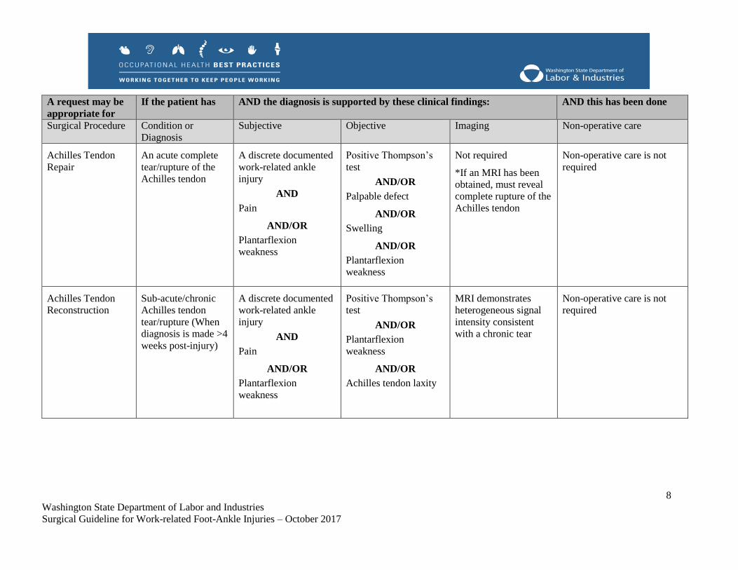
Achilles Tendon
Repair
An acute complete
tear/rupture of the
Achilles tendon
A discrete documented
work-related ankle
injury
AND
Pain
AND/OR
Plantarflexion
weakness
Positive Thompson’s
test
AND/OR
Palpable defect
AND/OR
Swelling
AND/OR
Plantarflexion
weakness
Not required
*If an MRI has been
obtained, must reveal
complete rupture of the
Achilles tendon
Non-operative care is not
required
Achilles Tendon
Reconstruction
Sub-acute/chronic
Achilles tendon
tear/rupture (When
diagnosis is made >4
weeks post-injury)
A discrete documented
work-related ankle
injury
AND
Pain
AND/OR
Plantarflexion
weakness
Positive Thompson’s
test
AND/OR
Plantarflexion
weakness
AND/OR
Achilles tendon laxity
MRI demonstrates
heterogeneous signal
intensity consistent
with a chronic tear
Non-operative care is not
required

9
Washington State Department of Labor and Industries
Surgical Guideline for Work-related Foot-Ankle Injuries – October 2017
A request may be
appropriate for
If the patient has AND the diagnosis is supported by these clinical findings: AND this has been done
Surgical Procedure Condition or
Diagnosis
Subjective Objective Imaging Non-operative care
Insertional Achilles
Tendon
Reconstruction
Ostectomy
Flexor Hallucis
Longus Transfer
Gastroc Slide
Insertional Achilles
tendinopathy, with
or without Haglund
deformity
A discrete documented
work-related injury
from either singular
direct trauma, or
repetitive traumatic
exposure, or acute
strain to the insertion
of the Achilles tendon
AND
Pain
Pain with palpation
OR
Swelling
OR
Warmth/redness
MRI demonstrates
abnormal signal in the
bone, bursa, or tendon
At least 12 weeks of any of
the following:
Activity modification Non-
opioid analgesics, Bracing
Immobilization
Posterior Tibialis
Tendon
Reconstruction
Medializing
Calcaneal
Osteotomy
Spring Ligament
Reconstruction
Gastroc Slide
FDL Transfer
Subtalar Fusion
Triple Arthrodesis
Posterior tibialis
tendon insufficiency/
dysfunction
Spring ligament
sprain or rupture
Hindfoot/midfoot
deformity and post-
traumatic arthritis
A discrete documented
work-related injury, the
mechanism of which
transmits enough force
to cause insufficiency
of the posterior tibial
tendon, or sprain or
rupture of the spring
ligament.
AND
Medial ankle or mid-
foot pain
In the patient with
congenital hindfoot
Swelling
AND
One of the following:
Inability to do a single
foot heel rise
AND/OR
Asymmetric pes planus
with heel valgus and
the too-many-toes sign
MRI demonstrates
posterior tibial
tendinopathy/
tenosynovitis
AND/OR
Spring ligament tear
At least 12 weeks of any of
the following:
Activity modification Non-
opioid analgesics Bracing
Immobilization Orthotics
Continued next page

10
Washington State Department of Labor and Industries
Surgical Guideline for Work-related Foot-Ankle Injuries – October 2017
A request may be
appropriate for
If the patient has AND the diagnosis is supported by these clinical findings: AND this has been done
Surgical Procedure Condition or
Diagnosis
Subjective Objective Imaging Non-operative care
Medial Cuneiform
Osteotomy
Lateral Column
Lengthening
Kidner Procedure
valgus and pes planus
posture of the foot, this
condition does not
arise from and is not
worsened by
cumulative weight
bearing in the
workplace
Tarsal Tunnel
Release
Tarsal tunnel
syndrome (TTS)
A discrete documented
work-related foot/ankle
injury
AND
Pain/paresthesias in the
distribution of the
medial and/or lateral
plantar nerves
If a compressive lesion
is not present on MRI,
a positive nerve
conduction study
(NCS) consistent with
tarsal tunnel syndrome*
*NCSs of this nerve are
typically difficult to
perform and interpret
A MRI is required for
all surgical candidates;
AND
A compressive lesion
is seen on the MRI
affecting the tibial
nerve (e.g. cyst)
* An MRI
demonstrates a
compressive lesion
affecting the tibial
nerve OR if there is no
evidence of a
compressive lesion, a
positive NCS
consistent with tarsal
tunnel syndrome
6 weeks of non-operative
care UNLESS MRI shows
space occupying lesion

11
Washington State Department of Labor and Industries
Surgical Guideline for Work-related Foot-Ankle Injuries – October 2017
A request may be
appropriate for
If the patient has AND the diagnosis is supported by these clinical findings: AND this has been done
Surgical Procedure Condition or
Diagnosis
Subjective Objective Imaging Non-operative care
Elective
Amputation when
pain is the primary
indication
If amputation is being requested, after all conservative care efforts have been exhausted, the patient must be evaluated at a
Department of Labor and Industries designated amputation Center of Excellence. See Amputations Contemplated in the Setting of
Chronic Pain for further discussion.

12
Washington State Department of Labor and Industries
Surgical Guideline for Work-related Foot-Ankle Injuries – October 2017
II. Introduction
This guideline reflects a best practice standard for surgical treatment of certain foot and ankle conditions
sustained by injured workers treated in the Washington State workers’ compensation system under Title
51 Revised Code of Washington (RCW). Providers who are in the department’s Medical Provider
Network are required to follow this guideline when treating injured workers.a The surgical criteria are
used in the department’s utilization review program as the supporting evidence has shown these provide
the best chance for injured workers to have a good surgical outcome. To help ensure that diagnosis and
treatment of foot and ankle conditions are of the highest quality, this guideline emphasizes:
Conducting a thorough assessment and making an accurate diagnosis.
Appropriately determining work-relatedness.
Making the best treatment decisions that are curative or rehabilitative.b
Facilitating the worker’s return to health, productivity, and work.
The guideline was developed in 2016-2017 by a subcommittee of the Industrial Insurance Medical
Advisory Committee (IIMAC). The subcommittee was comprised of practicing physicians in
rehabilitation medicine, occupational medicine, orthopedic surgery, and podiatry. The guideline
recommendations are based on the weight of the best available clinical and scientific evidence from a
systematic review of medical literature, and on a consensus of expert opinion when scientific evidence
was insufficient or inconclusive. Visit the department’s Medical Treatment Guidelines webpage for
detailed information on the guideline development process.c
A. Background and Prevalence
Workplace accidents (such as falls, slips, and machinery entrapment), that result in traumatic foot and
ankle injuries (such as fractures, sprains, and crush injuries or amputations), are the most recognizable. At
the same time, non-work related congenital problems (e.g. flat feet), joint instability (such as from an old
sport injury), and chronic conditions (e.g. diabetic peripheral neuropathy) can predispose a person to
being injured on the job and can complicate recovery. The goal after a workplace injury is to return the
worker to as close to pre-injury status as possible and maximize function and the ability to return to work.
The Bureau of Labor Statistics reports that the incidence rate for injuries to the ankle is 5.6 per 10,000 full
time workers, and the incidence rate for foot injuries as 4.8 per 10,000 full time workers.1 Sprains,
strains, and tears in the ankle were the injury type with the highest incidence at 3.7 per 10,000 full time
workers. Most of these injuries are first treated in hospital emergency departments. One study found the
estimated incidence of ankle sprains or strains was 206 per 100,000 patients reporting to an emergency
department.2 In the same study, the estimated incidence of ankle fractures was 49 per 100,000 reporting,
and for foot contusions or abrasions, the estimated incidence was 50 per 100,000 reporting.
a http://app.leg.wa.gov/RCW/default.aspx?cite=51.36.010 b http://app.leg.wa.gov/wac/default.aspx?cite=296-20-01002 c http://www.lni.wa.gov/ClaimsIns/Providers/TreatingPatients/TreatGuide/?F=MainFooter&source=FF
13
Washington State Department of Labor and Industries
Surgical Guideline for Work-related Foot-Ankle Injuries – October 2017
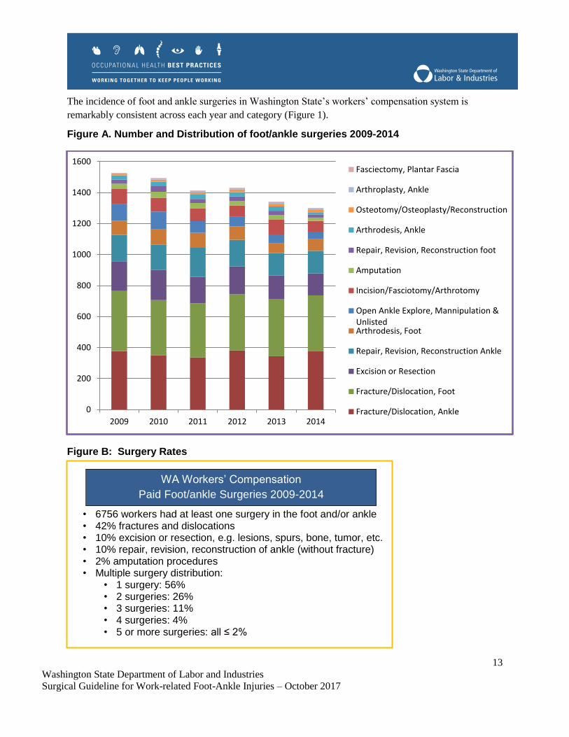
The incidence of foot and ankle surgeries in Washington State’s workers’ compensation system is
remarkably consistent across each year and category (Figure 1).
Figure A. Number and Distribution of foot/ankle surgeries 2009-2014
Figure B: Surgery Rates
0
200
400
600
800
1000
1200
1400
1600
2009 2010 2011 2012 2013 2014
Fasciectomy, Plantar Fascia
Arthroplasty, Ankle
Osteotomy/Osteoplasty/Reconstruction
Arthrodesis, Ankle
Repair, Revision, Reconstruction foot
Amputation
Incision/Fasciotomy/Arthrotomy
Open Ankle Explore, Mannipulation &UnlistedArthrodesis, Foot
Repair, Revision, Reconstruction Ankle
Excision or Resection
Fracture/Dislocation, Foot
Fracture/Dislocation, Ankle
• 6756 workers had at least one surgery in the foot and/or ankle
• 42% fractures and dislocations
• 10% excision or resection, e.g. lesions, spurs, bone, tumor, etc. • 10% repair, revision, reconstruction of ankle (without fracture) • 2% amputation procedures
• Multiple surgery distribution: • 1 surgery: 56%
• 2 surgeries: 26%
• 3 surgeries: 11%
• 4 surgeries: 4%
• 5 or more surgeries: all ≤ 2%
WA Workers’ Compensation
Paid Foot/ankle Surgeries 2009-2014

14
Washington State Department of Labor and Industries
Surgical Guideline for Work-related Foot-Ankle Injuries – October 2017
III. Establishing Work-relatedness
An injury sustained during the course of employment is defined in Washington State statute as “a sudden
and tangible happening, of a traumatic nature, producing an immediate or prompt result, and occurring
from without, and such physical conditions as result therefrom.” d A legal test for whether the department
or self-insured employer is liable for the care that a worker receives for an injury is whether the
workplace injury is “a proximate cause” of the accepted condition being treated. An injury may be a
proximate cause of a condition being treated only if “but for” the injury, the treatment would not be
necessary and proper. One approach to determining whether an injury meets this test is to determine
whether the worker’s need for treatment is any different than it would have been had, the work-related
injury never occurred. In the case of degenerative conditions, consider whether the duties and tasks of the
job significantly contributed to aggravation of the condition beyond what would normally be expected for
the worker’s age and genetics. If the answer is no, then it is likely not work related. If a proposed
treatment would have been needed regardless of the industrial injury, the injury would not be a proximate
cause of the need for treatment.
Occupational disease is defined in RCW 51.08.140 as a “disease or infection that arises naturally and
proximately out of employment.” e Establishing an occupational disease diagnosis requires that all of the
following criteria be met:
1. Exposure: Workplace activities that contribute to or cause foot or ankle conditions, and
2. Outcome: Diagnosis of a condition that meets the diagnostic criteria in this guideline, and
3. Relationship: Documentation that based on generally accepted scientific evidence, the work
exposures created a risk of contracting or worsening the condition relative to the risks in everyday
life, on a more-probable-than-not basis (Dennis v. Dept. of Labor and Industries, 1987). In
epidemiological studies, this will usually translate to an Odds Ratio (OR) ≥ 2.
A thorough occupational and non-occupational exposure history is essential for determining whether a
condition is work-related and whether it is due to an acute or chronic exposure. For chronic exposures, it
is important to document where, when, and for how long they occurred, as they could span multiple
employers who would then share liability for an occupational disease. Providers should submit the
Occupational Disease and Employment History form to the department or self-insurer as soon as possible
(a second form must be used for continuation of the occupational disease history).f
IV. Pre-existing Non-work-related Conditions
A. Pes Planus
Pes planus, also known as flat feet or fallen arches, is a foot condition characterized by a flattened,
pronated foot in the subtalar neutral position.3 In general, humans are born with flat feet, and the medial
d http://app.leg.wa.gov/RCW/default.aspx?cite=51.08.100 e http://app.leg.wa.gov/RCW/default.aspx?cite=51.08.140 f http://www.lni.wa.gov/Forms/pdf/F242-071-000.pdf; http://www.lni.wa.gov/Forms/pdf/F242-071-111.pdf

15
Washington State Department of Labor and Industries
Surgical Guideline for Work-related Foot-Ankle Injuries – October 2017
arch of the foot typically develops during the first decade of life.4 In some cases, an absent or abnormally
low arch persists through adolescence, or is acquired in adulthood.5
Classification of pes planus generally refers to flexible or rigid varieties, characterized by a qualitative
assessment of the stiffness of the foot during dynamic loading and/or physical exam.6 Flexible pes planus
is usually considered a continuation of a congenital condition, is generally asymptomatic, and can resolve
with time.4 In some cases, the condition is symptomatic and pathological, and may require treatment.4
When treatment is required, conservative interventions have been shown to help manage the condition,7
though surgical intervention may be necessary if conservative options fail.8, 9
Acquired pes planus is often asymptomatic and is characterized by an abducted forefoot and valgus
hindfoot.10 Acquired pes planus has been attributed to inflammatory arthritis, trauma, and most
commonly posterior tibial tendon dysfunction (PTTD).11, 12 PTTD will be addressed later in the guideline
within the posterior tibialis tendon reconstruction section.
Pes planus may predispose a worker to having foot pain if his or her job involves prolonged periods of
weight bearing. While an association between prolonged standing and increased risks of musculoskeletal
pain has been shown, there is insufficient evidence to demonstrate that prolong weight bearing is
associated with pes planus.13
B. Pes Cavus
Pes cavus refers to a foot deformity in which the medial longitudinal arch of the foot is abnormally high
and does not flatten with weight bearing.14 Gait deficits and foot pain have been associated with pes
cavus resulting from the decreased ability to absorb ground reaction forces due to the characteristic shape
of a cavus foot.14 Presenting in either childhood or adulthood, a cavus foot is a relatively common
finding, occurring in one fifth to one quarter of the general population.15 Several primary origins have
been associated with pes cavus: neuromuscular, congenital, idiopathic, and traumatic.14, 16 Neurological
conditions such as cerebral palsy, muscular dystrophy, Charcot-Marie-Tooth disease, and poliomyelitis
account for 75% of all pes cavus cases.16 As such, pes cavus is not considered a work-related condition.
Symptoms of pes cavus can include recurrent ankle sprain, tendon disorders, instability in gait, callous
formation and stress fractures to the lateral border of the foot.14, 16-19 This type of symptomology is
thought to derive from abnormal pressure distribution across the sole of the foot and can contribute to
significant disability.14, 20 The presence of this type of variant foot mechanics could predispose a worker
to mechanical stresses that may contribute to foot complaints. Treatment of pes cavus is primarily
conservative, with orthotics commonly used to reduce and redistribute pressure on the sole of the foot.14
Additional conservative treatment methods include balance improvement, stretching and strengthening of
weak muscles.19 When conservative measures fail, surgical treatment may be required.21

16
Washington State Department of Labor and Industries
Surgical Guideline for Work-related Foot-Ankle Injuries – October 2017
C. Plantar Fasciitis
The plantar fascia is a broad, fibrous tissue band that extends from the heel bone (calcaneus) to the toes
(metatarsals), providing support to the arch and stabilizing it during normal weight bearing. Plantar
fasciitis often presents as progressive and persistent deep, dull ache-like pain on the plantar surface of the
heel. Other symptoms can include a burning sensation, pain and/or stiffness when getting out of bed, pain
when getting up to walk after a period of inactivity, pain toward the end of the day, increased pain in the
heel or arch, and overall tired feet.22-24
Acute trauma (e.g. jumping or falling from a high perch onto a hard surface) may injure or rupture the
plantar fascia and could possibly be associated with occupational-related plantar fasciitis.25 However, the
evidence base suggesting associations of other, more chronic occupational exposures and the onset of
occupational-related plantar fasciitis is unclear. Examples of potential chronic occupational exposures
include long periods of standing or walking on certain surfaces (e.g. cement floors),22, 25, 26 wearing non-
supportive footwear,27-29 or prolonged stressful activity (e.g. long distance running or unusual rapid
walking such as a “forced march”).29 The diagnosis of occupational-related plantar fasciitis thus remains
controversial, and data are insufficient at this time to determine the risk posed by some specific chronic
occupational exposures. Allowance of occupational-related plantar fasciitis would usually only be
considered when the exposure is acute and related to significant occupational-related trauma to the heel or
planter fascia.
Treatment of plantar fasciitis may include orthotics (pads), heel cord stretching exercises, anti-
inflammatory medication, activity modification, night splinting, steroid injection to the site, or surgery.23,
30 Surgical treatment (e.g. fasciotomy) is rarely indicated, with less than 5% of patients undergoing
surgery.31
D. Osteoarthritis
A complicating factor when trying to establish work-relatedness is the presence of osteoarthritis (OA).
Osteoarthritis is a normal degenerative process and a progressive condition that results from loss or
deterioration of articular cartilage. It is the most common arthritic disease, it is the most common cause
of long-term disability in persons older than 65, and it is expected to become the world’s 4th leading cause
of disability by 2020.32
While osteoarthritis is an important consideration for treatment of foot and ankle conditions, the ankle
joint is much less likely to experience symptomatic OA through normal risk factors alone, such as age.33
Brown et al estimated that nearly 80% of all ankle osteoarthritis is post-traumatic; and the estimated
yearly incidence of symptomatic ankle OA in the same study was 1,113 cases in Iowa alone.34 In patients
seeking treatment, symptomatic osteoarthritis with radiographic changes occurs about 8-9.4 times more
often in knee than in the ankle.35, 36

17
Washington State Department of Labor and Industries
Surgical Guideline for Work-related Foot-Ankle Injuries – October 2017
Therefore, in the vast majority of arthritic conditions attributed to an occupation, a substantial, discrete
work-related traumatic event can be identified. A contention that an industrial injury caused, aggravated,
or accelerated an osteoarthritic condition should be supported by adequate documentation, to include:
1. A careful prior medical history, and a careful history of the present condition, to document as
clearly as possible any pre- or post-injury presence or progression, objectively or subjectively, of
an osteoarthritic condition;
2. An evidence-based explanation supporting the contention that the osteoarthritis would not
presently require treatment had the industrial injury or occupational disease not have occurred.
V. Assessment
With 33 joints, 26 bones, and over 100 muscles, tendons, and ligaments, the foot and ankle are a complex
structure. Clinical assessment should include:
Observing the foot and ankle for shape, swelling/edema, and color.
Checking the skin temperature.
Palpating for tenderness, knowing that assessing for bony injury may be limited in acute injuries
due to bruising and diffuse soft tissue swelling.
Evaluating range of motion and weight bearing ability.
Observing the workers gait and stance.
Checking posterior, anterior, medial, and lateral angles for anatomical alignment and position of
trunk and hip, hip and knee, knee and foot, and arch position.
Condition-specific tests such as Thompson, Squeeze, External Rotation Stress, Anterior Drawer,
and the Talar Tilt test.37
In addition to performing a thorough exam, it is critical to get an accurate history of injury occurrence,
previous injuries, and underlying comorbidities/ risk factors, which may predispose the worker to further
foot and ankle injury.
A. Imaging
The recommended imaging procedures for various foot and ankle surgeries are specified in the criteria
table. Weight bearing x-rays are recommended when determining the presence or extent of degenerative
disease. Given the complex three dimensional anatomy of the foot and ankle, CT scans or CT-SPECT
scans are used at times when finer detail is needed e.g. when suspecting an occult fracture or to further
study characteristics of a known fracture, deformity, or arthritis. Magnetic resonance imaging (MRI) is
most commonly used to diagnose injuries to the ligaments or tendons but is not recommended for every
case of acute pain or for degenerative joint disease (L&I requires prior authorization for all MRIs; visit
the Advanced Imaging Guidelines web page for complete information).g
g http://www.lni.wa.gov/ClaimsIns/Providers/TreatingPatients/TreatGuide/imaging.asp

18
Washington State Department of Labor and Industries
Surgical Guideline for Work-related Foot-Ankle Injuries – October 2017
B. Preventing Complications
Worsening conditions can lead to treatment that is more drastic and even amputation, especially if careful
monitoring and timely assessment and care are not done. It is critical to conduct a thorough assessment of
risk factors prior to surgery to evaluate the risks and benefits of the procedure. Risk factors include:
vascular disease
diabetes and the extent of diabetic complications
tobacco use
age-related system changes such as immune status
degree of soft tissue trauma and swelling
expected duration of surgical time
mental health status
psychosocial status
Examples of this workup include determining the level of diabetic control through HbA1c values,
performing appropriate vascular studies, consulting with a vascular surgeon, and initiating a preoperative
tobacco cessation program.h
Refer to the “Conservative Care Options for Work-Related Foot and Ankle Conditions” occupational
health practice resource published by the Industrial Insurance Chiropractic Advisory Committee and
Labor &Industries Office of the Medical Director for a thorough discussion of patient presentation,
prognostic indicators, and clinical examination recommendations.i
C. Measuring Functional Improvement
From the time of injury to full restoration, from conservative care to post-operative assessment, it’s
critical to measure the patient’s functional improvement and changes in their pain experience using
validated tools in a consistent fashion. Regular assessment not only helps to guide appropriate care, it
helps prevent and/or identify risk factors and symptoms for developing Complex Regional Pain
Syndrome (CRPS), which can be particularly difficult to treat.j The authors of this guideline recommend
using the following validated tools for measuring pain and functional improvement:
The Foot and Ankle Ability Measure (FAAM) is a validated tool used to assess functional measure
related to musculoskeletal disorders of the lower leg, foot, and ankle.38 k
h http://www.lni.wa.gov/ClaimsIns/Providers/TreatingPatients/ByCondition/TobaccoCessation.asp i http://www.lni.wa.gov/ClaimsIns/Files/OMD/IICAC/2015WorkRelatedMechanicalFootAnkleConditions44.pdf j http://www.lni.wa.gov/ClaimsIns/Files/OMD/MedTreat/ComplexRegionalPain2011.pdf k http://www.aaos.org/uploadedFiles/PreProduction/Quality/Measures/Foot%20and%20Ankle%20Ability%20Measure.pdf
19
Washington State Department of Labor and Industries
Surgical Guideline for Work-related Foot-Ankle Injuries – October 2017
The Short Musculoskeletal Function Assessment (SMFA) questionnaire can be used with several types of
musculoskeletal injuries, is comparable to other assessment scales such as the FAAM, and is freely
available.39-41 l
The PROMIS scale (Patient-Reported Outcomes Measurement Information System) measures function in
lower extremities using computer-assisted technology. Having been integrated into the Epic electronic
medical record software, the PROMIS scale has been validated in multiple settings.42-44 m
VI. Specific Conditions and Surgical Procedures
A. Ankle Arthroscopy and Cheilectomy
Not counting arthroscopic approaches to other surgical procedures, arthroscopy for removing a loose
body is a covered procedure when there is evidence of the ankle catching and/or locking and/or effusion
(presence of joint fluid and swelling). Arthroscopy, for removing bone spurs (cheilectomy), is also a
covered procedure when there is evidence of bony impingement. A 2015 systematic review reported
good clinical outcomes in 64-100% of patients undergoing arthroscopic treatment for anterior ankle
impingement, with complications seen in only 5% of cases.45 The same review also found that good to
excellent patient satisfaction was seen in 74-100% of cases, and the lowest satisfaction was reported in
studies with follow-up >5 years.45
B. Ankle Arthrodesis/Fusion or Arthroplasty/Replacement
Work related ankle injuries can be sudden and traumatic. They can result from a crush injury; or start as a
soft tissue injury, such as a sprain, and worsen over time if proper treatment is not provided at the start or
if significant comorbidities exist. Ankle arthrodesis and ankle arthroplasty are both surgical options when
the ankle cartilage is so damaged by arthritis that joint movement and weight bearing activities cause
intolerable pain. Fusing the tibia, fibula, and talus (arthrodesis) or replacing the ankle joint (arthroplasty)
are major procedures and require inpatient hospitalization and several weeks of protected weight bearing
followed by a structured rehabilitation program. Full recovery generally takes several months. These
procedures are usually reserved for advanced stages of impairment or disability when:
Osteoarthritis has resulted in an unacceptable level of impairment in the performance of normal
daily activities, or has created a disability that, without treatment, prevents gainful employment
There are deformities (congenital or developmental) in the joint, and
Prior conservative and surgical treatments have been unsuccessful in restoring function.
Determining whether arthrodesis or arthroplasty is preferable is a matter of clinical judgment. Generally,
ankle arthrodesis can include a broader set of patients (e.g. with comorbidities) than arthroplasty, which
requires more careful patient selection to achieve desirable outcomes.
l https://www.ortho.umn.edu/research/mfa-smfa-resources m http://www.healthmeasures.net/explore-measurement-systems/promis

20
Washington State Department of Labor and Industries
Surgical Guideline for Work-related Foot-Ankle Injuries – October 2017
Arthrodesis/Fusion
Arthrodesis is a permanent fusion of the tibia, fibula, and talus. The expected outcome is reduced or
absent pain, but because fusion fixes the ankle in one position, it will no longer be able to move. All
conservative measures should be exhausted before resorting to an arthrodesis. Risk factors to evaluate
before deciding on a fusion include the availability of viable bone to support the fusion, adequate nerve
and blood supply to allow healing, and deformities or problems from earlier injuries and treatments that
could compromise the effectiveness of the surgery.46-49
In a systematic review, revision rates following ankle arthrodesis was found to be 9% (95% CI, 5.5% to
11.6%), with the primary reason for revision being nonunion (65%).50 High-risk patients, including those
with diabetes and obesity, may be at an even higher risk for nonunion and other complications. One study
found that obese arthrodesis patients (BMI ≥30) had significantly increased risks of major and minor
complications within 90 days of surgery.51 A study of risk factors for nonunion following arthrodesis
found that noninsulin dependent diabetic patients were 18.7 times more likely to have a malunion
arthrodesis.52
Arthroplasty/Joint Replacement
Arthroplasty replaces the entire ankle joint with a prosthetic implant. Similar to arthrodesis, all
conservative measures should be exhausted before resorting to an ankle arthroplasty. Diabetes and
obesity pose significant risks for post-surgical complications and treatment failure. Strong consideration
should be given to alternative treatments (i.e. nonsurgical, arthrodesis) or deferring surgery until these
risk factors are appropriately treated and optimized. One study found diabetic patients to have greater
incidence of poor clinical outcomes, early-onset of osteolysis, and an overall negative impact on short to
medium-term results.53 Another study found diabetes to be independently associated with an increased
risk of perioperative complications, discharge to a facility versus home, and a longer hospital stay.54
Obese patients have been found to have significantly greater odds of implant failure by final follow-up
(adjusted OR 2.8, 95% CI 1.04 -7.53), and lower functional outcome scores when compared to non-obese
patients.55, 56 Werner et.al found that obesity was associated with significantly increased rates of all
complications, including revision (OR=1.6, 95% CI 1.2 - 2.2) after arthroplasty.51
As with any joint replacement, there is always the possibility of having to revise/replace the prosthesis.
Patients <50 years of age are likely to have better clinical results and comparable complication and
survivorship rates compared to those >50 years of age.57 In a study of national revision rates using
registry data for different implants, the average revision rate was 21.8% after 5 years, and 43.5% after 10
years.58 Another study of registry data found that survival rates for total ankle replacement was 0.87
(95% CI, 0.82–0.91) at 5 years, and 0.81 (95% CI, 0.74–0.88) at 10 years.59
C. Subtalar Arthrodesis
The subtalar joint in located just below the ankle joint between the talus bone and calcaneus (heel) bone.
A subtalar arthrodesis or fusion is performed to relieve pain during movement of the subtalar joint, and is
indicated when a work related injury results in post-traumatic arthritis of the hindfoot.60, 61

21
Washington State Department of Labor and Industries
Surgical Guideline for Work-related Foot-Ankle Injuries – October 2017
The hindfoot has several points of articulation with concave and convex curvatures, cavities, chambers,
and canals. The ways in which these spaces and surfaces are shaped and positioned are not the same in
everyone. The decision to fuse two or more joints depends on how severely a deformity produces
instability and pain, and how likely it is that surgery will improve and relieve them. Correction of
deformities helps to relieve symptoms and improve surgical prognosis and functionality.62
The subtalar joint enables the foot to adjust to uneven ground and rotates in multiple directions during
walking and standing.63 Arthrodesis of the subtalar joint (fusing the talus and calcaneus) can be done as a
single stand-alone procedure, whereas a double (adding the talonavicular joint) and triple (adding the
calcaneocuboid joint) arthrodesis are done in addition to the subtalar fusion.64
A double fusion preserves function of the midtarsal joint, is a simpler procedure, and avoids the risk of
midtarsal nonunion or malunion.65 The triple arthrodesis is usually reserved for correction of painful and
deforming conditions e.g. tarsal coalition (abnormal connection of two normally separate bones),
planovalgus (rigid flat foot), and cavovarus (high arch and inward turned heel) after conservative care has
failed.64, 66, 67
D. Debridement or Stabilization of a Medial or Lateral Talar Lesion
Injuries may cause chondral (cartilage) or osteochondral (bone and cartilage) lesions (OCLs) affecting the
tibia, fibula or talus. These defects may cause or aggravate degenerative conditions of the structures that
comprise the joints of the ankle. Symptoms can present immediately following injury, or may appear
later if a chronic degenerative process is present. Nearly all lateral OCLs have a history of trauma,
whereas medial OCLs are less likely to result from a previous trauma.68 There are no physical findings
indicative of lateral OCLs.
Treatment of medial or lateral talar injuries is directed at stabilizing the ankle or restoring its functional
anatomy. Depending upon the nature of the lesion(s), in each individual case, treatment may be non-
surgical (e.g. immobilization, restricted weight bearing) or surgical (e.g. debridement, fixation,
microfracture). Specific to the medial lesion, is the need to have a positive diagnostic lidocaine injection
before surgery is considered to signify that surgery is likely to relieve pain.
A decision to proceed to surgery treatment should be made only after considering relative
contraindications such as age, BMI, the extent of ankle trauma, and the presence of osteophytes. There
appears to be a correlation between lesion properties and clinical outcome. A case series found that a
lesion diameter of >15mm was correlated with worse clinical outcomes following microfracture, and a
cohort study found that risk of clinical failure following marrow stimulation procedures was significantly
associated with a defect area >150 mm2.69, 70 A separate study found shoulder-type ankle lesions
(uncontained lesions with no peripheral cartilage border) experienced worse clinical outcomes regardless
of size or location.71

22
Washington State Department of Labor and Industries
Surgical Guideline for Work-related Foot-Ankle Injuries – October 2017
E. Lateral Ligament Repair or Reconstruction
Lateral ankle ligament injuries are common, with one study finding that among 6 million patients, 3.2%
reported lateral ankle ligament injury or instability.72 Acute injuries from initial inversion and
plantarflexion trauma may damage the lateral ligaments.73 Chronic ankle instability can result if the
injury is severe, left untreated, or is undertreated.74
Non-operative treatment such as rest, ice, compression, and elevation (RICE), functional treatment, or
short-term immobilization, is generally the first choice for such injuries.75-78 In most cases, non-operative
care can lead to a fully functional recovery.73
When surgery is indicated, repair of the ligament using the patient’s own tissue is generally considered
the first line of treatment (e.g. Brӧstrom procedure).79-81 When there is insufficient tissue or if persistent
laxity is present, reconstruction using an auto- or allograft, (e.g. Watson-Jones or Evans technique) should
be considered.74, 81-83 Studies have shown that lateral ankle instability treated by reconstruction, shows no
significant difference in ankle plantarflexion or dorsiflexion ROM when compared with the contralateral
extremity84 While there is concern over excessive stiffness and decreased ankle inversion after
reconstruction, the preservation of muscle strength allows for preservation of ankle eversion.83, 84
F. Peroneal Tendon Repair
The peroneal tendons, including the peroneus brevis and peroneus longus tendons are common sources of
tendon pathology in the ankle.85, 86 The peroneal tendons can be traumatized with a single injury, or
repeatedly over time by mechanisms such as acute inversion ankle sprains or peroneal subluxation.85-87
Peroneal tendon disorders can present as posterolateral ankle pain, and may include tenosynovitis, tendon
dislocation, subluxation, or rupture.88, 89
Anatomic features, such as a low-lying brevis muscle belly or a peroneus quartus may be contributing
factors to peroneal tendon injury.90-93 When considering repair for a work-related peroneal tendon injury
and underlying deformities (e.g. pes cavus) are present, correction of the deformity at the time of surgery
may be done if required for a successful surgical outcome.85, 86, 88, 94, 95
G. Achilles Tendon Repair or Reconstruction
The Achilles tendon is the largest tendon in the body. Made up of fibrous connective tissue, it may
become susceptible to injury over time due to routine body stressors, activity changes, and lack of blood
flow to the area. In a study of 697 patients presenting to a clinic with a foot or ankle complaint, 5.6% of
patients were found to have an Achilles tendinopathy.96
An Achilles tendon repair or reconstruction procedure is performed when injury causes a partial tear or
acute rupture of the tendon attaching the calf muscles (soleus and gastrocnemius) to the heel bone
(calcaneus). The ultimate goal of any Achilles tendon treatment is to have the ruptured ends of the tendon
rejoined, and the tendon restored to its full length and function.

23
Washington State Department of Labor and Industries
Surgical Guideline for Work-related Foot-Ankle Injuries – October 2017
Repair at Insertion
The clinical presentation of insertional Achilles tendinopathy includes pain in the back of the leg near the
heel.97 Some patients may present with a small lump right above the heel (Haglund’s deformity).98
Evidence suggests that both surgical and conservative treatments for insertional Achilles tendinopathy are
effective.97, 99 At least 12 weeks of conservative care, including activity modification, non-opioid
analgesics, and/or immobilization with braces should be tried prior to any surgical intervention for
insertional Achilles tendinopathy.99 Corticosteroid injections are generally discouraged in treatment of
Achilles tendinopathy. While some studies have shown short-term benefits, the adverse effects of
corticosteroid injections such as atrophy, rupture, and decreased strength of the tendon, suppress any
potential value.100
Surgical repair of the Achilles tendon at the calcaneal insertion point is done for inflammation of the
sheath surrounding the tendon and the deterioration of the associated connective tissue.97, 101 The damaged
portion of the tendon is removed and, after preparing the heel bone surface, the healthy portion is
reattached.
Repair
The primary surgical procedure performed for acute ruptures of the Achilles tendon, an Achilles tendon
repair, can be performed with the first instance of a combination of clinical findings. Clinical findings
indicative of an Achilles repair include: acute rupture of the tendon, pain, swelling, positive Thompson’s
sign, plantarflexion weakness, and a palpable defect. Acute tears may be treated surgically or
conservatively with functional rehabilitation. Conservative treatment appears to have a higher incidence
of re-ruptures but avoids potential complications of surgery such as infections and wound healing
complications.102, 103
Reconstruction
Chronic Achilles tendon tears are more difficult to diagnosis as scarring may replace the palpable defect
seen in acute tears. Due to retraction of the tendon, primary repair is not always feasible for chronic tears.
A preoperative MRI is required to better define the anatomy of a chronic tear. Currently there is no gold
standard for repairing chronic tears; but multiple options exist, such as: flap tissue turn down, local
tendon transfer, autologous free tendon grafts, and allografts.104
H. Posterior Tibialis Tendon Reconstruction
Posterior tibialis tendon insufficiency/dysfunction (PTTD) occurs when the posterior tibial tendon
becomes inflamed or torn. The posterior tibialis attaches to the navicular on the medial aspect of the foot
helping to support the arch. Diagnosis of PTTD is supported by the clinical findings of:
1. Swelling along the medial aspect of the foot and ankle,105
2. Changes in the shape of foot with gradual collapse of the medial arch and increasing hindfoot
valgus,106 and
3. Inability to do a single foot heel rise (stand of toes) due to pain and weakness.107

24
Washington State Department of Labor and Industries
Surgical Guideline for Work-related Foot-Ankle Injuries – October 2017
Conservative treatment prior to surgical intervention is highly effective, with a recent study showing
87.5% of patients achieving success with non-operative treatment.108 If conservative treatment is not
successful, posterior tibialis tendon reconstruction may be accomplished through a variety of surgical
procedures such as:109-111 gastrocnemius recession,111 tenosynovectomy,112 tendon transfer,113
osteotomy,111, 114 and arthrodesis/fusion.113 The procedure of choice is determined by the severity and
degree of tendon disease.111, 115 Recovery after one of these procedures is likely to require 12 weeks of
non-weight bearing.
PTTD and Work-relatedness
As previously discussed in the Pre-existing Non-work related Conditions section, PTTD, or pes planus in
adults, it is unlikely that cumulative weight bearing in the workplace would arise or worsen such a
condition. The consensus of the guideline authors is that PTTD is closely correlated with genetic and
congenital predisposition, and does not normally result from cumulative workplace exposure. In the
patient with congenital hindfoot valgus and pes planus posture of the foot, PTTD is unlikely to arise from,
or be worsened by cumulative weight bearing in the workplace. As such, PTTD is generally not
considered an occupational disease.
I. Tarsal Tunnel Release
Tarsal Tunnel Syndrome (TTS) occurs when systemic disease, injury, or body structure entraps or
compresses the posterior tibial nerve or one of its branches (e.g. the lateral and medial plantar nerves).
Examples include space-occupying lesions, fractures of the medial calcaneal wall, direct trauma to the
heel, and traction neuritis.116 TTS is a relatively rare condition, and other causes of medial ankle and
plantar foot pain include but are not limited to plantar fasciitis, intervertebral disk lesion, interdigital
neuroma, plantar fibromatosis, peripheral vascular disease, valgus hindfoot, rheumatoid arthritis,
tenosynovitis, and diabetic neuropathy.117 It is important to rule out etiologies that are more common
prior to arriving upon the diagnosis of TTS. Care should be taken in patients with idiopathic or traumatic
etiologies, as they have been shown to be associated with worse clinical outcomes following surgery.118
Systemic comorbidities such as back pain, depression, COPD, or total previous systemic surgeries have
also been shown to be associated with worse clinical outcomes.119
Tarsal tunnel release exposes the tibial nerve and removes anatomical structures causing the entrapment
or compression. It is a covered procedure when all the following criteria are met:
1. A documented discrete work-related foot/ankle injury has occurred.
2. Presence of pain and/or paresthesias in the distribution of the medial and/or lateral plantar
nerves.118
3. An MRI demonstrates a compressive lesion (e.g. a cyst) affecting the tibial nerve OR if there
is no evidence of a compressive lesion, a positive nerve conduction study (NCS) consistent
with tarsal tunnel syndrome.

25
Washington State Department of Labor and Industries
Surgical Guideline for Work-related Foot-Ankle Injuries – October 2017
A positive NCS consistent with tarsal tunnel syndrome is an acceptable diagnostic tool, as it has been
found that NCS may be abnormal in some patients with suspected TTS.120 A positive NCS will generally
show a > 1msec difference between the terminal latency of the medial plantar nerve (MPN) to the
abductor hallucis muscle and terminal latency of the lateral plantar nerve (LPN) to the abductor digiti
quinti muscle. Normal findings of these terminal latencies are MPN of < 6.2msec and LPN of < 7 msec.
Increases in these latencies and those of sensory nerve conduction velocities are also indicative of TTS.121
J. Amputations
Amputation is a significant life-altering event. It should only be considered if the limb cannot be
reconstructed and/or removal will help achieve the best possible outcome. An amputation may be
necessary for preventing the spread of recalcitrant infections, severe peripheral vascular disease,
persistent nonunion of bone, and when there is insufficient bone, muscle, or nerve for reconstruction.
Amputation when pain is the primary indication is generally discouraged; see Amputations Contemplated
in the Setting of Chronic Pain.
Throughout treatment for lower limb injuries, patients should be monitored for risk factors that could lead
to amputation. If all conservative care options have been exhausted and amputation is the only choice,
the level of the planned amputation should be thoroughly evaluated for the limb’s ability to heal and
achieve maximum function. If the severity of risk factors and the potential for major complications such
as deep wound infection and supplementary limb amputation outweigh the benefit of the planned
procedure, delaying amputation until the risk factors have improved or proceeding with an alternative
non-surgical treatment plan may be necessary. For example, consider consulting with an Orthotist and/or
Prosthetist to evaluate the need for specialized ankle foot orthoses. One such device is the Dynamic
Offloading Brace, which has the potential to prevent amputations.n
Amputations Contemplated in the Setting of Chronic Pain
The decision algorithm to proceed with a partial foot or lower limb amputation is complex and not within
the scope of this guideline. Amputation for the purpose of intractable pain, however, is controversial and
merits discussion. While cases are relatively infrequent, such amputations can result in significant
morbidity, including continued pain. A recent study of patients with a diagnosis of Complex Regional
Pain Syndrome (CRPS) found that 22% of patients undergoing amputations experienced postoperative
complications, and 72% of patients experienced phantom limb pain within 3 months, increasing to 77% of
patients at ≥1 year post amputation.122
While there is no absolute marker for the source of pain, every effort should be taken to ensure that a
patient’s pain correlates with the diagnosis, and that any proposed procedure is likely to effectively
address the source of pain and increase the chance of improved functional outcomes. The provider should
watch for the development of unresolved chronic pain or sudden changes in the type and level of pain,
nhttp://www.lni.wa.gov/ClaimsIns/Providers/TreatingPatients/ByCondition/CovMedDev/SpecCovDec/DynamicOffloadingBrac
es.asp

26
Washington State Department of Labor and Industries
Surgical Guideline for Work-related Foot-Ankle Injuries – October 2017
and consult with any specialists needed to establish an effective pain management plan. If prescribing
opioids are part of that plan, the prescriber should follow L&I’s evidence based Guideline for Prescribing
Opioids to Treat Pain in Injured Workers.o When, as a last resort, amputation is being considered
primarily for the purpose of reducing pain, the worker must be evaluated at Labor & Industries designated
Amputation Center of Excellence.p
VII. Return to Work
Return to work (RTW) is expected after most occupational foot and ankle injuries. Duration of disability
or time off work depend on many factors such as the severity of the injury, type of treatment, comorbid
conditions, and job class type. Multiple resources are available through L&I’s RTW program to help
providers in their interactions with workers, employers, and claim managers to discuss and coordinate the
best ways to help with return to work.q There is a particularly useful “Return to Work Desk Reference”
for attending providers with guidance on how to talk with workers and their employers (and get paid for
it), online publications to inform the patient how returning to work can reduce disability, descriptions of
best practices, checklists, algorithms, vignettes, and a list of ways L&I staff can assist.r
L&I has also published a Functional Recovery Interventions Tracking Sheet that is designed as a checklist
for engaging the patient in discussions of recovery and returning to work.s
o http://www.lni.wa.gov/ClaimsIns/Files/OMD/MEDTreat/FINALOpioidGuideline010713.pdf p http://www.uwmedicine.org/harborview/services/burn-center q http://www.lni.wa.gov/ClaimsIns/Providers/TreatingPatients/RTW/default.asp r http://www.lni.wa.gov/IPUB/200-002-000.pdf s http://www.lni.wa.gov/forms/pdf/F245-420-000.pdf

27
Washington State Department of Labor and Industries
Surgical Guideline for Work-related Foot-Ankle Injuries – October 2017
VIII. Acknowledgements
Acknowledgement and gratitude go to all subcommittee members, clinical experts, consultants, and L&I
staff who contributed to this important guideline:
IIMAC Committee Members
Chris Howe, MD (Chair) – Proliance Orthopedic Associates, Valley Medical Center
Andrew Friedman, MD – Virginia Mason Medical Center
Kirk Harmon, MD – MultiCare
Subcommittee Clinical Experts
Janna Friedly, MD – Harborview Medical Center
Jonathan Hall, MD – ProOrtho, Proliance Surgeons
Byron Hutchinson, DPM – Franciscan Foot and Ankle Associates
Anne McCormack, MD – Providence Everett Healthcare
Erik Novak, MD, PhD – Proliance Orthopedic Associates, Valley Medical Center
Bruce Sangeorzan, MD – UW and Director VA Center for Limb Loss Prevention and Prosthetic
Engineering
Eugene Pepper Toomey, MD – Swedish Hospital
Qualis Health Consultants
Shari Fowler-Koorn, RN
Ken O’Bara, MD
John Sparks, MD
Margaret Baker, MD
Labor and Industries Staff
Gary M. Franklin, MD, MPH, Medical Director
Lee Glass, MD, JD, Associate Medical Director
Nicholas Reul, MD, MPH, Associate Medical Director
Zachary Gray, MPH, Epidemiologist
Simone P. Javaher, RN, BSN MPA, Clinical Manager of Health Policy
Angela Jones, BSN, RN
Aquila Doore, RN, BSN-BC, MBA, JD, Occupational Nurse Consultant

28
Washington State Department of Labor and Industries
Surgical Guideline for Work-related Foot-Ankle Injuries – October 2017
IX. References
1. Bureau of Labor Statistics, Number of nonfatal occupational injuries and illnesses requiring days
away from work, 2014, U.D.o. Labor, Editor. 2015: Washington, DC.
2. Lambers, K., D. Ootes, and D. Ring, Incidence of patients with lower extremity injuries
presenting to US emergency departments by anatomic region, disease category, and age. Clin
Orthop Relat Res, 2012. 470(1): p. 284-90.
3. Walters, J.L. and S.S. Mendicino, The flexible adult flatfoot: anatomy and pathomechanics. Clin
Podiatr Med Surg, 2014. 31(3): p. 329-36.
4. Ozan, F., et al., Symptomatic flexible flatfoot in adults: subtalar arthroereisis. Therapeutics and
clinical risk management, 2015. 11: p. 1597.
5. Shibuya, N., et al., Characteristics of adult flatfoot in the United States. The Journal of foot and
ankle surgery, 2010. 49(4): p. 363-368.
6. Lee, M.S., et al., Diagnosis and treatment of adult flatfoot. J Foot Ankle Surg, 2005. 44(2): p. 78-
113.
7. Scharer, B.M., B.E. Black, and N. Sockrider, Treatment of painful pediatric flatfoot with
Maxwell-Brancheau subtalar arthroereisis implant a retrospective radiographic review. Foot &
ankle specialist, 2010. 3(2): p. 67-72.
8. Brancheau, S.P., K.M. Walker, and D.R. Northcutt, An analysis of outcomes after use of the
Maxwell-Brancheau Arthroereisis implant. The Journal of Foot and Ankle Surgery, 2012. 51(1):
p. 3-8.
9. Nelson, S.C., D.M. Haycock, and E.R. Little, Flexible flatfoot treatment with arthroereisis:
radiographic improvement and child health survey analysis. The Journal of foot and ankle
surgery, 2004. 43(3): p. 144-155.
10. Abousayed, M.M., et al., Classifications in Brief: Johnson and Strom Classification of Adult-
acquired Flatfoot Deformity. 2016, Springer.
11. Erol, K., et al., An important cause of pes planus: the posterior tibial tendon dysfunction. Clinics
and practice, 2015. 5(1).
12. Zaw, H. and J.D.F. Calder, Operative management options for symptomatic flexible adult
acquired flatfoot deformity: A review. Knee Surgery, Sports Traumatology, Arthroscopy, 2010.
18(2): p. 135-142.
13. McCulloch, J., Health risks associated with prolonged standing. Work, 2002. 19(2): p. 201-205.
14. Burns, J., et al., Interventions for the prevention and treatment of pes cavus. The Cochrane
Library, 2007.
15. Aminian, A. and B.J. Sangeorzan, The anatomy of cavus foot deformity. Foot and ankle clinics,
2008. 13(2): p. 191-198.
16. Statler, T.K. and B.L. Tullis, Pes cavus. Journal of the American Podiatric Medical Association,
2005. 95(1): p. 42-52.
17. Burns, J., et al., The effect of pes cavus on foot pain and plantar pressure. Clinical Biomechanics,
2005. 20(9): p. 877-882.
18. Desai, S.N., R. Grierson, and A. Manoli, The cavus foot in athletes: fundamentals of examination
and treatment. Operative Techniques in Sports Medicine, 2010. 18(1): p. 27-33.
19. Manoli, A. and B. Graham, The subtle cavus foot,“the underpronator,” a review. Foot & ankle
international, 2005. 26(3): p. 256-263.
20. Groner, T.W. and L.A. DiDomenico, Midfoot osteotomies for the cavus foot. Clinics in podiatric
medicine and surgery, 2005. 22(2): p. 247-264.

29
Washington State Department of Labor and Industries
Surgical Guideline for Work-related Foot-Ankle Injuries – October 2017
21. Zhou, Y., et al., A prospective study of midfoot osteotomy combined with adjacent joint sparing
internal fixation in treatment of rigid pes cavus deformity. Journal of orthopaedic surgery and
research, 2014. 9(1): p. 44.
22. Irving, D.B., J.L. Cook, and H.B. Menz, Factors associated with chronic plantar heel pain: a
systematic review. J Sci Med Sport, 2006. 9(1-2): p. 11-22; discussion 23-4.
23. Cutts, S., et al., Plantar fasciitis. The Annals of The Royal College of Surgeons of England,
2012. 94(8): p. 539-542.
24. Beeson, P., Plantar fasciopathy: revisiting the risk factors. Foot Ankle Surg, 2014. 20(3): p. 160-
5.
25. Waclawski, E.R., et al., Systematic review: Plantar fasciitis and prolonged weight bearing.
Occupational Medicine, 2015. 65(2): p. 97-106.
26. Riddle, D.L., et al., Risk factors for Plantar fasciitis: a matched case-control study. J Bone Joint
Surg Am, 2003. 85-a(5): p. 872-7.
27. Thomas, J.L., et al., The diagnosis and treatment of heel pain: a clinical practice guideline–
revision 2010. The Journal of Foot and Ankle Surgery, 2010. 49(3): p. S1-S19.
28. Rajput, B. and R.J. Abboud, Common ignorance, major problem: the role of footwear in plantar
fasciitis. The Foot, 2004. 14(4): p. 214-218.
29. Singh, D., et al., Fortnightly review. Plantar fasciitis. BMJ: British Medical Journal, 1997.
315(7101): p. 172.
30. Orchard, J., Plantar fasciitis. Bmj, 2012. 345: p. e6603.
31. Davis, P.F., E. Severud, and D.E. Baxter, Painful heel syndrome: results of nonoperative
treatment. Foot & Ankle International, 1994. 15(10): p. 531-535.
32. Woolf, A.D. and B. Pfleger, Burden of major musculoskeletal conditions. Bulletin of the World
Health Organization, 2003. 81(9): p. 646-656.
33. Peyron, J. and R. Altman, The epidemiology of osteoarthritis. Osteoarthritis. Diagnosis and
Treatment. Philadelphia, PA: WB Saunders, 1984: p. 9-27.
34. Brown, T.D., et al., Posttraumatic osteoarthritis: a first estimate of incidence, prevalence, and
burden of disease. J Orthop Trauma, 2006. 20(10): p. 739-44.
35. Cushnaghan, J. and P. Dieppe, Study of 500 patients with limb joint osteoarthritis. I. Analysis by
age, sex, and distribution of symptomatic joint sites. Annals of the rheumatic diseases, 1991.
50(1): p. 8-13.
36. Wilson, M., et al. Idiopathic symptomatic osteoarthritis of the hip and knee: a population-based
incidence study. in Mayo Clinic proceedings. 1990.
37. Young, C.C., et al., Clinical examination of the foot and ankle. Primary Care: Clinics in Office
Practice, 2005. 32(1): p. 105-132.
38. Martin, R.L., et al., Evidence of validity for the Foot and Ankle Ability Measure (FAAM). Foot &
Ankle International, 2005. 26(11): p. 968-983.
39. Swiontkowski, M.F., et al., Short musculoskeletal function assessment questionnaire: validity,
reliability, and responsiveness. The Journal of Bone & Joint Surgery, 1999. 81(9): p. 1245-60.
40. Egol, K.A., et al., Predictors of short-term functional outcome following ankle fracture surgery. J
Bone Joint Surg Am, 2006. 88(5): p. 974-979.
41. Goldstein, C.L., et al., Comparison of different outcome instruments following foot and ankle
trauma. Foot & ankle international, 2010. 31(12): p. 1075-1080.
42. Cella, D., et al., The Patient-Reported Outcomes Measurement Information System (PROMIS)
developed and tested its first wave of adult self-reported health outcome item banks: 2005–2008.
Journal of clinical epidemiology, 2010. 63(11): p. 1179-1194.

30
Washington State Department of Labor and Industries
Surgical Guideline for Work-related Foot-Ankle Injuries – October 2017
43. Liu, H., et al., Representativeness of the patient-reported outcomes measurement information
system internet panel. Journal of clinical epidemiology, 2010. 63(11): p. 1169-1178.
44. Rothrock, N.E., et al., Relative to the general US population, chronic diseases are associated
with poorer health-related quality of life as measured by the Patient-Reported Outcomes
Measurement Information System (PROMIS). Journal of clinical epidemiology, 2010. 63(11): p.
1195-1204.
45. Zwiers, R., et al., Arthroscopic treatment for anterior ankle impingement: A systematic review of
the current literature. Arthroscopy - Journal of Arthroscopic and Related Surgery, 2015. 31(8): p.
1585-1596.
46. Frey, C., et al., A review of ankle arthrodesis: predisposing factors to nonunion. Foot & Ankle
International, 1994. 15(11): p. 581-584.
47. Collman, D.R., M.H. Kaas, and J.M. Schuberth, Arthroscopic ankle arthrodesis: factors
influencing union in 39 consecutive patients. Foot Ankle Int, 2006. 27(12): p. 1079-85.
48. Saragas, N.P., Results of arthroscopic arthrodesis of the ankle. Foot and ankle surgery, 2004.
10(3): p. 141-143.
49. DiGiovanni, C.W., et al., The Importance of Sufficient Graft Material in Achieving Foot or Ankle
Fusion. J Bone Joint Surg Am, 2016. 98(15): p. 1260-1267.
50. Haddad, S.L., et al., Intermediate and long-term outcomes of total ankle arthroplasty and ankle
arthrodesis. A systematic review of the literature. J Bone Joint Surg Am, 2007. 89(9): p. 1899-
905.
51. Werner, B.C., et al., Obesity Is Associated With Increased Complications After Operative
Management of End-Stage Ankle Arthritis. Foot Ankle Int, 2015. 36(8): p. 863-70.
52. Chahal, J., et al., Factors associated with outcome after subtalar arthrodesis. J Orthop Trauma,
2006. 20(8): p. 555-61.
53. Choi, W.J., et al., The impact of diabetes on the short- to mid-term outcome of total ankle
replacement. Bone Joint J, 2014. 96-b(12): p. 1674-80.
54. Schipper, O.N., et al., Effect of diabetes mellitus on perioperative complications and hospital
outcomes after ankle arthrodesis and total ankle arthroplasty. Foot Ankle Int, 2015. 36(3): p.
258-67.
55. Schipper, O.N., et al., Effect of obesity on total ankle arthroplasty outcomes. Foot and Ankle
International, 2016. 37(1): p. 1-7.
56. Gross, C.E., et al., The Effect of Obesity on Functional Outcomes and Complications in Total
Ankle Arthroplasty. Foot Ankle Int, 2016. 37(2): p. 137-41.
57. Rodrigues-Pinto, R., et al., Total ankle replacement in patients under the age of 50. Should the
indications be revised? Foot Ankle Surg, 2013. 19(4): p. 229-33.
58. Labek, G., et al., Revision rates after total ankle arthroplasty in sample-based clinical studies and
national registries. Foot Ankle Int, 2011. 32(8): p. 740-5.
59. Bartel, A.F. and T.S. Roukis, Total Ankle Replacement Survival Rates Based on Kaplan-Meier
Survival Analysis of National Joint Registry Data. Clin Podiatr Med Surg, 2015. 32(4): p. 483-94.
60. Vilá-Rico, J., et al., Subtalar arthroscopic arthrodesis: Technique and outcomes. Foot and Ankle
Surgery, 2015.
61. Munoz, M.A., R. Augoyard, and F. Canovas, Surgical treatment of hindfoot inflammatory
diseases: 107 arthrodesis. Orthopaedics and Traumatology: Surgery and Research, 2012.
98(SUPPL. 6).
62. Chung, H.J., S.Y. Bae, and J.W. Choo, Mid-term follow up results of subtalar distraction
arthrodesis using a double bone-block for calcaneal malunion. Yonsei Medical Journal, 2014.
55(4): p. 1087-1094.

31
Washington State Department of Labor and Industries
Surgical Guideline for Work-related Foot-Ankle Injuries – October 2017
63. Beimers, L., Subtalar joint kinematics and arthroscopy: insight in the subtalar joint range of
motion and aspects of subtalar joint arthroscopy. 2012.
64. Walter, R., S. Parsons, and I. Winson, Arthroscopic Subtalar, Double, and Triple Fusion. Foot
and Ankle Clinics, 2016. 21(3): p. 681-693.
65. Joveniaux, P., et al., Long-term results of in situ subtalar arthrodesis. International Orthopaedics,
2010. 34(8): p. 1199-1205.
66. DeVries, J.G. and B. Scharer, Hindfoot Deformity Corrected With Double Versus Triple
Arthrodesis: Radiographic Comparison. J Foot Ankle Surg, 2015. 54(3): p. 424-7.
67. Lee, W.T., et al., Instability of the Medial Column Post Triple Arthrodesis for Stage III Posterior
Tibial Tendon Dysfunction. Foot & Ankle Orthopaedics, 2016. 1(1): p. 2473011416S00204.
68. Flick, A. and N. Gould, Osteochondritis dissecans of the talus (transchondral fractures of the
talus): review of the literature and new surgical approach for medial dome lesions. Foot & ankle,
1985. 5(4): p. 165-185.
69. Chuckpaiwong, B., E.M. Berkson, and G.H. Theodore, Microfracture for osteochondral lesions
of the ankle: outcome analysis and outcome predictors of 105 cases. Arthroscopy: The Journal of
Arthroscopic & Related Surgery, 2008. 24(1): p. 106-112.
70. Choi, W.J., et al., Osteochondral lesion of the talus: Is There a critical defect size for poor
outcome? American Journal of Sports Medicine, 2009. 37(10): p. 1974-1980.
71. Choi, G.W., et al., Osteochondral lesions of the talus: are there any differences between
osteochondral and chondral types? Am J Sports Med, 2013. 41(3): p. 504-10.
72. Werner, B.C., et al., Trends in Ankle Arthroscopy and Its Use in the Management of Pathologic
Conditions of the Lateral Ankle in the United States: A National Database Study. Arthroscopy,
2015. 31(7): p. 1330-7.
73. Kerkhoffs, G.M., et al., Surgical versus conservative treatment for acute injuries of the lateral
ligament complex of the ankle in adults. The Cochrane Library, 2007.
74. Shakked, R.J., S. Karnovsky, and M.C. Drakos, Operative treatment of lateral ligament
instability. Curr Rev Musculoskelet Med, 2017. 10(1): p. 113-121.
75. Verhagen, R.A.W., G. de Keizer, and C.N. van Dijk, Long-term follow-up of inversion trauma of
the ankle. Archives of Orthopaedic and Trauma Surgery, 1995. 114(2): p. 92-96.
76. Kerkhoffs, G.M., et al., Immobilisation and functional treatment for acute lateral ankle ligament
injuries in adults. The cochrane library, 2002.
77. Kerkhoffs, G.M., et al., Different functional treatment strategies for acute lateral ankle ligament
injuries in adults. The cochrane library, 2002.
78. Petersen, W., et al., Treatment of acute ankle ligament injuries: a systematic review. Archives of
orthopaedic and trauma surgery, 2013. 133(8): p. 1129-1141.
79. Broström, L., Sprained ankles. VI. Surgical treatment of" chronic" ligament ruptures. Acta
chirurgica Scandinavica, 1966. 132(5): p. 551.
80. Gould, N., D. Seligson, and J. Gassman, Early and late repair of lateral ligament of the ankle.
Foot and Ankle, 1980. 1(2): p. 84-89.
81. Baumhauer, J.F. and T. O'brien, Surgical considerations in the treatment of ankle instability.
Journal of athletic training, 2002. 37(4): p. 458.
82. Younes, C., et al., Long-term results of surgical reconstruction for chronic lateral instability of
the ankle: Comparison of watson-jones and evans techniques. Journal of Trauma - Injury,
Infection and Critical Care, 1988. 28(9): p. 1330-1334.
83. Ventura, A., et al., Lateral ligament reconstruction with allograft in patients with severe chronic
ankle instability. Archives of Orthopaedic and Trauma Surgery, 2014. 134(2): p. 263-268.

32
Washington State Department of Labor and Industries
Surgical Guideline for Work-related Foot-Ankle Injuries – October 2017
84. Hsu, A.R., et al., Intermediate and Long-Term Outcomes of the Modified Brostrom-Evans
Procedure for Lateral Ankle Ligament Reconstruction. Foot Ankle Spec, 2016. 9(2): p. 131-9.
85. Cerrato, R.A. and J.T. Campbell, Tenodesis and transfer procedures for peroneal tears and
tendinosis. Techniques in Foot and Ankle Surgery, 2009. 8(3): p. 119-125.
86. Cerrato, R.A. and M.S. Myerson, Peroneal tendon tears, surgical management and its
complications. Foot Ankle Clin, 2009. 14(2): p. 299-312.
87. Dombek, M.F., et al., Peroneal tendon tears: A retrospective review. Journal of Foot and Ankle
Surgery, 2003. 42(5): p. 250-258.
88. Bojanić, I., et al., Peroneal tendoscopy - more than just a solitary procedure: Case-series.
Croatian Medical Journal, 2015. 56(1): p. 57-62.
89. Lee, S.J., et al., Ultrasound and MRI of the peroneal tendons and associated pathology. Skeletal
Radiol, 2013. 42(9): p. 1191-200.
90. Rosenberg, Z.S., et al., MR features of longitudinal tears of the peroneus brevis tendon. AJR Am
J Roentgenol, 1997. 168(1): p. 141-7.
91. Khoury, N.J., et al., Peroneus longus and brevis tendon tears: MR imaging evaluation.
Radiology, 1996. 200(3): p. 833-41.
92. Zammit, J. and D. Singh, The peroneus quartus muscle. Bone & Joint Journal, 2003. 85(8): p.
1134-1137.
93. Freccero, D.M. and M.J. Berkowitz, The relationship between tears of the peroneus brevis tendon
and the distal extent of its muscle belly: an MRI study. Foot Ankle Int, 2006. 27(4): p. 236-9.
94. Vega, J., et al., Tendoscopic procedure associated with peroneal tendons. Techniques in Foot and
Ankle Surgery, 2013. 12(1): p. 39-48.
95. Vega, J., et al., Tendoscopic groove deepening for chronic subluxation of the peroneal tendons.
Foot Ankle Int, 2013. 34(6): p. 832-40.
96. Waldecker, U., G. Hofmann, and S. Drewitz, Epidemiologic investigation of 1394 feet:
coincidence of hindfoot malalignment and Achilles tendon disorders. Foot Ankle Surg, 2012.
18(2): p. 119-23.
97. Wiegerinck, J.I., et al., Treatment for insertional Achilles tendinopathy: a systematic review.
Knee Surg Sports Traumatol Arthrosc, 2013. 21(6): p. 1345-55.
98. Schepsis, A.A., H. Jones, and A.L. Haas, Achilles tendon disorders in athletes. American Journal
of Sports Medicine, 2002. 30(2): p. 287-305.
99. Kearney, R. and M.L. Costa, Insertional achilles tendinopathy management: a systematic review.
Foot Ankle Int, 2010. 31(8): p. 689-94.
100. Li, H.-Y. and Y.-H. Hua, Achilles Tendinopathy: Current Concepts about the Basic Science and
Clinical Treatments. BioMed Research International, 2016. 2016.
101. van Dijk, C.N., et al., Terminology for Achilles tendon related disorders. Knee Surg Sports
Traumatol Arthrosc, 2011. 19(5): p. 835-41.
102. Wu, Y., et al., Is surgical intervention more effective than non-surgical treatment for acute
Achilles tendon rupture? A systematic review of overlapping meta-analyses. International Journal
of Surgery, 2016.
103. Erickson, B.J., et al., Is operative treatment of achilles tendon ruptures superior to nonoperative
treatment?: A systematic review of overlapping meta-analyses. Orthopaedic Journal of Sports
Medicine, 2015. 3(4).
104. Maffulli, N., A.G. Via, and F. Oliva, Chronic Achilles tendon disorders: tendinopathy and
chronic rupture. Clinics in sports medicine, 2015. 34(4): p. 607-624.
105. Churchill, R.S. and J.J. Sferra, Posterior tibial tendon insufficiency. Its Diagnosis, Management,
and Treatment. Am J Orthop (Belle Mead NJ), 1998. 27(5): p. 339-47.

33
Washington State Department of Labor and Industries
Surgical Guideline for Work-related Foot-Ankle Injuries – October 2017
106. Yao, K., T.X. Yang, and W.P. Yew, Posterior Tibialis Tendon Dysfunction: Overview of
Evaluation and Management. Orthopedics, 2015. 38(6): p. 385-391.
107. Laughlin, T., Evaluation and management of posterior tibial tendon dysfunction. Foot Ankle
Quart, 2000. 13(1): p. 1-9.
108. Nielsen, M.D., et al., Nonoperative care for the treatment of adult-acquired flatfoot deformity. J
Foot Ankle Surg, 2011. 50(3): p. 311-4.
109. Hentges, M.J., et al., Procedure selection for the flexible adult acquired flatfoot deformity. Clin
Podiatr Med Surg, 2014. 31(3): p. 363-79.
110. Marks, R.M., et al., Surgical reconstruction of posterior tibial tendon dysfunction: prospective
comparison of flexor digitorum longus substitution combined with lateral column lengthening or
medial displacement calcaneal osteotomy. Gait Posture, 2009. 29(1): p. 17-22.
111. Coetzee, J.C. and M.D. Castro, The indications and biomechanical rationale for various hindfoot
procedures in the treatment of posterior tibialis tendon dysfunction. Foot and Ankle Clinics,
2003. 8(3): p. 453-459.
112. Teasdall, R.D. and K.A. Johnson, Surgical Treatment of Stage I Posterior Tibial Tendon
Dysfunction. Foot & Ankle International, 1994. 15(12): p. 646-648.
113. Van Gestel, L., S. Van Bouwel, and J. Somville, Surgical treatment of the adult acquired flexible
flatfoot. Acta Orthop Belg, 2015. 81(2): p. 172-83.
114. Myerson, M.S., A. Badekas, and L.C. Schon, Treatment of stage II posterior tibial tendon
deficiency with flexor digitorum longus tendon transfer and calcaneal osteotomy. Foot Ankle Int,
2004. 25(7): p. 445-50.
115. Johnson, K.A. and D.E. Strom, Tibialis posterior tendon dysfunction. Clinical Orthopaedics and
Related Research, 1989(239): p. 196-206.
116. Gould, J.S., Tarsal tunnel syndrome. Foot Ankle Clin, 2011. 16(2): p. 275-86.
117. McSweeney, S.C. and M. Cichero, Tarsal tunnel syndrome-A narrative literature review. Foot
(Edinb), 2015. 25(4): p. 244-50.
118. Takakura, Y., et al., Tarsal tunnel syndrome. Causes and results of operative treatment. J Bone
Joint Surg Br, 1991. 73(1): p. 125-8.
119. Gondring, W.H., B. Shields, and S. Wenger, An outcomes analysis of surgical treatment of tarsal
tunnel syndrome. Foot and Ankle International, 2003. 24(7): p. 545-550.
120. Patel, A.T., et al., Usefulness of electrodiagnostic techniques in the evaluation of suspected tarsal
tunnel syndrome: An evidence‐based review. Muscle & nerve, 2005. 32(2): p. 236-240.
121. Coughlin, M.J., C.L. Saltzman, and R.A. Mann, Disorders of the Nerves, in Mann's Surgery of
the Foot and Ankle. 2013, Elsevier Health Sciences.
122. Bodde, M.I., et al., Informed decision-making regarding amputation for complex regional pain
syndrome type I. J Bone Joint Surg Am, 2014. 96(11): p. 930-934.