foraminifera promote calcification by elevating their ... · foraminifera promote calcification by...
TRANSCRIPT
Foraminifera promote calcification by elevatingtheir intracellular pHLennart Jan de Nooijer1, Takashi Toyofuku, and Hiroshi Kitazato
Institute of Biogeosciences, Japan Agency for Marine-Earth Science and Technology, 2-15 Natsushima-cho, Yokosuka, 237-0061 Japan
Edited by Steven M. Stanley, University of Hawaii at Manoa, Honolulu, HI, and approved July 27, 2009 (received for review April 19, 2009)
Surface seawaters are supersaturated with respect to calcite, buthigh concentrations of magnesium prevent spontaneous nucle-ation and growth of crystals. Foraminifera are the most wide-spread group of calcifying organisms and generally produce calcitewith a low Mg content, indicating that they actively remove Mg2�
from vacuolized seawater before calcite precipitation. However,one order of foraminifera has evolved a calcification pathway, bywhich it produces calcite with a very high Mg content, suggestingthat these species do not alter the Mg/Ca ratio of vacuolizedseawater considerably. The cellular mechanism that makes it pos-sible to precipitate calcite at high Mg concentrations, however, hasremained unknown. Here we demonstrate that they are able toelevate the pH at the site of calcification by at least one unit aboveseawater pH and, thereby, overcome precipitation-inhibition atambient Mg concentrations. A similar result was obtained forspecies that precipitate calcite with a low Mg concentration,suggesting that elevating the pH at the site of calcification is awidespread strategy among foraminifera to promote calcite pre-cipitation. Since the common ancestor of these two groups datesback to the Cambrian, our results would imply that this physio-logical mechanism has evolved over half a billion years ago. Sinceforaminifera rely on elevating the intracellular pH for their calci-fication, our results show that ongoing ocean acidification canresult in a decrease of calcite production by these abundantcalcifyers.
benthic foraminifera � foraminiferal evolution � ocean acidification
A large variety of organisms that form skeletons of calcium-carbonate have evolved over the last half billion years. Some
groups precipitate predominantly aragonite, such as scleractin-ian corals (1) and calcareous chlorophytes (2), others mostlycalcite, such as foraminifera (3), coccolithophores (4), andcorraline Rhodophytes (2), and some a chimera of the two (5,6).The geological prevalence of the different groups is thought tobe caused by successions in sea water chemistry: periods withrelatively high Ca2� concentrations and low Mg2� concentra-tions (i.e., with low Mg/Ca ratios) have favored organismsprecipitating calcite, while periods with relatively high Mg/Caratios (e.g., during the Neogene) have favored those formingaragonite (7–9). For foraminifera, the relation between oceanchemistry and their evolution is less clear (10) and possiblyobscured by the existence of different calcification strategies inthis group.
Calcifying foraminifera are commonly divided into two groupsaccording to their test (i.e., shell) structure: miliolid and hyaline.Miliolids precipitate calcite in the form of needles with a lengthof 2–3 �m within cytoplasmic vesicles (11, 12) (see: 13 for theonly known exception in this taxon). Before chamber formation,these needles accumulate in the cell and form a new chamberafter simultaneous transport outside the test and assembly withinan organic matrix (14). The needles forming the outer layer ofthe wall are arranged in dense rows that gives the wall of thesespecies an opaque appearance and provided the name for thewall structure of this taxon: porcelaneous. Hyaline species(including the Rotaliids, Buliminids, and all planktonic forami-nifera) store calcium and carbonate in separate intracellular
pools that are used to precipitate new chambers extracellularly(15–17). Chamber formation starts with the production of aprimary organic sheet (POS) in the shape of the new chamberthat provides nucleation sites for the initial calcite precipitation(16, 18, 19).
Parallel to differences between their calcification pathways,composition of miliolid and hyaline calcite differs considerably(20). The needles precipitated by miliolid species contain rela-tively high Mg/Ca ratios (100–150 mmol/mol) (21), comparableto calcites precipitated inorganically from seawater (22). Thecalcite precipitated by most hyaline species has much lowerMg/Ca ratios (1–20 mmol/mol), although exceptions exist (23,24). This low Mg calcite can only be precipitated by effectivediscrimination between Mg2� and Ca2� after seawater vacuol-ization. This discrimination is suggested to lead to the productionof an intracellular Ca-pool with a very low Mg/Ca that is used forthe precipitation of new calcite (25). This reduction in theintracellular Mg/Ca ratio may enhance calciumcarbonate pre-cipitation, but a mechanism to elevate the carbonate concen-tration at the site of calcification remains unknown. Also, theadditional mechanism that overcomes the inhibition of calciteprecipitation by magnesium in miliolid species is not yet found.
A recent application of the ratiometric f luorescent probeHPTS shows its potential in visualising the intracellular pH inmany foraminiferal species (26). We present results thus ob-tained showing that both groups are able to elevate the pH at thesite of calcification and can thereby promote calcification despitelow ambient carbonate concentrations and in the presence ofrelatively high Mg concentrations.
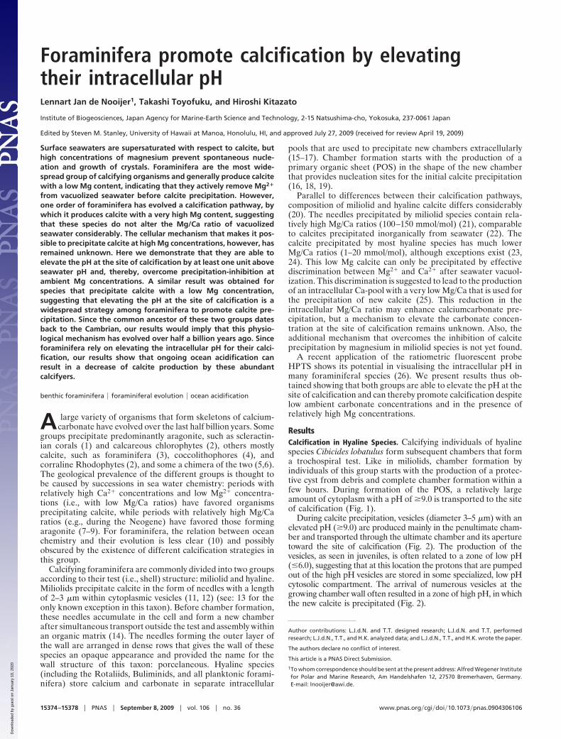
ResultsCalcification in Hyaline Species. Calcifying individuals of hyalinespecies Cibicides lobatulus form subsequent chambers that forma trochospiral test. Like in miliolids, chamber formation byindividuals of this group starts with the production of a protec-tive cyst from debris and complete chamber formation within afew hours. During formation of the POS, a relatively largeamount of cytoplasm with a pH of �9.0 is transported to the siteof calcification (Fig. 1).
During calcite precipitation, vesicles (diameter 3–5 �m) with anelevated pH (�9.0) are produced mainly in the penultimate cham-ber and transported through the ultimate chamber and its aperturetoward the site of calcification (Fig. 2). The production of thevesicles, as seen in juveniles, is often related to a zone of low pH(�6.0), suggesting that at this location the protons that are pumpedout of the high pH vesicles are stored in some specialized, low pHcytosolic compartment. The arrival of numerous vesicles at thegrowing chamber wall often resulted in a zone of high pH, in whichthe new calcite is precipitated (Fig. 2).
Author contributions: L.J.d.N. and T.T. designed research; L.J.d.N. and T.T. performedresearch; L.J.d.N., T.T., and H.K. analyzed data; and L.J.d.N., T.T., and H.K. wrote the paper.
The authors declare no conflict of interest.
This article is a PNAS Direct Submission.
1To whom correspondence should be sent at the present address: Alfred Wegener Institutefor Polar and Marine Research, Am Handelshafen 12, 27570 Bremerhaven, Germany.E-mail: [email protected].
15374–15378 � PNAS � September 8, 2009 � vol. 106 � no. 36 www.pnas.org�cgi�doi�10.1073�pnas.0904306106
Dow
nloa
ded
by g
uest
on
Janu
ary
10, 2
020
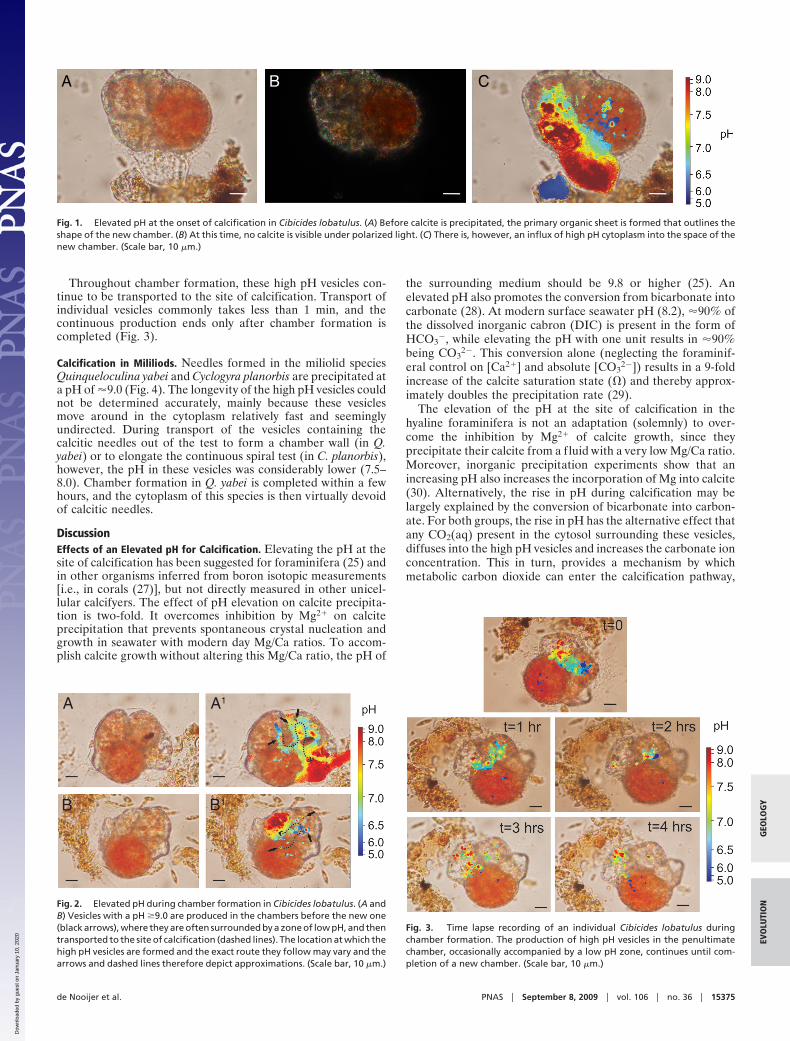
Throughout chamber formation, these high pH vesicles con-tinue to be transported to the site of calcification. Transport ofindividual vesicles commonly takes less than 1 min, and thecontinuous production ends only after chamber formation iscompleted (Fig. 3).
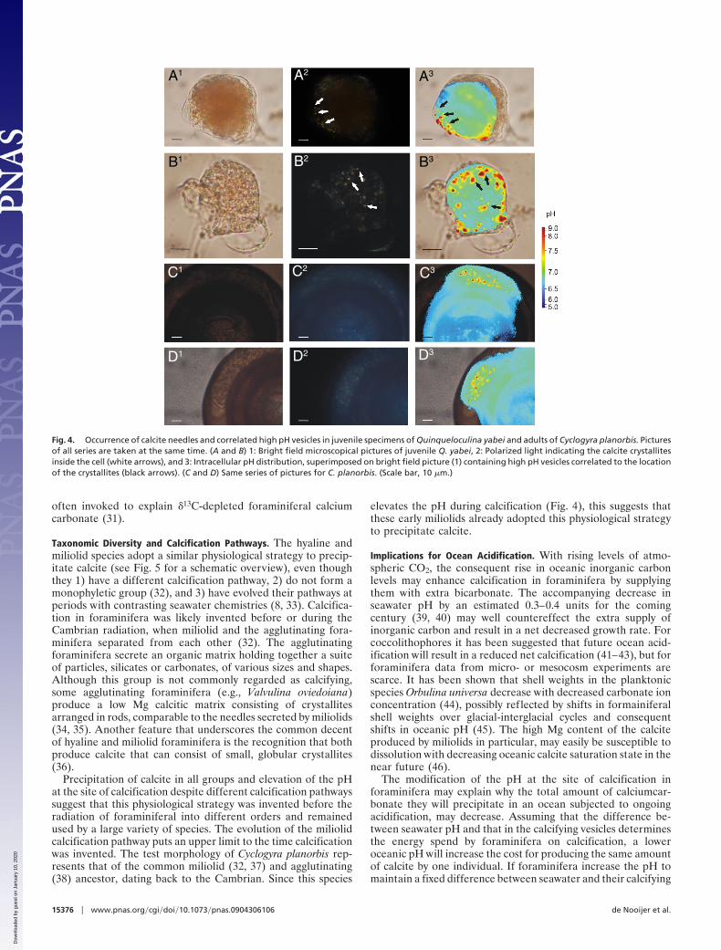
Calcification in Mililiods. Needles formed in the miliolid speciesQuinqueloculina yabei and Cyclogyra planorbis are precipitated ata pH of �9.0 (Fig. 4). The longevity of the high pH vesicles couldnot be determined accurately, mainly because these vesiclesmove around in the cytoplasm relatively fast and seeminglyundirected. During transport of the vesicles containing thecalcitic needles out of the test to form a chamber wall (in Q.yabei) or to elongate the continuous spiral test (in C. planorbis),however, the pH in these vesicles was considerably lower (7.5–8.0). Chamber formation in Q. yabei is completed within a fewhours, and the cytoplasm of this species is then virtually devoidof calcitic needles.
DiscussionEffects of an Elevated pH for Calcification. Elevating the pH at thesite of calcification has been suggested for foraminifera (25) andin other organisms inferred from boron isotopic measurements[i.e., in corals (27)], but not directly measured in other unicel-lular calcifyers. The effect of pH elevation on calcite precipita-tion is two-fold. It overcomes inhibition by Mg2� on calciteprecipitation that prevents spontaneous crystal nucleation andgrowth in seawater with modern day Mg/Ca ratios. To accom-plish calcite growth without altering this Mg/Ca ratio, the pH of
the surrounding medium should be 9.8 or higher (25). Anelevated pH also promotes the conversion from bicarbonate intocarbonate (28). At modern surface seawater pH (8.2), �90% ofthe dissolved inorganic cabron (DIC) is present in the form ofHCO3
�, while elevating the pH with one unit results in �90%being CO3
2�. This conversion alone (neglecting the foraminif-eral control on [Ca2�] and absolute [CO3
2�]) results in a 9-foldincrease of the calcite saturation state (�) and thereby approx-imately doubles the precipitation rate (29).
The elevation of the pH at the site of calcification in thehyaline foraminifera is not an adaptation (solemnly) to over-come the inhibition by Mg2� of calcite growth, since theyprecipitate their calcite from a fluid with a very low Mg/Ca ratio.Moreover, inorganic precipitation experiments show that anincreasing pH also increases the incorporation of Mg into calcite(30). Alternatively, the rise in pH during calcification may belargely explained by the conversion of bicarbonate into carbon-ate. For both groups, the rise in pH has the alternative effect thatany CO2(aq) present in the cytosol surrounding these vesicles,diffuses into the high pH vesicles and increases the carbonate ionconcentration. This in turn, provides a mechanism by whichmetabolic carbon dioxide can enter the calcification pathway,
A A1
B B1
Fig. 2. Elevated pH during chamber formation in Cibicides lobatulus. (A andB) Vesicles with a pH �9.0 are produced in the chambers before the new one(black arrows), where they are often surrounded by a zone of low pH, and thentransported to the site of calcification (dashed lines). The location at which thehigh pH vesicles are formed and the exact route they follow may vary and thearrows and dashed lines therefore depict approximations. (Scale bar, 10 �m.)
Fig. 3. Time lapse recording of an individual Cibicides lobatulus duringchamber formation. The production of high pH vesicles in the penultimatechamber, occasionally accompanied by a low pH zone, continues until com-pletion of a new chamber. (Scale bar, 10 �m.)
A B C
Fig. 1. Elevated pH at the onset of calcification in Cibicides lobatulus. (A) Before calcite is precipitated, the primary organic sheet is formed that outlines theshape of the new chamber. (B) At this time, no calcite is visible under polarized light. (C) There is, however, an influx of high pH cytoplasm into the space of thenew chamber. (Scale bar, 10 �m.)
de Nooijer et al. PNAS � September 8, 2009 � vol. 106 � no. 36 � 15375
GEO
LOG
YEV
OLU
TIO
N
Dow
nloa
ded
by g
uest
on
Janu
ary
10, 2
020
often invoked to explain �13C-depleted foraminiferal calciumcarbonate (31).
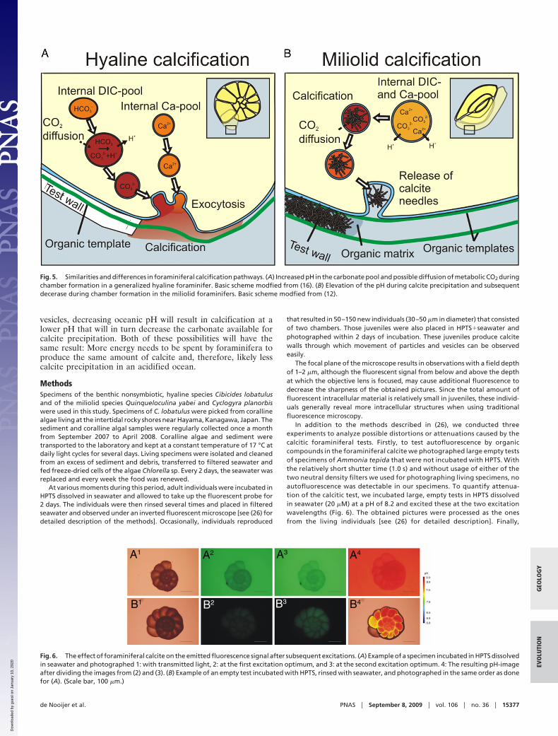
Taxonomic Diversity and Calcification Pathways. The hyaline andmiliolid species adopt a similar physiological strategy to precip-itate calcite (see Fig. 5 for a schematic overview), even thoughthey 1) have a different calcification pathway, 2) do not form amonophyletic group (32), and 3) have evolved their pathways atperiods with contrasting seawater chemistries (8, 33). Calcifica-tion in foraminifera was likely invented before or during theCambrian radiation, when miliolid and the agglutinating fora-minifera separated from each other (32). The agglutinatingforaminifera secrete an organic matrix holding together a suiteof particles, silicates or carbonates, of various sizes and shapes.Although this group is not commonly regarded as calcifying,some agglutinating foraminifera (e.g., Valvulina oviedoiana)produce a low Mg calcitic matrix consisting of crystallitesarranged in rods, comparable to the needles secreted by miliolids(34, 35). Another feature that underscores the common decentof hyaline and miliolid foraminifera is the recognition that bothproduce calcite that can consist of small, globular crystallites(36).
Precipitation of calcite in all groups and elevation of the pHat the site of calcification despite different calcification pathwayssuggest that this physiological strategy was invented before theradiation of foraminiferal into different orders and remainedused by a large variety of species. The evolution of the miliolidcalcification pathway puts an upper limit to the time calcificationwas invented. The test morphology of Cyclogyra planorbis rep-resents that of the common miliolid (32, 37) and agglutinating(38) ancestor, dating back to the Cambrian. Since this species
elevates the pH during calcification (Fig. 4), this suggests thatthese early miliolids already adopted this physiological strategyto precipitate calcite.
Implications for Ocean Acidification. With rising levels of atmo-spheric CO2, the consequent rise in oceanic inorganic carbonlevels may enhance calcification in foraminifera by supplyingthem with extra bicarbonate. The accompanying decrease inseawater pH by an estimated 0.3–0.4 units for the comingcentury (39, 40) may well countereffect the extra supply ofinorganic carbon and result in a net decreased growth rate. Forcoccolithophores it has been suggested that future ocean acid-ification will result in a reduced net calcification (41–43), but forforaminifera data from micro- or mesocosm experiments arescarce. It has been shown that shell weights in the planktonicspecies Orbulina universa decrease with decreased carbonate ionconcentration (44), possibly reflected by shifts in formainiferalshell weights over glacial-interglacial cycles and consequentshifts in oceanic pH (45). The high Mg content of the calciteproduced by miliolids in particular, may easily be susceptible todissolution with decreasing oceanic calcite saturation state in thenear future (46).
The modification of the pH at the site of calcification inforaminifera may explain why the total amount of calciumcar-bonate they will precipitate in an ocean subjected to ongoingacidification, may decrease. Assuming that the difference be-tween seawater pH and that in the calcifying vesicles determinesthe energy spend by foraminifera on calcification, a loweroceanic pH will increase the cost for producing the same amountof calcite by one individual. If foraminifera increase the pH tomaintain a fixed difference between seawater and their calcifying
A1
B1
A3
B3
A2
B2
C1 C2 C3
D1 D2 D3
Fig. 4. Occurrence of calcite needles and correlated high pH vesicles in juvenile specimens of Quinqueloculina yabei and adults of Cyclogyra planorbis. Picturesof all series are taken at the same time. (A and B) 1: Bright field microscopical pictures of juvenile Q. yabei, 2: Polarized light indicating the calcite crystallitesinside the cell (white arrows), and 3: Intracellular pH distribution, superimposed on bright field picture (1) containing high pH vesicles correlated to the locationof the crystallites (black arrows). (C and D) Same series of pictures for C. planorbis. (Scale bar, 10 �m.)
15376 � www.pnas.org�cgi�doi�10.1073�pnas.0904306106 de Nooijer et al.
Dow
nloa
ded
by g
uest
on
Janu
ary
10, 2
020
vesicles, decreasing oceanic pH will result in calcification at alower pH that will in turn decrease the carbonate available forcalcite precipitation. Both of these possibilities will have thesame result: More energy needs to be spent by foraminifera toproduce the same amount of calcite and, therefore, likely lesscalcite precipitation in an acidified ocean.
MethodsSpecimens of the benthic nonsymbiotic, hyaline species Cibicides lobatulusand of the miliolid species Quinqueloculina yabei and Cyclogyra planorbiswere used in this study. Specimens of C. lobatulus were picked from corallinealgae living at the intertidal rocky shores near Hayama, Kanagawa, Japan. Thesediment and coralline algal samples were regularly collected once a monthfrom September 2007 to April 2008. Coralline algae and sediment weretransported to the laboratory and kept at a constant temperature of 17 °C atdaily light cycles for several days. Living specimens were isolated and cleanedfrom an excess of sediment and debris, transferred to filtered seawater andfed freeze-dried cells of the algae Chlorella sp. Every 2 days, the seawater wasreplaced and every week the food was renewed.
At various moments during this period, adult individuals were incubated inHPTS dissolved in seawater and allowed to take up the fluorescent probe for2 days. The individuals were then rinsed several times and placed in filteredseawater and observed under an inverted fluorescent microscope [see (26) fordetailed description of the methods]. Occasionally, individuals reproduced
that resulted in 50–150 new individuals (30–50 �m in diameter) that consistedof two chambers. Those juveniles were also placed in HPTS�seawater andphotographed within 2 days of incubation. These juveniles produce calcitewalls through which movement of particles and vesicles can be observedeasily.
The focal plane of the microscope results in observations with a field depthof 1–2 �m, although the fluorescent signal from below and above the depthat which the objective lens is focused, may cause additional fluorescence todecrease the sharpness of the obtained pictures. Since the total amount offluorescent intracellular material is relatively small in juveniles, these individ-uals generally reveal more intracellular structures when using traditionalfluorescence microscopy.
In addition to the methods described in (26), we conducted threeexperiments to analyze possible distortions or attenuations caused by thecalcitic foraminiferal tests. Firstly, to test autofluorescence by organiccompounds in the foraminiferal calcite we photographed large empty testsof specimens of Ammonia tepida that were not incubated with HPTS. Withthe relatively short shutter time (1.0 s) and without usage of either of thetwo neutral density filters we used for photographing living specimens, noautofluorescence was detectable in our specimens. To quantify attenua-tion of the calcitic test, we incubated large, empty tests in HPTS dissolvedin seawater (20 �M) at a pH of 8.2 and excited these at the two excitationwavelengths (Fig. 6). The obtained pictures were processed as the onesfrom the living individuals [see (26) for detailed description]. Finally,
Fig. 5. Similarities and differences in foraminiferal calcification pathways. (A) Increased pH in the carbonate pool and possible diffusion of metabolic CO2 duringchamber formation in a generalized hyaline foraminifer. Basic scheme modfied from (16). (B) Elevation of the pH during calcite precipitation and subsequentdecerase during chamber formation in the miliolid foraminifers. Basic scheme modfied from (12).
B2 B3
A1 A2 A3 A4
B4B1
9.0
8.0
7.5
6.5
6.0
5.0
7.0
pH
Fig. 6. The effect of foraminiferal calcite on the emitted fluorescence signal after subsequent excitations. (A) Example of a specimen incubated in HPTS dissolvedin seawater and photographed 1: with transmitted light, 2: at the first excitation optimum, and 3: at the second excitation optimum. 4: The resulting pH-imageafter dividing the images from (2) and (3). (B) Example of an empty test incubated with HPTS, rinsed with seawater, and photographed in the same order as donefor (A). (Scale bar, 100 �m.)
de Nooijer et al. PNAS � September 8, 2009 � vol. 106 � no. 36 � 15377
GEO
LOG
YEV
OLU
TIO
N
Dow
nloa
ded
by g
uest
on
Janu
ary
10, 2
020
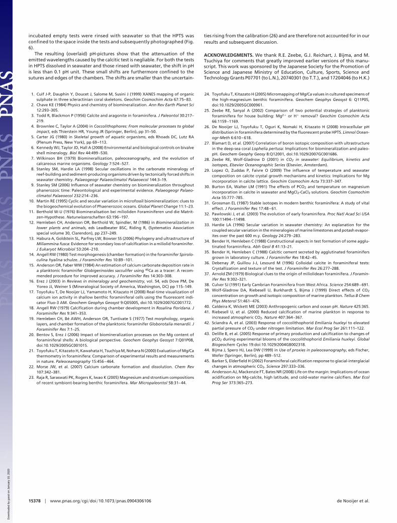
incubated empty tests were rinsed with seawater so that the HPTS wasconfined to the space inside the tests and subsequently photographed (Fig.6).
The resulting (overlaid) pH-pictures show that the attenuation of theemitted wavelengths caused by the calcitic test is negliable. For both the testsin HPTS dissolved in seawater and those rinsed with seawater, the shift in pHis less than 0.1 pH unit. These small shifts are furthermore confined to thesutures and edges of the chambers. The shifts are smaller than the uncertain-
ties rising from the calibration (26) and are therefore not accounted for in ourresults and subsequent discussion.
ACKNOWLEDGMENTS. We thank R.E. Zeebe, G.J. Reichart, J. Bijma, and M.Tsuchiya for comments that greatly improved earlier versions of this manu-script. This work was sponsored by the Japanese Society for the Promotion ofScience and Japanese Ministry of Education, Culture, Sports, Science andTechnology Grants P07701 (to L.N.), 20740301 (to T.T.), and 17204046 (to H.K.)
1. Cuif J-P, Dauphin Y, Doucet J, Salome M, Susini J (1999) XANES mapping of organicsulphate in three scleractinian coral skeletons. Geochim Cosmochim Acta 67:75–83.
2. Chave KE (1984) Physics and chemistry of biomineralization. Ann Rev Earth Planet Sci12:293–305.
3. Todd R, Blackmon P (1956) Calcite and aragonite in foraminifera. J Paleontol 30:217–219.
4. Brownlee C, Taylor A (2004) in Coccolithophores: From molecular processes to globalimpact, eds Thierstein HR, Young JR (Springer, Berlin), pp 31–50.
5. Carter JG (1980) in Skeletal growth of aquatic organisms, eds Rhoads DC, Lutz RA(Plenum Press, New York), pp 69–113.
6. Kennedy WJ, Taylor JD, Hall A (2008) Environmental and biological controls on bivalveshell mineralogy. Biol Rev 44:499–530.
7. Wilkinson BH (1979) Biominerallization, paleoceanography, and the evolution ofcalcareous marine organisms. Geology 7:524–527.
8. Stanley SM, Hardie LA (1998) Secular oscillations in the carbonate mineralogy ofreef-building and sediment-producing organisms driven by tectonically forced shifts inseawater chemistry. Palaeogeogr Palaeoclimatol Palaeoecol 144:3–19.
9. Stanley SM (2006) Influence of seawater chemistry on biomineralization throughoutphanerozoic time: Paleontological and experimental evidence. Palaeogeogr Palaeo-climatol Palaeoecol 232:214–236.
10. Martin RE (1995) Cyclic and secular variation in microfossil biomineralization: clues tothe biogeochemical evolution of Phaenerozoic oceans. Global Planet Change 11:1–23.
11. Berthold W-U (1976) Biomineralisation bei milioliden Foraminiferen und die Matrit-zen-Hypothese. Naturwissenschaften 63:196–197.
12. Hemleben CH, Anderson OR, Berthold W, Spindler, M (1986) in Biomineralization inlower plants and animals, eds Leadbeater BSC, Riding R, (Systematics Associationspecial volume 30, Clarendon), pp 237–249.
13. Habura A, Goldstein SL, Parfrey LW, Bowser SS (2006) Phylogeny and ultrastructure ofMiliammina fusca: Evidence for secondary loss of calcification in a miliolid foraminifer.J Eukaryot Microbiol 53:204–210.
14. Angell RW (1980) Test morphogenesis (chamber formation) in the foraminifer Spirolo-culina hyalina schulze. J Foraminifer Res 10:89–101.
15. Anderson OR, Faber WW (1984) An estimation of calcium carbonate deposition rate ina planktonic foraminifer Globigerinoides sacculifer using 45Ca as a tracer: A recom-mended procedure for improved accuracy. J Foraminifer Res 14:303–308.
16. Erez J (2003) in Reviews in mineralogy and geochemistry, vol. 54, eds Dove PM, DeYoreo JJ, Weiner S (Mineralogical Society of America, Washington, DC) pp 115–149.
17. Toyofuku T, De Nooijer LJ, Yamamoto H, Kitazato H (2008) Real-time visualization ofcalcium ion activity in shallow benthic foraminiferal cells using the fluorescent indi-cator Fluo-3 AM. Geochem Geophys Geosyst 9:Q05005, doi:10.1029/2007GC001772.
18. Angell RW (1979) Calcification during chamber development in Rosalina floridana. JForaminifer Res 9:341–353.
19. Hemleben CH, Be AWH, Anderson OR, Tuntivate S (1977) Test morphology, organiclayers, and chamber formation of the planktonic foraminifer Globorotalia menardii. JForaminifer Res 7:1–25.
20. Bentov S, Erez J (2006) Impact of biomineralization processes on the Mg content offoraminiferal shells: A biological perspective. Geochem Geophys Geosyst 7:Q01P08,doi:10.1029/2005GC001015.
21. Toyofuku T, Kitazato H, Kawahata H, Tsuchiya M, Nohara N (2000) Evaluation of Mg/Cathermometry in foraminifera: Comparison of experimental results and measurementsin nature. Paleoceanography 15:456–464.
22. Morse JW, et al. (2007) Calcium carbonate formation and dissolution. Chem Rev107:342–381.
23. Raja R, Saraswati PK, Rogers K, Iwao K (2005) Magnesium and strontium compositionsof recent symbiont-bearing benthic foraminifera. Mar Micropaleontol 58:31–44.
24. Toyofuku T, Kitazato H (2005) Micromapping of Mg/Ca values in cultured specimens ofthe high-magnesium benthic foraminifera. Geochem Geophys Geosyst 6: Q11P05,doi:10.1029/2005GC000961.
25. Zeebe RE, Sanyal A (2002) Comparison of two potential strategies of planktonicforaminifera for house building: Mg2� or H� removal? Geochim Cosmochim Acta66:1159–1169.
26. De Nooijer LJ, Toyofuku T, Oguri K, Nomaki H, Kitazato H (2008) Intracellular pHdistribution in foraminifera determined by the fluorescent probe HPTS. Limnol Ocean-ogr-Meth 6:610–618.
27. Blamart D, et al. (2007) Correlation of boron isotopic composition with ultrastructurein the deep-sea coral Lophelia pertusa: Implications for biomineralization and paleo-pH. Geochem Geophy Geosy 8:Q12001, doi:10.1029/2007GC001686.
28. Zeebe RE, Wolf-Gladrow D (2001) in CO2 in seawater: Equilibrium, kinetics andisotopes, Elsevier Oceanographic Series (Elsevier, Amsterdam).
29. Lopez O, Zuddas P, Faivre O (2009) The influence of temperature and seawatercomposition on calcite crystal growth mechanisms and kinetics: Implications for Mgincorporation in calcite lattice. Geochim Cosmochim Acta 73:337–347.
30. Burton EA, Walter LM (1991) The effects of PCO2 and temperature on magnesiumincorporation in calcite in seawater and MgCl2-CaCl2 solutions. Geochim CosmochimActa 55:777–785.
31. Grossman EL (1987) Stable isotopes in modern benthic foraminifera: A study of vitaleffect. J Foraminifer Res 17:48–61.
32. Pawlowski J, et al. (2003) The evolution of early foraminifera. Proc Natl Acad Sci USA100:11494–11498.
33. Hardie LA (1996) Secular variation in seawater chemistry: An explanation for thecoupled secular variation in the mineralogies of marine limestones and potash evapor-ites over the past 600 m.y. Geology 24:279–283.
34. Bender H, Hemleben C (1988) Constructional aspects in test formation of some agglu-tinated foraminifera. Abh Geol B 41:13–21.
35. Bender H, Hemleben C (1988) Calcitic cement secreted by agglutinated foraminifersgrown in laboratory culture. J Foraminifer Res 18:42–45.
36. Debenay JP, Guillou J-J, Lesourd M (1996) Colloidal calcite in foraminiferal tests:Crystallization and texture of the test. J Foraminifer Res 26:277–288.
37. Arnold ZM (1979) Biological clues to the origin of miliolidean foraminifera. J Foramin-ifer Res 9:302–321.
38. Culver SJ (1991) Early Cambrian Foraminifera from West Africa. Science 254:689–691.39. Wolf-Gladrow DA, Riebesell U, Burkhardt S, Bijma J (1999) Direct effects of CO2
concentration on growth and isotopic composition of marine plankton. Tellus B ChemPhys Meterol 51:461–476.
40. Caldeira K, Wickett ME (2003) Anthropogenic carbon and ocean pH. Nature 425:365.41. Riebesell U, et al. (2000) Reduced calcification of marine plankton in response to
increased atmospheric CO2. Nature 407:364–367.42. Sciandra A, et al. (2003) Response of coccolithophorid Emiliania huxleyi to elevated
partial pressure of CO2 under nitrogen limitation. Mar Ecol Prog Ser 261:111–122.43. Delille B, et al. (2005) Response of primary production and calcification to changes of
pCO2 during experimental blooms of the coccolithophorid Emiliania huxleyi. GlobalBiogeochem Cycles 19:doi:10.1029/2004GB002318.
44. Bijma J, Spero HJ, Lea DW (1999) in Use of proxies in paleoceanography, eds Fischer,Wefer (Springer, Berlin), pp 489–512.
45. Barker S, Elderfield H (2002) Foraminiferal calcification response to glacial-interglacialchanges in atmospheric CO2. Science 297:333–336.
46. Andersson AJ, Mackenzie FT, Bates NR (2008) Life on the margin: Implications of oceanacidification on Mg-calcite, high latitude, and cold-water marine calcifiers. Mar EcolProg Ser 373:365–273.
15378 � www.pnas.org�cgi�doi�10.1073�pnas.0904306106 de Nooijer et al.
Dow
nloa
ded
by g
uest
on
Janu
ary
10, 2
020