foreign body ingestion 2012 jcfs
TRANSCRIPT

8/2/2019 Foreign Body Ingestion 2012 JCFS
http://slidepdf.com/reader/full/foreign-body-ingestion-2012-jcfs 1/5
ovoid to round nuclei, immersed in a collagenous stroma. Betweenthose granular cells, islands of odontogenic epithelium were present.In addition, cementum-like bodies and dystrophic calcification wereseen, like in previous reports.4,5,13,14
Immunohistochemical staining is a valuable tool in elucidating the possible origin and nature of the granular cells.13 In the case reported,
the vimentin positivity and S-100 protein and cytokeratin negativ-ity, by the granular cells, confirmed the mesenchymal origin whilemitigating against epithelial or Schwann cell origin. Nevertheless,the literature reveals inconsistent findings regarding the expressionof S-100 by the granular cells, with some authors reporting slight positivity.15 Y 17 Yet, a histiocytic differentiation of the granular cellsis proposed by the strong expression of the CD68, previouslyreported 4,5,7,12,18 and confirmed in the current case. However, it issuggested that the expression of CD68 by the tumor cells does not necessarily imply histiocytic origin because other studies have shownthat CD68 is also expressed by non Y macrophage-derived cells.19,20
In addition, the epithelial islands had strong positive staining for CK14 and AE1/AE3, as previously reported.5,7,18 Moreover, our studyagrees with the hypothesis of mesenchymal origin owing to the in-tense immunoreactivity to CD68 and vimentin and absent immunos-
taining for CK14 and AE1/AE3 by the granular cells.The first malignant case related by Piatelli et al7 revealed similar immunohistochemical findings to the present case, suggesting that the distinction between a benign and a malignant lesion should be based on the clinical and histopathologic features.
The treatment of GCOT consists of conservative surgical pro-cedures, most often enucleation or curettage. Clinical, radiographic,and the follow-up data led to the conclusion that this lesion has a benign biologic behavior.5 However, long-term follow-up is recom-mended because a malignant counterpart of the CGCOT has already been reported,7 and 1 case that was treated with curettage recurred 13 years after it was initially removed.5
In conclusion, the immunohistochemical profile of CGCOT inthis study and in other publications showed that the granular cellsare mesenchymal in origin, with a possible histiocytic cell lineage.Yet, the present case shows a particular feature of large extension
with perforation of the maxillary cortical plates, resembling a ma-lignancy, which was excluded according to microscopy findings.However, this feature highlights the possibility of aggressive be-havior by these lesions. In this sense, the clinical and imaging fea-tures of the lesion, associated to the histopathologic aspects, should be carefully evaluated to perform the correct diagnosis of CGCOTand exclude its various differential diagnoses.
REFERENCES
1. Werthemann A. Uber spongiocytares adamantinoma [in German].Oncologia 1950;3:193 Y 207
2. Couch RD, Morris EE, Vellios F. Granular cell ameloblastic fibroma.Report of 2 cases in adults, with observations of its similarityto congenital epulis. Am J Clin Pathol 1962;37:398 Y 404
3. Vincent SD, Hammond HL, Ellis GL, et al. Central granular cellodontogenic fibroma. Oral Surg Oral Med Oral Pathol 1987;63:715 Y 721
4. Gomes CC, Naves MD, Pereira MV, et al. Granular cell odontogenictumour: case report and review of literature. Oral Oncol 2006;42:277 Y 280
5. Brannon RB, Goode RK, Eversole LR, et al. The central granular cell odontogenic tumor: report of 5 new cases. Oral Surg Oral
Med Oral Pathol Oral Radiol Endod 2002;94:641 Y 621
6. Brannon RB. Central odontogenic fibroma, myxoma (odontogenicmyxoma, fibromyxoma), and central odontogenic granular cell tumor.Oral Maxillofacial Surg Clin North Am 2004;16:359 Y 374
7. Piatelli A, Rubini C, Goteri G, et al. Central granular cell odontogenictumour: report of the first malignant case and review of theliterature. Oral Oncol 2003;39:78 Y 82
8. Martınez-Mata G, Mosqueda-Taylor A, Carlos-Bregni R, et al.Odontogenic myxoma: clinico-pathological, immunohistochemical and ultrastructural findings of a multicentric series. Oral Oncol 2008;44:601 Y 607
9. Toro C, Millesi W, Zerman N, et al. A case of aggressiveossifying fibroma with massive involvement of the mandible:
differential diagnosis and management options. Int J Pediatr Otorhinolaryngol Extra 2006;1:167 Y 172
10. Sopta J, Draºi( R, Tuli( G, et al. Cemento-ossifying fibromaof jaws V correlation of clinical and pathological findings.Clin Oral Investig 2011;15:201 Y 207
11. Lotay HS, Kalmar J, DeLeeuw K. Central odontogenic fibroma withfeatures of central g ranular cell odontogenic tumor. Oral Surg Oral Med Oral Pathol Oral Radiol Endod 2010;109:63 Y 66
12. Calvo N, Alonso D, Prieto M, et al. Central odontogenic fibromagranular cell variant: a case report and review of literature.
J Oral Maxillofac Surg 2002;60:1192 Y 1194
13. Yih WY, Thompson C, Meshul CK, et al. Central odontogenic granular cell tumor of the jaw: report of case and immunohistochemical and electron microscopy study. J Oral Maxillofac Surg 1995;53:453 Y 459
14. Gesek DJ Jr, Adrian JC, Reid EN. Central granular cellodontogenic tumor: a case report including light microscopy,immunohistochemistry, and literature review. J Oral Maxillofac Surg
1995;53:945 Y 94915. Regezi JA, Kerr DA, Courtney RM. Odontogenic tumors: analysis
of 706 cases. J Oral Surg 1978;36:771 Y 778
16. Shiro BC, Jacoway JR, Mirmiran SA, et al. Centralodontogenic fibroma, granular cell variant. A case report withS-100 immunohistochemistry and a review of the literature.Oral Surg Oral Med Oral Pathol 1989;67:725 Y 730
17. Mirchandani R, Sciubba JJ, Mir R. Granular cell lesions of the jaws and oral cavity: a clinicopathologic, immunohistochemical, and ultrastructural s tudy. J Oral Maxillofac Surg 1989;47:1248 Y 1255
18. de Sousa SO, de Arau jo NS, Melhado RM, et al. Central odontogenicgranular cell tumor: immunohistochemical study of two cases.
J Oral Maxillofac Surg 1998;56:787 Y 791
19. Facchetti F, Bertalot G, Grigolato PG. KPl (CD68) staining of malignant melanomas. Histopathology 1991;19:141 Y 145
20. Doussis IA, Gatter KC, Mason DY. CD68 reactivity of nonmacrophagederived tumors in cytological specimens. J Clin Pathol 1993;46:334 Y 336
Foreign Body Ingestion DuringDental Implant Procedures
Thiago de Santana Santos, DDS, Msc,*
Antonio Azoubel Antunes, DDS,* Andre Vajgel, DDS, Msc,Þ
Thames Bruno Barbosa Cavalcanti, DDS,þ
Luiz Ricardo Gomes de Caldas Nogueira, DDS,§
Jose Rodrigues Laureano Filho, DDS, Msc, PhDÞ
From the *Oral and Maxillofacial Surgery Program, Faculdade de Odonto-
logia de Ribeirao Preto, Universidade de Sao Paulo, Sao Paulo; †Oral and Maxillofacial Surgery Program, ‡Faculdade de Odontologia de Per-nambuco, Universidade de Pernambuco; and §Associa0ao Brasile ira deOdontologia de Pernambuco, Pernambuco, Brazil.
Received July 21, 2011.Accepted for publication October 9, 2011.Address correspondence and reprint requests to Thiago de Santana Santos,
DDS, Msc, Faculdade de Odontologia de Pernambuco, Av General Newton Cavalcanti, 1650, 54.753-220. Camaragibe, Pernambuco,Brazil; E-mail: [email protected]
The authors report no conflicts of interest.Copyright * 2012 by Mutaz B. Habal, MDISSN: 1049-2275DOI: 10.1097/SCS.0b013e31824cda32
The Journal of Craniofacial Surgery & Volume 23, Number 2, March 2012 Brief Clinical Studies
* 2012 Mutaz B. Habal, MD e119
Copyright © 2012 Mutaz B. Habal, MD. Unauthorized reproduction of this article is prohibited.
8/2/2019 Foreign Body Ingestion 2012 JCFS
http://slidepdf.com/reader/full/foreign-body-ingestion-2012-jcfs 2/5
Abstract: Two cases of swallowing of foreign material related to
dental implants during dental practice are described. A conservative
approach by clinical-radiographic follow-up was performed in both
cases; however, one of the patients required colonoscopy under gen-
eral anesthesia for the removal of the impacted foreign body from the
intestinal region. These complications not only have associated eco-nomic cost but also carry the risk of malpractice litigation against
the professional; thus, the surgeon was responsible for all the costs of
hospital and surgery management of this case. Details of the clinical
signs, radiographic examinations, type of treatment, and follow-up
are presented.
Key Words: Foreign bodies, deglutition, oropharynx,
emergencies, liability, legal
The passage of unusual materials through the oropharynx duringdental practice is the type of accident that may place the patient’s
life at risk.1 Y 3 Depending on the path followed, the object can be
either swallowed (more common) or aspirated. When swallowed, it can become embedded in any surface during its passage, therebycausing obstructive, inflammatory, or infectious complications.4
The diagnosis of foreign body aspiration is generally missed or delayed, and the patient later presents with long-term symptoms and complications, such as cough, stridor, wheezing, obstructive pneu-monitis, bronchiectasis, and abscess secondary to recurrent pulmo-nary infection.5
Approximately 80% of cases of swallowed foreign bodies oc-cur in the pediatric population, particularly between the ages of 6 months and 1 year.6 This occurrence is infrequent among adultsand is seen most often among elderly individuals.7 It is mainly seenin patients with psychiatric disorders and intoxicated individualsor secondarily to a specific health treatment.8 Cases of attempted suicide by ingestion of dentures, ingestion by patients with mentalincompetence, and a dislodged fixed partial denture while under-
going general anesthesia have been reported.9,10The swallowing of dental objects may accidentally occur for
various reasons and frequently involve pieces of prostheses.11,12
However, a number of other dental items can trigger this type of in- jury, especially small objects, such as endodontic files, dental clips, or brackets.13 Y 15 The passage of foreign bodies through the oropharynxand other accidents during dental treatment raise a number of ques-tions, initially of a clinical nature: Was the object swallowed or in-haled? Should an attempt be made to remove the object or should the patient be taken to a hospital? Is surgical intervention necessary?Moreover, depending on the course of events, ethical and legal issuesmay also arise: What is the professional’s liability? Who will bear theexpenses of themedical treatment? Canthe professional be sued by the patient because of the accident?
This article describes 2 clinical cases of foreign body ingestionand addresses what actions should be taken in the case of the disap-
pearance of an object from the oral cavity, emphasizing preventiveaspects and possible consequences of an ethical and legal nature.
CLINICAL REPORT
Patient 1
A 70-year-old man was referred for prosthetic rehabilitation with
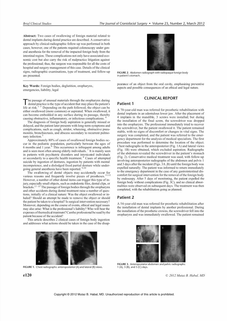
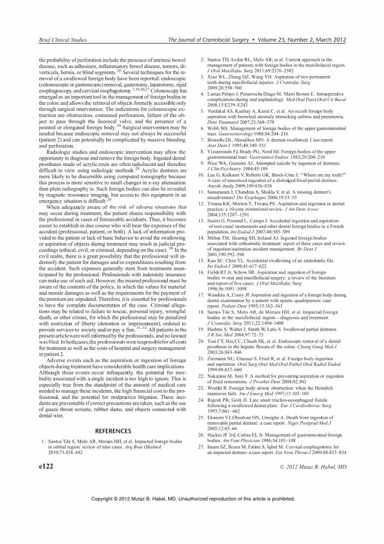
dental implants in an edentulous lower jaw. After the placement of 4 implants in the mandible, 3 screws were installed; but duringthe installation of the final screw, the screwdriver was dropped into the oropharynx. The professional immediately tried to recover the screwdriver, but the patient swallowed it. The patient remained stable, with no signs of discomfort or changes in vital signs. Thesurgery was completed, and the patient was referred to the emer-gency department for the analysis of medical specialists. The first procedure was performed to determine the location of the object.Chest radiographs in the anteroposterior (Fig. 1A) and lateral views(Fig. 1B) were obtained, which excluded aspiration. Radiographsof the abdomen revealed the screwdriver in the patient’s stomach(Fig. 2). Conservative medical treatment was used, with follow-upinvolving anteroposterior radiographs of the abdomen and pelvis 1and 3 days after the incident (Figs. 3A ,B) until the foreign body was
expelled naturally. The patient was informed to return immediatelyto the emergency department in the case of any gastrointestinal dis-comfort for surgical intervention for the removal of the foreign body by endoscopy. After 5 days of monitoring, the patient expelled theforeign body without complications (Fig. 3C), and no clinical abnor-malities were observed on subsequent days. The treatment was thencompleted, with the rehabilitation going as planned.
Patient 2
A 54-year-old man was referred for prosthetic rehabilitation after the installation of dental implants by another professional. Duringthe installation of the prosthetic crowns, the screwdriver fell into theoropharynx and was immediately swallowed. The patient remained
FIGURE 1. Chest radiographs: anteroposterior (A) and lateral (B) views.
FIGURE 2. Abdomen radiograph with radiopaque foreign bodyin patient’s stomach.
FIGURE 3. Anteroposterior abdomen and pelvis radiographs:1 (A), 3 (B), and 5 (C) days.
Brief Clinical Studies The Journal of Craniofacial Surgery & Volume 23, Number 2, March 2012
e120 * 2012 Mutaz B. Habal, MD
Copyright © 2012 Mutaz B. Habal, MD. Unauthorized reproduction of this article is prohibited.

8/2/2019 Foreign Body Ingestion 2012 JCFS
http://slidepdf.com/reader/full/foreign-body-ingestion-2012-jcfs 3/5
stable, with no signs of discomfort or changes in vital signs. The patient was referred to the emergency department for the analysis of medical specialists. On physical examination, vital signs and aus-cultation of the lungs were normal. Chest radiographs in the ante-roposterior and lateral views were obtained and excluded aspiration.Radiographs of the abdomen revealed the screwdriver in the patient’sstomach (Fig. 4). Conservative medical treatment was used, withfollow-up involving anteroposterior radiographs of the abdomen and
pelvis until the foreign body was expelled naturally. However, theradiographs revealed no differences in the position of the screw-driver between days 7 and 14 after the incident (Figs. 5A, B). The patient did not complain of gastrointestinal discomfort. Immediateintervention by colonoscopy under general anesthesia was used for the removal of the impacted foreign body (Fig. 6). The patient wasdischarged the next day and has since remained well.
DISCUSSION
The aspiration or ingestion of foreign bodies occurs most commonlyin procedures involving fixed prosthodontic dentistry V specificallythose involving the cementation of permanent crowns V and adjunc-tive procedures, such as the placement of cast posts, cores and onlays,as well as in implant-related procedures. The higher occurrence of
aspiration and ingestion could be attributed to the absence of rubber dams (barriers) and the ligation of objects with dental wire in the oralcavity during crown cementation and implant procedures. This hasobvious implications in occurrences of dislodgement.16
There is a relatively infrequent occurrence of adverse outcomesfrom the swallowing of foreign bodies in the special-needs and pe-diatric populations. This finding contradicts the widely held belief that these patients are at greater risk of aspirating or ingesting dentalforeign objects owing to their greater likelihood of having a neu-romuscular disease or physical handicap. These conditions can di-minish a patient’s protective airway reflexes, resulting in difficulty infollowing verbal commands from the professional.17
Although both aspiration and ingestion are infrequent occur-rences, ingestion occurs more often. This may be a direct result of the strong coughing that occurs when there is a foreign object in the
patient’s airway, thereby hampering aspiration.4,18 Approximately10% to 20% of patients having ingested a foreign body require anonoperative intervention, whereas 1% or less require surgery.6
The ingestion of a foreign body may not result in any signs or symptoms, and the object may be found totally by chance. However,it becomes apparent when complications arise, such as throat painor discomfort, persistent sensation of foreign body in the throat,retrosternal pain, tenderness in the neck, total dysphagia, pooling of saliva in the oropharynx, perforation, abscess, or formation of an
enterocolic fistula. The patient may also experience sweating and ahigh temperature and cough up blood.19 Foreign body ingestionshould be consideredas a differential diagnosis in patients whopresent with abdominal and constitutional symptoms and whose laboratoryexamination results for more common pathologies are negative.20
None of the cases reported here had any signs or symptoms.When an object falls into the oropharynx, the patient should first
be reassured and, if the professional judged it feasible to recover the piece, the use of a pincers or high-power vacuum are suitable for this purpose. The professional must not stimulate swallowing bytouching the regions adjacent to the isthmus of the oropharynx,such as the root of the tongue and the uvula.14 If swallowing occurs,the procedure should be interrupted or brought to a stable situation.The approach taken then depends on the severity of the case and the potential involvement of the upper airways. The patient should be positioned with the head reclined in reverse Trendelenburg position
(in which the upper part of the body is raised 20 Y 30 degrees) and usually responds immediately with the cough reflex. The elevationof the chair assists in the attempt to expel the object.21 If the object isnot expelled, signs of airway obstruction should be promptly identi-fied by monitoring the patient’s breathing and consciousness.22
If the patient has difficulty breathing, he/she should be admitted immediately to an emergency medical service for hospital care. Incases of airway obstruction, basic life support protocols recommend that, if the victim is conscious, is breathing well, and manages tocough effectively, the professional should stimulate coughing and monitor whether the obstruction is resolved. If coughing is inef-fective, the Heimlich maneuver should be performed with care not to produce traumatic injury. It is important not to give up if the first attempt fails and repeat the maneuver. As the victim is deprived of oxygen, the muscles of the trachea relax slightly, which may allow
the foreign object to be expelled on a second or third attempt.
23
Reported late-onset complications of undiagnosed swallowed dentures include extraluminal migration from the esophagus, caus-ing either a diverticulum or a perforation (once a perforation hasoccurred, further severe sequelae may be anticipated, eg, tracheo-esophageal fistula), enterocolonic fistula, sigmoid colon perforation,and death.24 A case is reported in which a removable partial denturewas found lodged in the midportion of the esophagus, with its lateralwings deeply embedded in the wall of the esophagus, causing lac-erations and severe hemorrhaging.25
Complications such as impaction, perforation, or obstructionmost often occur in regions with acute angles or physiologicalnarrowing in the gastrointestinal tract.20 Risk factors that increase
FIGURE 4. Abdomen radiograph with radiopaque foreign bodyin patient’s stomach.
FIGURE 5. Abdomen radiograph showing little displacement: 7 (A)and 14 (B) days.
FIGURE 6. Removing the foreign body by endoscopy: screwdriver (A)and capturing the screwdriver (B).
The Journal of Craniofacial Surgery & Volume 23, Number 2, March 2012 Brief Clinical Studies
* 2012 Mutaz B. Habal, MD e121
Copyright © 2012 Mutaz B. Habal, MD. Unauthorized reproduction of this article is prohibited.
8/2/2019 Foreign Body Ingestion 2012 JCFS
http://slidepdf.com/reader/full/foreign-body-ingestion-2012-jcfs 4/5
the probability of perforation include the presence of intrinsic boweldisease, such as adhesions, inflammatory bowel disease, tumors, di-verticula, hernia, or blind segments.26 Several techniques for the re-moval of a swallowed foreign body have been reported: endoscopic(colonoscopic or gastroscopic) removal, gastrotomy, laparotomy, rigid esophagoscopy, and cervical esophagotomy.7,19,20,27 Colonoscopy has
emerged as an important tool in the management of foreign bodies inthe colon and allows the retrieval of objects formerly accessible onlythrough surgical intervention. The indications for colonoscopic ex-traction are obstruction, contained perforation, failure of the ob- ject to pass through the ileocecal valve, and the presence of a pointed or elongated foreign body.20 Surgical intervention may beneeded because endoscopic removal may not always be successful(patient 2) and can potentially be complicated by massive bleedingand perforation.
Radiologic studies and endoscopic intervention may allow theopportunity to diagnose and remove the foreign body. Ingested dental prostheses made of acrylic resin are often radiolucent and thereforedifficult to view using radiologic methods.28 Acrylic dentures aremore likely to be discernible using computed tomography becausethis process is more sensitive to small changes in x-ray attenuation
than plain radiography is. Such foreign bodies can also be revealed by magnetic resonance imaging, but access to this equipment in anemergency situation is difficult.29
When adequately aware of the risk of adverse situations that may occur during treatment, the patient shares responsibility withthe professional in cases of foreseeable accidents. Thus, it becomeseasier to establish in due course who will bear the expenses of theaccident (professional, patient, or both). A lack of information pro-vided to the patient or lack of basic behavior to avoid the swallowingor aspiration of objects during treatment may result in judicial pro-ceedings (ethical, civil, or criminal, depending on the case).30 In thecivil realm, there is a great possibility that the professional will in-demnify the patient for damages and/or expenditures resulting fromthe accident. Such expenses generally stem from treatments unan-ticipated by the professional. Professionals with indemnity insurancecan make use of such aid. However, the insured professional must be
aware of the contents of the policy, in which the values for materialand morale damages as well as the requirements for the payment of the premium are stipulated. Therefore, it is essential for professionalsto have the complete documentation of the case. Criminal allega-tions may be related to failure to rescue, personal injury, wrongfuldeath, or other crimes, for which the professional may be penalized with restriction of liberty (detention or imprisonment), ordered to provide services to society and/or pay a fine.31 Y 33 All patients in the present article were well informed by the professionals, and no lawsuit was filed. In bothcases,the professionals were responsiblefor all costsfor treatment as well as the costs of hospital and surgery management in patient 2.
Adverse events such as the aspiration or ingestion of foreignobjects during treatment have considerable health care implications.Although these events occur infrequently, the potential for mor-
bidity associated with a single incident is too high to ignore. This isespecially true from the standpoint of the amount of medical careneeded to manage these incidents, the high financial cost to the pro-fessional, and the potential for malpractice litigation. These inci-dents are preventable if correct precautions are taken, such as the useof gauze throat screens, rubber dams, and objects connected withdental wire.
REFERENCES
1. Santos Tde S, Melo AR, Moraes HH, et al. Impacted foreign bodiesin orbital region: review of nine cases. Arq Bras Oftalmol 2010;73:438 Y 442
2. Santos TD, Avelar RL, Melo AR, et al. Current approach in themanagement of patients with foreign bodies in the maxillofacial region.
J Oral Maxillofac Surg 2011;69:2376 Y 2382
3. Xiao WL, Zhang DZ, Wang YH. Aspiration of two permanent teeth during maxillofacial injuries. J Craniofac Surg 2009;20:558 Y 560
4. Lamas Pelayo J, Penarrocha Diago M, Marti Bowen E. Intraoperativecomplications during oral implantology. Med Oral Patol Oral Cir Bucal 2008;13:E239 Y E243
5. Yurdakul AS, Kanbay A, Kurul C, et al. An occult foreign bodyaspiration with bronchial anomaly mimicking asthma and pneumonia.
Dent Traumatol 2007;23:368 Y 370
6. Webb WA. Management of foreign bodies of the upper gastrointestinaltract. Gastroenterology 1988;94:204 Y 216
7. Brunello DL, Mandikos MN. A denture swallowed. Case report. Aust Dent J 1995;40:349 Y 351
8. Vizcarrondo FJ, Brady PG, Nord HJ. Foreign bodies of the upper gastrointestinal tract. Gastrointest Endosc 1983;29:208 Y 210
9. Price WA, Giannini AJ. Attempted suicide by ingestion of dentures. J Clin Psychiatry 1984;45:189
10. Lau G, Kulkarni V, Roberts GK, Brock-Utne J. ‘‘Where are my teeth?’’A case of unnoticed ingestion of a dislodged fixed partial denture.
Anesth Analg 2009;109:836 Y 838
11. Samarasam I, Chandran S, Shukla V, et al. A missing denture’smisadventure! Dis Esophagus 2006;19:53 Y 55
12. Tiwana KK, Morton T, Tiwana PS. Aspiration and ingestion in dental practice: a 10-year institutional review. J Am Dent Assoc2004;135:1287 Y 1291
13. Susini G, Pommel L, Camps J. Accidental ingestion and aspirationof root canal instruments and other dental foreign bodies in a French
population. Int Endod J 2007;40:585 Y 589
14. Milton TM, Hearing SD, Ireland AJ. Ingested foreign bodiesassociated with orthodontic treatment: report of three cases and reviewof ingestion/aspiration incident management. Br Dent J 2001;190:592 Y 596
15. Kuo SC, Chen YL. Accidental swallowing of an endodontic file. Int Endod J 2008;41:617 Y 622
16. Fields RT Jr, Schow SR. Aspiration and ingestion of foreign bodies in oral and maxillofacial surgery: a review of the literatureand report of five cases. J Oral Maxillofac Surg 1998;56:1091 Y 1098
17. Wandera A, Conry JP. Aspiration and ingestion of a foreign body duringdental examination by a patient with spastic quadriparesis: casereport. Pediatr Dent 1993;15:362 Y 363
18. Santos Tde S, Melo AR, de Moraes HH, et al. Impacted foreign bodies in the maxillofacial region V diagnosis and treatment. J Craniofac Surg 2011;22:1404 Y 1408
19. Hashmi S, Walter J, Smith W, Latis S. Swallowed partial dentures. J R Soc Med 2004;97:72 Y 75
20. Tsai CY, Hsu CC, Chuah SK, et al. Endoscopic removal of a dental prosthesis in the hepatic flexure of the colon. Chang Gung Med J 2003;26:843 Y 846
21. Zitzmann NU, Elsasser S, Fried R, et al. Foreign body ingestionand aspiration. Oral Surg Oral Med Oral Pathol Oral Radiol Endod 1999;88:657 Y 660
22. Nakajima M, Sato Y. A method for preventing aspiration or ingestion
of fixed restorations. J Prosthet Dent 2004;92:30323. Westfal R. Foreign body airway obstruction: when the Heimlich
maneuver fails. Am J Emerg Med 1997;15:103 Y 105
24. Rajesh PB, Goiti JJ. Late onset tracheo-oesophageal fistulafollowing a swallowed dental plate. Eur J Cardiothorac Surg 1993;7:661 Y 662
25. Ekanem VJ, Obuekwe ON, Unuigbe A. Death from ingestion of removable partial denture: a case report. Niger Postgrad Med J 2005;12:65 Y 66
26. Hacker JF 3rd, Cattau EL Jr. Management of gastrointestinal foreign bodies. Am Fam Physician 1986;34:101 Y 108
27. Imam SZ, Ikram M, Fatimi S, Iqbal M. Cervical esophagotomy for an impacted denture: a case report. Ear Nose Throat J 2009;88:833 Y 834
Brief Clinical Studies The Journal of Craniofacial Surgery & Volume 23, Number 2, March 2012
e122 * 2012 Mutaz B. Habal, MD
Copyright © 2012 Mutaz B. Habal, MD. Unauthorized reproduction of this article is prohibited.
8/2/2019 Foreign Body Ingestion 2012 JCFS
http://slidepdf.com/reader/full/foreign-body-ingestion-2012-jcfs 5/5
28. de Ruiter MH, Van Damme PA, Drenth JP. Serious complicationsfollowing (removal a fter) ingestion of a partial denture [in Dutch].
Ned Tijdschr Tandheelkd 2008;115:267 Y 270
29. McLaughlin MG, Swayne LC, Caruana V. Computed tomographicdetection of a swallowed denture. Comput Med Imaging Graph1989;13:161 Y 163
30. D’Ovidio C, Carnevale A, Pantaleone G. A case of accidentalaspiration of a dental cutter into the bronchopulmonary tree: clinicalimplications and legal considerations. Minerva Stomatol 2008;57:535 Y 547
31. Montagna F, Cortesini C, Manca R, et al. Epidemiology of dental professional liability. Minerva Stomatol 2011;60:179 Y 193
32. Wells C, Thomas D. Deaths in the dental surgery: individualand organisational criminal liability. Br Dent J 2008;204:497 Y 502
33. Killila BA. Dental professional liability issues. J Indiana Dent Assoc1993;72:22 Y 24
Giant Maxillary MucoceleOccurring After Reduction
Malarplasty Hyo-In Kim, MD, Si-Gyun Roh, MD, Nae-Ho Lee, MD,
Kyung-Moo Yang, MD
Abstract: Reduction malarplasty for patients with a prominent ma-
lar complex is a popular procedure in Asia. However, a range of
complications have been reported after reduction malarplasty, such
as hematoma, orbital complications, asymmetric face, and non-
union. A medially displaced fracture or bony fragment can induce si-
nusitis and subsequent trauma to bones in combination with chronic
inflammatory processes, which can lead to chronic obstruction of
mucus-secreting glands. In our case, 46-year-old man presented with
a large mucocele in the maxillary sinus after malar reduction ap-
proximately 20 years ago.
Key Words: Mucocele, reduction malarplasty, maxillary sinus
In Asia, reduction malarplasty is quite popular for patients with a prominent malar complex. However, complications after reduction
malarplasty have rarely been described in the literature. Reductionmalarplasty was first introduced by Onizuka et al in 1983 using anintraoral approach.1 In 1991, Baek et al2 described a method based on a coronal approach, which was devised to increase the limited surgical field and reduce complications, such as cheek drooping.Since then, many new methods have been introduced to improve thedisadvantages of previous methods. Cheek drooping, orbital defor-
mity, soft tissue descent, facial contour asymmetry, inferior orbitalnerve paresthesia, facial nerve injuries, malunion, and maxillary si-nusitis have all been described as complications of reduction malar- plasty.3,4 Here, we describe a rare case of mucocele, which occurred 20 years after reduction malarplasty.
CLINICAL REPORT
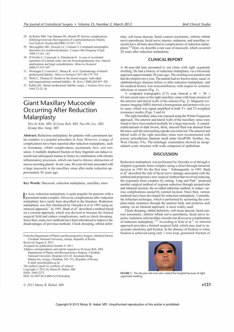
A 46-year-old man presented to our clinic with right zygomaticswelling. He had a history of reduction malarplasty via a bicoronalapproach approximately 20 years ago. The swelling was painless and had developed over a year. The patient had no known sinus, nasal, or ophthalmologic diseases before or after reduction malarplasty, and his medical history was noncontributory with respect to systemicinfections or tumors (Fig. 1).
A computed tomographic (CT) scan showed a 40 Â 50 Â43-mm-sized mass in the right maxillary sinus with bone erosion of the anterior and lateral walls of the sinuses (Fig. 2). Magnetic res-onance imaging (MRI) showed a homogenous and protein-rich cys-tic mass, which was signal amplified in both T1- and T2-weighted resonance modes (Figs. 3 and 4).
The right maxillary sinus was exposed using the Weber-Fergussonapproach. The anterior and lateral walls of the maxillary sinus werefound to have been pushed medially by a large mucocele. A consid-erable amount of dark brown, thick, mucoid fluid was drained fromthe mass, and the surrounding capsule was removed. The anterior and lateral walls of the right maxillary sinus were reconstructed with porous polyethylene titanium mesh plate (SynPOR; Synthes, Inc,West Chester, PA). The histologic examination showed an encap-sulated cystic structure with walls composed of epithelium.
DISCUSSION
Reduction malarplasty was performed by Onizuka et al through achipped zygomatic bone complex using a chisel through intraoralincision in 1983 for the first time.1 Subsequently, in 1991, Baek
et al
2
described the risk of facial nerve damage associated with themethod and proposed a new surgical method that involved reducingthe zygomatic bone complex by cutting. Yang and Park 5 proposed another surgical method of zygoma reduction through preauricular and intraoral incision, the so-called infracture method, to reduce var-ious complications caused by coronal incision. Since then, variousmethods have been developed for reduction malarplasty. Currently,the infracture technique, which is performed by sectioning the com- plete malar eminence through the anterior body and posterior archcutting via an intraoral approach, is most widely used.
Cheek drooping, orbital deformity, soft tissue descent, facial con-tour asymmetry, inferior orbital nerve paresthesia, facial nerve in- juries, malunion, and maxillary sinusitis can all occuras complicationsof reduction malarplasty.3,4 According to Kim et al,6 an intraoralapproach provides a limited surgical field, which may lead to in-accurate osteotomy and fixation. In the absence of fixation or when
fixation is achieved using only 1 wire loop, greenstick fracture of From the Department of Plastic and Reconstructive Surgery, Medical School,
Chonbuk National University, Jeonju, Republic of Korea.Received August 4, 2011.Accepted for publication October 9, 2011.Address correspondence and reprint requests to Si-Gyun Roh, MD,
Department of Plastic and Reconstructive Surgery, Chonbuk, National University Hospital, 634-18, Keumam-Dong,Dukjin-Gu, Jeonju, Chonbuk, 561-712, Republic of Korea;E-mail: [email protected]
The authors report no conflicts of interest.Copyright * 2012 by Mutaz B. Habal, MDISSN: 1049-2275DOI: 10.1097/SCS.0b013e31824cda4a
FIGURE 1. The 46-year-old man who visited the hospital because of rightzygomatic swelling.
The Journal of Craniofacial Surgery & Volume 23, Number 2, March 2012 Brief Clinical Studies
* 2012 Mutaz B. Habal, MD e123
C i ht © 2012 M t B H b l MD U th i d d ti f thi ti l i hibit d