forensic investigation of a shawl linked to the 'jack the
TRANSCRIPT

This is a repository copy of Forensic Investigation of a Shawl Linked to the "Jack the Ripper" Murders..
White Rose Research Online URL for this paper:http://eprints.whiterose.ac.uk/143909/
Version: Accepted Version
Article:
Louhelainen, J and Miller, D orcid.org/0000-0002-1709-108X (2020) Forensic Investigationof a Shawl Linked to the "Jack the Ripper" Murders. Journal of Forensic Sciences, 65 (1). pp. 295-303. ISSN 0022-1198
https://doi.org/10.1111/1556-4029.14038
[email protected]://eprints.whiterose.ac.uk/
Reuse
Items deposited in White Rose Research Online are protected by copyright, with all rights reserved unless indicated otherwise. They may be downloaded and/or printed for private study, or other acts as permitted by national copyright laws. The publisher or other rights holders may allow further reproduction and re-use of the full text version. This is indicated by the licence information on the White Rose Research Online record for the item.
Takedown
If you consider content in White Rose Research Online to be in breach of UK law, please notify us by emailing [email protected] including the URL of the record and the reason for the withdrawal request.

Forensic Investigation of a Shawl Linked to the “Jack The Ripper” Murders
Jari Louhelainen,1 Ph.D.; and David Miller,2 Ph.D.
1Pharmacy and Biomolecular Sciences, James Parsons Building Byrom Street, Room 10.06,
Liverpool, L3 3AF, United Kingdom.
2Reproduction and Early Development Group, University of Leeds, Institute of Genetics,
Health and Therapeutics, Clarendon Way, Leeds, LS2 9JT, United Kingdom.
Corresponding Author: Jari Louhelainen, Ph.D. E-mail: [email protected]
ABSTRACT: A set of historic murders, known as the “Jack the Ripper murders”, started in
London in August 1888. The killer’s identity has remained a mystery to date. Here, we
describe the investigation of, to our knowledge, the only remaining physical evidence linked
to these murders, recovered from one of the victims at the scene of the crime. We applied
novel, minimally destructive techniques for sample recovery from forensically relevant stains
on the evidence and separated single cells linked to the suspect, followed by phenotypic
analysis. The mtDNA profiles of both the victim and the suspect matched the corresponding
reference samples, fortifying the link of the evidence to the crime scene. Genomic DNA from
single cells recovered from the evidence was amplified, and the phenotypic information
acquired matched the only witness statement regarded as reliable. To our knowledge, this is
the most-advanced study to date regarding this case.
KEYWORDS: forensic science, laser capture microdissection, whole-genome amplification,
historic murders, fabric sampling, Jack the Ripper, single cell analysis
In 1888, five women were murdered within a window of 3 months, and ever since, these
murders have been referred as the case of Jack the Ripper (1). Since 2011, we have been
involved in the analysis of a silk shawl, which has been linked to one of these murders, which
took place more than 130 years ago. The victim linked to this shawl, Catherine
Eddowes, had, amongst other wounds, her left kidney and uterus cut out and removed (2).
The outcry caused by the first murders gave rise to the Whitechapel Vigilance Committee,
formed from volunteers, and George Lusk was elected as the president of the committee. Two
weeks after the murder of Eddowes, Lusk received a cardboard box containing a longitudinally
divided kidney and a letter claiming that the kidney was taken from the victim. Although
believed at the time, the majority of modern criminologists believe that this was a hoax, as
were most, if not all, of the letters signed by Jack the Ripper. It has also been suggested that
the serial killer removed the kidney, reflecting the belief in the Old Testament that conscience,
emotions, wisdom and desires reside in the kidney (2).
To the best of our knowledge, the shawl referred in this paper is the only piece of physical
evidence known to be associated with these murders. In 1888, Acting Sergeant Amos
Simpson originally recovered the shawl from the scene of one of the murders, and more
recently, it was stored in the Metropolitan Police Crime Museum, also known as the Black

Museum. The location and movements of the shawl are recorded in the provenance letter,
written by a direct relative of Amos Simpson (Figure 1). In 1888, blood was not considered
hazardous due to the extremely limited medical knowledge compared to today; hence, in
Victorian times, recovery of clothing from scenes of violent crimes was not unusual. According
to the records, the rest of the possessions belonging to Eddowes were destroyed. Because
forensic analysis methods in 1888 were very limited, there was no justification to preserve
items belonging to the victim. At the time of this murder, fingerprinting and photography were
in very early stages, and DNA had not even been discovered. The shawl is screen printed and
approximately 8 ft long, with some sections cut and torn (Figure 2). For the study presented
here, most of the shawl was available for analysis, excluding a very small piece. To the best
of our knowledge, this missing small section of the shawl was destroyed during a research
project at another research laboratory. A segment of the shawl was also destroyed by the wife
of Sergeant Amos Simpson, as this section was covered in blood.
In this paper, we describe for the first time systematic, molecular level analysis of the only
surviving physical evidence linked to the Jack the Ripper murders. We obtained mitochondrial
DNA sequences for identification of both the victim and the suspect candidate. Thus, finding
both matching profiles in the same piece of evidence enhances the statistical probability of its
overall identification and reinforces the claim that the shawl is authentic. Furthermore,
phenotypic information about the suspect was derived from the genomic DNA of single cells
extracted from the shawl. A custom set of in-house-designed assays was used for examination
of single nucleotide polymorphisms (SNPs) known to be linked to human hair, skin and eye
colour. As in most historic cases involving genetic material, the age of the biological material
and its limited availability remain bottlenecks of the forensic analysis. In the case presented
here, all the data collected support the hypothesis that the shawl contains biological material
from Catherine Eddowes and that the mtDNA sequences obtained from semen stains match
the sequences of one of the main police suspects, Aaron Kosminski. The phenotypic

information derived from the genomic DNA also matches with the only eyewitness account,
which has generally been considered reliable.
Materials and Methods
Visual Inspection of the Shawl (Evidence)
The evidence used for this study was originally obtained from a descendant of Acting Sergeant
Amos Simpson, who was present at the murder scene of Catherine Eddowes at the time her
body was discovered. The evidence was loaned for research purposes by the current, verified
owner. During the loan, a full Chain of Custody was maintained with limited and secured
access to the authors of this paper only. The evidence provided was a shawl that had two
separate parts. Both parts were measured, and preliminary mapping of the potentially
important stains was performed.
Forensic Imaging
Routine forensic imaging methods were applied during the investigation of the shawl. Both
parts of the shawl were photographed in sections using a standard DSLR camera (Nikon D90)
equipped with an AF-S Micro NIKKOR 2.8G/60 mm lens at maximum resolution of the camera
(12 Mpixel). The sections were examined further with Quaser (Foster + Freeman, Evesham,
UK) using reflected UV with corresponding filtering for the wavelengths used (UV exclusion,
visible wavelengths pass). For infrared photography, a full-spectrum modified Nikon D40x
DSLR camera was used with a NIKKOR 18-55 mm lens and an adapted ElNIKKOR 2.8G/80
mm lens, together with a 950 nm infrared high wavelength pass filter. A custom, infrared-
modified Vivitar 285HV flash was used (low wavelength cut off at 720 nm) for IR imaging.
Based on these investigations, all stains and imprints judged to be forensically important were
mapped for sampling at later stages.
Samples and Sampling Method

After initial optimisation attempts, the final sampling was performed with a two-step procedure.
The contaminating surface material was removed from stained areas using the double swab
method, as described in (4). The actual sampling for DNA analysis was performed from the
pre-treated stains using a “vacuum” method by means of an automated pipette with a modified
tip and a small (20-40 µl) volume of standard phosphate-buffered saline (PBS) with added
NaCl to release the material bound inside the fabric.
Spectrophotometry
The silk dye colour measurements were performed using an Implen NanoPhotometer (Implen
Gmbh, München, Germany) with an Implen Microliter Cell and 2 µl of total volume. A full
wavelength scan was performed from 200 nm to 950 nm.
Preparation of the Slides for Microscopy
One hundred microliters of physiological saline solution was added to each sample tube. The
tubes were centrifuged at 400 x g for 10 minutes at RT, and 10 µl of supernatant was retained
in which the pellet was resuspended. The suspension from each tube was smeared onto
coated glass slides and left to dry for 2 hours. The slides were briefly immersed in saline to
remove salt crystals and then fixed for 30 minutes in 4% paraformaldehyde. The slides were
immersed in modified Giemsa stain for one hour and then rinsed with ddH2O and dried.
Microscopy
The Giemsa-stained slides were examined and photographed at 400x using a standard upright
microscope. Prior to the laser capture microscope (LCM), the slides were scanned with Aperio
ScanScope, and the slides were examined with Aperio ImageScope v.11.2.0.780 software
(ScanScope and software both from Leica Microsystems, Milton Keynes, UK). The coordinates
of the cells suspected to have retained nuclei were catalogued, and those coordinates were
used with a Zeiss MicroBeam 4 LCM microscope. The cells of interest were captured by

defocusing the laser and by sending a photon pulse, physically propelling the cell upwards off
the microscope glass to the cap of the transfer (capture) tube.
Sampling and DNA Isolation
The samples obtained from the shawl were stored at -80°C prior to the analysis t o minimise
further biological degradation. For the forensic stains suspected originating from the victim,
either a direct PCR or Qiagen DNA Investigator Kit was used with the manufacturer’s protocol
(Qiagen, Hilden, Germany). The reference samples were obtained from the current owner of
the item, laboratory personnel working on the shawl and from maternal descendants of the
victim and suspect. Prior to this, all participants had received and signed informed consent
forms, and the study was approved by the LJMU ethical committee (14/PBS/001, valid until
Mach 31, 2019). As only very minute amounts of genetic material were expected from the
shawl samples, 2 µl of linear acrylamide (5 mg/ml) was added as a co-precipitant for all ethanol
precipitations. Both reference samples and the samples extracted from the shawl were
analysed using Sanger sequencing (see “DNA sequencing”).
The semen stain-related samples were isolated using a laser microdissection microscope
(LCM; Zeiss MicroBeam 4). Prior to the Whole Genome Amplification (WGA) reactions, the
single cells were collected individually to the caps of standard Zeiss collection tubes, as
described in the manufacturer’s guidelines. To remove the cells for extraction and WGA steps,
D2 and DLB (Qiagen, Germany) buffers were carefully added to the caps of the LCM tubes,
and the tubes were centrifuged at 1,000 g upside down for five minutes at RT. The cells were
processed in individual tubes but in batches of eight for convenience. The “master mixes” were
prepared following the guidelines of the manufacturer of the WGA kit (Repli-g,
Qiagen, Germany). The success of the WGA was verified using quantitative PCR (qPCR;
Qiagen RotorGene HRM, Germany) with human genomic DNA-specific primers with
appropriate negative controls (first and last sample in all runs), and only those samples that
were clearly positive were selected for further analysis. For qPCR, 40 cycles were performed,
followed by melting curve analysis for QC. In the final reaction mix, iTaq Universal SYBR

Green SuperMix (Bio-Rad, Hercules, USA) was used. All reactions were performed in a 10 µl
reaction volume with a primer concentration of 400 nM for each primer and 0.5 µl of genomic
DNA. All work was performed in a custom-built laboratory equipped with positive air pressure
and a dedicated PCR hood with UV and HEPA sterilisation. All laboratory-sourced or prepared
chemicals were dry autoclaved and treated in a UV cross linker (Stratagene 1000, Stratagene,
USA) at full power for 20 minutes. Dedicated sets of pipettes were also used and treated with
UV. The reference samples were prepared in a separate PCR hood to avoid cross-
contamination issues. All hood surfaces were also treated with DNA Away (Molecular
BioProducts, USA).
DNA Sequencing
Due to the suspected fragmentation of the biological material in the shawl, DNA sequencing
of mitochondrial HVI and HVII regions was performed in short segments in both directions.
These combinations are shown in Table 1. The primer sequences and target regions are listed
in Table 2. The DNA sequencing was partially outsourced to GATC Biotech (GATC Germany);
some reactions were performed using AB 3130 with BigDye chemistry. Primers were custom
synthesised and supplied by Eurofins (Ebersberg, Germany). The DNA concentrations were
determined with Hoechst fluorometry and an Invitrogen Qubit spectrophotometer (Invitrogen,
USA) prior to sequencing. For primer and DNA concentrations, the manufacturer’s guidelines
were followed. In cases where samples were too dilute, maximum recommended volumes
were used.
Automatic base calling of the sequences was performed using the DNA sequencers’
proprietary software packages. Re-sequencing of those sequences containing unclear
(verified with visual inspection of the sequencing electropherograms) or N-calls was attempted
in another direction or using another primer combination. After custom sampling, virtually no
contamination of modern DNA sequences was detected. The main issue, however, was the
length of the readable sequence, which was typically less than 100 bp. Using the traditional

double swab technique, a couple of reference sequences matching a person known to have
handled the shawl were obtained from the areas near the corners of the shawl. However, this
was not the case with “vacuum” sampling from any of the stains regarded to have forensic
value.
Haplogroup Matching
The sequenced samples were compared to the revised Cambridge Reference Sequence
(rCRS), and differences were catalogued for both regions HV1 and HV2 (5).
SNP and Sex Analysis
For the phenotype analysis, a set of SNPs known to be linked to human hair, skin and eye
colour were used. The genes corresponding to the SNPs include OCA2, MC1R, TUBB3 and
HERC. The lists of the successful SNPs analysed are given in Table 3. Ten of the
WGAamplified samples were subjected to SNP analysis using in-house qPCR assays (Qiagen
Rotor-Gene, Germany). Sex determination was based on an in-house SYBR Green-based
SNP assay, which utilises qPCR melting curve analysis. The primers used for this assay
were forward: TCCCAGTTTAAGCTCTGATGGT and reverse:
CTTACRGCCATATTTAGGAGGA. This assay produces a 102 bp amplicon from both the X
and Y chromosomes, but as the sequence between the primer sequence is not identical, the
resulting melting curve analysis produces two peaks that can be easily distinguished.
Results
The shawl is made of silk and consists of a larger piece that measured approximately 178 x
62 cm and a smaller piece that measured approximately 90 x 30 cm (Figure 2). The larger
piece of shawl had two clearly different sections: a blue floral pattern at one end and a brown
single-colour section with lighter colour runs at the other end. Some stains could be seen with
the naked eye. The shawl was initially examined for forensically relevant stains using ambient
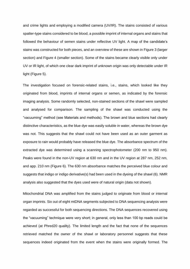
and crime lights and employing a modified camera (UV/IR). The stains consisted of various
spatter-type stains considered to be blood, a possible imprint of internal organs and stains that
followed the behaviour of semen stains under reflective UV light. A map of the candidate’s
stains was constructed for both pieces, and an overview of these are shown in Figure 3 (larger
section) and Figure 4 (smaller section). Some of the stains became clearly visible only under
UV or IR light, of which one clear dark imprint of unknown origin was only detectable under IR
light (Figure 5).
The investigation focused on forensic-related stains, i.e., stains, which looked like they
originated from blood, imprints of internal organs or semen, as indicated by the forensic
imaging analysis. Some randomly selected, non-stained sections of the shawl were sampled
and analysed for comparison. The sampling of the shawl was conducted using the
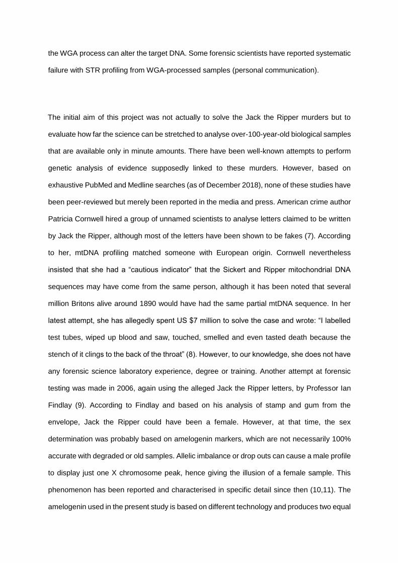
“vacuuming” method (see Materials and methods). The brown and blue sections had clearly
distinctive characteristics, as the blue dye was easily soluble in water, whereas the brown dye
was not. This suggests that the shawl could not have been used as an outer garment as
exposure to rain would probably have released the blue dye. The absorbance spectrum of the
extracted dye was determined using a scanning spectrophotometer (200 nm to 950 nm).
Peaks were found in the non-UV region at 630 nm and in the UV region at 287 nm, 252 nm,
and app. 210 nm (Figure 6). The 630 nm absorbance matches the perceived blue colour and
suggests that indigo or indigo derivative(s) had been used in the dyeing of the shawl (6). NMR
analysis also suggested that the dyes used were of natural origin (data not shown).
Mitochondrial DNA was amplified from the stains judged to originate from blood or internal
organ imprints. Six out of eight mtDNA segments subjected to DNA sequencing analysis were
regarded as successful for both sequencing directions. The DNA sequences recovered using
the “vacuuming” technique were very short; in general, only less than 100 bp reads could be
achieved (at Phred20 quality). The limited length and the fact that none of the sequences
retrieved matched the owner of the shawl or laboratory personnel suggests that these
sequences indeed originated from the event when the stains were originally formed. The

completed DNA sequences displayed an overall match for both the suspect candidate and the
victim, with their corresponding DNA sequencing results found in more than 70% of the stains
analysed. This suggests that the stains originated from a single source. In other words, the
victim’s stains are from one individual, and stains linked to the suspect are similarly from a
single person. As some of these profiles belong to living individuals, the exact nucleotide
variations from the revised Cambridge Reference Sequence are presented as variant blocks
in Figure 7. The suspected sequence from the evidence contains two blocks that could not be
determined with high confidence; however, for all other mtDNA markers, an identical profile
was produced. For the victim, an identical mtDNA profile was acquired from both the shawl
and the reference individual from the maternal line. Additionally, the mtDNA sequence for the
same region of the shawl owner and laboratory operator was analysed, and neither of these
matched the mtDNA sequences isolated from the shawl. We used the frequencies published
in EMPOP (v4/R12) to calculate the frequencies of the mtDNA profiles extracted from the
shawl for the victim and the suspect using the variations from the rCRS. The frequency for the
suspect was 1.9 x 10⁻² and for the victim was 1.3 x 10⁻³.
These frequencies are based on EMPOP database material for pure European population.
However, as a detailed census (including ethnicity) is not available for London in year 1888
these numbers might not be fully accurate. Frequency as odds finding both profiles in the same
piece of evidence is therefore 2.5 x 10⁻⁵.
When analysing the other sections of the shawl using traditional “double swab” sampling
methods, two mtDNA segments were found, originating from apparent contamination from
fresh DNA and matching with one of the reference samples who was known to handle the
shawl in the past. These samples were recovered near the corners of the shawl. This is to be
expected and has been shown in other studies, as fresh, non-fragmented DNA amplifies much
more readily than old DNA.

Shawl stains that illustrated semen-like behaviour during the initial inspection were sampled,
transferred to microscope slides and stained with modified Giemsa (Figure 8). The slides
produced this way were pre-scanned using an Aperio ScanScope, which is capable of
producing high-quality images of full microscope slides. The scanned images of the slides
were then examined remotely to locate cells suspected to contain intact nuclei. These cells
were then selected for closer inspection using LCM and transferred using the laser capture
from the microscope slides to the caps of the transfer tubes. The transfers were quality
controlled by comparing the images before and after laser capture. All transfers attempted
were judged to be successful as the cellular material disappeared from the microscope slide.
The isolated single cells were subjected to whole-genome amplification (WGA), and twelve
samples were selected for further genetic analysis. The allelic status of a selection of SNPs
linked to visual characteristics was determined using qPCR analysis. The qPCR analysis also
made possible sex determination, demonstrated in Figure 9, using an amelogeninbased assay
producing two distinct peaks in the melting curve analysis. The results suggest that the donor
of these cells is a male and has brown eyes and brown hair.
Discussion
Due to the historic value of the shawl, it was decided early on that only noninvasive/minimally
destructive methods would be applied. This decision created both limitations and challenges
for the analysis, as many of the routine approaches became unavailable with this decision.
The first challenge was to avoid surface contamination whilst ensuring that only “old” biological
material was used for genetic analysis. It is known that cellular material, including semen
heads, can attach to fabrics quite rigidly, therefore requiring harsh mechanical treatment (such
as shaking overnight in solution). In the paper presented here, neutral biological buffer and
microsampling using liquid pressure were applied to the pre-treated fabric.

The originality of the stains on the shawl was initially tested in several ways to reveal any
attempts to forge the forensic stain patterns, for example, with an acid phosphatase test that
proved negative, as would be expected for old semen stains. The shawl’s stains were also
tested for the presence of pig and horse DNA with species-specific mtDNA primers. These
reactions were all negative, implying that the stains were not artificially created with animal
blood from pigs or horses.
It is known that the PFA used for cell fixation during microscopy procedures can affect
downstream analysis requirements, especially for RNA applications. Therefore, the procedure
presented in this paper may not be directly suitable for casework where tissue type needs to
be determined using RNA-based markers. However, in this study, no problems were
encountered with DNA work from PFA samples.
The most common concerns regarding DNA analysis of old or ancient samples are the
degradation of DNA to shorter fragments and contamination from modern DNA. The
degradation can render the DNA to such short fragments that the sequence information cannot
be easily analysed using routine methods. On the other hand, this can help the investigator
differentiate modern DNA from old DNA. Another QC measure could be the abundance of the
DNA in the sample: abundant, long DNA sequences suggest modern contamination.
Deamination is another well-known, DNA-modifying type of damage. DNA repair kits and
enzymes are commercially available, but based on our prior experience with these
approaches, the idea was rejected as the repair procedures would require too much
optimisation of individual reactions, exhausting our very limited samples. Instead, we aimed to
optimise the sampling of the shawl to such a level that the modern DNA would not interfere
with the old-DNA analysis. Although the material available to us is clearly very limited, further
analysis could be attempted using next-generation sequencing combined with a droplet digital
PCR (ddPCR) method. This method can resolve DNA samples that contain DNA from multiple
or mixed sources (such as old and new samples). Additionally, in theory, the WGA-amplified
samples could be used for genomic DNA profiling. However, it is yet unknown at what level
the WGA process can alter the target DNA. Some forensic scientists have reported systematic
failure with STR profiling from WGA-processed samples (personal communication).
The initial aim of this project was not actually to solve the Jack the Ripper murders but to
evaluate how far the science can be stretched to analyse over-100-year-old biological samples
that are available only in minute amounts. There have been well-known attempts to perform
genetic analysis of evidence supposedly linked to these murders. However, based on
exhaustive PubMed and Medline searches (as of December 2018), none of these studies have
been peer-reviewed but merely been reported in the media and press. American crime author
Patricia Cornwell hired a group of unnamed scientists to analyse letters claimed to be written
by Jack the Ripper, although most of the letters have been shown to be fakes (7). According
to her, mtDNA profiling matched someone with European origin. Cornwell nevertheless
insisted that she had a “cautious indicator” that the Sickert and Ripper mitochondrial DNA
sequences may have come from the same person, although it has been noted that several
million Britons alive around 1890 would have had the same partial mtDNA sequence. In her
latest attempt, she has allegedly spent US $7 million to solve the case and wrote: “I labelled
test tubes, wiped up blood and saw, touched, smelled and even tasted death because the
stench of it clings to the back of the throat” (8). However, to our knowledge, she does not have
any forensic science laboratory experience, degree or training. Another attempt at forensic
testing was made in 2006, again using the alleged Jack the Ripper letters, by Professor Ian
Findlay (9). According to Findlay and based on his analysis of stamp and gum from the
envelope, Jack the Ripper could have been a female. However, at that time, the sex
determination was probably based on amelogenin markers, which are not necessarily 100%
accurate with degraded or old samples. Allelic imbalance or drop outs can cause a male profile
to display just one X chromosome peak, hence giving the illusion of a female sample. This
phenomenon has been reported and characterised in specific detail since then (10,11). The
amelogenin used in the present study is based on different technology and produces two equal

length amplicons from both chromosomes, thus avoiding the typical allele dropout by
preferential amplification of the shorter allele. Furthermore, as discussed before, most Jack
the Ripper letters have been shown to be fakes, and two people have been convicted of forging
some of them (Maria Coroner and Miriam Howells). As both are female, the forensic trace
DNA would indicate female as well. Additionally, in the past, postal officials were often females,
and quite normally they would seal the stamps and envelopes for the customers using their
own saliva. Our study initially started as an academic exercise, which later evolved to a test
case against one of the most well-known suspects, as identified by multiple persons linked to
Scotland Yard.
LCM is a method that has been traditionally used for tumour samples in pathology research.
Although the first report of successful WGA from single cells was published in 1992, in some
cases, LCM has been successfully combined with WGA only after fairly extensive optimisation
(12,13). LCM is not routinely used in forensics and to our knowledge, it has not been attempted
from WGA-amplified single-cell aged samples. Since our initial reports in 2014 and 2015, other
research groups have successfully applied similar approaches for more recent forensic cases
(14).
The SNP analysis was performed from the amplified genomic DNA derived from the semen.
A selection of qPCR assays developed in-house were used to generate an “Irisplex”-type
panel using previously described targets (15-17). The phenotypic characteristics were
deduced from these nucleotide allele data.
Conclusions
In summary, we present in this paper the most systematic and most advanced genetic analysis
to date regarding the Jack the Ripper murders and show that the presence of mtDNA on the
shawl matches the female victim’s mtDNA derived from stains on it and that mtDNA also on
the shawl matches the suspect candidate’s mtDNA. Furthermore, both are on the same piece
of evidence and originate from specific, forensically relevant stains that are in concordance
with Jack the Ripper’s modus operandi. The results clearly suggest that the owner or
laboratory personnel have not contributed to the samples; hence, surface contamination can
be successfully avoided by using the novel technique presented in this paper. According to
the SWGDAM 2013 guidelines, if samples have two or more nucleotide position differences,
they can be excluded as coming from the same source or maternal lineage, except when
heteroplasmy is encountered (18). The mtDNA sequencing results are compiled in Figure 7
as variation blocks for easy readability. There are several reasons why we wanted to use this
graphical format. First, we expect this paper to be interesting to forensic scientists but also to
the general public, especially for those interested in true crime. Second, due to the restrictions
set by the Data Protection Act, detailed nucleotide-level DNA information of living individuals
should not be published.
During the analysis of the shawl, the theory that the shawl was not in fact property of the victim
but belonged to the murderer was strengthened by the fact that the blue indigo dye in the floral
parts of the silk shawl was water soluble. Thus, this expensive silk shawl could not have been
used as an everyday outer garment by the victim who reportedly had a very low income and
was constantly struggling to afford accommodation.
One of the strengths of this paper is the demonstration of the use of aged single cells as a
source of genomic DNA. This was taken further by using in-house assays for amelogenin sex
determination and phenotypic SNP markers. The results were in full accordance with one of
the very few witness statements considered reliable: a male with brown eyes and brown hair.
Although these characteristics are surely not unique, they fully support our hypothesis. We
have no reliable information on how common these phenotypic features were with males in
London in 1888, but at the moment, blue eyes are more common than brown in England.
The approaches presented in this paper should be useful for other similar cases where there
is a high risk of contamination. Although we have demonstrated these techniques in the
context of historic murders, they should be directly transferable to more modern cases with
similar issues, and some police forces have already adapted them.
Acknowledgements
Dr. Haggart provided technical help with the Laser Capture Microdissection and Dr S McColl
with presumptive blood testing.
References
1. Eckert WG. The Whitechapel murders: the case of Jack the Ripper. Am J Forensic
Med Pathol 1981;2(1):53–60.
2. Wolf G. A kidney from hell? A nephrological view of the Whitechapel murders in 1888.
Nephrol Dial Transplant 2008;23(10):3343–9.
3. Latest details of the Whitechapel murders. The Illustrated Police News [London] 22
September 1888, Front page.
4. Pang BC, Cheung BK. Double swab technique for collecting touched evidence. Leg
Med (Tokyo) 2007;9(4):181–4.
5. Andrews RM, Kubacka I, Chinnery PF, Lightowlers RN, Turnbull DM, Howell N.
Reanalysis and revision of the Cambridge reference sequence for human mitochondrial DNA.
Nat Genet 1999;23(2):147.
6. Rondão R, Seixas de Melo J, Schaberle FA, Voss G. Excited state characterization of
a polymeric indigo. Phys Chem Chem Phys 2012;14(5):1778–83.

7. Cornwell P. Crochet work and flowers. In: Cornwell. Portrait of a killer: Jack the Ripper
– case closed. New York, NY: Putnam Publishing Group, 2002;168–74.
8. Patricia Cornwell: I spent $7 million solving the Jack the Ripper case, 2017 Feb 24;
https://www.telegraph.co.uk/women/life/patricia-cornwell-spent-7-million-solving-jack-
rippercase/ (accessed November 15, 2018).
9. Jack the Ripper ‘may have been female’; 2006 May 17;
https://www.smh.com.au/national/jack-the-ripper-may-have-been-female-
20060517gdnkai.html (accessed November 15, 2018).
10. Wang J, McCord B. The application of magnetic bead hybridization for the recovery
and STR amplification of degraded and inhibited forensic DNA. Electrophoresis
2011;32(13):1631–8.
11. Fattorini P, Previderè C, Sorçaburu-Cigliero S, Marrubini G, Alù M, Barbaro AM, et al.
The molecular characterization of a depurinated trial DNA sample can be a model to
understand the reliability of the results in forensic genetics. Electrophoresis
2014;35(2122):3134–44.
12. Zhang L, Cui X, Schmitt K, Hubert R, Navidi W, Arnheim N. Whole genome
amplification from a single cell: implications for genetic analysis. Proc Natl Acad Sci U S A
1992;89(13):5847–51.
13. Aaltonen KE, Ebbesson A, Wigerup C, Hedenfalk I. Laser capture microdissection
(LCM) and whole genome amplification (WGA) of DNA from normal breast tissue -
optimization for genome wide array analyses. BMC Research Notes 2011;4(1):69.
14. DiVA portal. Mixed DNA profiles from single-donors, 2015;
http://www.divaportal.org/smash/record.jsf?pid=diva2%3A853689&dswid=-9427
(accessed December 21, 2018).

15. Duffy DL, Montgomery GW, Chen W, Zhao ZZ, Le L, James MR et al. A three-
singlenucleotide polymorphism haplotype in intron 1 of OCA2 explains most human
eye-color variation. Am J Hum Genet 2007;80(2):241–52.
16. PoWpiech E, Draus-Barini J, Kupiec T, Wojas-Pelc A, Branicki W. Gene-gene
interactions contribute to eye colour variation in humans. J Hum Genet
2011;56(6):447–55.
17. Walsh S, Liu F, Wollstein A, Kovatsi L, Ralf A, Kosiniak-Kamysz A et al. The HIrisPlex
system for simultaneous prediction of hair and eye colour from DNA. Forensic Sci
Int Genet 2013;7(1):98–115.
18. Butler JM. SWGDAM mitochondrial DNA interpretation guidelines. In: Advanced topics
in forensic DNA typing: interpretation. San Diego, CA: Academic Press, 2012;407–8.

TABLE 1—List of human mtDNA primer combinations used for sequencing. A total of 8 prime
sets were used to sequence the target region in both directions.
L48 R159 F120 H285
F220 R377
L15997 R16153
L16097 R16233
F16112 R16322
L16190 R16322 F16268 H16401
TABLE 2—Primer sequences of the
mtDNA sequencing primers used.
Primer name Primer sequence Region
F120 CGCAGTATCTGTCTTTGATTCC HV2 of mtDNA
F16112 CACCATGAATATTGTACGGT HV1 of mtDNA
F16268 CACTAGGATACCAACAAACC HV1 of mtDNA

F220 TGCTTGTAGGACATAATAAT HV2 of mtDNA
H16401 TGATTTCACGGAGGATGGTG HV1 of mtDNA
H285 GGGGTTTGGTGGAAATTTTTTG HV2 of mtDNA
L15997 CACCATTAGCACCCAAAGCT HV1 of mtDNA
L16097 TACATTACTGCCAGCCACCA HV1 of mtDNA
L16190 CCCCATGCTTACAAGCAAGT HV1 of mtDNA
L48 CTCACGGGAGCTCTCCATGC HV2 of mtDNA
R159 AAATAATAGGATGAGGCAGGAATC HV2 of mtDNA
R16153 CAGGTGGTCAAGTATTTATGG HV1 of mtDNA
R16233 TGATAGTTGAAGGTTGATTGCTGT HV1 of mtDNA
R16322 TGGCTTTATGTACTATGTAC HV1 of mtDNA
R377 GTGTTAGGGTTCTTTGTTTT HV2 of mtDNA
TABLE 3—List of primers used for SNP analysis. Ancestral allele indicated with a star (*).
SNP ID Main characteristics Alleles Chromosome
RS12913832 Blue eyes A*/G 15
RS1805005 Blond hair / fair skin G*/T 16

RS1805006 Melanoma susceptibility C*/A/G 16
RS1805007 Red hair / fair skin C*/G/T 16
RS1805008 Red hair / fair skin C*/T 16
RS1805009 Red hair / fair skin G*/A/C 16
RS28777 Black / Blond hair C*/A 5
RS12821256 Blond / Brown hair T*/C 12
RS1800407 Blue / Non-blue eyes C*/T 15
RS12203592 Brown hair C*/T 6
RS2228479 Red hair / fair skin G*/A/C 16
Figure Legends
FIG. 1—Letter of provenance from the Great Great Nephew of the Acting Sergeant Amos
Simpson who originally recovered the shawl from the scene of the crime. Some details have
been blurred after scanning of the document, for protection of personal details.

FIG. 2—Images of the shawl parts. Upper left: largest piece of the shawl with the blue and
brown sections. Lower left: the floral detail on the shawl. Right: smaller piece of the shawl from
the blue side.
FIG. 3—Overview map of the most prominent and forensically relevant stains and features on
the larger piece of the shawl. 1: Large stain, non-homogenous with variable intensities, visible
by eye. 2: Another larger stain, less prominent but similar features as stain 1. 3: Two smaller
stains, visible with cross-polarising light and UV. 4: A set of smaller stains, with features
compatible with blood stains.
FIG. 4—Overview map of the forensically relevant stains and features on the smaller piece of
the shawl. 1: Section with candidates for semen stains. 2: Unidentified stains absorbing UV.
3: Smaller fluorescent stains compatible with semen stains.
FIG. 5—Stain region of unknown origin, which was invisible to the naked eye but very clear
under IR light. The centre part of the stain was surrounded by clear but less IR visible stain
“rings”.
FIG. 6—Absorbance of the water-soluble stain extracted from the blue section of the shawl.
The absorbance peak in the visible region is app. 630 nm. The vertical line indicates 575 nm
absorbance.
FIG. 7— Sequencing results of mtDNA, presented as graphical blocks when deviations from
the Human revised Cambridge reference sequence (rCRS) have been recorded. Colour

coding is used to highlight the results (victim = blue, suspect = red, owner = green and
laboratory operator = gray).
FIG. 8—Examples of the cells recovered from the shawl. Modified Giemsa staining, 400x.
The “bubbling” in the images is due to the staining method used.
FIG. 9—A qPCR melting curve analysis of the human amelogenin gene. The DNA sequence
differences of the AMELX and AMELY genes produce two peaks with different melting
temperatures. Panel 1 demonstrates the result from WGA-amplified cells, two peaks
demonstrating the simultaneous presence of X and Y chromosomes. Panel 2 is a control
female sample, and Panel 3 is a negative control, showing only the formation of primer dimers.