formaldehyde reference exposure levels - oehha · pdf filedraft tsd for noncancer rels, srp4...
TRANSCRIPT

Draft TSD for Noncancer RELs, SRP4 September 2008
Appendix D Formaldehyde 1
Formaldehyde Reference Exposure Levels DRAFT
(Methanal, oxomethane, methylene oxide)
CAS 50-00-0
CH2 O 1. Summary The non-cancer adverse health effects of formaldehyde are largely a manifestation of its ability to irritate mucous membranes. As a result of its solubility in water and high reactivity, formaldehyde is efficiently absorbed into the mucus layers protecting the eyes and respiratory tract where it rapidly reacts, leading primarily to localized irritation. Acute high exposure may lead to eye, nose and throat irritation, and in the respiratory tract, nasal obstruction, pulmonary edema and dyspnea. Prolonged or repeated exposures have been associated with allergic sensitization, respiratory symptoms (coughing, wheezing, shortness of breath), histopathological changes in respiratory epithelium, and decrements in lung function. Children, especially those with diagnosed asthma, may be more likely to show impaired pulmonary function and symptoms than are adults following chronic exposure to formaldehyde. The studies reviewed for this document include those published through the Spring of 2008.
1.1 Formaldehyde Acute REL Reference Exposure Level 55 μg/m³ (44 ppb) Critical effect(s) Mild and moderate eye irritation Hazard Index target(s) Eye irritation
1.2 Formaldehyde 8-Hour REL Reference Exposure Level 9 μg/m³ (7 ppb) Critical effect(s) Nasal obstruction and discomfort, lower airway discomfort, and eye irritation Hazard Index target(s) Respiratory
1.3 Formaldehyde Chronic REL Reference Exposure Level 9 μg/m³ (7 ppb) Critical effect(s) Nasal obstruction and discomfort, lower airway discomfort, and eye irritation Hazard Index target(s) Respiratory

Draft TSD for Noncancer RELs, SRP4 September 2008
Appendix D Formaldehyde 2
2. Physical & Chemical Properties (ATSDR, 1999)
Description Colorless gas Molecular formula CH2O Molecular weight 30.03 g/mol Density 0.815 g/L @ -20° C Boiling point -19.5° C Melting point -92° C Vapor pressure 3883 mm Hg @ 25° C Flashpoint 300° C Explosive limits 7% - 73% Solubility soluble in water, alcohol, ether and other polar solvents Odor threshold 0.05-0.5 ppm Metabolites formic acid Conversion factor 1 ppm in air = 1.24 mg/m3 @ 25° C
3. Occurrence and Major Uses Formaldehyde has four major applications: as an intermediate in the manufacture of melamine, polyacetal, and phenolic resins; as an intermediate in the production of industrial chemicals; as a bactericide or fungicide; and as a component in the manufacture of end-use consumer products. Phenol-formaldehyde resins are used in the production of plywood, particleboard, foam insulation, and a wide variety of molded or extruded plastic items. Formaldehyde is also used as a preservative, a hardening and reducing agent, a corrosion inhibitor, a sterilizing agent, and in embalming fluids. Indoor sources include upholstery, permanent press fabrics, carpets, pesticide formulations, urea-formaldehyde foam insulation, and cardboard and paper products. Outdoor sources include emissions from fuel combustion (motor vehicles), industrial fuel combustion (power generators), oil refining processes, and other uses (copper plating, incinerators, etc.). The largest portion of outdoor ambient formaldehyde results from photochemical oxidation of a number of reactive organic gases in the atmosphere (CARB, 2006). According to the California Toxics Inventory (CARB, 2005a), the mean statewide ambient level of formaldehyde in 2004 was 2.69 ppb, with the highest levels (3.76 ppb) reported for the South Coast Air Basin. The California Air Resources Board (CARB) reported statewide emissions of 20,251 tons from stationary and mobile sources (CARB, 2005b). 4. Metabolism Inhaled formaldehyde reacts rapidly at the site of contact and is efficiently absorbed in the respiratory tract. A portion of the formaldehyde entering the mucous layer of the respiratory tract is reversibly hydrated to methylene glycol. Both the hydrated and free formaldehyde may be absorbed into the epithelial layer where formaldehyde may bind reversibly to glutathione to form S-hydroxymethylglutathione. This in turn is oxidized to S-formylglutathione by formaldehyde dehydrogenase. Hydrolysis of S-formylglutathione yields formate and glutathione. Formic acid may be eliminated in urine and feces, or dehydrogenated to CO2 and exhaled. The presence of glutathione and formaldehyde dehydrogenase in epithelial cells of the

Draft TSD for Noncancer RELs, SRP4 September 2008
Appendix D Formaldehyde 3
respiratory tract varies with location and influences the amount of formaldehyde reaching the blood. While glutathione-bound formaldehyde is rapidly metabolized, free formaldehyde in cells can form DNA-protein cross-links (Franks, 2005). Formaldehyde dehydrogenase (ADH3), although central to the metabolism of formaldehyde, has a broad specificity that includes the structurally related compound, S-nitrosoglutathione (GSNO), an endogenous bronchodilator and reservoir of nitric oxide (NO) activity (Jensen et al., 1998). In cultured cells, formaldehyde appears to trigger ADH3-mediated GSNO reduction by enzyme-bound cofactor recycling (Staab et al., 2008). As shown in Figure 1, the S-hydroxymethylglutathione (HMGSH) formed spontaneously from formaldehyde and glutathione is oxidized by ADH3 with the formation of NADH that may then participate in the ADH3-mediated reduction of GSNO (Thompson and Grafstrom, 2008). (Because of its participation in this reaction, ADH3 is also known as GSNO reductase.) This reductive pathway results in low levels of GSNO that in turn stimulate the production and activity of 5-lipoxygenase, the rate-limiting enzyme in the synthesis of powerful bronchoconstrictors, the cysteinyl leukotrienes. On the other hand, high levels of GSNO inhibit this enzyme and thus the synthesis of the bronchoconstrictors (Zaman et al., 2006). Up-regulation of the degradation of GSNO has been demonstrated in mouse lung following inhalation of formaldehyde (Yi et al., 2007), while low levels of GSNO in the lungs have been associated with severe asthma attacks in children (Gaston et al., 1998) and airway hyperactivity in mice (Que et al., 2005). These results suggest that the potential association of formaldehyde exposure with asthma-like respiratory symptoms is in part due to its effects on NO via the enhanced degradation of GSNO. Nitric oxide has multiple functions in the lungs, from its participation in the regulation of airway and vascular tone to mucin secretion and mucociliary clearance (Reynaert et al., 2005). The dysregulation of NO by formaldehyde helps to explain the variety and variability in the toxic manifestations following formaldehyde inhalation.

Draft TSD for Noncancer RELs, SRP4 September 2008
Appendix D Formaldehyde 4
Figure 1 Formaldehyde Driven Reduction of GSNO
GSNO
L-Arg
NOS
NO + GSH
S-NO-Protein bronchiole dilation
S-formyl GSH
HMGSH CH2O + GSH
NAD+
ADH3NADH
glutathione sulfonimide
5-lipoxygenase cysteinyl leukotriene (bronchoconstrictor)High GSNO
Low GSNO
X
Oxidation of the glutathione conjugate of formaldehyde, HMGSH, by ADH3 generates NADH that drives the reduction of GSNO, also by ADH3, thereby reducing the nitric oxide available for bronchiole dilation. Low GSNO levels stimulate, but high GSNO levels inhibit 5-lipoxygenase production of cysteinyl leukotriene.
5. Acute Toxicity of Formaldehyde The acute effects of formaldehyde exposure appear to be largely a result of its irritant properties. However, some individuals experience symptoms following acute exposures that are a result of previous sensitization following acute high formaldehyde exposure, or long term low level exposures. For this reason, some of the studies included in this section describe manifestations of toxicity in which acute exposure was the precipitating event but in which the contribution of previous exposures or sensitization is unknown. Sensitization may be of an immunological nature with the development of formaldehyde-specific IgE or IgG (e.g. Thrasher et al. 1987), or may be neurologically mediated with involvement of the hypothalamic/pituitary/adrenal axis (Sorg et al., 2001a,b). In addition, genetic variation among individuals in the alcohol dehydrogenases mentioned above affects individual responses to formaldehyde. This is especially germane to studies in which the effects include symptoms such as bronchoconstriction and airway hyperreactivity, and in which there is unexpected individual variation.
Many of the studies described in this document have evaluated the relationship between formaldehyde inhalation and clinically-diagnosed asthma or asthma-like symptoms. Asthma is a chronic disease of airway obstruction resulting in variable airflow that has classically been considered to involve both airway inflammation and airway hyperresponsiveness. Outwardly asthma manifests as a characteristic cough, wheeze, and shortness of breath due to spasmodic contractions of the bronchi and mucus hypersecretion. These symptoms may or may not reflect an underlying allergic response. As shown in the study by Que et al. (2005), the hyperresponsiveness and the inflammation are not necessarily coupled. Although the RELs presented in this document are not based on studies that used asthma as the critical endpoint, uncertainty factors were applied in the REL estimates to explicitly consider the potential of formaldehyde to cause or exacerbate asthma-like wheeze and cough symptoms, especially in asthmatic children. We have therefore included discussion of recent work that provides a

Draft TSD for Noncancer RELs, SRP4 September 2008
Appendix D Formaldehyde 5
biochemical mechanism by which formaldehyde exposure is linked to at least one symptom of asthma, bronchoconstriction. The bronchoconstrictive effects of formaldehyde exposure may be the partially responsible for the lower airway discomfort reported in the study upon which the 8-hour and chronic RELs are based.
5.1 Acute Toxicity to Adult Humans In small human studies, exposure to formaldehyde (1-3 ppm) has resulted in eye and upper respiratory tract irritation (Weber-Tschopp et al., 1977; Kulle et al., 1987). Most people cannot tolerate exposures to more than 5 ppm formaldehyde in air; above 10-20 ppm symptoms become severe and shortness of breath occurs (Feinman, 1988). High concentrations of formaldehyde may result in nasal obstruction, pulmonary edema, choking, dyspnea, and chest tightness (Porter, 1975; Solomons and Cochrane, 1984). A few human case studies report severe pulmonary symptoms. A medical intern with known atopy and exposure to reportedly high (but unspecified) levels of formaldehyde over a period of 1 week developed dyspnea, chest tightness, and edema, following a subsequent 2 hour exposure to formaldehyde (Porter, 1975). Five workers exposed to formaldehyde from newly installed urea-formaldehyde chipboard in a poorly ventilated basement experienced intolerable eye and upper respiratory tract irritation, choking, marked dyspnea, and nasal obstruction (Solomons and Cochrane, 1984). However, the concentration of formaldehyde and the contribution of other airborne chemicals were unknown in both reports. Numerous acute controlled and occupational human exposure studies have been conducted with both asthmatic and normal subjects to investigate formaldehyde’s irritative and pulmonary effects (Frigas et al., 1984; Sheppard et al., 1984; Sauder et al., 1986; Schachter et al., 1986; Kulle et al., 1987; Sauder et al., 1987; Schachter et al., 1987; Witek et al., 1987; Uba et al., 1989; Harving et al., 1990; Akbar-Khanzadeh et al., 1994). Short exercise sessions during exposure on a bicycle ergometer were included in some of the studies. Concentrations of formaldehyde in the human exposure studies ranged as high as 3 ppm for up to 3 hours. The major findings in these studies were mild to moderate eye and upper respiratory tract irritation typical of mild discomfort from formaldehyde exposure. Chemosensory irritation and subjective symptoms following exposure to formaldehyde at concentrations relevant to the workplace were examined by Lang et al. (2008) in 11 male and 10 female volunteers. Each subject was exposed for 4 hours to a randomized sequence of ten exposure conditions. These included exposures at concentrations of 0, 0.15, 0.3 and 0.5 ppm, exposures at 0.3 and 0.5 ppm that included four transient peak exposures at 0.6 and 1.0 ppm, respectively, and exposures in the presence of 10 ppm ethyl acetate of 0, 0.3, 0.5, and 0.5 ppm with 1.0 ppm peaks. Objective measures of irritation included conjunctival redness, blinking frequency, nasal flow resistance, pulmonary function, and reaction times. The participant’s subjective evaluation of physical and mental wellbeing was assessed by questionnaire before, during and after each day’s exposure. To assess the potential influence of personality traits on subjective responses, each subject’s positive or negative affectivity was evaluated. Conjunctival irritation and blinking frequency were significantly elevated only with exposure to 0.5 ppm with peaks of 1.0 ppm (p < 0.05). The authors considered this level to be a LOAEL, and 0.5 ppm
Draft TSD for Noncancer RELs, SRP4 September 2008
Appendix D Formaldehyde 6
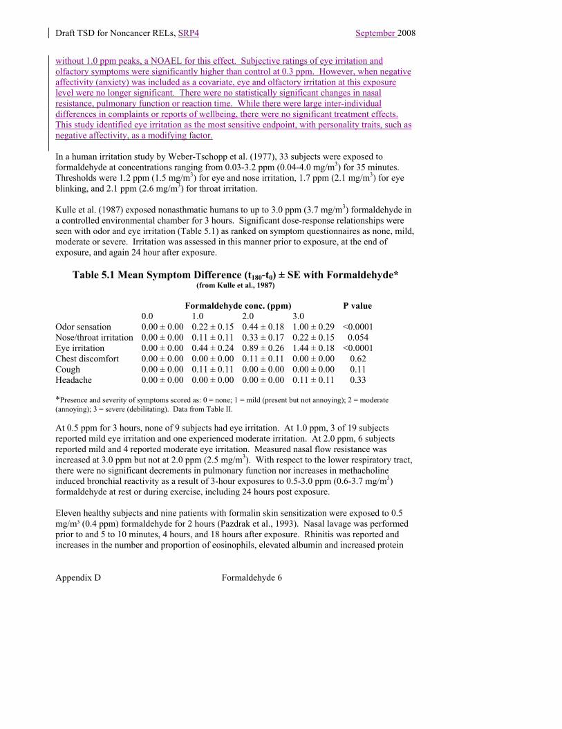
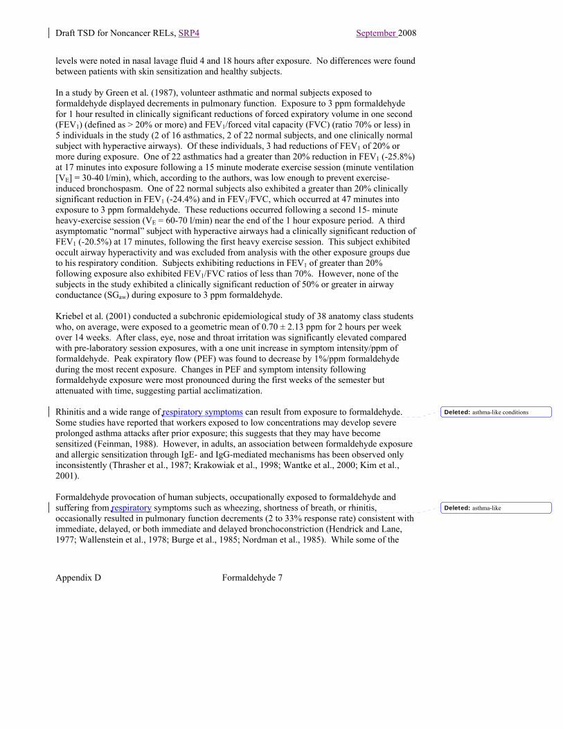
without 1.0 ppm peaks, a NOAEL for this effect. Subjective ratings of eye irritation and olfactory symptoms were significantly higher than control at 0.3 ppm. However, when negative affectivity (anxiety) was included as a covariate, eye and olfactory irritation at this exposure level were no longer significant. There were no statistically significant changes in nasal resistance, pulmonary function or reaction time. While there were large inter-individual differences in complaints or reports of wellbeing, there were no significant treatment effects. This study identified eye irritation as the most sensitive endpoint, with personality traits, such as negative affectivity, as a modifying factor. In a human irritation study by Weber-Tschopp et al. (1977), 33 subjects were exposed to formaldehyde at concentrations ranging from 0.03-3.2 ppm (0.04-4.0 mg/m3) for 35 minutes. Thresholds were 1.2 ppm (1.5 mg/m3) for eye and nose irritation, 1.7 ppm (2.1 mg/m3) for eye blinking, and 2.1 ppm (2.6 mg/m3) for throat irritation. Kulle et al. (1987) exposed nonasthmatic humans to up to 3.0 ppm (3.7 mg/m3) formaldehyde in a controlled environmental chamber for 3 hours. Significant dose-response relationships were seen with odor and eye irritation (Table 5.1) as ranked on symptom questionnaires as none, mild, moderate or severe. Irritation was assessed in this manner prior to exposure, at the end of exposure, and again 24 hour after exposure.
Table 5.1 Mean Symptom Difference (t180-t0) ± SE with Formaldehyde* (from Kulle et al., 1987)
Formaldehyde conc. (ppm) P value
0.0 1.0 2.0 3.0 Odor sensation 0.00 ± 0.00 0.22 ± 0.15 0.44 ± 0.18 1.00 ± 0.29 <0.0001Nose/throat irritation 0.00 ± 0.00 0.11 ± 0.11 0.33 ± 0.17 0.22 ± 0.15 0.054 Eye irritation 0.00 ± 0.00 0.44 ± 0.24 0.89 ± 0.26 1.44 ± 0.18 <0.0001Chest discomfort 0.00 ± 0.00 0.00 ± 0.00 0.11 ± 0.11 0.00 ± 0.00 0.62 Cough 0.00 ± 0.00 0.11 ± 0.11 0.00 ± 0.00 0.00 ± 0.00 0.11 Headache 0.00 ± 0.00 0.00 ± 0.00 0.00 ± 0.00 0.11 ± 0.11 0.33 *Presence and severity of symptoms scored as: 0 = none; 1 = mild (present but not annoying); 2 = moderate (annoying); 3 = severe (debilitating). Data from Table II. At 0.5 ppm for 3 hours, none of 9 subjects had eye irritation. At 1.0 ppm, 3 of 19 subjects reported mild eye irritation and one experienced moderate irritation. At 2.0 ppm, 6 subjects reported mild and 4 reported moderate eye irritation. Measured nasal flow resistance was increased at 3.0 ppm but not at 2.0 ppm (2.5 mg/m3). With respect to the lower respiratory tract, there were no significant decrements in pulmonary function nor increases in methacholine induced bronchial reactivity as a result of 3-hour exposures to 0.5-3.0 ppm (0.6-3.7 mg/m3) formaldehyde at rest or during exercise, including 24 hours post exposure. Eleven healthy subjects and nine patients with formalin skin sensitization were exposed to 0.5 mg/m³ (0.4 ppm) formaldehyde for 2 hours (Pazdrak et al., 1993). Nasal lavage was performed prior to and 5 to 10 minutes, 4 hours, and 18 hours after exposure. Rhinitis was reported and increases in the number and proportion of eosinophils, elevated albumin and increased protein
Draft TSD for Noncancer RELs, SRP4 September 2008
Appendix D Formaldehyde 7
levels were noted in nasal lavage fluid 4 and 18 hours after exposure. No differences were found between patients with skin sensitization and healthy subjects. In a study by Green et al. (1987), volunteer asthmatic and normal subjects exposed to formaldehyde displayed decrements in pulmonary function. Exposure to 3 ppm formaldehyde for 1 hour resulted in clinically significant reductions of forced expiratory volume in one second (FEV1) (defined as > 20% or more) and FEV1/forced vital capacity (FVC) (ratio 70% or less) in 5 individuals in the study (2 of 16 asthmatics, 2 of 22 normal subjects, and one clinically normal subject with hyperactive airways). Of these individuals, 3 had reductions of FEV1 of 20% or more during exposure. One of 22 asthmatics had a greater than 20% reduction in FEV1 (-25.8%) at 17 minutes into exposure following a 15 minute moderate exercise session (minute ventilation [VE] = 30-40 l/min), which, according to the authors, was low enough to prevent exercise-induced bronchospasm. One of 22 normal subjects also exhibited a greater than 20% clinically significant reduction in FEV1 (-24.4%) and in FEV1/FVC, which occurred at 47 minutes into exposure to 3 ppm formaldehyde. These reductions occurred following a second 15- minute heavy-exercise session (VE = 60-70 l/min) near the end of the 1 hour exposure period. A third asymptomatic “normal” subject with hyperactive airways had a clinically significant reduction of FEV1 (-20.5%) at 17 minutes, following the first heavy exercise session. This subject exhibited occult airway hyperactivity and was excluded from analysis with the other exposure groups due to his respiratory condition. Subjects exhibiting reductions in FEV1 of greater than 20% following exposure also exhibited FEV1/FVC ratios of less than 70%. However, none of the subjects in the study exhibited a clinically significant reduction of 50% or greater in airway conductance (SGaw) during exposure to 3 ppm formaldehyde. Kriebel et al. (2001) conducted a subchronic epidemiological study of 38 anatomy class students who, on average, were exposed to a geometric mean of 0.70 ± 2.13 ppm for 2 hours per week over 14 weeks. After class, eye, nose and throat irritation was significantly elevated compared with pre-laboratory session exposures, with a one unit increase in symptom intensity/ppm of formaldehyde. Peak expiratory flow (PEF) was found to decrease by 1%/ppm formaldehyde during the most recent exposure. Changes in PEF and symptom intensity following formaldehyde exposure were most pronounced during the first weeks of the semester but attenuated with time, suggesting partial acclimatization. Rhinitis and a wide range of respiratory symptoms can result from exposure to formaldehyde. Some studies have reported that workers exposed to low concentrations may develop severe prolonged asthma attacks after prior exposure; this suggests that they may have become sensitized (Feinman, 1988). However, in adults, an association between formaldehyde exposure and allergic sensitization through IgE- and IgG-mediated mechanisms has been observed only inconsistently (Thrasher et al., 1987; Krakowiak et al., 1998; Wantke et al., 2000; Kim et al., 2001). Formaldehyde provocation of human subjects, occupationally exposed to formaldehyde and suffering from respiratory symptoms such as wheezing, shortness of breath, or rhinitis, occasionally resulted in pulmonary function decrements (2 to 33% response rate) consistent with immediate, delayed, or both immediate and delayed bronchoconstriction (Hendrick and Lane, 1977; Wallenstein et al., 1978; Burge et al., 1985; Nordman et al., 1985). While some of the
Deleted: asthma-like conditions
Deleted: asthma-like

Draft TSD for Noncancer RELs, SRP4 September 2008
Appendix D Formaldehyde 8
concentrations of formaldehyde that elicited a positive response following provocation tests (6 to 20.7 ppm) were quite high, the authors of these studies suggested that formaldehyde-induced bronchial hyperreactivity is due to specific sensitization to the gas. However, none of these studies was able to detect antibodies to formaldehyde which would support that sensitization to formaldehyde occurs through an immunologic pathway. Alternatively, the wheezing and shortness of breath maybe related to the formaldehyde-stimulated depletion of the bronchodilator, GSNO, in the airways. In controlled studies with asthmatics from urea-formaldehyde insulated homes, formaldehyde concentrations equal to or greater than those found in indoor environments have not resulted in hematologic or immunologic abnormalities. These tests include: blood count and differential, erythrocyte sedimentation rate; lymphocyte subpopulations (E-rosetting, T3, T4, T8, B73.1, Fc receptor positive lymphocytes and large granular lymphocytes); lymphocyte response to phytohemagglutinin and formalin-treated red blood cells; serum antibody against the Thomsen-Friedenrich RBC antigen and against formalin-RBC; and natural killer, interferon-boosted natural killer, and antibody-dependent cell-mediated cytotoxicity (Pross et al., 1987). While six of the studies cited above reported decrements in lung function associated with short-term formaldehyde exposure among at least some of the asthmatic subjects, a number of other exposure studies of patients with asthma have failed to demonstrate that exposure to formaldehyde results in onset or aggravation of the patients’ asthmatic symptoms (Sheppard et al., 1984; Sauder et al., 1987; Harving et al., 1990; Krakowiak et al., 1998). The effects of formaldehyde on asthmatics may be dependent on previous, repeated exposure to formaldehyde. Burge et al. (1985) found that 3 out of 15 occupationally exposed workers challenged with formaldehyde vapors at concentrations from 1.5 ppm to 20.6 ppm for brief durations exhibited late asthmatic reactions. Six other subjects had immediate asthmatic reactions likely due to irritant effects. Asthmatic responses (decreased PEF, FVC, and FEV1) were observed in 12 occupationally-exposed workers challenged with 2.0 ppm (2.5 mg/m3) formaldehyde (Nordman et al., 1985). Similarly, asthmatic responses were observed in 5 of 28 hemodialysis workers occupationally exposed to formalin and challenged with formaldehyde vapors (concentration not measured) (Hendrick and Lane, 1977). In asthmatics not occupationally exposed to formaldehyde, Sheppard et al. (1984) found that a 10-minute challenge with 3 ppm formaldehyde coupled with moderate exercise did not induce significant changes in airway resistance or thoracic gas volume. Gorski et al. (1992) evaluated the production of active oxygen species by neutrophils in 18 persons exposed to 0.5 mg/m3 formaldehyde for 2 hours. All 13 subjects who had allergic contact dermatitis (tested positive to formaldehyde in skin patch) exhibited significantly higher chemiluminescence of granulocytes isolated from whole blood 30 minutes and 24 hours post-exposure than the individuals who were not formaldehyde sensitive. Thus, the immune cellular response of skin-sensitized individuals to an inhalation exposure to formaldehyde indicates increased production of active oxygen species. This is consistent with increasing evidence that endogenous or exogenous reactive oxygen and reactive nitrogen species are responsible for the airway inflammation of asthma (Sugiura and Ichinose, 2008).
Deleted: The significance of this result is unclear but may have repercussions for toxicological effects mediated by active oxygen species.

Draft TSD for Noncancer RELs, SRP4 September 2008
Appendix D Formaldehyde 9
In addition to its effects on the respiratory tract, the irritant properties of formaldehyde also manifest as ocular irritation. In an anatomy dissecting laboratory, formaldehyde levels were found to peak at 0.62 ppm, with a gradual decrease to 0.11 ppm. Formaldehyde-related irritation of the eyes, nose, throat, airways and skin was reported by 59% of the students. These effects were significantly (p < 0.001) higher among wearers of contact lenses compared with students without glasses or wearing glasses (Tanaka et al., 2003). The ability of contact lenses to trap and concentrate volatile compounds, and to extend the exposure time by limiting the eye’s normal self-cleansing, may make contact lens wearers more susceptible to ocular exposure and irritation by formaldehyde.
5.2 Acute Toxicity to Infants and Children No studies of the effects of acute exposure to formaldehyde in children or young experimental animals were located. However, as noted above for adults, there is evidence that following acute exposure to formaldehyde, asthmatics and others previously sensitized to formaldehyde may be more likely to show respiratory symptoms such as wheezing, shortness of breath, rhinitis, and/or decrements in pulmonary function consistent with immediate and/or delayed bronchoconstriction (Nordman et al., 1985; Burge et al., 1985; Hendrick and Lane, 1977; Wallenstein et al., 1978). Furthermore, some asthmatics may respond with significant reductions in lung function due to the irritant effects on asthma, sensitized or not. Additionally, the depletion of the endogenous bronchodilator, GSNO, following formaldehyde exposure may be particularly important in children. Gaston et al. (1998) compared concentrations of tracheal S-nitrosothiol concentrations in eight asthmatic children in respiratory failure with those of 21 non-asthmatic children undergoing elective surgery. In asthmatic children, the metabolism of GSNO was accelerated and the mean S-nitrosothiol concentrations significantly lower compared to normal children (65 ± 45 vs 502 ± 429 nmol/l). Thus asthmatic children, with low levels of GSNO, are expected to be unusually vulnerable to any further depletion of GSNO caused by formaldehyde. The potential association between formaldehyde exposure and asthma is of special concern for children since, as noted in OEHHA (2001): “OEHHA considers asthma to impact children more than adults. Children have higher prevalence rates of asthma than do adults (Mannino et al., 1998). In addition, asthma episodes can be more severe due to the smaller airways of children, and result in more hospitalizations in children, particularly from the ages of 0 to 4 years, than in adults (Mannino et al., 1998).” Thus children, particularly asthmatic children, may be at greater risk from acute exposure to formaldehyde.
5.3 Acute Toxicity to Experimental Animals Acute exposures of experimental animals to formaldehyde are associated with changes in pulmonary function (decreased respiratory rate, increased airway reactivity and resistance) at low concentrations, while pulmonary edema and death have been reported at high concentrations. Neurochemical and neurobehavioral changes have also been observed. In 72 rats exposed to approximately 600-1,700 mg/m3 (500-1,400 ppm) formaldehyde vapor for 30 minutes, the LC50 was found to be 1,000 mg/m3 (800 ppm) (Skog, 1950). The first deaths did
Deleted: ¶Predisposing Conditions for Formaldehyde Toxicity¶¶Medical: Persons with eye, skin, respiratory, or allergic conditions (especially asthma) may be more sensitive to the effects of formaldehyde (ATSDR, 1999). Asthmatics sensitized to formaldehyde may be more sensitive to formaldehyde at low concentrations than non-sensitized individuals.¶¶

Draft TSD for Noncancer RELs, SRP4 September 2008
Appendix D Formaldehyde 10
not occur until 6 hours after cessation of exposure. Respiratory difficulty lasted several days after exposure and the last of 49 rats died after 15 days of purulent bronchitis and diffuse bronchopneumonia. Three weeks following exposure, histological examinations of the 23 surviving animals revealed bronchitis, pulmonary microhemorrhages, and edema. No changes were seen in other organs. A multispecies study by Salem and Cullumbine (1960) showed that a 10-hour exposure to 15.4 ppm (19 mg/m3) formaldehyde vapor killed 3 out of 5 rabbits, 8 of 20 guinea pigs, and 17 of 50 mice. The report stated that formaldehyde exposure resulted in delayed lethality. Alarie (1981) determined the 10 minute LC50 for formaldehyde in mice to be 2,162 ppm (95% confidence interval, 1,687-2,770 ppm). The post-exposure observation period was 3 hours. From the concentration mortality graph provided in the report, an MLE05 and BC05 of 1,440 ppm and 778 ppm, respectively, could be estimated for a 10-minute formaldehyde exposure. However, as indicated in the previous reports, delayed deaths occur with formaldehyde which suggests that the 3-hour post-exposure observation period used in this study may not have been long enough. In other lethality studies, Nagornyi et al.(1979) determined a 4-hour formaldehyde LC50 in rats and mice to be 588 mg/m3 (474 ppm) and 505 mg/m3 (407 ppm), respectively. However, the raw data for this study were not included in the report. Horton et al. (1963) observed that a 2-hour exposure of mice to 0.9 mg/l (900 mg/m3) formaldehyde resulted in deaths from massive pulmonary hemorrhage and edema, but a 2 hour exposure to 0.14 mg/l (140 mg/m3) did not produce signs of “substantial distress.” Swiecichowski et al., (1993) exposed groups of five to seven guinea pigs to 0.86, 3.4, 9.4, 31.1 ppm (1.1, 4.2, 11.6, 38.6 mg/m3) formaldehyde for 2 hours, or to 0.11, 0.31, 0.59, 1.05 ppm (0.14, 0.38, 0.73, 1.30 mg/m3) formaldehyde for 8 hours. An 8-hour exposure to levels greater than or equal to 0.3 ppm (> 0.4 mg/m3) formaldehyde was sufficient to produce a significant increase in airway reactivity. Similar effects occurred after greater than 9 ppm (> 11 mg/m3) formaldehyde for the 2-hour exposure group. Formaldehyde exposure also heightened airway smooth muscle responsiveness to acetylcholine (or carbachol) ex vivo. No inflammation or epithelial damage was seen up to 4 days after exposure. The researchers suggest that duration of exposure is important to the induction of airway hyperreactivity and that prolonged (8-hour), low-level exposures may generate abnormal physiologic responses in the airways not detectable after acute (2-hour) exposures. Male F-344 rats, 7-9 weeks old, were exposed to 0.5, 2, 6 or 15 ppm formaldehyde for 6 hours per day for 1 to 4 days (Monteiro-Riviere and Popp, 1986). Effects noted in the rat nasal respiratory epithelium with 0.5 or 2 ppm were limited to altered cilia with occasional wing-like projections on the ends of the ciliary shafts. Effects noted at 6 ppm for 1 day were autophagic vacuoles in some basal cells, neutrophils in the basal and suprabasal layers, and hypertrophy of goblet and ciliated cells. Loss of microvilli in ciliated cells was noted at all exposure concentrations.

Draft TSD for Noncancer RELs, SRP4 September 2008
Appendix D Formaldehyde 11
Rats were exposed to 0, 5, 10 or 20 ppm formaldehyde for 3 hours per day on 2 consecutive days (Boja et al., 1985). Decreased motor activity and neurochemical changes in dopamine and 5-hydroxytryptamine neurons were reported. The effects of formaldehyde inhalation on open-field behavior in mice were examined by Malek et al. (2004) 2 and 24 hours after a single 2-hour exposure to 0, 1.1, 2.3 or 5.2 ppm. Two hours after exposure there were significant decreases in rearing and in several measures of exploratory behavior, with evidence of dose-dependence in all dose groups compared with controls. At 24 hours, there were still significant differences between dosed and control mice but the dose-dependence was no longer evident. Nielson et al. (1999) analyzed the breathing patterns of Balb/c mice exposed to 0.2-13 ppm formaldehyde and found a concentration-dependent decrease in respiratory rate of 32.9%/log concentration. In the range of 0.3-4.0 ppm, the decrease in respiratory rates was attributable to sensory irritation. Above 4.0 ppm, bronchoconstriction also contributed to the decreased breathing rate. The authors suggest a NOEL of 0.3 ppm for these effects in mice. Amdur (1960) exposed groups of 4 to 18 guinea pigs to formaldehyde at 0.05, 0.31, 0.58, 1.22, 3.6, 11.0, or 49 ppm formaldehyde for one hour. Resistance to flow and lung compliance were calculated from measures of intrapleural pressure, tidal volume, and rate of flow to the lungs at the end of exposure and one hour later. Resistance and compliance were significantly different from the control level for the 0.31 ppm exposure (p<0.05) and increasingly significant at higher concentrations. One hour later, only the 49 ppm exposure remained significant (p<0.01). In addition, the tracheas of groups of 6 to 10 guinea pigs were cannulated and exposed for one hour to 0.90, 5.2, 20, or 50 ppm formaldehyde, and 1.14 or 3.6 ppm formaldehyde with 10 mg/m3 sodium chloride. With the protective effect of the trachea bypassed, the resistance and compliance changed substantially. The addition of sodium chloride further enhanced the effect, including a significant effect after one hour for the 1.14 ppm formaldehyde exposure. These results show that formaldehyde that reaches the lungs has a marked effect on airways resistance and compliance in addition to an effect on the upper airways. Riedel et al. (1996) studied the influence of formaldehyde exposure on allergic sensitization in guinea pigs. Three groups of guinea pigs (12/group) were exposed to clean air or two different formaldehyde concentrations (0.13 and 0.25 ppm) over five consecutive days. Following exposure, the animals were sensitized to allergen by inhalation of 0.5% ovalbumin (OA). Three weeks later the animals were subjected to bronchial provocation with OA and specific anti-OA-IgGl (reaginic) antibodies in serum were measured. In another group of six animals, the respiratory tract was examined histologically for signs of inflammation directly after the end of formaldehyde or clean air exposure. In the group exposed to 0.25 ppm formaldehyde, 10/12 animals were found to be sensitized to OA (positive reaction on specific provocation) vs. 3/12 animals in the control group (P < 0.01). Furthermore, compressed air measurements of specific bronchial provocation and serum anti-OA-antibodies were significantly higher in the 0.25 ppm formaldehyde group than in controls. The median for compressed air measurement was 0.35 ml for the formaldehyde-exposed group vs. 0.09 ml for the controls (p< 0.01), indicating increased bronchial obstruction. The median for the anti-OA-IgGl measured in the formaldehyde-exposed group was 13 vs. less than 10 EU in the controls, (p < 0.05), indicating enhanced sensitization.

Draft TSD for Noncancer RELs, SRP4 September 2008
Appendix D Formaldehyde 12
In the group exposed to 0.13 ppm formaldehyde, no significant difference was found compared to the control group. Histological examination found edema of the bronchial mucosa, but there was no sign of inflammation of the lower airways in formaldehyde-exposed guinea pigs. The investigators concluded that short-term exposure to a low concentration of formaldehyde (0.25 ppm) can significantly enhance sensitization to inhaled allergens in the guinea pig. As described in Section 5, the main formaldehyde-metabolizing enzyme, ADH3, also reduces the endogenous bronchodilator GSNO. To examine the role of GSNO and ADH3 (known in this study as GSNO reductase, GSNOR) in airway tone and asthma, Que et al. (2005) used wild type mice and mice with a targeted deletion of GSNOR (GSNOR-/-). Following a challenge with allergen (ovalbumin), GSNOR activity in bronchoalveolar lavage fluid from wild type mice increased significantly (p < 0.05) compared to buffer (PBS) controls, while as expected, no GSNOR activity was detected in the GSNOR-/- mice with either treatment. Levels of S-nitrosothiols (SNO) were assayed in homogenates of lung tissues from both types of mouse and found to be barely detectable with PBS treatment. However, after ovalbumin challenge, SNO levels were significantly higher (p < 0.02) in GSNOR-/- mice compared to wild type, indicating metabolism of SNOs by GSNOR under “asthmatic-like” conditions in wild type mice. Metabolism of GSNO results in a loss of bronchodilation capacity. Deletion of GSNOR had no effect on NO generation by NO synthase as there were no differences between wild type and GSNOR-/- mice in nitrate or nitrite levels regardless of treatment. To investigate the effects of deletion of GSNOR on airway hyper-responsivness, pulmonary resistance was measured at baseline (PBS) and after methacoline challenge, with and without ovalbumin treatment. At baseline, there was no difference among mouse types and treatments, while at higher methacoline levels (100-1000 µg/kg), pulmonary resistance was found to be significantly lower (p < 0.001) in GSNOR-/- mice than in wild type, presumably due to higher GSNO levels that enhance bronchodilation. Importantly, ovalbumin caused a marked increase in airway responsiveness in wild type mice but had little effect in GSNOR-/- mice. This indicates that GSNOR regulates basal airway tone as well as hyper-responsiveness to both allergen challenge and bronchoconstrictor agonists. It is also noteworthy that the total number and composition of leukocytes, levels of interleukin-13 and total serum IgE were comparable between wild type and GSNOR-/- mice. This indicates that protection from asthma in the GSNOR-/- mice is not a result of a suppressed response to allergen, and that SNOs, especially GSNO, can preserve airway patency in the presence of inflammation. Thus the inflammatory response is not linked to hyper-responsiveness as long as adequate levels of GSNO are maintained. A connection between formaldehyde and the activity of GSNOR described in the study above by Que et al., was outlined by Thompson and Grafstrom (2008) and supported by Yi et al. (2007) and Staab et al. (2008). In the study by Yi and associates, groups of 6 mice were exposed to formaldehyde at 0, 1, or 3 mg/m3 continuously for 72 hours. Following exposure, lungs were isolated to allow measurement of GSNOR mRNA levels by RT-PCR, and enzymatic activity with GSNO. Formaldehyde at 3 mg/m3 significantly increased the numbers of GSNOR transcripts compared to control (0.58 vs 0.4 GSNOR/ß actin; p < 0.05), while GSNOR reduction of GSNO showed a significant dose-dependent increase with formaldehyde concentration (p < 0.01). The stimulation of GSNO reduction by formaldehyde was also observed by Staab et al. (2008) in an in vitro study using recombinant human GSNOR. In this study, GSNO levels in buccal carcinoma cells were reduced in a dose-dependent fashion following a 1 hour exposure to

Draft TSD for Noncancer RELs, SRP4 September 2008
Appendix D Formaldehyde 13
formaldehyde in the 1-5 mM range with significance at 5 mM (p < 0.05). The results from this study support a model in which formaldehyde (as the glutathione conjugate, HMGSH) is oxidized by GSNOR (ADH3) in the presence of high levels of NAD+, producing NADH. This process was found to be accelerated by high levels of GSNO. GSNO is in turn reduced with the oxidation of NADH to form glutathione sulfonimide. Formaldehyde thus depletes cellular SNO (in the form of GSNO) which results in dysregulation of NO signaling pathways. 6. Chronic Toxicity of Formaldehyde
6.1 Chronic Toxicity to Adult Humans Formaldehyde primarily affects the mucous membranes of the upper airways and eyes. Exposed populations that have been studied include embalmers, residents in houses insulated with urea-formaldehyde foam, anatomy class students, histology technicians, wood and pulpmill workers, and asthmatics. A number of studies describing these effects have been briefly summarized below. For the sake of brevity, only the studies that best represent the given effects are presented. Formaldehyde is also a recognized carcinogen (IARC, 2006), however, this document will address only its non-carcinogenic properties. In the study chosen for determination of the 8-hour and chronic RELs, nasal obstruction and discharge, and frequency of cough, wheezing, and symptoms of bronchitis were reported in 66 workers in a formaldehyde production plant exposed for 1-36 years (mean = 10 years) to a mean concentration of 0.21 ppm (0.26 mg/m3) formaldehyde (Wilhelmsson and Holmstrom, 1992). All workers were exposed almost exclusively to formaldehyde, the concentrations of which were measured in the ambient air of the worksite with personal sampling equipment. Referents consisted of 36 office workers in a government office with exposure to a mean concentration of 0.07 ppm (0.09 mg/m3) formaldehyde, and no industrial solvent or dust exposure. Symptom data, collected by questionnaire, were separated into general and work-related, and allowed identification of individuals with atopy and mucosal hyperreactivity. The critical effects from chronic exposure to formaldehyde in this study included nasal obstruction, lower airway discomfort, and eczema or itching. The frequency of reported lower airway discomfort (intermittent cough, wheezing, or symptoms of chronic bronchitis) was significantly higher among formaldehyde-exposed vs non-exposed workers (44 vs 14%; p < 0.01) (Table 6.1). Work-related nasal discomfort also was significantly higher in the formaldehyde group (53%) compared with the referent group (3%; p < 0.001). Similarly, work-related eye discomfort was 20% in the formaldehyde group but nonexistent among referents. The significant increase in symptoms of nasal discomfort in exposed workers did not correlate with total serum IgE antibody levels. However, two exposed workers, who complained of nasal discomfort, had elevated IgE levels. The investigators concluded that formaldehyde can induce nonspecific nasal hypersensitivity.

Draft TSD for Noncancer RELs, SRP4 September 2008
Appendix D Formaldehyde 14
Table 6.1.1 Symptoms of Formaldehyde Exposure vs Reference Group (from Wilhelmsson and Holmstrom, 1992)
Formaldehyde Reference Rate difference % (n=66) % (n=36) % 95% CI
General nasal discomfort 67 25 42 24-60 Workplace nasal discomfort 53 3 50 37-63 General lower airway discomfort 44 14 30 14-47 Workplace lower airway discomfort 33 3 28 15-40 General eye discomfort 24 6 18 6-36 General skin discomfort 36 11 25 10-41 In a cross-sectional study supportive of these results, Edling et al. (1988) reported histopathological changes in nasal mucosa of workers (n=75) occupationally exposed to formaldehyde (one wood laminating plant) or formaldehyde plus wood dust (two particle board plants). Ambient formaldehyde measurements in these three composite wood processing plants between 1975 and 1983 gave a time-weighted average (TWA) of 0.1-1.1 mg/m3 (0.08- 0.89 ppm) with peaks of up to 5 mg/m3 (4 ppm). The exposed workers were compared on the basis of medical and work histories, clinical examinations and nasal biopsies to 25 workers selected with regard to age and smoking habits but without occupational formaldehyde exposure. Based on the histories, there was a high frequency of eye and upper airway symptoms among workers. Nasal symptoms (running nose and crusting) associated with formaldehyde exposure were reported in 60% of the workers, while 75% complained of lacrimation. Clinical examinations revealed grossly normal nasal mucosa in 75% of the cases while 25% had swollen or dry changes, or both, to the nasal mucosa. Histological examination (Table 6.2) revealed that only 3 of the 75 formaldehyde-exposed workers had normal, ciliated pseudostratified epithelium. Squamous metaplasia was reportedly observed in 59, while 6 showed mild dysplasia, and in 8 there was loss of ciliated cells and goblet cell hyperplasia. The histological grading showed a significantly higher score for nasal lesions among workers with formaldehyde exposure when compared with the referents (2.9 versus 1.8; p < 0.05). Exposed smokers had a higher, but non-significant, score than ex-smokers and non-smokers. While the mean exposure time was 10.5 years (range 1-39 yr), there was no discernable difference among histology scores as a function of years of employment. The histology scores were also not different between workers in the particle board plants, exposed to both formaldehyde and wood dust, and workers in the laminate plant with exposure only to formaldehyde. The authors thus attribute the pathological changes in the nasal mucosa and the other adverse effects to formaldehyde alone in the 0.1-1.1 mg/m3 range.

Draft TSD for Noncancer RELs, SRP4 September 2008
Appendix D Formaldehyde 15
Table 6.1.2 Distribution of Histological Characteristics Associated with Formaldehyde Exposure (from Edling et al., 1988)
Histological characteristic Grading score Point score Workers % Normal respiratory epithelium 0 0 3 4 Loss of ciliated cells 1 1 8 11 Mixed cuboidal/squamous epithelium, metaplasia
2 2 24 32
Stratified squamous epithelium 3 3 18 24 Keratosis 4 4 16 21 Budding of epithelium add 1 5 0 0 Mild or moderate dysplasia 6 6 6 8 Severe dysplasia 7 7 0 0 Carcinoma 8 8 0 0
Histological changes in the nasal mucosa of formaldehyde-exposed workers were also reported by Boysen et al. (1990). In this study, nasal biopses were collected from 37 workers with 5 or more years of occupational formaldehyde exposure (0.5 - > 2 ppm) and compared with age-matched, unexposed controls who otherwise had similar environmental exposures and smoking habits. Histological changes in the nasal epithelium were scored as indicated in Table 6.1.3.
Table 6.1.3 Types of Nasal Epithelia and Scoring (from Boysen et al., 1990)
Types of epithelia Histological scorePseudostratified columnar 0 Stratified cuboidal 1 Mixed stratified cuboidal/stratified squamous 2 Stratified squamous, non-keratinizing 3 Stratified squamous, keratinizing 4 Dysplasia 5
As shown by the histological scoring in Table 6.1.4 below, metaplastic changes in the nasal epithelium were more pronounced in the formaldehyde-exposed workers although this difference did not reach statistical significance.
Table 6.1.4 Histological Scores of Nasal Epithelia
Histological score No 0 1 2 3 4 5 MeanExposed 37 3 16 5 9 1 3 1.9 Controls 37 5 17 10 5 0 0 1.4
Rhinoscopical examination revealed hyperplastic nasal mucosa in 9 of 37 formaldehyde-exposed workers but in only 4 of the controls. In addition, the incidence of subjective nasal complaints

Draft TSD for Noncancer RELs, SRP4 September 2008
Appendix D Formaldehyde 16
was significantly (p < 0.01) higher in the exposed group. While the small size of this study, and the small amount of the nasal mucosa accessible to biopsy limited its ability to detect formaldehyde- related histopathology, the results are consistent with the histopathologies reported by Edling et al. above. In another occupational health study (Grammer et al., 1990), 37 workers, who were exposed for an unspecified duration to formaldehyde concentrations in the range of 0.003 to 0.073 ppm, reported ocular irritation. However, no significant serum levels of IgE or IgG antibodies to formaldehyde-human serum albumin were detected. Kerfoot and Mooney (1975) reported that estimated formaldehyde exposures of 0.25-1.39 ppm evoked numerous complaints of upper respiratory tract and eye irritation among seven embalmers at six different funeral homes. Three of the seven embalmers in this study reportedly had asthma. Levine et al. (1984) examined the death certificates of 1477 Ontario undertakers. Exposure measurements taken from a group of West Virginia embalmers were used as exposure estimates for the embalming process, ranging from 0.3-0.9 ppm (average 1-hour exposure) and 0.4-2.1 ppm (peak 30-minute exposure). Mortality due to non-malignant diseases was significantly elevated due to a two-fold excess of deaths related to the digestive system. The authors suggest increased alcoholism could have contributed to this increase. Ritchie and Lehnen (1987) reported a dose-dependent increase in health complaints (eye and throat irritation, and headaches) in 2000 residents living in 397 mobile and 494 conventional homes. Complaints of symptoms of irritation were noted at concentrations of 0.1 ppm formaldehyde or above. Similarly, Liu et al. (1991) found that exposure to 0.09 ppm (0.135 mg/m3) formaldehyde exacerbated chronic respiratory and allergy problems in residents living in mobile homes. Employees of mobile day-care centers (66 subjects) reported increased incidence of eye, nose and throat irritation, unnatural thirst, headaches, abnormal tiredness, menstrual disorders, and increased use of analgesics as compared to control workers (Olsen and Dossing, 1982). The mean formaldehyde concentration in these mobile units was 0.29 ppm (0.43 mg/m3) (range = 0.24 - 0.55 mg/m3). The exposed workers were exposed in these units for a minimum of 3 months. A control group of 26 subjects in different institutions was exposed to a mean concentration of 0.05 ppm (0.08 mg/m3) formaldehyde. Occupants of houses insulated with urea-formaldehyde foam insulation (UFFI) (1726 subjects) were compared with control subjects (720 subjects) for subjective measures of irritation, measures of pulmonary function (FVC, FEV1, FEF25-75, FEF50), nasal airway resistance, odor threshold for pyridine, nasal cytology, and hypersensitivity skin-patch testing (Broder et al., 1988). The mean length of time of exposure to UFFI was 4.6 years. The mean concentration of formaldehyde in the UFFI-exposed group was 0.043 ppm, compared with 0.035 ppm for the controls. A significant increase in symptoms of eye, nose and throat irritation was observed in subjects from UFFI homes, compared with controls. No other differences from control measurements were observed.

Draft TSD for Noncancer RELs, SRP4 September 2008
Appendix D Formaldehyde 17
Alexandersson and Hedenstierna (1989) evaluated symptoms of irritation, spirometry, and immunoglobulin levels in 34 wood workers exposed to formaldehyde over a four-year period. Exposure to 0.4 - 0.5 ppm formaldehyde resulted in significant decreases in FVC, FEV1, and FEF25-75. Removal from exposure for four weeks allowed for normalization of lung function in the non-smokers. Kriebel et al. (2001) conducted a subchronic epidemiological study of 38 anatomy class students who, on average, were exposed to a geometric mean of 0.70 ± 2.13 ppm formaldehyde for two hours per week over fourteen weeks. After class, eye, nose and throat irritation was significantly elevated compared with pre-laboratory session exposures, with a one unit increase in symptom intensity/ppm formaldehyde. Peak respiratory flow (PEF) was found to decrease by 1%/ppm formaldehyde during the most recent exposure. Changes in PEF and symptom intensity following formaldehyde exposure were most pronounced during the first week of the semester but attenuated with time, suggesting partial acclimatization. Histology technicians (280 subjects) were shown to have reduced pulmonary function, as measured by FVC, FEV1, FEF25-75, and FEF75-85, compared with 486 controls (Kilburn et al., 1989). The range of formaldehyde concentrations was 0.2 - 1.9 ppm, volatilized from formalin preservative solution. Malaka and Kodama (1990) investigated the effects of formaldehyde exposure in plywood workers (93 exposed, 93 controls) exposed for 26.6 years, on average, to 1.13 ppm (range = 0.28 - 3.48 ppm). Fifty-three smokers were present in both exposed and control groups. Exposure assessment was divided into three categories: high (> 5 ppm), low (< 5 ppm), and none (reference group). Subjective irritation and pulmonary function tests were performed on each subject, and chest x-rays were taken of ten randomly selected volunteers from each group. Respiratory symptoms of irritation were found to be significantly increased in exposed individuals, compared with controls. In addition, exposed individuals exhibited significantly reduced FEV1, FEV1/FVC, and forced expiratory flow rate at 25% through 75% of FVC (FEF25-
75), compared with controls. Forced vital capacity was not significantly reduced. Pulmonary function was not found to be different after a work shift, compared to the same measurement taken before the shift. No differences in chest x-rays were observed between exposed and control workers. Occupational exposure to formaldehyde concentrations estimated to be 0.025 ppm (0.038 mg/m3) for greater than six years resulted in complaints by 22 exposed workers of respiratory, gastrointestinal, musculoskeletal, and cardiovascular problems, and in elevated formic acid excretion in the urine (Srivastava et al., 1992). A control group of twenty seven workers unexposed to formaldehyde was used for comparison. A significantly higher incidence of abnormal chest x-rays was also observed in formaldehyde-exposed workers compared with controls. Chemical plant workers (70 subjects) were exposed to a mean of 0.17 ppm (0.26 mg/m3) formaldehyde for an unspecified duration (Holmstrom and Wilhelmsson, 1988). Compared with 36 control workers not exposed to formaldehyde, the exposed subjects exhibited a higher
Draft TSD for Noncancer RELs, SRP4 September 2008
Appendix D Formaldehyde 18
frequency of eye, nose, and deep airway discomfort. In addition, the exposed subjects had diminished olfactory ability, delayed mucociliary clearance, and decreased FVC. Alexandersson et al. (1982) compared the irritant symptoms and pulmonary function of 47 carpentry workers exposed to a mean concentration of formaldehyde of 0.36 ppm (range = 0.04 - 1.25 ppm) with 20 unexposed controls. The average length of employment for the exposed workers was 5.9 years. Symptoms of eye and throat irritation as well as airway obstruction were more common in exposed workers. In addition, a significant reduction in FEV1, FEV1/FVC, and MMF was observed in exposed workers compared with controls. Horvath et al. (1988) compared subjective irritation and pulmonary function in 109 workers exposed to formaldehyde with similar measures in a control group of 254 subjects. The formaldehyde concentrations for the exposed and control groups were 0.69 ppm (1.04 mg/m3) and 0.05 ppm (0.08 mg/m3), respectively. Mean formaldehyde concentration in the pre-shift testing facility and the state (Wisconsin) ambient outdoor - formaldehyde level were both 0.04 ppm (0.06 mg/m3). Duration of formaldehyde exposure was not stated. Subjects were evaluated pre- and post work-shift and compared with control subjects. Significant differences in symptoms of irritation, FEV1, FEV1/FVC ratio, FEF50, FEF25, and FEF75 were found when comparing exposed subjects’ pre- and post work-shift values. However, the pre-workshift values were not different from controls. The binding of formaldehyde to endogenous proteins creates haptens that can elicit an immune response. Chronic exposure to formaldehyde has been associated with immunological hypersensitivity as measured by elevated circulating IgG and IgE autoantibodies to human serum albumin (Thrasher et al., 1987). In addition, a decrease in the proportion of T-cells was observed, indicating altered immunity. Thrasher et al. (1990) later found that long-term exposure to formaldehyde was associated with autoantibodies, immune activation, and formaldehyde-albumin adducts in patients occupationally exposed, or residents of mobile homes or of homes containing particleboard sub-flooring. The authors suggest that the hypersensitivity induced by formaldehyde may account for a mechanism for asthma and other health complaints associated with formaldehyde exposure. An epidemiological study of the effects of formaldehyde on 367 textile and shoe manufacturing workers employed for a mean duration of 12 years showed no significant association between formaldehyde exposure, pulmonary function (FVC, FEV1, and PEF) in normal or asthmatic workers, and occurrence of specific IgE antibodies to formaldehyde (Gorski and Krakowiak, 1991). The concentrations of formaldehyde did not exceed 0.5 ppm (0.75 mg/m3). Workers (38 total) exposed for a mean duration of 7.8 years to 0.11 - 2.12 ppm (mean = 0.33 ppm) formaldehyde were studied for their symptomatology, lung function, and total IgG and IgE levels in the serum (Alexandersson and Hedenstierna, 1988). The control group consisted of 18 unexposed individuals. Significant decrements in pulmonary function, FVC (p < 0.01) and FEV1 (p < 0.05)) were observed, compared with the controls. Eye, nose, and throat irritation was also reported more frequently by the exposed group. No correlation was found between duration of exposure, or formaldehyde concentration, and the presence of IgE and IgG antibodies.

Draft TSD for Noncancer RELs, SRP4 September 2008
Appendix D Formaldehyde 19
As described in section 5.1, chronic or repeated exposure to formaldehyde may influence the response of asthmatics to acute or short-term challenges. In the study by Burge et al. (1985) late asthmatic reactions were noted in 3 out of 15 occupationally exposed workers after short-duration exposure to 1.5 – 20.6 ppm formaldehyde. Similarly, among workers with occupational exposure to formaldehyde, asthmatic responses (decreased PEF, FVC, and FEV1) were reported in 12 workers challenged with 1.67 ppm (2.5 mg/m3) formaldehyde (Nordman et al., 1985) and in 5 of 28 hemodialysis workers following challenge with formaldehyde vapors (concentration not measured) (Hendrick and Lane, 1977). In contrast, Sheppard et al. (1984) found that in asthmatics not occupationally exposed to formaldehyde, a 10-minute challenge with 3 ppm formaldehyde coupled with moderate exercise did not induce significant changes in airway resistance or thoracic gas volume. Thus individuals with chronic formaldehyde exposure may be at greater risk for adverse responses to acute exposures. These individuals may have been sensitized immunologically, as in the cases of elevated circulating antibodies, or neurologically, following repeated or chronic exposures to formaldehyde (Sorg et al., 2001a,b).
6.2 Chronic Toxicity to Infants and Children There are few studies that compare the effects of chronic formaldehyde exposure on children versus adults. Among those that do there is evidence that children are more susceptible to the adverse effects of chronic exposure. Krzyzanowski et al. (1990) assessed chronic pulmonary symptoms and function in 298 children (6-15 years of age) and 613 adults (> 15 years of age) in relation to measured formaldehyde levels in their homes. Information on pulmonary symptoms and doctor-diagnosed asthma and chronic bronchitis was collected by questionnaire. Pulmonary function was assessed as peak expiratory flow rates (PEFR) measured up to four times a day. The prevalence of chronic respiratory symptoms in children was not related to formaldehyde levels measured in tertiles (< 40, 41-60, > 60 ppb). However, doctor-diagnosed asthma and chronic bronchitis were more prevalent in houses with elevated formaldehyde (p for trend < 0.02). This effect was driven by the high disease prevalence observed in homes with kitchen formaldehyde levels >60 ppb, and was especially pronounced among children with concomitant exposure to environmental tobacco smoke (Table 6.2.1). By comparison, in adults, while the prevalence rates of chronic cough and wheeze were somewhat higher in houses with higher formaldehyde, none of the respiratory symptoms or diseases was significantly related to formaldehyde levels.

Draft TSD for Noncancer RELs, SRP4 September 2008
Appendix D Formaldehyde 20
Table 6.2.1 Prevalence Rate (per 100) of Diagnosed Bronchitis and Asthma in Children with Formaldehyde (from Krzyzanowski et al., 1990)
Formaldehyde (ppb) P value
Bronchitis ≤ 40 (N) 41-60 (N) >60 (N) X2 trend Household mean 3.5 (258) 17.2 (29) 9.1 (11) <0.02 Main room mean 3.2 (253) 15.6 (32) 9.1 (11) <0.01 Bedroom mean 3.8 (262) 16.0 (25) 9.1 (11) <0.04 Subject’s bedroom 4.7 (256) 6.7 (30) 11.1 (9) >0.35 Kitchen 3.5 (255) 0 (22) 28.6 (21) <0.001 No ETS 4.3 (141) 0 (12) 10.0 (10) >0.40 ETS 1.9 (106) 0 (10) 45.5 (11) <0.001
Asthma All children 11.7 (256) 4.2 (24) 23.8 (21) <0.03 No ETS 8.5 (142) 8.3 (12) 0 (10) >0.50 ETS 15.1 (106) 0 (12) 45.5 (11) <0.05
In a random effects model, Krzyzanowski et al. (1990) reported that lung function (PEFR) in children, but not adults, was significantly decreased by formaldehyde (coefficient ± SE: -1.28 ± 0.46 vs 0.09 ± 0.27). Measurements of PEFR in the morning suggested that children with asthma (n = 4) were more severely affected than healthy children (coefficient ± SE: -1.45 ± 0.53 vs 0.09 ± 0.15) (Table 6.2.2). Compared to children, the effects of formaldehyde on pulmonary function in adults were smaller, transient, limited to morning measurements, and generally most pronounced among smokers exposed to the higher levels of formaldehyde. These studies suggest that children may be more susceptible to the effects of chronic formaldehyde exposure on lung function than are adults.
Table 6.2.2 Relation of PEFR (L/min) to Indoor Formaldehyde (from Krzyzanowski et al., 1990)
Factor Child coefficient ± SE Adult coefficient ± SEHCHO house mean -1.28 ± 0.46 0.09 ± 0.27 Morning vs bedtime -6.10 ± 3.0 -5.90 ± 1.10 HCHO bdrm mean/morning 0.09 ± 0.15 -0.07 ± 0.04 HCHO bdrm mean/morning/asthma -1.45 ± 0.53 Among studies of children only, a case-control study by Rumchev et al. (2002) examined risk factors for asthma among young children (6 mo- 3 yr). Cases included children with clinically-diagnosed asthma, and controls were children of the same age group without such a diagnosis. Formaldehyde levels were measured in the homes, once in summer and once in winter. Questionnaires were used to assess potential risk factors for asthma and to collect parental reports of respiratory symptoms characteristic of asthma (cough, shortness of breath, wheeze, runny nose, trouble breathing, and hay fever) in their children. Formaldehyde levels were higher in the homes of children exhibiting respiratory symptoms. Estimates of the relative risk for clinically-diagnosed asthma (odds ratios) were adjusted for measured indoor air pollutants,

Draft TSD for Noncancer RELs, SRP4 September 2008
Appendix D Formaldehyde 21
relative humidity, temperature, atopy, family history of asthma, age, gender, socioeconomic status, pets, smoke exposure, air conditioning, and gas appliances. Compared with children exposed to < 8 ppb, children in homes with formaldehyde levels > 49 ppb had a 39% higher risk of asthma (p < 0.05) after adjusting for common asthma risk factors. Franklin et al. (2000) measured exhaled nitric oxide (eNO) levels in 224 children 6-13 years of age as an indicator of inflammation of the lower airways following chronic low-level formaldehyde exposure in the home. While there was no effect of formaldehyde on lung function measured by spirometry, eNO was significantly higher in children from homes with average formaldehyde levels ≥ 50 ppb compared with those from homes with levels ≤ 50 ppb (15.5 ppb eNO vs 8.7; p = 0.02). Garrett et al. (1999) examined the association between formaldehyde levels at home (median 15.8 µg/m3; maximum 139 µg/m3) and atopy and allergic sensitization in 148 children, 7-14 years of age. The risk of atopy increased by 40% with each 10 µg/m3 increase in bedroom formaldehyde. Two measures of allergic sensitization to twelve common environmental allergens, the number of positive skin prick tests and maximum wheal size, both showed linear associations with increasing maximum formaldehyde exposure levels. After adjusting for parental asthma and allergy, there was no evidence of an association between asthma in the children and formaldehyde levels. However, these data do suggest that formaldehyde levels commonly found in homes can enhance sensitization of children to common aeroallergens. Of the numerous, primarily occupational, studies in adults, the NOAEL and LOAEL are 17 μg/m3 (14 ppb) and 101 μg/m3 (81 ppb), respectively, after adjustment for exposure continuity. These values are based on data on nasal and eye irritation as observed in Wilhelmsson and Holstrom (1992), and histological lesions in the nasal cavity as documented in Edling et al. (1988). However, studies in children, including the Krzyzanowski study above, indicate adverse health impacts in children at concentrations as low as 30 ppb. Wantke et al. (1996) reported that formaldehyde-specific IgE and respiratory symptoms were reduced when children transferred from schools with formaldehyde concentrations of 43 to 75 ppb to schools with concentrations of 23 to 29 ppb. While these human studies are not entirely consistent with each other, and there is potential for confounding in each, nevertheless, taken together, they suggest that children may be more sensitive to formaldehyde toxicity than adults. A potential role for formaldehyde, GSNO and its metabolizing enzyme, GSNOR, in asthma is described in Section 5 above. The activity of GSNOR tends to be higher, and the levels of GSNO lower, in the lungs of asthmatics compared to non-asthmatics. This connection prompted Wu et al. (2007) to investigate whether genetic variation in GSNOR is associated with childhood asthma and atopy. The study group included 532 children, aged 4 to 17 with clinically diagnosed asthma, and their parents. Seven single nucleotide polymorphisms (SNPs) in GSNOR were genotyped in DNA extracted from lymphocytes to examine the relationship between common haplotypes and asthma. Atopy was determined with skin prick tests using a collection of 25 aeroallergens. Two of the GSNOR SNPs were associated with increased risk of asthma, but none was associated with atopy. Whereas a lower risk for asthma was associated with one (RR 0.77; 95% CI 0.61-0.97) or two (RR 0.66; 95% CI 0.44-0.99) copies of the minor A allele of SNP rs1154404, homozygosity for the major T allele of this SNP carried an increased risk of asthma. Homozygosity for the minor allele of SNP re28730619 also carried an increased risk of

Draft TSD for Noncancer RELs, SRP4 September 2008
Appendix D Formaldehyde 22
asthma (RR 1.60; 95% CI 1.13-2.26; p = 0.0077). In the haplotype analysis, children with the most common GSNOR haplotype (GTCGG), that contained the major T allele of rs1154404 and the minor G allele of rs28730619, were at increased risk of childhood asthma. These results thus suggest that variants in GSNOR genotype influence childhood asthma susceptibility.
6.3 Chronic Toxicity to Experimental Animals Studies of the effects of chronic formaldehyde exposure in experimental animals tend to focus on lesions in the upper respiratory tract and the hyperplastic or metaplastic changes observed in the respiratory epithelium. Systemic effects, such as changes in body or organ weight, or blood chemistry, appear to be secondary to the effects of the olfactory irritation on feeding behavior. There is also evidence that repeated or long-term exposure to formaldehyde may cause neurologically-based sensitization (Sorg et al., 2001a) and altered expression of stress hormones (Sorg et al., 2001b). In studies examining respiratory effects, Fischer-344 rats and B6C3F1 mice (120 animals/sex) were exposed to concentrations of 0, 2.0, 5.6, or 14.3 ppm formaldehyde vapor for 6 hours/day, 5 days/week for 24 months (Kerns et al., 1983). The exposure period was followed by up to six months of non-exposure. Interim sacrifices were conducted at 6, 12, 18, 24, 27, and 30 months. Both male and female rats in the 5.6 and 14.3 ppm groups demonstrated decreased body weights over the two-year period. At the 6 month sacrifice, the rats exposed to 14.3 ppm formaldehyde had non-neoplastic lesions of epithelial dysplasia in the nasal septum and turbinates. As the study progressed, epithelial dysplasia, squamous dysplasia, and mucopurulent rhinitis increased in severity and distribution in all exposure groups. In mice, cumulative survival decreased in males from 6 months to the end of the study. Serous rhinitis was detected at 6 months in the 14.3 ppm group of mice. Metaplastic and dysplastic changes were noted at 18 months in most rats in the 14.3 ppm group and in a few mice in the 5.6 ppm exposure group. By 24 months, the majority of mice in the 14.3 ppm group had metaplastic and dysplastic changes associated with serous rhinitis, in contrast to a few mice in the 5.6 ppm group and a few in the 2 ppm group (exact number not given). Woutersen et al. (1989) exposed male Wistar rats (60 animals/group) 6 hours/day for 5 days/week to 0, 0.1, 1.0 and 10 ppm formaldehyde vapor for 28 months. Compound-related nasal lesions of the respiratory and olfactory epithelium were observed only in the 10 ppm group. In the respiratory epithelium, the lesions consisted of rhinitis, squamous metaplasia and basal cell/pseudoepithelial hyperplasia. In the olfactory region, the lesions included epithelial degeneration and rhinitis. No differences in behavior or mortality were noted among the various groups. However, growth retardation was observed in the 10 ppm group from day 14 onwards. In a parallel study, male Wistar rats were exposed to 0, 0.1, 1.0 and 10 ppm formaldehyde for 3 months followed by a 25-month observation period. Compound-related histopathological changes were found only in the noses of the 10 ppm group and comprised increased incidence of squamous metaplasia of the respiratory epithelium, and rhinitis. In a chronic exposure study that primarily investigated aspects of nasal tumor development, Monticello et al. (1996) examined nasal cavities of male F-344 rats (0-10 ppm, 90 animals/group; 15 ppm, 147 animals) following exposure to 0, 0.7, 2, 6, 10, and 15 ppm

Draft TSD for Noncancer RELs, SRP4 September 2008
Appendix D Formaldehyde 23
formaldehyde for 6 hours/day, 5 days/week for 24 months. Treatment-related decreases in survival were apparent only in the 15 ppm group. Nasal lesions at the two highest doses included epithelial hypertrophy and hyperplasia, squamous metaplasia, and a mixed inflammatory cell infiltrate. Lesions in the 6 ppm group were minimal to absent and limited to focal squamous metaplasia in the anterior regions of the nasal cavity. No formaldehyde-induced lesions were observed in the 0.7 or 2 ppm groups. Kamata et al. (1997) exposed 32 male F-344 rats/group to gaseous formaldehyde at 0, 0.3, 2, and 15 ppm 6 hours/day, 5 days/week for up to 28 weeks. A room control, non-exposed group was also included in the study. Five animals per group were randomly selected at the end of the 12, 18, and 24 months, and surviving animals at 28 months were sacrificed for full pathological evaluation. Behavioral effects related to sensory irritation were evident in the 15 ppm group. Significant decreases in food consumption, body weight and survival were also evident in this group. No exposure-related hematological findings were observed. Biochemical and organ weight examination revealed decreased triglyceride levels and absolute liver weights at the highest exposure, but was likely related to reduced food consumption. Abnormal histopathological findings were confined to the nasal cavity. Inflammatory cell infiltration, erosion or edema of the nasal cavity was evident in all groups, including controls. Significantly increased incidence of non-proliferative (squamous cell metaplasia without epithelial cell hyperplasia) and proliferative lesions (epithelial cell hyperplasia with squamous cell metaplasia) were observed in the nasal cavities beginning at 2 ppm. In the 0.3 ppm group, a non-significant increase in proliferative nasal lesions (4/20 animals) were observed in rats that were either sacrificed or died following the 18th month of exposure. Rusch et al. (1983) exposed groups of 6 male cynomolgus monkeys, 20 male or female rats, and 10 male or female hamsters to 0, 0.2, 1.0, or 3.0 ppm (0, 0.24, 1.2, or 3.7 mg/m3) formaldehyde vapor for 22 hours/day, 7 days/week for 26 weeks. There was no treatment-related mortality during the study. In monkeys, the most significant findings were hoarseness, congestion and squamous metaplasia of the nasal turbinates in 6/6 monkeys exposed to 2.95 ppm. There were no signs of toxicity in the lower exposure groups. In the rat, squamous metaplasia and basal cell hyperplasia of the nasal epithelia were significantly increased in rats exposed to 2.95 ppm. The same group exhibited decreased body weights and decreased liver weights. In contrast to monkeys and rats, hamsters did not show any signs of response to exposure, even at 2.95 ppm. Kimbell et al. (1997) exposed male F-344 rats (< 6/group) to 0, 0.7, 2, 6, 10, and 15 ppm 6 hr/day, 5 days/week for 6 months. Squamous metaplasia was not observed in any regions of the nasal cavity in any of the control, 0.7, or 2 ppm groups. However, the extent and incidence of squamous metaplasia in the nasal cavity increased with increasing dose beginning at 6 ppm. In subchronic studies, Wilmer et al. (1989) found that intermittent (8 hours/day, 5 days/week) exposures of rats to 4 ppm formaldehyde for 13 weeks resulted in significant histological changes in the nasal septum and turbinates. In contrast, continuous exposure of rats for 13 weeks to 2 ppm formaldehyde did not produce significant lesions. This study revealed the concentration dependent nature of the nasal lesions caused by formaldehyde exposure. Zwart et al. (1988) exposed male and female Wistar rats (50 animals/group/sex) to 0, 0.3, 1, and 3 ppm formaldehyde vapor for 6 hr/day, 5 days/week for 13 weeks. Compound related

Draft TSD for Noncancer RELs, SRP4 September 2008
Appendix D Formaldehyde 24
histopathological nasal changes varying from epithelial disarrangement to epithelial hyperplasia and squamous metaplasia were found in the 3 ppm group, and were restricted to a small area of the anterior respiratory epithelium. These changes were confirmed by electron microscopy and were not observed in other groups. Woutersen et al. (1989) exposed rats (20 per group) to 0, 1, 10, or 20 ppm formaldehyde 6 hours/day, 5 days/week for 13 weeks. Rats exposed to 20 ppm displayed retarded growth, yellowing of the fur, and significant histological lesions in the respiratory epithelium. Exposure to 10 ppm did not affect growth, but resulted in significant histological lesions in the respiratory tract. No effects on specific organ weights, blood chemistries, liver glutathione levels, or urinalysis were detected at any level. No significant adverse effects were seen at the 1.0 ppm exposure level. Appelman et al. (1988) found significant nasal lesions in rats (20 per group; 0, 0.1, 1.0, or 10.0 ppm) exposed to 10 ppm formaldehyde 6 hours/day, 5 days/week for 52 weeks, but exposure to 1.0 ppm or less for this period did not result in nasal histological lesions. However, the rats exposed to formaldehyde displayed decreased body weight in all groups compared with controls. Apfelbach and Weiler (1991) determined that rats (5 exposed, 10 controls) exposed to 0.25 ppm (0.38 mg/m3) formaldehyde for 130 days lost the olfactory ability to detect ethyl acetate odor. Maronpot et al. (1986) exposed groups of 20 mice to 0, 2, 4, 10, 20, or 40 ppm formaldehyde 6 hours/day, 5 days/week, for 13 weeks. Histological lesions in the upper respiratory epithelium were seen in animals exposed to 10 ppm or greater. Exposure to 40 ppm was lethal to the mice. A six-month exposure of rats to 0, 0.5, 3, and 15 ppm formaldehyde (3 rats per group) resulted in significantly elevated total lung cytochrome P450 in all formaldehyde-exposed groups (Dallas et al., 1989). The degree of P450 induction was highest after 4 days exposure and decreased slightly over the course of the experiment. A series of studies have addressed the effects of long-term repeated exposures to formaldehyde on altered functioning of the hypothalamic-pituitary-adrenal (HPA) axis (Sorg et al., 2001b) and on neurobehavioral changes in rats (Sorg et al., 2001a). To study formaldehyde’s effects on the HPA, Sorg et al. (2001b) measured corticosterone levels in the trunk blood of male Sprague-Dawley rats 20 or 60 min following acute chamber exposures to air or formaldehyde (0.7 or 2.4 ppm). All groups showed increased corticosterone levels above naive basal levels at 20 min followed by a return to baseline by 60 min, with no differences between treatment groups. A second experiment assessed the effects of repeated formaldehyde exposure (1 h/day, 5 days/week for 2 or 4 weeks) on basal corticosterone levels and those after a final challenge. Basal corticosterone levels were increased above naive values after 2 week exposure to air or 0.7 ppm formaldehyde. By 4 weeks, corticosterone levels in the air group returned to naive values, but remained elevated in the 0.7 ppm formaldehyde group. There were no differences in basal corticosterone levels among either 2.4 ppm exposed groups. After a final air or formaldehyde challenge, the 2 and 4 week air and 0.7 ppm formaldehyde groups had elevated corticosterone levels similar to their acute response, while in the 2 and 4 week 2.4 ppm formaldehyde groups, corticosterone levels were higher than their acute response levels, indicating enhanced reactivity

Draft TSD for Noncancer RELs, SRP4 September 2008
Appendix D Formaldehyde 25
of the HPA axis to subsequent formaldehyde. It thus appears that repeated low-level formaldehyde exposure alters HPA axis functioning and the release of stress hormones. Since glucocorticoids may stimulate or inhibit the synthesis of surfactant-associated proteins in the lung (Liley et al., 1988), the alteration of HPA function may represent another pathway by which formaldehyde affects pulmonary function. For example, the pulmonary surfactants that regulate surface tension in the lungs are in turn regulated by surfactant-associated proteins. Reports of lower airway discomfort associated with chronic formaldehyde exposure may be related to the altered release or activity of these surfactant-associated proteins in the lung. In another study of the effects of formaldehyde and the hypothalamus-pituitary-adrenal (HPA) axis, Sari et al. (2004) exposed female C3H/He mice to formaldehyde (0, 80, 400, 2000 ppb) by inhalation for 16 h/day, 5 days/week, for 12 weeks. Immunocytochemistry was used to examine corticotropin releasing hormone (CRH)-immunoreactive (ir) neurons in the hypothalamus, and adrenocorticotropin hormone (ACTH)-ir cells in the pituitary. RT-PCR was used to quantify ACTH rnRNA in the pituitary. Two groups of female mice were exposed, one of which comprised control mice with no allergen exposure. The other group was made allergic by injection of ovalbumin and alum prior to exposure to formaldehyde. Animals in the second group were further exposed to aerosolized ovalbumin as a booster four times during the exposure period. In the non-allergic group, formaldehyde caused a dose-dependent increase in the number of CRH-ir neurons with a similar pattern of increases in ACTHir cells and ACTH mRNA. The allergic mice showed an increase in basal levels of all these markers of HPA activity, and were responsive to the lowest concentration of formaldehyde. Thus at low levels of exposure, allergen and formaldehyde exposure exacerbate each other’s effects on the stress response of the HPA. 7. Developmental and Reproductive Toxicity In humans there are few data on the association of teratogenicity or adverse reproductive effects with formaldehyde exposure. Existing data do not suggest that formaldehyde, by inhalation or oral routes, produces significant teratogenic or reproductive effects (ATSDR, 1999). A developmental toxicity study on formaldehyde was conducted by Martin (1990). Pregnant rats (25 per group) were exposed to 0, 2, 5, or 10 ppm formaldehyde for 6 hours/day, during days 6-15 of gestation. Although exposure to 10 ppm formaldehyde resulted in reduced food consumption and body weight gain in the maternal rats, no effects on the number, viability or normal development of the fetuses were seen. In addition, Saillenfait et al. (1989) exposed pregnant rats (25 per group) to 0, 5, 10, 20, or 40 ppm formaldehyde from days 6 - 20 of gestation. Maternal weight gain and fetal weight were significantly reduced in the 40 ppm exposure group. No significant fetotoxicity or teratogenic defects were observed at formaldehyde levels that were not also maternally toxic. Evidence of embryotoxicity was reported by Kitaeva et al. (1990) in embryos of rats that had been exposed to formaldehyde by inhalation 4 h/d, 5 d/wk for 4 months. At 1.5 mg/m3, but not at 0.5 mg/m3, there was a significant increase in the proportion of degenerate embryos. By comparison, the bone marrow cells of the mothers appeared to be more sensitive to formaldehyde as shown by significant increases in the numbers of cells with aberrations, and the numbers of chromosomes with aberrations and aneuploidy at both dose levels.

Draft TSD for Noncancer RELs, SRP4 September 2008
Appendix D Formaldehyde 26
8. Derivation of Reference Exposure Levels
8.1 Formaldehyde Acute Reference Exposure Level Study Kulle et al., 1987 Study population 19 nonasthmatic, nonsmoking humans Exposure method Whole body to 0.5-3.0 ppm Exposure continuity Single exposure per concentration Exposure duration 3 hr Critical effects mild and moderate eye irritation LOAEL 1 ppm NOAEL 0.5 ppm Benchmark concentration 0.44 ppm Time-adjusted exposure not applied Human Equivalent Concentration not applied LOAEL uncertainty factor (UFL) not applied Subchronic uncertainty factor (UFs) not applied Interspecies uncertainty factor
Toxicokinetic (UFA-k) 1 (default, human study) Toxicodynamic (UFA-d) 1 (default, human study)
Intraspecies uncertainty factor Toxicokinetic (UFH-k) 1 (site of contact; no systemic effects) Toxicodynamic (UFH-d) 10 (asthma exacerbation in children)
Cumulative uncertainty factor 10 Reference Exposure Level 55 μg/m3 (44 ppb)
Acute Reference Exposure Levels are levels at which intermittent one-hour exposures are not expected to result in adverse health effects (see Section 5 of the Technical Support Document).
Kulle et al (1987) was chosen as the critical study for the determination of the acute REL as it used a sensitive endpoint, eye irritation. It featured human subjects showing significant (p < 0.05) responses with short-term exposures to a range of formaldehyde concentrations, and the data permitted the use of a benchmark concentration (BMC) approach. As described in the technical support document, OEHHA recommends the use of the BMC approach whenever the available data support it as the BMC method provides a more statistically sound estimate of the point of departure in the REL determination. The proposed acute REL was based on a BMCL05 for eye irritation, estimated using log-probit analysis (Crump, 1984). The BMCL05 is defined as the 95% lower confidence limit of the concentration expected to produce a response rate of 5%. The resulting BMCL05 from this analysis was 0.44 ppm (0.53 mg/m3) formaldehyde. The endpoint of eye irritancy appears to be more a function of formaldehyde concentration rather than duration of exposure (Yang et al., 2001), so no time correction factor was applied. An uncertainty factor (UFH-k) of 1 was used since sensory irritation is not expected to involve large toxicokinetic differences among individuals. Although the toxicological endpoint is eye irritation, the REL should protect against all possible adverse effects. The respiratory irritant effect, with documented potential to exacerbate asthma, is clearly an effect with the potential to differentially impact infants and

Draft TSD for Noncancer RELs, SRP4 September 2008
Appendix D Formaldehyde 27
children. The toxicodynamic component of the intraspecies uncertainty factor UFH-d is therefore assigned an increased value of 10 to account for potential asthma exacerbation. These considerations are applied equally to the acute, 8-hour and chronic REL. As noted in Section 5.1, contact lens wearers appear to be at greater risk for ocular irritation with formaldehyde exposure. However, since contact lens users, and infants and children are generally mutually exclusive groups, it is expected that with the ten-fold toxicodynamic UFH-d described above, the acute REL should be adequately protective of these individuals as well.
8.2 Formaldehyde 8-Hour Reference Exposure Level Study Wilhelmsson and Holmstrom, 1992 Study population 66 chemical plant workers Exposure method Discontinuous occupational exposure Exposure continuity 8 hr/day, 5 days/week (assumed) Exposure duration 10 years (average); range 1-36 years Critical effects Nasal obstruction and discomfort, lower airway
discomfort, and eye irritation. LOAEL Mean 0.26 mg/m3 (range 0.05 – 0.6 mg/m3)
(described as exposed group) NOAEL Mean of 0.09 mg/m3 (described as control group of
office workers) Benchmark concentration not derived Time-adjusted exposure 0.09 mg/m3 (time adjustment not applied) Human Equivalent Concentration not applied LOAEL uncertainty factor (UFL) 1 (NOAEL observed) Subchronic uncertainty factor (UFs) not applied Interspecies Uncertainty Factor
Toxicokinetic (UFA-k) 1 (default, human study) Toxicodynamic (UFA-d) 1 (default, human study)
Intraspecies Uncertainty Factor Toxicokinetic (UFH-k) 1 (site of contact; no systemic effects) Toxicodynamic (UFH-d) 10 (asthma exacerbation in children)
Cumulative uncertainty factor 10 Reference Exposure Level 9 μg/m3 (7 ppb)
The 8-hour Reference Exposure Level is a concentration at or below which adverse noncancer health effects would not be anticipated for repeated 8-hour exposures (see Section 6 in the Technical Support Document). The 8-hour REL is based on the occupational study by Wilhelmsson and Holmstrom (1992). This study evaluated the effects of formaldehyde on the upper airways of adult human subjects exposed to a mean formaldehyde concentration of 0.26 mg/m3 during the work day compared with a referent group exposed to 0.09 mg/m3. The critical effects in this study included nasal obstruction and discomfort, lower airway discomfort, and eye irritation. A NOAEL and a LOAEL may be derived from these data but no other dose-response information was provided.

Draft TSD for Noncancer RELs, SRP4 September 2008
Appendix D Formaldehyde 28
This study included only adults, but there is evidence that children may be more susceptible to long term exposures to formaldehyde than are adults. Thus, in the absence of child-specific data, an intraspecies uncertainty factor of 10 for toxicodynamic variability was applied. For comparison, the 8-hour REL of 9 μg/m³ is similar to the value of 10 µg/m3 based on increased pulmonary resistance in guinea pigs following an 8 hr exposure to 0.11 – 1.05 ppm formaldehyde (Swiecichowski et al., 1993). The NOAEL of 0.59 ppm in guinea pigs was adjusted to a Human Equivalent Concentration (HEC) of 0.49 ppm with a regional gas dose ratio (RGDR) of 0.826. Use of the HEC adjustment entails an interspecies uncertainty factor of 6, while an intraspecies uncertainty factor of 10 addresses toxicodynamic variability.
Study Swiecichowski et al., 1993 Study population 25-35 adult male guinea pigs Exposure method Whole body exposure Exposure continuity Exposure duration 8 hr Critical effects Increased specific pulmonary resistance LOAEL 1.0 ppm NOAEL 0.59 ppm Benchmark concentration not derived Time-adjusted exposure not applied Human Equivalent Concentration 0.49 ppm (610 µg/m3) (0.59 * RGDR 0.826 for
pulmonary effects) LOAEL uncertainty factor (UFL) 1 (default: NOAEL observed) Subchronic uncertainty factor (UFs) not applied Interspecies Uncertainty Factor
Toxicokinetic (UFA-k) 6 (with HEC adjustment) Toxicodynamic (UFA-d) 1 (with HEC adjustment)
Intraspecies Uncertainty Factor Toxicokinetic (UFH-k) 1 (no systemic effect) Toxicodynamic (UFH-d) 10 (potential asthma exacerbation in children)
Cumulative uncertainty factor 60 Reference Exposure Level 10 μg/m³ (8 ppb)

Draft TSD for Noncancer RELs, SRP4 September 2008
Appendix D Formaldehyde 29
8.3 Formaldehyde Chronic Reference Exposure Level Study Wilhelmsson and Holmstrom, 1992
supported by Edling et al., 1988 Study population 66 human chemical plant workers Exposure method Discontinuous occupational exposure Exposure continuity 8 hr/day, 5 days/week (assumed) Exposure duration 10 years (average); range 1-36 years Critical effects Nasal obstruction and discomfort, lower airway
discomfort. LOAEL Mean 0.26 mg/m3 (range 0.05 – 0.6 mg/m3)
(described as exposed group) NOAEL Mean of 0.09 mg/m3 (described as control group of
office workers) Benchmark concentration not derived Time-adjusted exposure 0.09 mg/m3 for NOAEL group Human Equivalent Concentration not applied LOAEL uncertainty factor (UFL) not applied Subchronic uncertainty factor (UFs) not applied Interspecies uncertainty factor
Toxicokinetic (UFA-k) 1 (default, human study) Toxicodynamic (UFA-d) 1 (default, human study)
Intraspecies uncertainty factor Toxicokinetic (UFH-k) 1 (no systemic effects) Toxicodynamic (UFH-d) 10 (potential asthma exacerbation in children)
Cumulative uncertainty factor 10 Reference Exposure Level 9 µg/m3 (7 ppb)
The chronic Reference Exposure Level is a concentration at which adverse noncancer health effects would not be expected from chronic exposures (see Section 7 in the Technical Support Document). The study by Wilhelmsson and Holmstrom (1992) was selected for development of the chronic REL as it investigated long-term exposure to formaldehyde relatively free of other confounding exposures. From this study it was possible to determine both a NOAEL and a LOAEL. Since this study included only adults, a combined intraspecies uncertainty factor of 10 for toxicodynamic variability was applied to account for the possibly greater susceptibility of children with long term exposures to formaldehyde. The susceptibility of young children was examined in a study by Rumchev et al. (2002) that compared children (mean age 25 mo) with a clinical diagnosis of asthma to children without this diagnosis. The LOAEL used (60 µg/m3) represents the formaldehyde level at which the authors found a statistically elevated risk for asthma-related respiratory symptoms. For this comparison, the NOAEL was taken to be 30 µg/m3, the lower end of the NOAEL range. Intraspecies uncertainty factors of 3.16 for potential toxicodynamic variability and 1 for toxicokinetic differences give a cumulative uncertainty factor of 3.16 for an inhalation chronic REL of 10 µg/m3 (8 ppb), similar to the chronic REL calculated from the critical study.
Deleted: ; histopathological nasal lesions including rhinitis, squamous metaplasia, and dysplasia

Draft TSD for Noncancer RELs, SRP4 September 2008
Appendix D Formaldehyde 30
Study Rumchev et al., 2002 Study population 88 asthmatic children (mean age 25 mo);
104 nonasthmatic controls (mean age 20 mo) Exposure method Ambient in home Exposure continuity Continuous assumed Exposure duration range 0.5-3 years Critical effects Parent-reported respiratory symptoms (cough,
shortness of breath, wheeze, trouble breathing) LOAEL 60 µg/m3 NOAEL 30 µg/m3 (lower limit of NOAEL range) Benchmark concentration not derived Time-adjusted exposure not applied Human Equivalent Concentration 30 µg/m3 LOAEL uncertainty factor (UFL) 1 Subchronic uncertainty factor (UFs) not applied Interspecies uncertainty factor
Toxicokinetic (UFA-k) 1 (default, human study) Toxicodynamic (UFA-d) 1 (default, human study)
Intraspecies uncertainty factor Toxicokinetic (UFH-k) 1 (study performed in children) Toxicodynamic (UFH-d) √10 (inter-individual variation)
Cumulative uncertainty factor √10 Reference Exposure Level 10 μg/m3 (8 ppb)
The Rumchev study supports an association with exposure to formaldehyde and the observation of asthma-like symptoms (cough, shortness of breath, wheeze, trouble breathing) in children. However, it was not selected for REL development due to the difficulties in distinguishing asthma from other wheezing conditions in the clinical diagnoses in such a young population. There are additional uncertainties associated with the exposure continuity, and the possibility of observational and/or recall bias in the parental reports of respiratory symptoms characteristic of asthma. For comparison with the chronic REL of 9 μg/m3 (7 ppb) presented above, Table 8.3.1 below presents a summary of potential formaldehyde RELs based on chronic and subchronic animal studies originally presented in OEHHA (2000). The toxicological endpoint was nasal lesions, consisting principally of rhinitis, squamous metaplasia, and dyplasia of the respiratory epithelium. The most striking observation is the similarity of potential RELs among the rat chronic studies (exposures > 26 weeks) that contain a NOAEL. The range of RELs from these animal studies, 1.5 – 24.9 ppb, includes the proposed REL (7 ppb) based on a human study. Another related observation is that the NOAEL and LOAEL are similar among all the studies, regardless of exposure duration. The NOAEL and LOAEL are generally in the range of 1 - 4 ppm and 1 – 10 ppm, respectively, with the exception of the study by Kamata et al. (1997) that may be due to the absence of a dose level between 2 and 0.3 ppm. It is also of interest that the studies of Rusch et

Draft TSD for Noncancer RELs, SRP4 September 2008
Appendix D Formaldehyde 31
al (1983) indicate that monkeys and rats are of about the same sensitivity. In addition, the results of the Rusch studies suggest that, at least for the endpoint of squamous metaplasia, formaldehyde concentration is more important than the total dose since these animals, receiving more continuous exposure, exhibited the same adverse effects seen in studies using more intermittent exposures. ATSDR has estimated minimum risk levels (MRLs), defined as “an estimate of daily human exposure to a substance that is likely to be without an appreciable risk of adverse effects (noncarcinogenic) over a specified duration of exposure” (ATSDR, 1999). For formaldehyde inhalation exposures they describe as “acute” (≤ 14 days), the MRL is 40 ppb based on a LOAEL of 0.4 ppm from a study by Pazdrak et al. (1993), and a 9-fold uncertainty factor (3 for use of a LOAEL; 3 for intraspecies variability). This exposure period is much longer than the acute REL of one hour, but the acute REL represents possibly repeated exposures. The MRL for an “intermediate” exposure period of 15-364 days is 30 ppb based on a NOAEL of 0.98 ppm for clinical signs of nasopharyngeal irritation and lesions in the nasal epithelium in monkeys (Rusch et al., 1983). A chronic MRL (≥ 365 d) of 8 ppb was developed based on damage to nasal epithelium in chemical factory workers (Holmstrom et al., 1989). This number is similar to the chronic REL of 7 ppb reported here. The MRLs are more similar to the chronic RELs developed by OEHHA in that they assume continuous exposure over the specified time period rather than regular but periodic exposures, as assumed for the 8-hour RELs considered above.
8.4 Formaldehyde as a Toxic Air Contaminant Formaldehyde was identified by the ARB as a toxic air contaminant (TAC) in accordance with sections 39660-39662 of the California Health and Safety Code on March 12, 1992 (Title 17, California Code of Regulations, section 93001)(CCR, 2007). In view of the differential impacts on infants and children identified in Section 6.2, OEHHA recommends that formaldehyde be listed as a toxic air contaminant which may disproportionately impact children pursuant to Health and Safety Code, Section 39669.5(c).

Draft TSD for Noncancer RELs, SRP2 June 2008
Appendix D Formaldehyde 32
Table 8.3.1. Summary of Chronic and Subchronic Formaldehyde Studies in Experimental Animals
Study Animal Duration Exposure LOAEL
ppm NOAELppm
Time adj DAF
LOAELUF UFak UFad UFhk UFhd UFsc
Cum UF
REL ppb
REL µg/m3
Woutersen 89 rat 28 mo 6 h 5 d 9.8 1 0.179 0.148 1 1 3.16 1 10 1 30 4.9 6.1 Kerns 83 rat 24 mo 6 h 5 d 2 n/a 0.357 0.296 6 1 3.16 1 10 1 200 1.5 1.8 Monticello 96 rat 24 mo 6 h 5 d 6.01 2.05 0.366 0.304 1 1 3.16 1 10 1 30 10.1 12.6 Kamata 97 rat 24-28 mo 6 h 5 d 2 0.3 0.054 0.044 1 1 3.16 1 10 1 30 1.5 1.8 Appelman 88 rat 52 wk 6 h 5 d 9.4 1 0.179 0.148 1 1 3.16 1 10 1 30 4.9 6.1 Rusch 83 rat 26 wk 22 h 7d 2.95 0.98 0.898 0.746 1 1 3.16 1 10 1 30 24.9 30.8 Kimbell 97 rat 26 wk 6 h 5 d 6 2 0.357 0.296 1 1 3.16 1 10 1 30 9.9 12.3 Wilmer 89 rat 13 wk 8 h 5 d 4 2 0.238 0.198 1 1 3.16 1 10 1 30 6.6 8.2 Woutersen 87 rat 13 wk 6 h 5 d 9.7 1 0.179 0.148 1 1 3.16 1 10 1 30 4.9 6.1 Zwart 88 rat 13 wk 6 h 5 d 2.98 1.01 0.180 0.15 1 1 3.16 1 10 1 30 5.0 6.2 Kerns 83 mouse 24 mo 6 h 5 d 5.6 2 0.357 0.296 1 2 3.16 1 10 1 60 4.9 6.1 Maronpot 86 mouse 13 wk 6 h 5 d 10.1 4.08 0.729 0.605 1 2 3.16 1 10 1 60 10.1 12.5
Rusch 83 monkey 26 wk 22 h 7d 2.95 0.98 0.898not used 1 2 2 1 10 1 40 22.5 27.8
Draft TSD for Noncancer RELs, SRP2 June 2008
Appendix D Formaldehyde 33
9.0 References Akbar-Khanzadeh F, Vaquerano MU, Akbar-Khanzadeh M and Bisesi MS (1994). Formaldehyde exposure, acute pulmonary response, and exposure control options in a gross anatomy laboratory. Am J Ind Med 26(1): 61-75. Alarie Y (1981). Toxicological evaluation of airborne chemical irritants and allergens using respiratory reflex reactions. . Proceedings of the inhalation toxicology and technology symposium.Ann Arbor Sciences, Inc. 207-231. Kalamazoo, MI, October 23-24, 1980. Alexandersson R and Hedenstierna G (1988). Respiratory hazards associated with exposure to formaldehyde and solvents in acid-curing paints. Arch Environ Health 43(3): 222-7. Alexandersson R and Hedenstierna G (1989). Pulmonary function in wood workers exposed to formaldehyde: a prospective study. Arch Environ Health 44(1): 5-11. Alexandersson R, Hedenstierna G and Kolmodin-Hedman B (1982). Exposure to formaldehyde: effects on pulmonary function. Arch Environ Health 37(5): 279-84. Amdur MO (1960). The response of guinea pigs to inhalation of formaldehyde and formic acid alone and with a sodium chloride aerosol. Int J Air Pollut 3: 201-20. Apfelbach R and Weiler E (1991). Sensitivity to odors in Wistar rats is reduced after low-level formaldehyde-gas exposure. Naturwissenschaften 78(5): 221-3. Appelman LM, Woutersen RA, Zwart A, Falke HE and Feron VJ (1988). One-year inhalation toxicity study of formaldehyde in male rats with a damaged or undamaged nasal mucosa. J Appl Toxicol 8(2): 85-90. ATSDR. (1999). Toxicological profile for formaldehyde. Atlanta, GA: Agency for Toxic Substances and Disease Registry http://www.atsdr.cdc.gov/toxprofiles/tp111.pdf. Boja JW, Nielsen JA, Foldvary E and Truitt EB, Jr. (1985). Acute low-level formaldehyde behavioural and neurochemical toxicity in the rat. Prog Neuropsychopharmacol Biol Psychiatry 9(5-6): 671-4. Boysen M, Zadig E, Digernes V, Abeler V and Reith A (1990). Nasal mucosa in workers exposed to formaldehyde: a pilot study. Br J Ind Med 47(2): 116-121. Broder I, Corey P, Brasher P, Lipa M and Cole P (1988). Comparison of health of occupants and characteristics of houses among control homes and homes insulated with urea formaldehyde foam. III. Health and house variables following remedial work. Environ Res 45(2): 179-203. Burge PS, Harries MG, Lam WK, O'Brien IM and Patchett PA (1985). Occupational asthma due to formaldehyde. Thorax 40(4): 255-60.

Draft TSD for Noncancer RELs, SRP2 June 2008
Appendix D Formaldehyde 34
CARB (2005a). Annual Statewide Toxics Summary - Formaldehyde. Sacramento, CA. http://www.arb.ca.gov/adam/toxics/statepages/hchostate.html. CARB. (2005b). The California Almanac of Emissions and Air Quality - 2005 Edition. California Air Resources Board. http://www.arb.ca.gov/aqd/almanac/almanac05/almanac05.htm. CARB. (2006). The California Almanac of Emissions and Air Quality - 2006 Edition. California Air Resources Board. http://www.arb.ca.gov/aqd/almanac/almanac06/almanac2006all.pdf. CCR (2007). California Code of Regulations Section 93001 Hazardous Air Pollutants Identified as Toxic Air Contaminants. Sacramento, CA: California Office of Administrative Law. 8-20-07. http://ccr.oal.ca.gov/linkedslice/default.asp?SP=CCR-1000&Action=Welcome. Crump KS (1984). A new method for determining allowable daily intakes. Fundam Appl Toxicol 4(5): 854-71. Dallas CE, Badeaux P, Theiss JC and Fairchild EJ (1989). The influence of inhaled formaldehyde on rat lung cytochrome P450. Environ Res 49(1): 50-9. Edling C, Hellquist H and Odkvist L (1988). Occupational exposure to formaldehyde and histopathological changes in the nasal mucosa. Br J Ind Med 45(11): 761-5. Feinman SE (1988). Formaldehyde sensitivity and toxicity. Boca Raton (FL): CRC Press Inc. Franklin P, Dingle P and Stick S (2000). Raised exhaled nitric oxide in healthy children is associated with domestic formaldehyde levels. Am J Respir Crit Care Med 161(5): 1757-9. Franks SJ (2005). A mathematical model for the absorption and metabolism of formaldehyde vapour by humans. Toxicology and Applied Pharmacology 206(3): 309-320. Frigas E, Filley WV and Reed CE (1984). Bronchial challenge with formaldehyde gas: lack of bronchoconstriction in 13 patients suspected of having formaldehyde-induced asthma. Mayo Clin Proc 59(5): 295-9. Garrett MH, Hooper MA, Hooper BM, Rayment PR and Abramson MJ (1999). Increased risk of allergy in children due to formaldehyde exposure in homes. Allergy 54(4): 330-7. Gaston B, Sears S, Woods J, Hunt J, Ponaman M, McMahon T and Stamler JS (1998). Bronchodilator S-nitrosothiol deficiency in asthmatic respiratory failure. Lancet 351(9112): 1317-9. Gorski P and Krakowiak A (1991). Formaldehyde--induced bronchial asthma--does it really exist? [Abstract]. Pol J Occup Med Environ Health 4(4): 317-20.

Draft TSD for Noncancer RELs, SRP2 June 2008
Appendix D Formaldehyde 35
Gorski P, Tarkowski M, Krakowiak A and Kiec-Swierczynska M (1992). Neutrophil chemiluminescence following exposure to formaldehyde in healthy subjects and in patients with contact dermatitis. Allergol Immunopathol (Madr) 20(1): 20-3. Grammer LC, Harris KE, Shaughnessy MA, Sparks P, Ayars GH, Altman LC and Patterson R (1990). Clinical and immunologic evaluation of 37 workers exposed to gaseous formaldehyde. J Allergy Clin Immunol 86(2): 177-81. Green DJ, Sauder LR, Kulle TJ and Bascom R (1987). Acute response to 3.0 ppm formaldehyde in exercising healthy nonsmokers and asthmatics. Am Rev Respir Dis 135(6): 1261-6. Harving H, Korsgaard J, Pedersen OF, Molhave L and Dahl R (1990). Pulmonary function and bronchial reactivity in asthmatics during low-level formaldehyde exposure. Lung 168(1): 15-21. Hendrick DJ and Lane DJ (1977). Occupational formalin asthma. Br J Ind Med 34(1): 11-8. Holmstrom M and Wilhelmsson B (1988). Respiratory symptoms and pathophysiological effects of occupational exposure to formaldehyde and wood dust. Scand J Work Environ Health 14(5): 306-11. Holmstrom M, Wilhelmsson B and Hellquist H (1989). Histological changes in the nasal mucosa in rats after long-term exposure to formaldehyde and wood dust. Acta Otolaryngol 108(3-4): 274-83. Horton AW, Tye R and Stemmer KL (1963). Experimental carcinogenesis of the lung. Inhalation of gaseous formaldehyde or an aerosol of coal tar by C3H mice. J Natl Cancer Inst 30: 31-43. Horvath EP, Jr., Anderson H, Jr., Pierce WE, Hanrahan L and Wendlick JD (1988). Effects of formaldehyde on the mucous membranes and lungs. A study of an industrial population. JAMA 259(5): 701-7. IARC. (2006). Formaldehyde, 2-butoxyethanol and 1-tert-butoxypropan-2-ol; Summary of data reported and evaluation. International Agency for Research on Cancer. Lyon, France Jensen DE, Belka GK and Du Bois GC (1998). S-Nitrosoglutathione is a substrate for rat alcohol dehydrogenase class III isoenzyme. Biochem J 331 ( Pt 2): 659-68. Kamata E, Nakadate M, Uchida O, Ogawa Y, Suzuki S, Kaneko T, Saito M and Kurokawa Y (1997). Results of a 28-month chronic inhalation toxicity study of formaldehyde in male Fisher-344 rats. J Toxicol Sci 22(3): 239-54. Kerfoot EJ and Mooney TF (1975). Formaldehyde and paraformaldehyde study in funeral homes. Am Ind Hyg Assoc J 36(7): 533-7. Kerns WD, Pavkov KL, Donofrio DJ, Gralla EJ and Swenberg JA (1983). Carcinogenicity of formaldehyde in rats and mice after long-term inhalation exposure. Cancer Res 43(9): 4382-92.

Draft TSD for Noncancer RELs, SRP2 June 2008
Appendix D Formaldehyde 36
Kilburn KH, Warshaw R and Thornton JC (1989). Pulmonary function in histology technicians compared with women from Michigan: effects of chronic low dose formaldehyde on a national sample of women. Br J Ind Med 46(7): 468-72. Kim CW, Song JS, Ahn YS, Park SH, Park JW, Noh JH and Hong CS (2001). Occupational asthma due to formaldehyde. Yonsei Medical Journal 42(4): 440-445. Kimbell JS, Gross EA, Richardson RB, Conolly RB and Morgan KT (1997). Correlation of regional formaldehyde flux predictions with the distribution of formaldehyde-induced squamous metaplasia in F344 rat nasal passages. Mutat Res 380(1-2): 143-54. Kitaeva LV, Kitaev EM and Pimenova MN (1990). [The cytopathic and cytogenetic sequelae of chronic inhalational exposure to formaldehyde on female germ cells and bone marrow cells in rats]. Tsitologiia 32(12): 1212-6. Krakowiak A, Gorski P, Pazdrak K, Ruta U, Wantke F, Focke M, Hemmer W, Tschabitscher M, Gann M, Tappler P, Gotz M and Jarisch R (1998). Airway response to formaldehyde inhalation in asthmatic subjects with suspected respiratory formaldehyde sensitization. Formaldehyde and phenol exposure during an anatomy dissection course: a possible source of IgE-mediated sensitization? Am J Ind Med 33(3): 274-81. Kriebel D, Myers D, Cheng M, Woskie S and Cocanour B (2001). Short-term effects of formaldehyde on peak expiratory flow and irritant symptoms. Arch Environ Health 56(1): 11-8. Krzyzanowski M, Quackenboss JJ and Lebowitz MD (1990). Chronic respiratory effects of indoor formaldehyde exposure. Environ Res 52(2): 117-25. Kulle TJ, Sauder LR, Hebel JR, Green DJ and Chatham MD (1987). Formaldehyde dose-response in healthy nonsmokers. Japca 37(8): 919-24. Lang I, Bruckner T and Triebig G (2008). Formaldehyde and chemosensory irritation in humans: A controlled human exposure study. Regul Toxicol Pharmacol 50(1): 23-36. Levine RJ, Andjelkovich DA and Shaw LK (1984). The mortality of Ontario undertakers and a review of formaldehyde-related mortality studies. J Occup Med 26(10): 740-6. Liley HG, White RT, Benson BJ and Ballard PL (1988). Glucocorticoids both stimulate and inhibit production of pulmonary surfactant protein A in fetal human lung. Proc Natl Acad Sci U S A 85(23): 9096-100. Liu KS, Huang FY, Hayward SB, Wesolowski J and Sexton K (1991). Irritant effects of formaldehyde exposure in mobile homes. Environ Health Perspect 94: 91-4. Malaka T and Kodama AM (1990). Respiratory health of plywood workers occupationally exposed to formaldehyde. Arch Environ Health 45(5): 288-94.
Draft TSD for Noncancer RELs, SRP2 June 2008
Appendix D Formaldehyde 37
Malek FA, Moritz KU and Fanghanel J (2004). Effects of a single inhalative exposure to formaldehyde on the open field behavior of mice. Int J Hyg Environ Health 207(2): 151-8. Mannino DM, Homa DM, Pertowski CA, Ashizawa A, Nixon LL, Johnson CA, Ball LB, Jack E and Kang DS (1998). Surveillance for asthma--United States, 1960-1995. MMWR CDC Surveill Summ 47(1): 1-27. Maronpot RR, Miller RA, Clarke WJ, Westerberg RB, Decker JR and Moss OR (1986). Toxicity of formaldehyde vapor in B6C3F1 mice exposed for 13 weeks. Toxicology 41(3): 253-66. Martin WJ (1990). A teratology study of inhaled formaldehyde in the rat. Reprod Toxicol 4(3): 237-9. Monteiro-Riviere NA and Popp JA (1986). Ultrastructural evaluation of acute nasal toxicity in the rat respiratory epithelium in response to formaldehyde gas. Fundam Appl Toxicol 6(2): 251-62. Monticello TM, Swenberg JA, Gross EA, Leininger JR, Kimbell JS, Seilkop S, Starr TB, Gibson JE and Morgan KT (1996). Correlation of regional and nonlinear formaldehyde-induced nasal cancer with proliferating populations of cells. Cancer Res 56(5): 1012-22. Nagornyi PA, Sudakova Zh A and Shchablenko SM (1979). [General toxic and allergic action of formaldehyde]. Gig Tr Prof Zabol(1): 27-30. Nielsen GD, Hougaard KS, Larsen ST, Hammer M, Wolkoff P, Clausen PA, Wilkins CK and Alarie Y (1999). Acute airway effects of formaldehyde and ozone in BALB/c mice. Hum Exp Toxicol 18(6): 400-9. Nordman H, Keskinen H and Tuppurainen M (1985). Formaldehyde asthma--rare or overlooked? J Allergy Clin Immunol 75(1 Pt 1): 91-9. OEHHA. (2000). The Air Toxics Hot Spots Program Risk Assessment Guidelines Part III: Technical Support Document for the Determination of Noncancer Chronic Reference Exposure Levels. OEHHA. http://www.oehha.ca.gov/air/chronic_rels/pdf/relsP32k.pdf. Olsen JH and Dossing M (1982). Formaldehyde induced symptoms in day care centers. Am Ind Hyg Assoc J 43(5): 366-70. Pazdrak K, Gorski P, Krakowiak A and Ruta U (1993). Changes in nasal lavage fluid due to formaldehyde inhalation. Int Arch Occup Environ Health 64(7): 515-9. Porter JA (1975). Letter: Acute respiratory distress following formalin inhalation. Lancet 2(7935): 603-4.
Draft TSD for Noncancer RELs, SRP2 June 2008
Appendix D Formaldehyde 38
Pross HF, Day JH, Clark RH and Lees RE (1987). Immunologic studies of subjects with asthma exposed to formaldehyde and urea-formaldehyde foam insulation (UFFI) off products. J Allergy Clin Immunol 79(5): 797-810. Que LG, Liu L, Yan Y, Whitehead GS, Gavett SH, Schwartz DA and Stamler JS (2005). Protection from experimental asthma by an endogenous bronchodilator. Science 308(5728): 1618-21. Reynaert NL, Ckless K, Wouters EF, van der Vliet A and Janssen-Heininger YM (2005). Nitric oxide and redox signaling in allergic airway inflammation. Antioxid Redox Signal 7(1-2): 129-43. Riedel F, Hasenauer E, Barth PJ, Koziorowski A and Rieger CH (1996). Formaldehyde exposure enhances inhalative allergic sensitization in the guinea pig. Allergy 51(2): 94-9. Ritchie IM and Lehnen RG (1987). Formaldehyde-related health complaints of residents living in mobile and conventional homes. Am J Public Health 77(3): 323-8. Rumchev KB, Spickett JT, Bulsara MK, Phillips MR and Stick SM (2002). Domestic exposure to formaldehyde significantly increases the risk of asthma in young children. Eur Respir J 20(2): 403-8. Rusch GM, Clary JJ, Rinehart WE and Bolte HF (1983). A 26-week inhalation toxicity study with formaldehyde in the monkey, rat, and hamster. Toxicol Appl Pharmacol 68(3): 329-43. Saillenfait AM, Bonnet P and de Ceaurriz J (1989). The effects of maternally inhaled formaldehyde on embryonal and foetal development in rats. Food Chem Toxicol 27(8): 545-8. Salem H and Cullumbine H (1960). Inhalation toxicities of some aldehydes. Toxicol Appl Pharmacol 2: 183-7. Sari DK, Kuwahara S, Tsukamoto Y, Hori H, Kunugita N, Arashidani K, Fujimaki H and Sasaki F (2004). Effect of prolonged exposure to low concentrations of formaldehyde on the corticotropin releasing hormone neurons in the hypothalamus and adrenocorticotropic hormone cells in the pituitary gland in female mice. Brain Research 1013(1): 107-116. Sauder LR, Chatham MD, Green DJ and Kulle TJ (1986). Acute pulmonary response to formaldehyde exposure in healthy nonsmokers. J Occup Med 28(6): 420-4. Sauder LR, Green DJ, Chatham MD and Kulle TJ (1987). Acute pulmonary response of asthmatics to 3.0 ppm formaldehyde. Toxicol Ind Health 3(4): 569-78. Schachter EN, Witek TJ, Jr., Brody DJ, Tosun T, Beck GJ and Leaderer BP (1987). A study of respiratory effects from exposure to 2.0 ppm formaldehyde in occupationally exposed workers. Environ Res 44(2): 188-205.

Draft TSD for Noncancer RELs, SRP2 June 2008
Appendix D Formaldehyde 39
Schachter EN, Witek TJ, Jr., Tosun T, Leaderer BP and Beck GJ (1986). A study of respiratory effects from exposure to 2 ppm formaldehyde in healthy subjects. Arch Environ Health 41(4): 229-39. Sheppard D, Eschenbacher WL and Epstein J (1984). Lack of bronchomotor response to up to 3 ppm formaldehyde in subjects with asthma. Environ Res 35(1): 133-9. Skog E (1950). A toxicological investigation of lower aliphatic aldehydes. Acta Pharmacol 6: 299-318. Solomons K and Cochrane JW (1984). Formaldehyde toxicity. Part I. Occupational exposure and a report of 5 cases. S Afr Med J 66(3): 101-2. Sorg BA, Bailie TM, Tschirgi ML, Li N and Wu W-R (2001b). Exposure to repeated low-level formaldehyde in rats increases basal corticosterone levels and enhances the corticosterone response to subsequent formaldehyde. Brain Research 898(2): 314-320. Sorg BA, Tschirgi ML, Swindell S, Chen L and Fang J (2001a). Repeated formaldehyde effects in an animal model for multiple chemical sensitivity. Ann NY Acad Sci 933(Role of Neural Plasticity in Chemical Intolerance): 57-67. Srivastava AK, Gupta BN, Bihari V, Gaur JS, Mathur N and Awasthi VK (1992). Clinical studies of employees in a sheet-forming process at a paper mill. Vet Hum Toxicol 34(6): 525-7. Staab CA, Alander J, Brandt M, Lengqvist J, Morgenstern R, Grafstrom RC and Hoog JO (2008). Reduction of S-nitrosoglutathione by alcohol dehydrogenase 3 is facilitated by substrate alcohols via direct cofactor recycling and leads to GSH-controlled formation of glutathione transferase inhibitors. Biochem J 413(3): 493-504. Sugiura H and Ichinose M (2008). Oxidative and nitrative stress in bronchial asthma. Antioxid Redox Signal 10(4): 785-97. Swiecichowski AL, Long KJ, Miller ML and Leikauf GD (1993). Formaldehyde-induced airway hyperreactivity in vivo and ex vivo in guinea pigs. Environ Res 61(2): 185-99. Tanaka K, Nishiyama K, Yaginuma H, Sasaki A, Maeda T, Kaneko SY, Onami T and Tanaka M (2003). [Formaldehyde exposure levels and exposure control measures during an anatomy dissecting course]. Kaibogaku Zasshi 78(2): 43-51. Thompson CM and Grafstrom RC (2008). Mechanistic considerations for formaldehyde-induced bronchoconstriction involving S-nitrosoglutathione reductase. J Toxicol Environ Health A 71(3): 244-8. Thrasher JD, Broughton A and Madison R (1990). Immune activation and autoantibodies in humans with long-term inhalation exposure to formaldehyde. Arch Environ Health 45(4): 217-23.

Draft TSD for Noncancer RELs, SRP2 June 2008
Appendix D Formaldehyde 40
Thrasher JD, Wojdani A, Cheung G and Heuser G (1987). Evidence for formaldehyde antibodies and altered cellular immunity in subjects exposed to formaldehyde in mobile homes. Arch Environ Health 42(6): 347-50. Uba G, Pachorek D, Bernstein J, Garabrant DH, Balmes JR, Wright WE and Amar RB (1989). Prospective study of respiratory effects of formaldehyde among healthy and asthmatic medical students. Am J Ind Med 15(1): 91-101. Wallenstein G, Rebohle E, Bergmann I, Voigt U and Schneider WD (1978). [Occupational diseases of the respiratory system due to chemical substances with potential allergen effects]. Dtsch Gesundheitsw 33(24): 1119-23. Wantke F, Demmer CM, Tappler P, Gotz M and Jarisch R (1996). Exposure to gaseous formaldehyde induces IgE-mediated sensitization to formaldehyde in school-children. Clin Exp Allergy 26(3): 276-80. Wantke F, Focke M, Hemmer W, Bracun R, Wolf-Abdolvahab S, Gotz M, Jarisch R, Gotz M, Tschabitscher M, Gann M and Tappler P (2000). Exposure to formaldehyde and phenol during an anatomy dissecting course: sensitizing potency of formaldehyde in medical students. Allergy 55(1): 84-87. Weber-Tschopp A, Fischer T and Grandjean E (1977). [Irritating effects of formaldehyde on man (author's transl)]. Int Arch Occup Environ Health 39(4): 207-18. Wilhelmsson B and Holmstrom M (1992). Possible mechanisms of formaldehyde-induced discomfort in the upper airways. Scand J Work Environ Health 18(6): 403-7. Wilmer JW, Woutersen RA, Appelman LM, Leeman WR and Feron VJ (1989). Subchronic (13-week) inhalation toxicity study of formaldehyde in male rats: 8-hour intermittent versus 8-hour continuous exposures. Toxicol Lett 47(3): 287-93. Witek TJ, Jr., Schachter EN, Tosun T, Beck GJ and Leaderer BP (1987). An evaluation of respiratory effects following exposure to 2.0 ppm formaldehyde in asthmatics: lung function, symptoms, and airway reactivity. Arch Environ Health 42(4): 230-7. Woutersen RA, van Garderen-Hoetmer A, Bruijntjes JP, Zwart A and Feron VJ (1989). Nasal tumours in rats after severe injury to the nasal mucosa and prolonged exposure to 10 ppm formaldehyde. J Appl Toxicol 9(1): 39-46. Wu H, Romieu I, Sienra-Monge JJ, Estela Del Rio-Navarro B, Anderson DM, Jenchura CA, Li H, Ramirez-Aguilar M, Del Carmen Lara-Sanchez I and London SJ (2007). Genetic variation in S-nitrosoglutathione reductase (GSNOR) and childhood asthma. J Allergy Clin Immunol 120(2): 322-8.

Draft TSD for Noncancer RELs, SRP2 June 2008
Appendix D Formaldehyde 41
Yang X, Zhang YP, Chen D, Chen WG and Wang R (2001). Eye irritation caused by formaldehyde as an indoor air pollution--a controlled human exposure experiment. Biomed Environ Sci 14(3): 229-36. Yi C, Ke K, Xiaohua L and Xu Y (2007). Up-regulation of GSNO reductase in mice lungs by formaldehyde inhalation. Bioinform Biomed Engineering 6-8: 294-297. Zaman K, Hanigan MH, Smith A, Vaughan J, Macdonald T, Jones DR, Hunt JF and Gaston B (2006). Endogenous S-nitrosoglutathione modifies 5-lipoxygenase expression in airway epithelial cells. Am J Respir Cell Mol Biol 34(4): 387-93. Zwart A, Woutersen RA, Wilmer JW, Spit BJ and Feron VJ (1988). Cytotoxic and adaptive effects in rat nasal epithelium after 3-day and 13-week exposure to low concentrations of formaldehyde vapour. Toxicology 51(1): 87-99.