formation of nervous systems and neural stem cells in ascidians
TRANSCRIPT

6
Formation of Nervous Systems and Neural Stem Cells in Ascidians
Kiyoshi Terakado Saitama University
Japan
1. Introduction
Phylum Chordata comprises three subphyla, Cephalochordata, Urochordata and Vertebrata.
Urochordates (tunicates) are morphologically very diverse, but recent phylogenetic analyses
revealed that urochordates and not chephalochordates are the closest living relatives of
vertebrates (Blair and Hedges, 2005; Delsuc et al., 2006, 2008; Putnam et al., 2008).
The central nervous system of the ascidian embryo is formed from the neural plate by its rolling into a hollow tube on the dorsal surface. This feature is unique to and common in chordates (synapomorphy). Cell number of mature tadpole remains nearly constant until metamorphosis begins. From earlier, two histological features are noticed in the tissues of the swimming larva; the first is the cessation of cell division in all the larval tissues and also in rudiments of adult organs, and the second is that functional cells are restricted to the larval organs. These cells differentiate almost synchronously in the larva. Larvae swim first for distribution and then for search the settlement places for metamorphosis without feeding. After onset of metamorphosis, the cells of larval organ disintegrate and/or rearrangement and those of adult rudiments begin to divide and differentiate into functional organs. Body wall muscle begins to contract intermittently, concomitant with the beginning of the feeding. That is, the cells of adult organs inhibited by some factors during swimming stage. The adult ascidian neural complex comprises the cerebral ganglion and the neural gland/its derived organ. The former is formed from the primodium of the cerebral ganglion constructed by rearrangement of the larval central nervous system during metamorphosis, and the latter is formed from a thin tube called the neurohypophyseal duct, respectively. The larval central nervous system contains functional neurons and glial cells, called the ependymal cells. Most of functional neurons and the glial cells in the tail region are lost during metamorphosis. Neurohypophyseal duct cells, located in the anterior left side of the sensory vesicle of swimming larvae, are derived from the anterior embryonic neural plate, which expresses common transcription factors in vertebrates and urochordates. After metamorphosis begins, the duct elongates anteriorly and fuses with the stomodeal ectoderm, where the dorsal tubercle, a large ciliated structure that opens into the upper part of the pharynx, later develops. The rudiment of the cerebral ganglion and the duct elongate posteriorly. The duct also differentiates into the neural gland. The dorsal wall of the neural gland in more developed ascidians has a thick epithelium (placode), the central part of which forms the dorsal strand by repeated invaginations along the visceral nerve. Both
www.intechopen.com

Neural Stem Cells and Therapy
122
gonadotropin-releasing hormone (GnRH) neurons and prolactin-like (non-GnRH) neurons are generated in the dorsal strand and migrate to the cerebral ganglion along the visceral nerve throughout adulthood. Thus, the epithelium of the dorsal strand derived from the neurohypophyseal duct possesses neurogenic potential for such neurons during life (neural stem cells). The GnRH and prolactin-like neurons are mutually in close contact in the dorsal strand and their concurrent seasonal changes also occur in relation to the reproduction. The generation of GnRH and prolactin-like neurons and their migration into the brain suggest that the ascidian dorsal strand is homologous to the craniate olfactory placode, and provide unequivocal support for the clade Olfactores.
2. Formation of central nervous systems
Recently, the notion about the formation of adult central nervous system in ascidians is
greatly changed; earlier view is that the neural complex comprising the cerebral ganglion
and the neural gland is formed from the neurohypophyseal duct positioned at left side of
the cerebral ganglion (Wiley, 1893; Mackie and Burighel, 2005; Manni et al., 2004; Manni et
al., 2005). However, Takamura (2002) using neuron-specific antibody and Horie et al. (2011)
using light-labeled fluorescent protein clearly revealed that the larval central nervous
system contributes to the formation of the adult central nervous system. The contributed
cells, called the ependymal cells, remain unchanged in number and state of differentiation
until metamorphosis. Based on the formation of the adult central nervous system from the
larval ependymal cells, these cells are claimed to act as neural stem cell-like cells (Horie et
al., 2011). The anterior-posterior axis of the larval central nervous system is also inherited to
form the adult central nervous system, indicating that the anterior-posterior axis of the
central nervous system is already determined by developmental regulatory genes (Wada et
al., 1998; Horie et al., 2011). Experimental ablation of the cerebral ganglion in C. intestinalis, it
regenerates in its entirety within a few weeks (Bollner et al., 1997). This indicates that the
regeneration of the adult central nervous system does not require preexisting cells (neurons
and glial cells) from the central nervous system; i.e., it accomplishes entirely by cell supply
from other tissues and organs than those of the central nervous system. The regenerated
central nervous system may also acquire the anterior-posterior axis identical to that of
normal development. In the regenerating brain, no mitotic figures were detected, indicating
that migration of post-mitotic cells to the site of ganglion regeneration (Bollner et al., 1995,
1997). Initial concentration of GnRH-like cells along the ventral surface of the regenerating
brain in C. intestinalis (Bollner et al., 1997) suggests that these cells originate in the dorsal
strand and migrate to the surface of regenerating brain as those in normal development.
Because the adult brain possesses various types of neurons (cholinergic, GABAergic,
glycinergic, and glutamatergic ones), trans-differentiation of the cells from existing tissue
must be involved in brain regeneration. The regeneration of the neural gland epithelium
and then the neural gland luminal cells after neural complex ablation suggests to be caused
by extensive cell divisions of the remaining epithelial cells of the dorsal strand by a heavy
BrdU labeling (Bollner et al., 1995).
In the larval central nervous system, non-functional cells are generated in excess as
compared with the functional neurons; i.e., larval functional neurons are approximately 100
and non-functional cells, called the ependymal cells, are 245 in Ciona central nervous system
www.intechopen.com

Formation of Nervous Systems and Neural Stem Cells in Ascidians
123
(Horie et al., 2011). With regard to the ependymal cells, another explanation may be possible
that they are neural progenitor cells that remain quiescent during larval stages and
differentiate into adult neurons (and provably also to glial cells) after metamorphosis. They
might not be neural stem cells because they occupy large numbers of cells (over 70%) of the
larval central nervous system. It has not also been ascertained whether these cells have the
ability of self-renew or persistent cell division to yield new neurons and/or glial cells, that
the ability is the primary characteristic of neural stem cells.
2.1 Formation of peripheral nervous system
There are two common routs of origin of neurons in vertebrates and urochordates, i.e., the
origin in central nervous system (most neurons) and that in peripheral nervous system (very
restricted neurons including GnRH neurons). The presence of the olfactory placode in
vertebrates and that of its homologous organ in ascidians which both generate the GnRH
neurons is a very conspicuous phenomenon in animal kingdom and makes an important
morphological characteristic of the clade Olfactores (vertebrates + tunicates).
GnRH is the hypothalamic neurohormone that activates the release of gonadotropin from
gonadotropes of the pituitary in vertebrates. These GnRH neurons originate from the
olfactory placode and migrate into the brain (GnRH1 or GnRH3 in some teleosts). The other
GnRH neurons that originate in the midbrain tegmentum (GnRH2) seem to have a co-
transmitter or a neuromodulater function. In ascidians, GnRH that is so called even in
invertebrates devoid of the pituitary, because of the composition of the identical number of
amino acid residues, conserved sequences, and may involved in reproduction (Powell et al.,
1996; Terakado 2001; Adams et al., 2003) or in neuromodulation (Tsutsui et al., 1998). GnRH
fibers distribute widely through the body, such as innerside of gonoduct, surface of gonads,
branchial basket, surface of muscle bands, ciliated epithelium of pharynx, tentacles, etc.,.
Synaptic button was not observed in fiber tips, suggesting that one of the functions of
ascidian GnRHs is a neuromodulator or a paracrine secretion. Collectively, there are, at
least, four common characteristics between vertebrate and ascidian GnRHs, such as
decapeptide, conserved sequence, relation to reproduction, origination in peripheral organ
and persistent neurogenesis throughout the adult life.
The evolutionary origin of neurogenic placodes remains controversial because of
morphological divergence in chordates. Despite the importance of neurogenic placodes for
understanding real functions and phylogenetic relationships among chordates,
morphological and developmental data remain scarce. In craniates, peripheral GnRH
neurons arise from the olfactory placode, whose cells are derived from the anterior region of
the embryonic neural plate (Okubo et al., 2006; Cariboni et al., 2007; Schwarting et al., 2007;
Bhattacharyya and Bronner-Fraser, 2008; Chen et al., 2009; Kanaho et al., 2009). Generation
of peripheral neurons is a unique phenomenon that does not conform to the central nervous
system origin of most neurons. The possible presence of a neurogenic placodal structure in
invertebrate chordates has long been debated (Manni et al., 2004, 2005, 2006; Mackie and
Burighel, 2005; Mazet et al., 2005, 2006; Schlosser, 2005). Recently, it was directly shown that,
using one of the biggest solitary ascidian Halocynthia roretzi (Fig.1), GnRH neurons are
generated in the dorsal strand and migrate into the cerebral ganglion (Terakado, 2009). It
was then hypothesized that the dorsal strand is homologous to the craniate olfactory
www.intechopen.com

Neural Stem Cells and Therapy
124
placode, an idea that are based on the topological relations and generation of peripheral
GnRH neurons, which are commonly derived from the anterior region of the embryonic
neural plate (Elwyn, 1937; Satoh, 1994; Cole and Meinertzhagen, 2001, 2004). The anterior
region of the embryonic neural plate has been suggested to be the territory of the
olfactory/adenohypophyseal placodes of vertebrates (Mazet et al., 2005) and expresses
certain transcription factors common in craniates. Similarly, prolactin (PRL)-like neurons
generated in close contact with GnRH neurons in the dorsal strand, which is formed from
the dorsal epithelium of the neural gland by repeated invaginations (Fig. 2A).
Fig. 1. Schematic drawing of the neural complex in a 3-year-old Halocynthia roretzi. The cerebral ganglion (cg) lies between the atrial and branchial siphons. The neural gland (ng) is located just beneath the ganglion, and its lumen opens anteriorly to upper part of the pharynx through the ciliated duct (cd). The neural gland extends posteriorly with the dorsal strand (ds).
www.intechopen.com

Formation of Nervous Systems and Neural Stem Cells in Ascidians
125
Fig. 2. Formation of the dorsal strand, localization of GnRH and PRL-like neurons and controls.
A. Formation of the dorsal strand (ds) from the placode (arrow) in the dorsal epithelium of the neural
gland (ng) by invagination facing the visceral nerve (vn). Aldehyde fuchsin stain. B and C. Occurrence of
PRL-like neurons in the cerebral ganglion (cg) and the dorsal strand (ds). PRL-like neurons in the cerebral
ganglion are located mostly in the cortical region and possess long neurites, while neurons in the dorsal
strand possess very short or lack neurites. Antibullfrog PRL stain. D and E. Specificity of GnRH
immunoreactivity. Anti-human GnRH reactivity (D) is completely abolished by treatment with antiserum
(E) that had been preabsorbed with the same antigen (2 ug/ml). F and G. Specificity of PRL-like
immunoreactivity. Antibullfrog PRL reactivity (F) is completely abolished by treatment with antiserum
(G) that had been preabsorbed with the same antigen (10 ug/ml). Bars: (A) 100 um; (B-G) 50 um.
www.intechopen.com

Neural Stem Cells and Therapy
126
Urochordates are now thought to be the sister group of vertebrates. Therefore, it is likely that developmental novelties of chordates (neural crest and placode) arose during evolution of the common ancestor of urochordates and vertebrates.
In cephalochordates (amphioxus), the similarity to vertebrates during embryonic development and in the expression of certain common transcription factors in the anterior region suggests the possible presence of a placode(s) that is homologous with those in craniates (Gorbman, 1995; Yasui et al., 2000; Boorman and Shimeld, 2002). However, the proposed placode generates neither neuroendocrine cells that give rise to GnRH neurons nor endocrine cells that give rise to adenohypophyseal endocrine cells. Thus, the first chordate may have lacked placodes (Meulemans and Bronner-Fraser, 2007). These observations provide support the clade Olfactores.
Several neuronal populations are generated from the olfactory placode of vertebrates. Recent results have demonstrated certain morphological, developmental (Burighel et al., 1998; Manni et al., 2004; Terakado, 2009), and molecular (such as Six, Pitx, Eya, Pax, Coe, Dach, POUIV gene families) commonalities between urochordates and vertebrates (Bassham and Postlethwait, 2005; Boorman and Shimeld, 2002; Christiaen et al., 2002; Mazet et al., 2005; Mazet and Shimeld, 2005; Schlosser, 2005), but information on the localization of these transcription factors and on the sites of emergence of neuroendocrine/endocrine cells remains controversial.
Because the novel structure, termed the dorsal strand placode, is for the first time histologically observed in invertebrate chordates, definition of the structure is seriously evaluated based the previous criteria (Northcut and Gans, 1983; Schlosser, 2005). The dorsal strand “placode” has the following characteristics; (1) generation from thickened epithelium. (2) invagination at the center of thickened epithelium. (3) generation of many types of cells including neurons. (4) delamination from the invaginated epithelium. (5) migration into the brain and to some other regions. All criteria are fit for the dorsal strand “placode”, indicating that generation of placode really occurred in ascidians as an important developmental novelty in a common ancestor of vertebrates and urochordates. Additionally, expression of some transcription factors suggest the establishment of gene network to yield neuroendocrine/endocrine cells, but are often contradict between expression site and real occurrence of cells (see review by Schlosser, 2005) by provably an incomplete establishment of network or by its regression.
The aim of this review is to describe the recent findings on the origin of adult nervous systems, the persistent proliferation of GnRH and PRL-like neurons in the dorsal strand through lifetime, the morphological features of GnRH and PRL-like non-GnRH neurons, and to provide the morphological bases for further cellular, molecular and neural stem cell studies.
2.1.1 Animals
Following species are used in most ascidian studies.
Ciona intestinalis (Enterogona) is a cosmopolitan solitary ascidian that has become the model species of urochordates. Generation time is about 3 months.
Halocynthia roretzi (Pleurogona) is one of the biggest solitary ascidian and produce large eggs. Generation time is about 3-4 years (Fig.1).
www.intechopen.com

Formation of Nervous Systems and Neural Stem Cells in Ascidians
127
Phallusia mammillata is a large solitary ascidian and found in the Atlantic and Mediterranean Sea. Generation time is about 8 months.
Botryllus schlosseri is a cosmopolitan colonial ascidian that has become the model species for the study of blastogenesis (asexual reproduction).
Description of conventional histological and immunohistlogical methods is omitted. Some sections were immunostained with a swine ACTH primary antiserum (Tanaka and Kurosumi, 1986) to illustrate the intimate topological relationships between the neural gland, the dorsal strand, and the visceral nerve.
2.1.2 Immunoelectron microscopy
The neural complexes were cut into small pieces and fixed for 4 hrs at 4°C in 0.2 M sodium phosphate buffer, pH 7.4, containing 4% paraformaldehyde (Merck) and 0.5% glutaraldehyde (TAAB). Tissue was washed overnight at 4°C in phosphate-buffered saline, post-fixed for 1 hr at 4°C with 1% osmium tetroxide in 0.15 M phosphate buffer, dehydrated, and embedded in Epon-Araldite. Ultrathin sections (approximately 8 nm thick) were collected and mounted on nickel grids (Nisshin EM, Tokyo) coated with Formvar (TAAB) which had been stored in a refrigerator at 20°C in order to ensure the adhesion of ultrathin sections, and treated with 1% meta-periodic acid for 10-30 min. As the primany antibody, an antiserum to bullfrog PRL (1:120) or to salmon GnRH (1:200) was used. The sections were then treated with gold-labeled (10 nm) secondary antibody (1:20; British BioCell International, Cardiff, UK) for 1 hr, washed, double-stained lightly with aqueous uranyl acetate and lead citrate, and examined with an electron microscope.
2.1.3 Specificity of immunoreactivity for PRL-like or GnRH neurons
Immunocytochemistry using antibullfrog PRL revealed numerous PRL-like neurons in the dorsal strand and the cerebral ganglion (Terakado et al., 1997; Figs. 2B, C). In control sections using preabsorbed antibody, immunoreactivity was undetectable (compare Figs. 2F and G). Using immunoelectron microscopy in control sections with the same preabsorbed antibody, no gold particles were observed in any neurons or endocrine cells. Because GnRH and PRL-like neurons are often localized side by side, the specificity of the GnRH antiserum for the dorsal strand or the cerebral ganglion was also examined. We observed no GnRH immunoreactivity when sections were stained with preabsorbed GnRH antiserum (compare Figs. 2D and E).
2.2 Development of the neurohypophyseal duct
The neurohypophyseal duct (ND) is a thin duct located on the anterior left side of the sensory vesicle of larvae, and remains a duct-like structure until the onset of metamorphosis (Satoh, 1994; Cole and Meinertzhagen, 2001, 2004; Manni et al., 2005). In metamorphosed juveniles, the rudiment of the cerebral ganglion appears as a small mass of cells on the dorsal side of the preexisting neurohypophyseal duct (Figs. 3A, B, C). Brain development proceeds slowly in H. roretzi, and thickening of the epidermis (placode formation) and delamination/migration of pioneer cells into the brain is not discernible with light microscopy (Figs. 3A, B, C). The ND elongates anteriorly and fuses with the stomodeal ectoderm (3B), later forming the dorsal tubercle. The cerebral ganglion elongates posteriorly to form a thin, long ganglion in young juveniles (Fig. 3D). Formation and development of the neural gland are more delayed than those of the adult central nervous sytem (Fig. 3D).
www.intechopen.com

Neural Stem Cells and Therapy
128
Fig. 3. Development of the neural complex and the topological relationships of the dorsal strand.
A, B, C. Early stages of neural complex formation. The rudiment of the cerebral ganglion (cg) appears on the dorsal side of the neurohypophyseal duct (nd), attaching closely to it (A, anterior is bottom). The neurohypophyseal duct elongates to fuse with the stomodial epithelium (B, anterior is top. cd; ciliated duct). The cerebral ganglion begins to elongate posteriorly (C, anterior is bottom). Hematoxylin/eosin stain. D. The cerebral ganglion in a 5-mm juvenile. The cerebral ganglion (cg) elongates further posteriorly to form a thin, long structure. The nerve fibers run along the long axis. The ciliated duct (cd) is seen in the anterior ventral side (anterior to the left). Hematoxylin stain. E. Schematic representation of the adult neural complex. The epithelium of the dorsal strand (ds) is continuous to the ciliated duct (cd) through the dorsal region of the neural gland (ng, anterior to the right. vn; visceral nerve). F.
Longitudinal section along the neural gland (ng)dorsal strand (ds)visceral nerve (vn) axis at the position indicated by 遖 in panel E. The dorsal strand (ds) is closely associated with the visceral nerve (vn). The luminal space of the neural gland lies just under the dorsal epithelium. Dense cells in the
dorsal strand are ACTH-positive cells. Antiswine ACTH stain. G. Transverse section at the position indicated by 遘 in panel E. The cerebral ganglion (cg) is separated by the dorsal epithelium of the neural gland (ng). The luminal space (ls) lies in the most dorsal region of the neural gland. Aldehyde fuchsin stain. Bars: 50 um.
www.intechopen.com

Formation of Nervous Systems and Neural Stem Cells in Ascidians
129
2.3 Formation of neural complex
The adult neural complex of H. roretzi is schematically described, with special reference to the continuation of the epithelium of dorsal strand (ds, Fig. 3E). The most anterior part is the dorsal tubercle, which opens into the upper part of the pharyngeal cavity, where it presents as a screw-like structure in which cilia on the outer surface move towards the pharyngeal cavity. In contrast, cilia on the inner surface move towards the interior of the body. Next to the dorsal tubercle towards the inside is the ciliated duct, in which cells are elongated. The ciliated duct contains both young cells, as judged by their small size and high nucleus/cytoplasm ratio, and degenerating cells, as judged by degenerating cells, as judged by disintegrating organelles. Cilia are embedded in prominent microvilli, and are arranged obliquely to point towards the interior. The region between the ciliated duct and the anterior part of the neural gland body is non-ciliated and has an exocrine function; some large granules (0.3-0.8 um in diameter) lie on the apical side and are often exposed to the lumen (Terakado et al., 1997). In the neural gland region, the epithelial structure is apparent in the dorsal side that faces the cerebral ganglion or the visceral nerve, but is indistinct in other regions. The luminal spaces that are continuous through the ciliated duct, the neural gland, and the dorsal strand terminate at the tips of the tubular structures of the dorsal strand (Figs. 3E, F, G).
Other than the dorsal epithelial cells, the cells of the neural gland are mostly, if not entirely, binucleate and loosely connected, and have no secretory granules. The neural gland elongates posteriorly along the visceral nerve and forms the dorsal strand from the dorsal epithelium by invagination towards the visceral nerve (Fig. 2A). There is an intimate topological relationship between the neural gland, the dorsal strand, and the visceral nerve (Figs. 3E, F). Why is the dorsal strand formed along the visceral nerve? One possibility may be that induction phenomenon might exist between them. Neuroendocrine cells containing GnRH and non-GnRH (PRL-like) neurons are localized in the dorsal strand and the cerebral ganglion. Adenohypophyseal-like cells such as adrenocorticotropic hormone-, growth hormone-, prolactin-, and gonadotropic hormone-immunoreactive cells were also present in the dorsal strand of H. roretzi (unpublished observation), which are compatible with the close proximal development of the olfactory and hypophyseal placodes in vertebrates (Mazet et al., 2005), though no such immunoreactivities are obtained in C. intestinalis which are compatible with the genome (Holland et al., 2008) and peptidomic (Kawada et al., 2011) analyses. Those differences between two species might be caused by loss of hypophyseal hormone genes in C. intestinalis. However, its exact understanding requires further studies.
2.4 Formation of the neural gland and the dorsal strand
The origin of the neural gland is unclear because of the absence of a specific marker (Takamura, 2002; Horie et al., 2011) and least studies on wide range of adult development. Our observations clearly revealed that the epithelium of the ciliated duct is continuous with the dorsal epithelium of the neural gland and further with that of the dorsal strand.
Therefore, it is evident that the ciliated funnel (ciliated duct)neural glanddorsal strand system is a single entity (Fig. 3E) that is derived from the neurohypophyseal duct. The rudiment of the cerebral ganglion in H. roretzi appears immediately on the dorsal side of the neurohypophyseal duct of metamorphosed larvae (Fig. 3A). Delamination/migration of cells from the neurohypophyseal duct to the rudiment of the cerebral ganglion was not
www.intechopen.com

Neural Stem Cells and Therapy
130
ascertained in our studies. The above results suggest that the adult neural complex is formed from dual origins and that its components may develop separately in an early phase (before migration of neurons to the cerebral ganglion) in normal development, although the rudiments of the cerebral ganglion and the neural gland are closely associated. This hypothesis is compatible with the proposition that the larval central nervous system contributes to the formation of the adult central nervous system (Takamura, 2002; Horie et al., 2011), rather than the hypothesis that the entire adult neural complex is generated from the neurohypophyseal duct (Willey, 1893). Peripheral GnRH and PRL-like neurons are generated in the dorsal strand, the epithelium of which is derived from the dorsal wall of the neurohypophyseal duct. In colonial ascidians, neurogenesis may also occur in the dorsal strand (if present), which remains fused with the cerebral ganglion for a long time, supplying neural cells (Manni et al., 1999; Koyama, 2002). The vertebrate olfactory epithelium (derived from the olfactory placode) is capable of prolonged neurogenesis that continues throughout adulthood (Beites et al., 2005; Murdoch and Roskams, 2007). This phenomenon also occurs during regeneration of neurons in the olfactory epithelium (Beites et al., 2005). This observation shows that the neurohypophyseal duct (and its derivatives) is homologous to the olfactory placode of vertebrates and can generate neural cells (neural stem cells) throughout the life of the organism, and that this phenomenon has been maintained throughout evolution from urochordates to mammals. Urochordate species that lack a dorsal strand, such as thaliaceans, appendicularians, and some colonial ascidians, seem not to generate peripheral neurons such as the GnRH and PRL-like neurons. These species may have been regressed the peripheral neurogenesis as an adaptation to asexual reproduction and/or perhaps because of a gross downsizing of body size.
It is well known that in appendicularians, cell division does not occur throughout the body
after metamorphosis, and adults of chephalochordate (amphioxus) do not regenerate when
injured, suggesting devoid of multipotent stem cells including neural ones.
2.5 Neurogenesis in the neural complex
Although ascidian neural complex contains several organs and distinct regions (cerebral
ganglion, neural gland, dorsal strand, ciliated duct, and non-ciliated duct), neurogenesis
occurs in the cerebral ganglion and the dorsal strand. The latter is probably the exclusive site
of peripheral neurogenesis under normal conditions. Using immunostaining with
antibullfrog PRL, we observed PRL-like cells along the dorsal strand and in the cerebral
ganglion (Terakado et al., 1997). Using electron microscopy, two types of morphologically
distinct neurons that occur side by side were discernible (Fig. 4A). In contrast to the
generation of GnRH neurons, which occurs both within and adjacent to the epithelium
(Terakado, 2009), PRL-like neurons were primarily generated adjacent to the epithelium
(Figs. 4A, B, 5A). GnRH neurons contained a single kind of moderately dense secretory
granules. On the other hand, PRL-like neurons contained both very dense and moderately
dense granules of similar diameter (Figs. 4A, B). Young PRL-like neurons, as judged by a
high nucleus/cytoplasm ratio and the presence of a few secretory granules, were frequently
found within cell masses lying beside the epithelium (Fig. 5B). Using immunoelectron
microscopy, PRL-immunoreactive material was detected in dense granules but not in
moderately dense ones (compare Figs. 6A, B). Granules of one cell were often
immunopositive for GnRH, whereas those of a neighboring cell were immunonegative (Fig.
www.intechopen.com

Formation of Nervous Systems and Neural Stem Cells in Ascidians
131
6C). Similarly, granules of one cell were frequently immunopositive for PRL, whereas those
of a neighboring cell were immunonegative (Fig. 6D). Most notably, GnRH and PRL
immunoreactivities were mutually exclusive, suggesting that these neurons are distinct.
Fig. 4. Electron micrographs of parts of the dorsal strand.
A. Many young and developing GnRH (GnRH) and PRL-like (PRL) neurons are localized beside the epithelium (ep) of the dorsal strand. B. Two types of neurons in the dorsal strand. PRL-like neurons possess dense and moderately dense secretory granules, while GnRH neurons contain similar, moderately dense granules. Uranyl acetate-lead citrate double stain. Bars: 1 um.
www.intechopen.com
Neural Stem Cells and Therapy
132
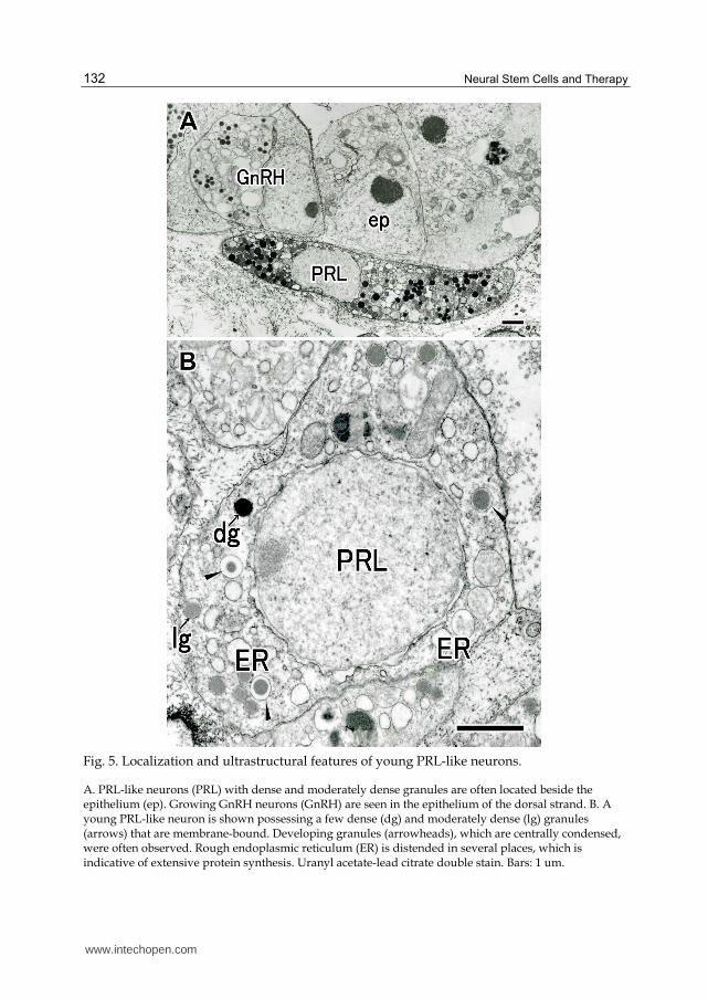
Fig. 5. Localization and ultrastructural features of young PRL-like neurons.
A. PRL-like neurons (PRL) with dense and moderately dense granules are often located beside the epithelium (ep). Growing GnRH neurons (GnRH) are seen in the epithelium of the dorsal strand. B. A young PRL-like neuron is shown possessing a few dense (dg) and moderately dense (lg) granules (arrows) that are membrane-bound. Developing granules (arrowheads), which are centrally condensed, were often observed. Rough endoplasmic reticulum (ER) is distended in several places, which is indicative of extensive protein synthesis. Uranyl acetate-lead citrate double stain. Bars: 1 um.
www.intechopen.com

Formation of Nervous Systems and Neural Stem Cells in Ascidians
133
Fig. 6. Distinction of PRL-like neurons from GnRH neurons.
A and B. Immunoelectron micrographs of PRL-like neurons in the cerebral ganglion (A) and the dorsal strand (B). Gold particles are localized on the dense granules (dg) in both neurons. Gold particles are
not localized in the moderately dense granules (lg). Antibullfrog PRL labeling. C and D. Comparison of anti-GnRH and anti-PRL immunoreactivity between GnRH neuron and probable PRL-like neuron, and between PRL-like neuron and probable GnRH neuron. C. GnRH immunoreactivity in the cerebral ganglion reveals that granules in one cell type are GnRH immunopositive (bottom), whereas those in the other cell type are GnRH immunonegative (top). D. PRL-like immunoreactivity in the cerebral ganglion reveals that granules in the cell on the top are PRL-like immunopositive, whereas those of the cell on the bottom are immunonegative. Bars: (A, B) 250 nm; (C, D) 500 nm.
www.intechopen.com

Neural Stem Cells and Therapy
134
2.6 Homology of the dorsal strand to the olfactory placode
The dorsal strand of H. roretzi is generated by repeated invaginations of the dorsal strand placode, and it produces GnRH and PRL-like neurons, some of which migrate into the cerebral ganglion through the visceral nerve (Terakado, 2009, 2010). Cells derived from the anterior region of the embryonic neural plate and their topological relationships indicate striking similarities between the dorsal strand and the olfactory placode, which suggests that the dorsal strand of urochordate ascidians is homologous to the olfactory placode of vertebrates (Terakado, 2009). Because the ascidian dorsal strand is a single organ, this notion is compatible with the formation of a single olfactory placode in agnathans (Uchida et al., 2003). The dorsal strand also generates many PRL-like (non-GnRH) neurons (Figs. 2C, 4A, B), a finding that has been reported in other species. Antisalmon PRL also stains some cells of the dorsal strand in H. roretzi (unpublished observation). The presence of PRL-like cells has been reported in the cerebral ganglion of Ciona (Fritsch et al., 1982) and Styela (Pestalino, 1983), and in the brain of vertebrates (Fuxe et al., 1977; Krieger and Liotta, 1979; Toubeau et al., 1979; Hansen and Hansen, 1982). We previously suggested that these PRL-immunoreactive neurons in the vertebrate brain may be homologous to those in the cerebral ganglion of ascidians (Terakado et al., 1997). It is well known that molecular features of prolactin in the adenohypophysis resemble those of vertebrate growth hormones (Kawauchi and Sower, 2006). Immunoreactivity of some neurons of the cerebral ganglion and some cells of the dorsal strand to anti-PRL and antigrowth hormone antisera (unpublished observation) suggests the presence of ancestral molecule(s) of the growth hormone family in ascidians. Even the presence of a single molecule in ascidians raises the possibility that multiple antisera raised against molecules belonging to growth hormone family members react to ascidian prolactin due to the presence of common epitopes. Additional informations are needed to elucidate this problem.
2.7 Significance of neurogenesis in the peripheral organ
Most neurons are generated in the central nervous system in vertebrates and invertebrates. However, the vertebrate olfactory placode (peripheral organ) commonly generates GnRH neurons as well as other non-GnRH neurons (Murakami and Arai, 1994; Hilal et al., 1996; Yamamoto et al., 1996). Prior to or during the breeding season, the number of secretory granules greatly increases in the visceral nerve (Fig. 8). Generation of PRL-like neurons in the peripheral organ of chordates has not been reported other than the present species. The reason(s) why the GnRH neurons originate peripherally in vertebrates and ascidians is unknown; however, it is evident that GnRH neurons generated in peripheral organs are crucial for reproduction. PRL-like neurons in H. roretzi also concomitantly originate with the GnRH neurons side by side and migrate into the brain. Quantity of both neurons reveals a year-round change on a large scale which provably accompanies neuron loss after breeding season or winter in mature individuals and recovery thereafter by cell supply to the cerebral ganglion from the dorsal strand.
2.8 Migration of GnRH and PRL-like neurons to the brain
The observation that granulated PRL-like neurons are often found among the fibers of the visceral nerve (Fig. 7) suggests that they migrate towards the cerebral ganglion, similar to the GnRH neurons (Terakado, 2009). The PRL-like neurons in the dorsal strand mostly oval, whereas those in the cerebral ganglion have long neurites (compare Figs. 2B and C). This
www.intechopen.com

Formation of Nervous Systems and Neural Stem Cells in Ascidians
135
suggests that PRL-like neurons elongate after entering the brain. Unattached GnRH and PRL-like neurons were numerous and were located also near the visceral nerve (Fig.7 left). The intimate morphological relationship between the dorsal strand and the visceral nerve (Figs. 3E, F) has long been emphasized (see review by Goodbody, 1974; Chiba et al., 2004). In H. roretzi, this relationship may now be explained by the possibility that GnRH and PRL-like neurons generated in the dorsal strand invade the bundles of visceral nerve fibers and migrate towards the brain. This close topological relationship may be very important for invasion and migration from the site of generation (the dorsal strand) to the cerebral ganglion. This morphological relationship may partly explain the observation that the brain of H. roretzi easily reverts from a thin, cord-like structure (after breeding season, in winter) to the normal brain shape via migration of neurons from the dorsal strand. Similar phenomena are observed during regeneration of other brain structures; for example, after extirpation of the brain in C. intestinalis, the regenerating brain contains GnRH neurons, suggesting that GnRH neurons originate in the dorsal strand and subsequently migrate into the regenerating brain (Bollner et al., 1997). Together with the previous demonstration which GnRH neurons are generated in the dorsal strand and migrate into the brain (Terakado, 2009), the current results reveal that PRL-like neurons are generated in the dorsal strand and migrate into the brain during normal development. They both maintain the brain function throughout the life of the organism with other neurons. Even in colonial ascidians, dorsal strand cells divide frequently and become incorporated directly into the cerebral ganglion (Koyama, 2002) and/or enter into the circulation and participate in the formation of blastozooids as neural stem cells.
Fig. 7. Contact region between the dorsal strand and the visceral nerve (vn).
GnRH neurons and PRL-like neurons are often attached to the nerve, elongated along the nerve fibers, and distributed continuously towards the cerebral ganglion. Unattached GnRH neurons (uGnRH) are seen in the left. Uranyl acetate-lead citrate double stain. Bar: 5 um.
The neurohypophyseal duct and its derived tissues and cells generate a number of cell types including the ciliated duct cells, epithelial cells of the neural gland, luminal cells of the neural gland, epithelial cells of the dorsal strand, and neuroendocrine/endocrine cells in the dorsal strand. Similarly, the rudiment of the adult central nervous system may generate
www.intechopen.com

Neural Stem Cells and Therapy
136
various kinds of neural cells in the developing brain. Of these cell types, those that are generated in the dorsal strand and migrate into the brain via the visceral nerve may be exclusively GnRH neurons in Ciona or GnRH and PRL-like neurons in Halocynthia. Abundance of both neurons in the cerebral ganglion and the dorsal strand in H. roretzi may correspond to the gigantism seen in this species and the corresponding necessity for a large neural network (provably relating to gametogenesis at expense of the body-wall muscle) to adjust to external/internal changes in the environment.
Fig. 8. Electron micrograph of the visceral nerve fibers at breeding season. The secretory granules in the fibers (vn) increase greatly prior to or during the breeding season. Bar: 1 um
2.9 Role of the neurohypophyseal duct
What is the significance of the neurohypophyseal duct in ascidians, in spite of solitary or colonial ones? From the neurohypophyseal duct, the neural gland and then the dorsal strand are formed in all the solitary ascidians and in some colonial ascidians. In the vegetative reproduction (the latter), migratory cells are produced from the dorsal strand (Manni et al, 1999; Koyama, 2002) and may participate in the formation of adult central nervous system of blastozooids as neural stem cells. Above results lead to the hypothesis that the neurohypophyseal duct is a cell reservoir that sets the undifferentiated cells aside for the post-metamorphic formation of the neural gland and the dorsal strand that contains neural stem cells. Peripheral organ origin of the GnRH neurons and their migration into the brain shares in vertebrates and urochordates, and may have evolved in the common ancestor of vertebrates and urochordates. In colonial ascidians that lack or reduced immunological staining to GnRH may be the result of regression due to the exceeding of asexual reproduction.
www.intechopen.com

Formation of Nervous Systems and Neural Stem Cells in Ascidians
137
3. Conclusion
Urochordate ascidians share some morphological and developmental characteristics with those of vertebrates. About one thirds of the larval central nervous system is functional neurons and the rest is glial cells, called the ependymal cells. At metamorphosis, most of functional neurons and glial cells in the tail region disappear. Adult central nervous system is generated from the rearranged ependymal cells (undifferentiated neural cells) of the larval central nervous system after onset of metamorphosis. On the other hand, the peripheral nervous system is later generated from the dorsal strand which is formed by repeated invaginations of the thickened dorsal epithelium of the neural gland. The olfactory placode in vertebrates and the dorsal strand in ascidians are both derived from the anterior region of embryonic neural plate and generate GnRH and some other neurons. In some solitary ascidians, GnRH and PRL-like neurons are continuously generated in the dorsal strand throughout life. Therefore, ascidians are very useful for neural stem cell studies in providing important informations about fundamental processes of neural stem cell formation.
4. Acknowledgment
I am grateful to Emer. Prof. Hideshi Kobayashi of The University of Tokyo for continuous encouragement during studies and to the members of the Department of Regulation Biology, Saitama University for facilitation of the study. The author is also grateful to Emer. Prof. Sakae Kikuyama of Waseda University for gift of the bullfrog prolactin PRL antiserum.
5. References
Adams BA, Tello JA, Erchegyi J, Warby C, Hong DJ, Akinsanya KO, Mackie GO, Vale W, Rivier JE, Sherwood NM (2003) Six novel gonadotropin-releasing hormones are encoded as triplets on each of two genes in the protochordate, Ciona intestinalis. Endocrinology 144: 1907-1919
Bassham S, Postlethwait JH (2005) The evolutionary history of placodes: a molecular genetic investigation of the larvacean urochordate Oikopleura dioica. Development 132: 4259–4272
Beites CL, Kawauchi S, Crocker CE, Calof AL (2005) Identification and molecular regulation of neural stem cells in the olfactory epithelium. Exp Cell Res 306: 309–316
Bhattacharyya S, Bronner-Fraser M (2008) Conpetence, specification and commitment to an olfactory placode fate. Development 135: 4165–4177
Blair JE, Hedges SB (2005) Molecular phylogeny and divergence times of deuterostome animals. Mol Biol Evol 22: 2275–2284
Bollner T, Beesley PW, Thorndyke MC (1997) Investigation of the contribution from peripheral GnRH-like immunoreactive ‘neuroblast’ to the regenerating central nervous system in the protochordate Ciona intestinalis. Proc Roy Soc Lond B264: 1117–1123
Boorman CJ, Shimeld SM (2002) Pitx homeobox genes in Ciona and amphioxus show left-right asymmetry is a conserved chordate character and defines the ascidian adenohypophysis. Evol Dev 4: 354–365
Burighel P, Lane NJ, Zaniolo G, Manni L (1998) Neurogenic role of the neural gland in the development of the ascidian, Botryllus schlosseri (Tunicata, Urochordata). J Comp Neurol 394: 230-241
Cariboni A, Maggi R, Parnavelas JG (2007) From nose to fertility: the long migratory journey of gonadotropin-releasing hormone neurons. Trends Neurosci 30: 638-644
www.intechopen.com

Neural Stem Cells and Therapy
138
Chen B, Kim EH, XU PX (2009) Initiation of olfactory placode development and neurogenesis is blocked in mice lacking both Six1and Six4. Bev Biol 326: 75-85
Chiba S, Sasaki A, Nakayama A, Takamura K, Satoh N (2004) Development of Ciona intestinalis juveniles (through 2nd ascidian stage). Zool Sci 21: 285-298
Christiaen L, Burighel P, Smith WC, Vernier P, Bourrat F, Joly J-S (2002) Pitx genes in Tunicates provide new molecular insight into the evolutionary origin of pituitary. Gene 287: 107-113
Cole AG, Meinertzhagen IA (2001) Tailbud embryogenesis and the development of the neurohypophysis in the ascidian Ciona intestinalis. In “Biology of ascidians” Ed by H Sawada, H Yokosawa, CC Lambert, Tokyo, Springer-Verlag pp 137-141
Cole AG, Meinertzhagen IA (2004) The central nervous system of the ascidian larva: mitotic history of cells forming the neural tube in late embryonic Ciona intestinalis. Dev Biol 271: 239-262
Delsuc F, Brinkmann H, Chourrout D, Philippe H (2006) Tunicate and not cephalochordates are the closest living relatives of vertebrates. Nature 439: 965-968
Delsuc F, Tsagkogeorga G, Lartillot N, Phillippe H (2008) Additional Molecular support for the new chordate phylogeny. Genesis 46: 592-604
Elwyn A (1937) Some stages in the development of the neural complex in Ecteinascida turbinate. Bull Neurol Inst NY6: 163-177
Fritsch HA, Van Noorden S, Pearse AG (1982) Gastro-intestinal and neurohormonal peptides in the alimentary tract and cerebral complex of Ciona intestinalis (Ascidiaceae). Cell Tissue Res 223: 369-402
Fuxe K, Haekfelt T, Eneroth P, Gustafsson JA, Skett P (1977) Prolactin-like immunoreactivity: localization in nerve terminals of rat hypothalamus. Science 196: 899-900
Goodbody I (1974) The physiology of ascidians. Adv Mar Biol 12: 1-140 Gorbman A (1995) Olfactory origins and evolution of the brain-pituitary endocrine system:
facts and speculation. Gen Comp Endocrinol 97: 171-178 Hansen BL, Hansen GN (1982) Immunocytochemical demonstration of somatotropin-like
and prolactin-like activity in the brain of Calamoichtes calabaricus (Actinopterygii). Cell Tissue Res 222: 615-6627
Hilal EM, Chen JH, Silverman AJ (1996) Joint migration of gonadotropin-releasing hormone (GnRH) and neuropeptide Y (NPY) neurons from olfactory placode to central nervous system. J Neurobiol 31: 487-502
Holland LZ, Albalat R, Azumi K, Benito-Gutierrez E, Blow MJ, Bronner-Fraser M, Brunet F, Butts T, Candiani S, Dishaw LJ, Ferrir DEK, Garcia-Fernandez J, Gibson-Brown JJ, Gissi C, Godzik A, Hallbook F, Hirose D, Hosomichi K, Ikuta T, Inoko H, Kasahara M, Kasamatsu L, Kawashima T, Kimura A, Kobayashi M, Kozmik Z, Kubokawa K, Laudet V, Litman GW, McHardy AC, Meulemans D, Nonaka M, Olinski RP, Pancer Z, Pennacchio LA, Pestarino M, Rast JP, Rigoutsos I, Robinson-Rechavi M, Roch G, Saiga H, Sasakura Y, Satake M, Satou Y, Schubert M, Sherwood N, Shiina T, Takatori N, Tello J, Vopalensky P, Wada S, Xu A, Ye Y, Yoshida K, Yoshizaki F, Yu Jr-K, Zhang Q, Zmasek CM, Putnam NH, Rokhsar DS, Satoh N, Holland PWH (2008) Genome Res 18: 1100-1111
Horie T, Shinki R, Ogura Y, Kusakabe TG, Satoh N, Sasakura Y (2011) Ependymal cells of chordate larvae are stem-like cells that form the adult nervous system. Nature 469: 525-528
Kanaho Y, Enomoto M, Endo D, Maehiro S, Park MK, Murakami S (2009) Neurotrophic effect of gonadotropin-releasing hormone of neurite extension and neuronal
www.intechopen.com

Formation of Nervous Systems and Neural Stem Cells in Ascidians
139
migration of embryonic gonadotropin-releasing hormone neurons in chick olfactory nerve bundle culture. J Neurosci Res 87: 2237-2244
Kawada K, Ogasawara M, Sekiguchi T, Aoyama M, Hotta K, Oka K, Satake H (2011) Peptidomic analysis of the central nervous system of the protochordate, Ciona intestinalis: Homologs and prototypes of vertebrate peptides and novel peptides. Endocrinology 152: 2416-2427
Kawauchi H, Sower SA (2006) The down and evolution of hormones in the adenohypophysis. Gen Comp Endocrinol 148: 3-14
Koyama H (2002) The dorsal strand of Polyandrocarpa misakiensis (Protochordata: Ascidiacea): a light and electron microscope study. Acta Zool (Stockh), 83: 231-243
Krieger DT, Liotta AS (1979) Pituitary hormones in brain: where, how, and why? Science 205: 366-372
Mackie GO, Burighel P (2005) The nervous system in adult tunicates: current research directions. Can J Zool. 83: 151-220
Manni L, Lane NJ, Sorrentino M, Zaniolo G, Burighel P (1999) Mechanism of neurogenesis during the embryonic development of a tunicate. J Comp Neurol 412: 527-541
Manni L, Lane NJ, Joly JS, Gasparini F, Tiozzo S, Caicci F, Zaniolo G, Burighel P (2004) Neurogenic and non-neurogenic placodes in ascidians. J Exp Zool (Mol Dev Evol) 302: 483-504
Manni L, Agnelletto A, Zaniolo G., Burighel P (2005) Stomodial and neurohypophysial placodes in Ciona intestinalis: Insights into the origin of the pituitary gland. J Exp Zool (Mol Dev Evol) 300B: 324-339
Manni L, Mackie GO, Caicci F, Zaniolo G, Burighel P (2006) Coronal organ of ascidians and the evolutionary significance of secondary sensory cells in chordates. J Comp Neurol 495: 363-37
Mazet F (2006) The evolution of sensory placodes. Sci World J 6: 1841-1850 Mazet F, Hutt JAA, Milloz J, Millard J, Graham A, Shimeld SM (2005) Molecular evidence
from Ciona intestinalis for the evolutionary origin of vertebrate sensoiry placodes. Dev Biol 282: 494-508
Mazet F, Shimeld SM (2005) Molecular evidence from ascidians for the evolutionary origin of vertebrate cranial sensory placodes. J Exp Zool (Mol Dev Evol) 304B:340-346
Meulemans D, Bronner-Fraser M (2007) The amphioxus Sox family: implications for the evolution of vertebrate placodes. Int J Biol Sci 3: 356-364
Murakami S, Arai Y (1994) Transient expression of somatostatin immunoreactivity in the olfactory-forebrain region in the chick embryo. Brain Res Dev Brain Res 82: 277-285
Murdoch B, Roskams AJ (2007) Olfactory epithelium progenitors: Insights from transgenic mice and in vitro biology. J Mol Histol 38: 581-899
Northcutt RG, Gans C (1983) The genesis of neural crest and epidermal placode: a reinterpretation of vertebrate origins. Q Rev Biol 58: 1-28
Okubo K, Sakai F, Lau EL, Yoshizaki G, Takeuchi Y, Naruse K, Aida K, Nagahama Y (2006) Forebrain gonadotropin-releasing hormone neuronal development: insights from transgenic medaka and the relevance to X-linked Kallman syndrome. Endocrinology 147: 1076-1084
Pestalino M (1983) Prolactinergic neurons in a protochordate. Cell Tissue Res 233: 471-474 Powell JF, Reska-Skinner SM, Prakash MO, Fischer WH, Park M, Rivier JE, Craig AG,
Mackie GO, Sherwood NM (1996) Two new forms gonadotropin-releasing hormone in a protochordate and the evolutionary implications. Proc Natl Acad Sci USA 93: 10461-10464
www.intechopen.com

Neural Stem Cells and Therapy
140
Putnam NH, Butts T, Ferrier DEK, Furlong RF, Hellsten U, Kawashima T, Robinson-Rechavi M, Shoguchi E, Terry A, Yu J-K, Benito-Gutierrez E, Dubchak I, Garcia-Fernandez J, Gibson-Brown JJ, Grigoriev IV, Horton AC, de Jong PJ, Jurka J, Kapitonov VV, Kohara Y, Kuroki Y, Lindquist E, Lucas S, Osoegawa K, Pennacchio LA, Salamov AA, Satou Y, Sauka-Spengler T, Schmutz J, Shin-I T, Toyoda A, Bronner-Fraser M, Fujiyama A, Holland LZ, Holland PWH, Satoh N, Rokhsar DS (2008) The amphioxus genome and the evolution of the chordate karyotype. Nature 453: 1064-1071
Satoh N (1994) Developmental biology of ascidians. Cambridge University Press, Cambridge, UK
Schlosser G (2005) Evolutionary origins of vertebrate placodes: insights from developmental studies and from comparisons with other deuterostomes. J Exp Zool (Mol Dev Evol) 304B: 347-399
Schwarting GA, Wierman ME, Tobet SA (2007) Gonadotropin-releasing hormone neuronal migration. Semin Reprod Med 25: 305-312
Takamura K (2002) Changes in the nervous systems from larva to juvenile in Ciona intestinalis. Rep Res Inst Mar Biores Fukuyama Univ 12: 27-35
Tanaka S, Kurosumi K (1986) Differential subcellular localization of ACTH and α-MSH in corticotropes of the rat anterior pituitary. Cell Tissue Res 243: 229-238
Terakado K (2010) Generation of prolactin-like neurons in the dorsal strand of ascidians. Zool Sci 27: 581-588
Terakado K (2009) Placode formation and generation of gonadotropin-releasing hormone (GnRH) neurons in ascidians. Zool Sci 26: 389-405
Terakado K (2001) Induction of gamete release by gonadotropin-releasing hormone in a protochordate, Ciona intestinalis. Gen Comp Endocrinol 124:277-284
Terakado K, Ogawa M, Inoue K, Yamamoto K, Kikuyama S (1997) Prolactin-like immunoreactivity in the granules of neural complex cells in the ascidian Halocynthia roretzi. Cell Tissue Res 289: 63-71
Toubeau G, Desclin J, Parmentier M, Pasteels JL (1979) Compared localization of prolactin-like and adrenocorticotropin immunoreactivity within the brain of the rat. Neuroendocrinology 29: 374-384
Tsutsui H, Yamamoto N, Ito H, Oka Y (1998) GnRH-immunoeactive neuronal system in the presumptive ancestral chordate, Ciona intestinalis (Ascidian). Gen Comp Endocrinol 112: 426-432
Uchida K, Murakami Y, Kuraku S, Hirano S, Kuratani S (2003) Development of the adenohypophysis in the lamprey: evolution of epigenetic patterning programs in organogenesis. J Exp Zool (Mol Dev Evol) 300B: 32-47
Wada H, Saiga H, Satoh N, Holland PW (1998) Tripartite organization of the ancestral chordate brain and the antiquity of placodes: insight from ascidian Pax-2/5/8, Hox and Otx genes. Development 125: 1113-1122
Willey A (1893) Studies on the Protochordata. II The development of neuro-hypophyseal system in Ciona intestinalis and Clavelina lepadiformis, with an account of the origin of sense organs in Ascidia mentula. Q J Microsc Sci 35: 295-334
Yamamoto N, Uchiyama H, Ohki-Hamazaki H, Tanaka H, Ito H (1996) Migration of GnRH-immunoreactive neurons from the olfactory placode to the brain: a study using avian embryonic chimeras. Brain Res Dev Brain Res 95: 234-244
Yasui K, Zhang S, Uemura M, Saiga H (2000) Left-right asymmetric expression of BbPtx-related gene, in a lancelet species and the developmental left-sidedness in deuterostomes. Development (Camb.) 127: 187-195
www.intechopen.com
Neural Stem Cells and TherapyEdited by Dr. Tao Sun
ISBN 978-953-307-958-5Hard cover, 440 pagesPublisher InTechPublished online 15, February, 2012Published in print edition February, 2012
InTech EuropeUniversity Campus STeP Ri Slavka Krautzeka 83/A 51000 Rijeka, Croatia Phone: +385 (51) 770 447 Fax: +385 (51) 686 166www.intechopen.com
InTech ChinaUnit 405, Office Block, Hotel Equatorial Shanghai No.65, Yan An Road (West), Shanghai, 200040, China
Phone: +86-21-62489820 Fax: +86-21-62489821
This book is a collective work of international experts in the neural stem cell field. The book incorporates thecharacterization of embryonic and adult neural stem cells in both invertebrates and vertebrates. It highlightsthe history and the most advanced discoveries in neural stem cells, and summarizes the mechanisms ofneural stem cell development. In particular, this book provides strategies and discusses the challenges ofutilizing neural stem cells for therapy of neurological disorders and brain and spinal cord injuries. It is suitablefor general readers, students, doctors and researchers who are interested in understanding the principles ofand new discoveries in neural stem cells and therapy.
How to referenceIn order to correctly reference this scholarly work, feel free to copy and paste the following:
Kiyoshi Terakado (2012). Formation of Nervous Systems and Neural Stem Cells in Ascidians, Neural StemCells and Therapy, Dr. Tao Sun (Ed.), ISBN: 978-953-307-958-5, InTech, Available from:http://www.intechopen.com/books/neural-stem-cells-and-therapy/formation-of-nervous-systems-and-neural-stem-cells-in-ascidians

© 2012 The Author(s). Licensee IntechOpen. This is an open access articledistributed under the terms of the Creative Commons Attribution 3.0License, which permits unrestricted use, distribution, and reproduction inany medium, provided the original work is properly cited.