formulation development and evaluation of...
TRANSCRIPT
FORMULATION DEVELOPMENT AND EVALUATION OF
TRANSDERMAL PATCH CONTAINING AMLODIPINE
BESYLATE
Dissertation submitted to
THE TAMILNADU Dr. M.G.R MEDICAL UNIVERSITY, CHENNAI
in partial fulfillment of the requirements
for the award of the degree of
MASTER OF PHARMACY
IN
PHARMACEUTICS
DEPARTMENT OF PHARMACEUTICS
PERIYAR COLLEGE OF PHARMACEUTICAL SCIENCES FOR
GIRLS, TIRUCHIRAPPALLI -620 021.
March 2008(AN ISO 9001 CERTIFIED INSTITUTION)
63

Prof.T.N.K.Suriyaprakash M.Pharm.,( Ph.D.)
Head, Department of Pharmaceutics,Periyar College of Pharmaceutical Sciences for Girls,Tiruchirapalli-620 021Tamil Nadu,India.
CERTIFICATE
This is to Certify that this dissertation entitled “FORMULATION
DEVELOPMENT AND EVALUATION OF TRANSDERMAL PATCH
CONTAINING AMLODIPINE BESYLATE” :by Mr Shaikh Imran for
the award of “Master Of Pharmacy” degree, comprises of the bonafide
work done by him in the Department of Pharmaceutics, Periyar College of
Pharmaceutical Sciences for Girls, Tiruchirapalli, under my supervision and
guidance and to my full satisfaction.
Place: Tiruchirapalli
Date: (Prof.T.N.K.Suriyaprakash)
64

Dr. R.Senthamarai, M.Pharm., Ph.D., Principal,Periyar College of Pharmaceutical Sciences for Girls,Tiruchirapalli-620 021Tamil Nadu,India.
CERTIFICATE
This is to Certify that this dissertation entitled “FORMULATION
DEVELOPMENT AND EVALUATION OF TRANSDERMAL PATCH
CONTAINING AMLODIPINE BESYLATE”, by Mr Shaikh Imran for
the award of “Master of Pharmacy” degree, comprises of the bonafide
work done by him in the Department of Pharmaceutics, Periyar College of
Pharmaceutical Sciences for Girls, Tiruchirapalli, his work was supervised
by Prof.T.N.K.Suriyaprakash M.Pharm., (Ph.D.), Head, Department of
Pharmaceutics, Periyar College of Pharmaceutical Sciences for Girls,
Tiruchirapalli.
I recommend this research work for acceptance as project for the
partial fulfillment of the degree of “Master of Pharmacy” of the
Department of Pharmaceutics, Periyar College of Pharmaceutical Sciences
for Girls, Tiruchirapalli, for the year March 2008.
Place : Tiruchirapalli
Date: (Dr.R.Senthamarai)
65

ACKNOWLEDGEMENT
Though words are seldom sufficient to express gratitude and feelings,
it some how gives me an opportunity to thank those who helped me during
the tenure of my study. The work of dissertation preparation was a daunting
task and a fascinating experience.
I take this opportunity to express my deep sense of gratitude to my
guide Prof.T.N.K.Suriyaprakash M.Pharm, (Ph.D.), Head, Department of
Pharmaceutics, Periyar College of Pharmaceutical Sciences for Girls,
Trichy-21, for his guidance, valuable suggestions and liberal encouragement
to complete this work successfully entitled “FORMULATION
DEVELOPMENT AND EVALUATION OF TRANSDERMAL PATCH
CONTAINING AMLODIPINE BESYLATE”,
It’s my privilege and honor to thank Dr.R.Senthamarai M.Pharm,
Ph.D., Principal, Periyar College of Pharmaceutical Sciences for Girls,
Trichy-21, for providing all the necessary facilities to do this thesis work.
I extend my sincere thanks to our honorable chairman
Man.K.Veeramani, MA.B.L., Chancellor, Periyar Maniyammai University
and Mr.Gnana Sebastian Correspondent, Periyar College of
Pharmaceutical Sciences for Girls, Trichy-21, for providing all the facilities.
My warmest thanks to Dr..S..Karpagam Kumarasundari M.Pharm,
Ph.D Head, Department of Pharmacology, Periyar College of
66

Pharmaceutical Sciences for Girls, Trichy-21, for his valuable suggestion
and help to complete thesis work.
I express my deep sense of gratitude to Prof.A.M.Ismail M.Pharm,
(Ph.D) Dean(PG), Periyar College of Pharmaceutical Sciences for Girls,
Trichy-21, for his suggestion to complete this work.
My sincere and heart felt thanks to Mrs.K.Reeta Vijaya Rani
M.Pharm, (Ph.D), Mr.M.Sakthivel M.Pharm., Ms.N.Pavala Rani
M.Pharm., and Mrs.R.Latha Eswari M.Pharm., Department of
Pharmaceutics, Periyar College of Pharmaceutical Sciences for Girls,
Trichy-21,for their valuable suggestion and help to complete thesis work.
Words are insufficient to express my feeling towards my classmates V.Narayanan, B.Senthilnathan, V.Dineshkumar, A.Sivakumar, Eugien Leo Prakash and other my friends who helped me directly and indirectly in the successful completion of this dissertation.
I convey my special thanks to Mr.K.A.S..Mohammed Shafique
M.Pharm., for his constant help and suggestion which were of great
importance to me to complete this thesis work.
Also express my heartfelt and sincere thanks to all teaching and non-
teaching staff members for their timely help.
It’s my privilege to express my sincere thanks to Mrs.K.Tamilselvi,
Librarian, for providing the library facilities and co-operations to complete
this work.
67

Last but not the least I am glad to express my warm gratitude to all
my Teacher, Juniors, Friends, and well wishers for their kind co-operation
encouragement and help rendered at various stages of this research work.
I express my heartfelt thanks to my parents, my sisters for their
support and encouragement throughout my course of study.
SHAIKH IMRAN
68
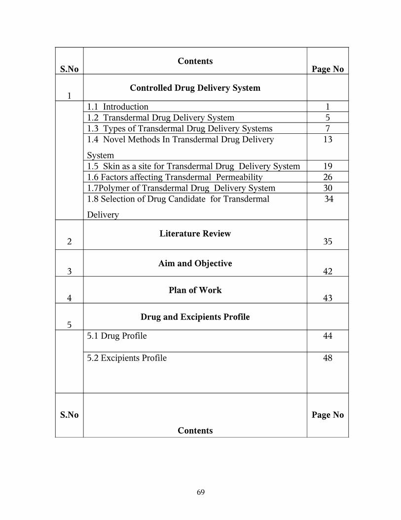
S.NoContents
Page No
1Controlled Drug Delivery System
1.1 Introduction 11.2 Transdermal Drug Delivery System 51.3 Types of Transdermal Drug Delivery Systems 71.4 Novel Methods In Transdermal Drug Delivery
System
13
1.5 Skin as a site for Transdermal Drug Delivery System 191.6 Factors affecting Transdermal Permeability 261.7Polymer of Transdermal Drug Delivery System 301.8 Selection of Drug Candidate for Transdermal
Delivery
34
2Literature Review
35
3Aim and Objective
42
4Plan of Work
43
5Drug and Excipients Profile
5.1 Drug Profile 44
5.2 Excipients Profile 48
S.No
Contents
Page No
69

6
Materials and Methods
6.1 List of Instrument 506.2 List of reagents 506.3 Materials 50 6.4 Methods 516.5 Standard Curve for Amlodipine 546.6 Procedure For Fabrication of Transdermal Film 556 .7 Evaluation of Transdermal Drug Delivery System 576.8 Accelerated Stability Studies 60
7 Result and Discussion 638 Summary and Conclusion 929 Bibliography 94
70

1. Controlled drug delivery system
1. 1 Introduction
Over the past twenty-five years, as the expense and complication involved
in marketing new drug entities have increased with concomitant recognition
of the therapeutic advantage of controlled drug delivery system. There are
several seasons for the attractiveness of these dosage forms. It is generally
recognized that for many disease states, a substantial number of
therapeutically effective compounds already exist. Side effects or the
necessity to administer the compound in a clinical setting, however, often
limits the effectiveness of this drugs.1
The goal in designing controlled drug delivery systems is to reduce
the frequency of dosing and increase effectiveness of the drug by
localization at the site of action .Reducing the dose required (or) providing
uniform drug delivery.
Controlled release drug administration means not only prolonged
release, but also implies predictability and reproducibility of drug release
kinetics. Controlled drug delivery system is the one, which delivers the drug
at a predetermined rate, systematically, for a specific period of time .2
71

FIGURE 1 Theoretical Illustration Comparing Blood Drug Concentration
profiles Of A Controlled-Release Drug Delivery System And Conventional
Dosage Forms Via Various Routes Of Administration.
Figure 1 show comparative blood drug level profiles obtained from
administration of conventional, controlled as well as prolonged release
dosage forms. Thus, the conventional tablet or capsule provides only a
single and transient burst of drug. As long as the amount of drug is above
the minimum effective concentration, a pharmacological response is
observed problems occur when the therapeutic range is very narrow or
when the peak is greater than the upper limit of this of this range. Indeed,
one of the main purposes of controlled release to improve safety and
minimize side effects of the drug by reducing fluctuations in drug level.3
72
MERITS OF CONTROLLED DRUG DELIVERY SYSTEM4
1. The potential merit that a controlled release drug delivery system may
bring to us can be appreciated by a consideration of prolonged and
efficient delivery of therapeutically effective dosages, and localization
of therapy.
2. The "peak and valley' pattern is more striking for drugs with
biological half-life less than4hrs. Which can be over come by
controlled release drug delivery system
3. It provides maximum utilization of drug enabling reduction in total
amount of dose administered.
4. Improved patient convenience and compliance due to less frequent
drug administration.
5. Reduction in fluctuation in steady state levels and therefore better
control of disease condition and reduced intensity of local or systemic
side effects.
6. Increased safety margin of high potency drugs due to better control of
plasma levels.
7. Reduction in health care cost through improved therapy, shorter time
period, less frequency of dosing and reduction in personal time to
dispense, administer and monitor patients.
73

DEMIRITS OF CONTROLLED DRUG DELIVERY SYSTEM5
1. Decreased systemic availability in comparison to immediate release
conventional dosage forms; this may due to incomplete release,
increased first pass metabolism, increased stability, insufficient
residence time for complete release, site-specific action, pH-
dependant solubility, etc.
2. Possibility of dose dumping due to food, physiologic or formulation
variables or chewing or grinding of oral formulation by the patient
and thus, increased risk of toxicity.
3. Retrieval of drug is difficult in case of toxicity, poisoning or
hypersensitivity reaction.
4. Reduced potential for dosage adjustment of drugs normally
administered in varying strengths.
5. It is higher cost of formulation.
74
1. 2 TRANSDERMAL DRUG DELIVERY SYSTEM
For many decades medication of acute disease as well as a chronic illness
has been accomplished dosage forms. Like tablets, capsules, ointments,
aerosol, injectables and suppositories, as carrier, recently a technical
advancement have resulted in the development of new techniques of drug
delivery which includes transdermal drug delivery system. Several
transdermal drug delivery systems have been developed to achieve the
objective of systemic medication through topical application on the skin
surface 4.
The principle of transdermal drug delivery system is that they could
provide controlled drug delivery (have constant drug concentration in
plasma) over a prolonged period of time. It is anticipated that transdermal
drug delivery system can be designed to input drugs at appropriate rates to
maintain suitable plasma drug levels for therapeutic efficacy, without the
periodic sojourns into plasma concentration that would accompany toxicity
(or) lack of efficacy.
Transdermal delivery of antihypertensive is one of the prime focus areas
of drug delivery systems. Various antihypertensive such as metoprolol,
clonidine, propranalol, bupranolol, isosorbide dinitrite, verapamil,
nifedipine, etc., have been studied for their suitability in transdermal
therapeutic systems.
75

ADVANTAGES OF TRANSDERMAL DRUG DELIVERY SYSTEMS2
1. Avoids problems associated with gastro-intestinal absorption due to
pH enzymatic activity, and drug food interactions.
2. It is a substitute for oral route.
3. Avoid the risks and inconveniences of I.V therapy.
4. Provides predictable extended duration of activity.
5. Extends the activity of drug with short half-life.
6. Multilayer therapy with single application.
7. Provides capacity to terminate.
8. Minimize inter and intra patient variation.
9. Provides suitability by self-administration.
10.Reduces daily dosing, thus improving patient compliance.
11.Enhance therapeutic efficacy, reduce side effects due to optimization
of blood concentration –time profile and elimination of pure entity of
drugs into systemic circulation.
LIMITATIONS OF TRANSDERMAL DRUG DELIVERY SYSTEMS8
1. Difficulty of permeation through human skin:
In addition to physical barrier, human skin functions as a
chemical barrier. The outer most layer of skin, the
stratum corneum is an excellent barrier to all chemicals
including drugs. If drug requirements are more than
10mg per day the transdermal delivery will be difficulty.
Only relatively potent drugs can be given through this
route.
76

2. Skin Irritation:
• Skin irritation or contact dermatitis due to
excipients and enhancers of the drug system used
for increasing percutaneous absorption is another
major limitation.
3. Clinical Need:
• It has to be examined carefully before developing a
transdermal product.
1.3 TYPES OF TRANSDERMAL DRUG DELIVERY SYSTEMS
Several approaches can be effectively utilized to control the release of
systematically active drugs for permeation at a programmed rate through the
skin tissues. The successfully launched commercially available transdermal
drug delivery systems may be classified into four types, depending on the
technological approach9.
Membrane permeation controlled transdermal therapeutic
system; (Drugs tried under this are: Scopolamine and
Nitroglycerine).
Adhesive dispersion type transderrmal system. (Drugs tried
under this is Nitroglycerine).
77
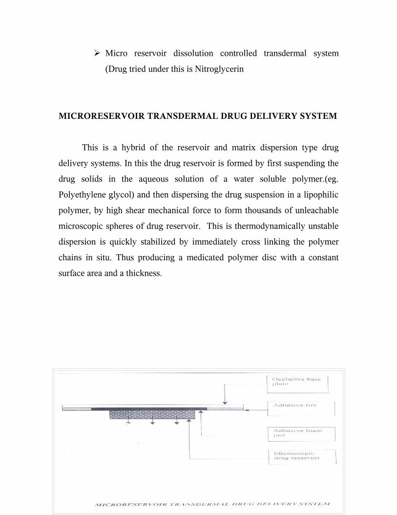
Micro reservoir dissolution controlled transdermal system
(Drug tried under this is Nitroglycerin
MICRORESERVOIR TRANSDERMAL DRUG DELIVERY SYSTEM
This is a hybrid of the reservoir and matrix dispersion type drug
delivery systems. In this the drug reservoir is formed by first suspending the
drug solids in the aqueous solution of a water soluble polymer.(eg.
Polyethylene glycol) and then dispersing the drug suspension in a lipophilic
polymer, by high shear mechanical force to form thousands of unleachable
microscopic spheres of drug reservoir. This is thermodynamically unstable
dispersion is quickly stabilized by immediately cross linking the polymer
chains in situ. Thus producing a medicated polymer disc with a constant
surface area and a thickness.
78

Figure No.2 Microreservoir Transdermal Drug Delivery System
MATRIX DIFFUSION TRANSDERMAL DRUG DELIVERY SYSTEM
Here, the drug reservoir is formed by homogenously dispersing the
drug solids in a hydrophilic or lipophilic polymer matrix, the medicated
polymer is then molded into a medicated disc with a defined surface area
and controlled thickness. This drug reservoir containing polymer disc is
mounted to an occlusive base plate in a compartment fabricated from a drug
impermeable plastic backing. The adhesive, in this is spread along with
circumference of the patch to form an adhesive rim.
Figure No.3 Matrix Diffusion Transdermal Drug Delivery System
79
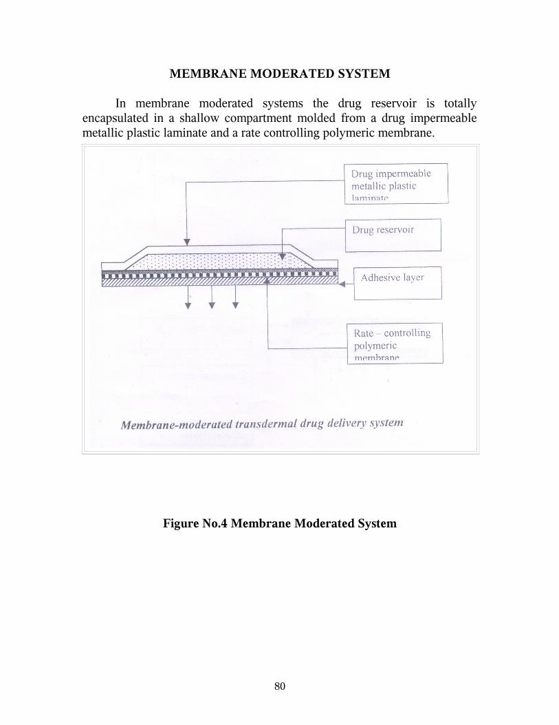
MEMBRANE MODERATED SYSTEM In membrane moderated systems the drug reservoir is totally encapsulated in a shallow compartment molded from a drug impermeable metallic plastic laminate and a rate controlling polymeric membrane.
Figure No.4 Membrane Moderated System
80

ADHESIVE DIFFUSION CONTROLLED SYSTEM: In this system the drug reservoir is formulated by directly dispersing the
drug in an adhesive polymer on a flat sheet of drug impermeable backing to
form a thin drug reservoir layer. On the top of the drug reservoir layer,
layers of non-mediated rate controlling adhesive polymer of constant
thickness are applied to produce an adhesive diffusion-controlled drug
delivery system
Figure No.5 Adhesive Diffusion Controlled System
81

Table. No.1 Commercially Available Transdermal Therapeutic Systems Drug/ Manufacturer
Trade Name
Duration Type of System
Therapeutic Use
Scopolamine Alza Ciba
Transderm - Scop
2 days Reservoir Alleviate motion sickness.
Nitroglycerine Alza/Ciba Hercom Searle Key Wyeth
Transderm –NitroNTSNitrodiscNitro-durDeponite
1 day Reservoir Matrix Matrix Matrix Sandwich
Treatment and prevention of Angina.
Isosorbide dinitrate Nitro electric industrial
Frandol Tape 1 day Matrix Treatment and prevention of Angina.
Clonidine Boehringer Ingelheim
Catpres -TTS 7 days Reservoir Treatment of hypertension.
Estradiol Ciba-Giegy Parke-davis
Estraderm 3 days Reservoir Relief of post-menopausal symptoms.
Nicotine1
Alza Ciba-Giegy
Nicoderm Habitrol prostep
1 day Reservoir Matrix Matrix
Aid in smoking cessation.
Fentanyl Janssens
Duagesic 3 days Reservoir Matrix
Relief from moderate severe pain.
Ketoprofen 2 Pacific Pharmaceuticals
Ketopatch 1 day Matrix Analgesic and anti-inflammatory.
1 The product available in India [Transderm – TTS; CIBA-GIEGY;
Top nitro; FULFORD(India)].
2. The product is very recently launched in India.
82
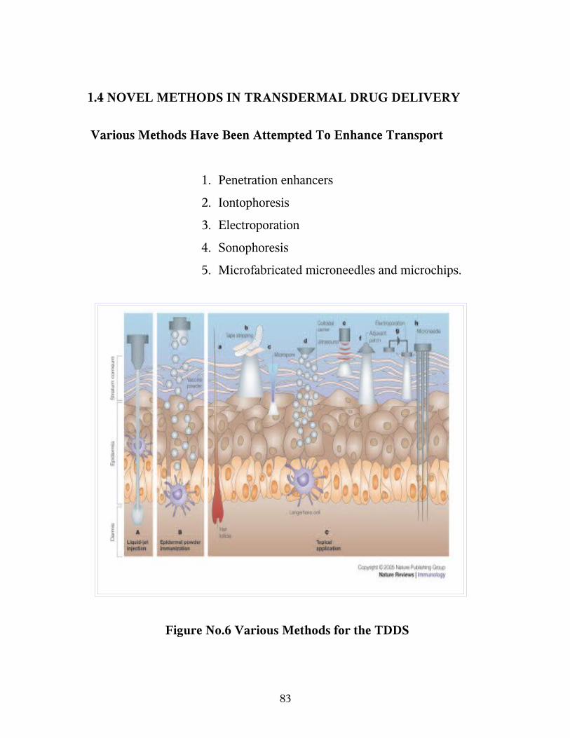
1.4 NOVEL METHODS IN TRANSDERMAL DRUG DELIVERY
Various Methods Have Been Attempted To Enhance Transport
1. Penetration enhancers
2. Iontophoresis
3. Electroporation
4. Sonophoresis
5. Microfabricated microneedles and microchips.
Figure No.6 Various Methods for the TDDS
83

PENETRATION ENCHANCERS APPROACH
An ideal enhancer should be pharmacologically, inactive, non-irritant
and should not damage the skin irreversibly. The effects of an enhancer on
the permeation of a drug usually depend upon the physico-chemical
characteristics of the permeate as well as the enhancer molecule. The
penetration of the enhancers into the stratum corneum is a basic requirement
for their efficacy. It is possible to facilitate the penetration of the drug by
appropriate pretreatment of the skin with penetration enhancer. The
lipoprotein partitioning theory of Barry offers the most acceptable
explanation for the skin with penetration enhancers and the stratum
corneum. Accordingly, the main reason for enhancement includes:
Interactions with the intercellular lipids and intracellular keratin.
Increased penetration of high amounts of enhancer or cosolvent
into the stratum corneum for to the improved dissolving capacity of
the barrier to the drugs. Many of the chemical enhancers such as
dimethyl sulfoxide, surfactants, alcohols, urea, and its derivatives
have been screened for their penetration enhacement. The adverse
effects caused by some of these enhancers restricts there use widely11.
84

IONTOPHORESIS TECHNOLOGY
Iontophoresis is a process or technique involving the transport of
ionic or charged molecules into a tissue by the passage of direct or periodic
electric current through an electrolyte solution containing the ionic
molecules to be delivered using an appropriate electrode polarity. The
process involves the transfer of ions into the body by an electromotive force.
Ions with positive charge are driven into the skin at the anode and those with
the negative charge at the cathode. In the conventional topical treatment by
iontophoresis, the drug is administrated through an electrode having the
same charge as the drug and a return electrode opposite in charge to the drug
is placed at a neutral site on the body surface. The operator then selects a
current intensity below the pain threshold level of the patient and allows the
current to flow for an appropriate period of time.
The current intensity should be increased slowly, maintained for the
length of the treatment and decreased slowly at the end of the treatment. The
current must have within comfortable toleration of the patient. A current
destiny less than 0.5mA/sq.cm, of the electrode surface has been found to be
tolerated by the patient. Interposition of a moist pad between the electrode
plate and the skin is necessary for making a perfect contact, preventing any
skin burns, overcoming skin resistance and protecting the skin from
absorbing a caustic metallic compound formed on the metal plate surface. It
is important that, the drug be applied through the electrode with correct
polarity may not result in penetration of the drug. The electrode must not
come in any direct contact with skin as it may cause burns.
85

Iontophoresis was found to be widely used in several clinical
situations; Iontophoretic delivery of ionizable drugs like, propranolol across
excised human skin from rabbit, pig and humans has been reported.
Iontophoretic delivery of a weakly basic analgesic, oxycodone across the
excised skin from human and several animals was investigated using pulse
current delivered from a newly developed transdermal iontotherapeutic
system66 .
ELECTROPORATION TECHNOLOGY
Electroporation or electropermeabilization involves changes in
membrane of cell due to application of transmembrane voltages. The change
in the membrane involves; structural rearrangement and conductance leading
to temporary loss of semipermeability of cell membranes. Suggesting
formation of pores. The pulses are normally used on the unilamellar
phospholipids bilayers of cell membranes. Approximately 100
multilammeller bilayer of the stratum corneum need about 100V pulses for
electroporation or 1V per bilayer. Electroporation of skin takes place at high
transdermal voltage (100V or more). There is considerable indirect evidence
that high voltage pulses cause changes in the skin structure.
Electroporation is a technique in which the drug encapsulated in
vesicles or particles is delivered in to the skin by applying pulse leading to
breakdown of the stratum corneum. Pressure medicated electroincorporation
has been used to deliver leuprolide acetate micropheres into hairless mouse
skin and human skin engrafted on immunodeficient nude mice. It has been
shown that application of continuous low voltage resulted in a calcein flux
with three orders of magnitude.
86

Besides, the modes compounds calcein, other drugs investigated for
transdermal delivery by electroporation includes; metaprolol flurbiprofen,
cyclosporine, heparin, fenanyl and oligonucleotides67.
SONOPHORESIS TECHNOLOGY68
Ultra sound has been used to treat a wide range of clinical condition
and to transport drugs to deeper tissues. The movement of drugs through
living perturbation is called Sonophoresis. Ultra sound was applied with a
sonicator (VCX-400 sonics and materials) operating at a frequency of 20
KHz. The sonicator were operated in the duty-cycle; that is pulsed mode
(0.1s on and 0.9s off or 1s on and 9s off or 5s on and 5s off).
Ultra sound may enhance transdermal drug delivery systems by
affecting the skin structure (through which enhanced diffusion may occur),
by inducing concection or by a combination of both effects. Because, skin
conductivity is an excellent indicator of the skin barrier properties.
The ultra sound application has resulted in modest permeation of
simple molecular. It was reported that, low frequency ultrasound could be
used to deliver insulin across rabbit skin in vivo, resulting in increased
plasma hormone levels and lowering of blood glucose.
Measurement of ultrasound intensity: A commonly used calorimetric
method was employed to calculate the power from the sonicator based on
the change in the temperature of water exposed to the sonicator.
87

Another method is aluminium foil measurement. In an attempt to
quantify the potential of the sonicator to induce transient cavitations in
aluminium foil paper. It was mounted onto the diffusion cell in a manner
identical to that of the skin sonication was performed for 20s [0.1 on and0.9s
off] and then aluminium foil was remove from the cell. The number of pits
on the foil was determined by visual inspection. The pits represent physical
evidence of the effects of cavitations bubble formation induced by
ultrasound.
MICROFABRICATED MICRONEEDLES AND MICROCHIPS
TECHNOLOGY18
The micro fabricated micro needles technology employs micro-sized
needles made from silicon. These micro needle arrays after insertion into
the skin create conditions for transport of drug across the stratum corneum.
The drug after crossing the stratum corneum diffuses rapidly through deeper
tissue and taken up by capillaries for systemic administration. Micro
needles were made using the micro fabrication technology similar as that of
making of integrated circuits. The micro fabrication technology is simple
for cheap and mass production of micron sized structures. For the drug
delivery, a three-dimensional array of sharp-tipped micro needles with
approximately 150micrometre, in lengths was fabricated. Deep reactive ion
etching technique is based on the black silicon method. Each micro needle
is about 1 micron in diameter or one hundredth of the diameters of a human
hair. These needles can be seen only under a microscope. A microprocessor
and pump for delivering tiny amounts of the drug.
88

The microprocessor and pump automatically inject the right dosage of the
drug. The micro needles have extremely sharp tips with radius of curvature
less than 1mm facilitating easy piercing into the skin.
1.5 SKIN AS A SITE FOR TRANSDERMAL DRUG DELIVERY
SYSTEMS
STRUCTURE OF THE SKIN
The skin consists of different tissues that are joined to perform
specific functions. The thickness of the skin cover most of the body is 1-2
mm thick.
Structurally the skin consists of two principal parts. The superficial,
thinner portion which is composed of epithelial tissue is the epidermis. The
deeper, thicker, connective tissue part is the dermis. Deep to the hypodermis
and not part of the skin is the subcutaneous layer. The hypodermis layer
consists of areolar and adipose tissues. The sub cutaneous layer serves as a
storage depot for fat and contains large blood vessels that supply the skin.
This region also contains nerve endings called lamellated corpuses that are
sensitive to pressure.
Epidermis
The epidermis is keratinized stratified squamous epithelium. It
contains four principal types of cells, keratinocytes, melanocytes, langerhans
cells and merkel-cells. About 90% of epidermal cells are keratinocytes
which produce the protein keratin20
89

Epidermis consists of following layers:
o Stratum basale,
o Stratum spinosum,
o Stratum granulosum,
o Stratum lucidum,
o Stratum Corneum.
Figure No.7 Structure of the Human Skin
90

Stratum Basale:
The deepest layer of the epidermis is the stratum basale, composed of
a single row of cubical or columnar keratinocytes.
Superficial to the stratum basale is the stratum spinosum where 8-10
layers of polyhedral keratinocytes fit closely together.
Stratum Granulosum:
At the middle of the epidermis the stratum granulosum consists of
three to five layers of flattened keratinocytes that are undergoing apoptosis.
Stratum corneum:
The stratum corneum consists of 25-30 layers of dead,flat
keratinocytes. The interior of the cells contains mostly densely packed
intermediate filaments and keratohyalien. Between the cells are lipids from
lamellar granules that help to make this layer water-repellent. These cells are
continuously shed and replaced by cells from the deeper strata. The stratum
corneum serves as an effective water-repellent barrier and also protects
against injury and microbes. Constant exposure of skin to friction stimulates
the formation of a callus an abnormal thickening of the epidermis21.
ROUTES OF PENETRATION
When a molecule reaches intact skin it contacts cellular debris,
microorganisms, sebum, and other materials. The diffusing then has three
potential entry routes to the viable tissue- through the hair follicles with their
associated sebaceous glands, via the sweat ducts, or across the continuous
stratum corneum between these appendages. We can summarize relevant
91

features arriving at a general conclusion27
Sebum and surface material
The layer of sebum mixed with sweat, bacteria, and dead cells is this
(0.4 – 10 µm), irregular, and discontinuous: it hardly affects percutaneous
absorption.
Skin appendages
Their fractional area available for absorption is small (about 0.1%)
and this route usually cannot contribute appreciably to the steady state flux.
However, the route may be important for ions and large polar molecules
which cross-intact stratum corneum with difficulty. Diseases, which disturb
the horny layer, such as eczema and exfoliate dermatitis, allow easy access.
Skin appendages may act as shunts, important at short times prior to
steady state diffusion, e.g. in bioassays which use pharmacological
reactions. Thus minute concentrations of nicotinates or corticosteroids
penetration rapidly down the shunt route may trigger erythema or blanching.
Epidermal route
The epidermal layers (particularly the epidermis) may metabolize and
inactivate a drug, or activate a prodrug. The dermal papillary layer contains
so many capillaries that the average residence time of a drug in the dermis
may only be about a minute. Usually, the deeper dermal layers do not
influence percutaneous absorption. However, the dermis may bind a
hormone such as testosterone, decreasing its systemic removal. If the
92

penetrant is very lipophililc, it crosses the horny layer to meet an aqueous
phase in which it is poorly soluble. The chemical potential immediately
below the barrier may then become high, approaching that in the barrier.
The potential gradient (stratum corneum to viable tissue) falls, together with
the flux. The rate-determining step in percutaneous absorption the becomes
barrier clearance not barrier penetration.
Within the stratum corneum, molecules penetrate either intercellular
or transcellularly. Electron micrographs of intercellular material suggest a
segregation of lipid between protein filaments. In hydrated tissues, these
lipid and polar regions would provide parallel pathways for diffusion.
Molecules would portion into and diffuse through, either network according
to their polarities. The intercellular route is rich in neutral lipid and this
pathway may be more important in percutaneous absorption than previously
thought.
Topically applied agents such as steroids, hexachlorophane,
griseofulvin, sodium fusidate and fusidic acid may form a depot or reservoir
by binding within the stratum corneum.
93

MECHANISM OF SKIN PERMEATION
A systematically active drug that will reach a target tissue from the
site of drug administration of the skin surface must posses some
physicochemical properties that are capable of facilitating the absorption of
drug through the stratum corneum the penetration of drug through viable
epidermis and also uptake of the drug by capillary network in the dermal
papillary layer. The sequence of transdermal permeation of drug is shown.
The rate of the permeation, dq/dt across the skin tissues can be expressed
mathematically by the following relationship.
dq/dt = Ps[Cd-Cr].
Where
Cd and Cr, respectively the concentration of a skin penetrant in the
donor compartment (e.g. Body),
Ps is the overall permeability coefficient of the skin.
Ps = Ks Dss/hs. (1)
Where
Ks is the partition-coefficient for the interfacial partitioning of a
penetrant molecule from the solution of medium or a transdermal drug
delivery system on to a stratum corneum;
Dss is the apparent diffusity for the steady state diffusion of the
94

penetrant molecule through a thickness of skin tissues.3
Analysis of (eq-1) suggests that to achieve a constant rate of drug
permeation one needs to maintain the drug concentration on the surface of
stratum corneum (Cd) consistency and substantially greater than the
drug concentration in the body (Cr),
i.e.., Cd>Cr;
Under such a condition eq.1 can be reduced to
dq/dt = PsCd
The rate of skin permeation dq/dt becomes a constant, if the
magnitude of Cd remains fairly constant throughout the course of skin
permeation to maintain Cd at a rate (Rd) that is either constant or always
greater than the rate of skin update (Ra), i.e. Rd> Ra. By making Rd greater
than Ra the drug concentration on the skin surface (Cd) is maintained at a
level equal to or greater than the equilibrium (or saturation) solubility of the
drug in the stratum corneum (Cse).ie., Cd.C>Cse and a maximum rate of skin
permeation
(dq/dt)m = PsCse
The other mechanism of permeation involves diffusion through
shunts particularly those offered by the relatively widely distributed hair
follicles and exocrine glands. Typically one square centimeter of human
skin yields 10 hair follicles, 15 sebaceous glands and 100 sweat glands.
However the appendages provide a small fractional surface area of
approximately 0.1% of the total skin area.
95

Recent studies indicate the importance of appending in percutaneous
absorption. The appendages route may be more significant for ions and large
polar molecules, which slowly permeates across the bulk of the intact horny
layer. The two potential micro pathways serve the stratum corneum through
the transcellular and intercellular route.
The principal pathway taken by the penetrant is decided mainly by
diffusant’s partition coefficient . Most of the diffusant’s permeate by both
the routes. The intercellular pathway is considered to provide the principal
route and the major barrier to the permeation of the drugs.
1.6 FACTORS AFFECTING TRANSDERMAL PERMEABILITY
Physiological and pathological conditions of the skin .28
1. Skin condition:
• Intact skin prevents penetration. When the skin is
exposed to mustard gas, hydrogen sulphide gas, acids,
alkalies or ultra violet radiation, skin turns to porous
form which will lead to enhanced drug penetration. Mild
burns will increase the penetration whereas severe burns
retard.
96

2. Skin hydration:
• Hydration results from water diffusing from underlying
epidermal layers or from perspiration accumulation after
application of an occlusive vehicle or covering on the
surface.
• Under occlusive conditions densely and closely packed
cells of the skin are opened up and increase its porosity.
Occlusion also reduces the “irreversible” binding
capacity of stratum corneum. When the skin undergoes
hydration, its resistance and capacitance may change. As
the time of hydration increases the low frequency
impedance of the excised skin decreases with time. A
much less activation energy is required to diffuse through
hydrated skin.
3. Skin Age:
• Fetal and infant skin appears more permeable than adult
skin. The stratum corneum of preterm infants is not well
developed and as such provides little barrier to the
ingress of substances. So this route of delivery is possible
for neonatal therapy, when difficulty is encountered in
oral or
4. Increased blood flow:
97

• Due to increased blood flow, a concentration gradient is
established which will promote drug absorption.
5. Skin Temperature:
• Raising skin temperature results in an increase in the rate
of skin permeation. This may be due to thermal energy
required differently solubility of drug in skin tissues.
Increased vasodilatation of skin vessels.
6.Regional skin sites:
Difference in the nature and thickness of the barrier layer
of the skin causes variation in permeability.
7. Cutaneous drug metabolism:
Catabolic enzymes present in the viable epidermis may
render a drug inactive by metabolism and thus affect the
topical bioavailability of the drug.
8.Species variation:
• Human and animals display wide differences in physical
characteristics such as the number of appendage openings
per unit area and the thickness of the stratum corneum.
The average permeability order is Monkey > Dog >Goat
> Rat > Guinea pig > Mouse > Human skin.
98

Physicochemical properties of the penetrant molecules29.
I. Partition coefficient: A lipid/water partition coefficient of 1 or
greater is generally required for optimal transdermal permeability.
The partition coefficient of a drug molecule may be altered by
chemical modification of its functional groups. Membrane partition
coefficient increases exponentially as the length of the lipophillic
alkyl chain increases.
II. pH conditions : Application of solutions whose pH values are very
high or very low can be destructive to the skin. With moderate pH
values, the flux of ionizable drugs can be affected by changes in pH
that alter the ratio of charged to uncharged species and their
transdermal permeability.
III. Drug concentration: The amount of drug percutaneously absorbed
per unit surface area over time interval increase as the concentration
of the drug in the vehicle is increased.
IV. Molecular characteristics of drug: An inverse relationship appears
to exist between absorption rate and molecular weight. Small
molecules penetrate more rapidly than large molecules. Drugs with
molecular weights of upto 500 Dalton can penetrate well. Drugs with
molecular weights above 500 Dalton can be delivered transversally by
iontophoresis.
99
Physicochemical properties of drug delivery systems30.
I. Vehicle: Vehicles serve as drug carriers. Lipophillic solvents
facilitate penetration. The absorption of water-soluble and lipid
soluble substances from terpene and terpene derivatives was better
than from alcoholic solutions. Solubility of drug in the vehicle
determines the rate.
II. Composition of drug delivery systems: It affects not only the rate
of drug release but also the permeability of stratum corneum by
means of hydration mixing with skin lipids or other sorption
promoting effects.
III. Enhancement of transdermal permeation: Transdermal
permeation can be increased by including some penetrates,
sorption promoters etc. Organic solvents like Dimethyl acetamide,
dimethyl formamide, dimethyl sulfoxide, ethylene glycol,
propylene glycol and polyethylene glycol.
IV. Surfactants: Anionic surfactants are the most effective. E.g.
Sodium lauryl sulphate. Chemical like Azone also promote
absorption.
100

1.7 POLYMERS OF TRANSDERMAL DELIVERY SYSTEM
POLYMERS:
The polymer controls the release of the drug from the devices. The
following criteria should be satisfied for a polymer to be used in a
transdermal system..31
1. Drug solubility and diffusivity in the polymer.
2. The desired drug loading and its effect on polymer
integrity.
3. Compatibility of the polymer with necessary
excepients, such as solvents and skin permeation
enhancer of the drug.
4. Skin compatibility: The effect of moisture occluded
under the polymer formulation.
5. Mechanical properties: Softness, Flexibility
Compatibility to skin and mechanical integrity.
6. Ease of fabrication.
7. Toxicity and purity i.e., compliance with safety
requirements of the FDA.
8. Cost and availability.
It is rare to find a commercial polymer that satisfies all the above
criteria for polymer selection. Hence various techniques have been
101

employed to modify the polymer properties and thus drug release rates.
Cross-linked polymers :
The higher the degree of cross-linking the more dense the polymer
and slower the diffusion of drug molecule through the matrix cross-linking
may be achieved chemically using cross-linking agent or by irradiation. This
approached has been applied to the preparation of the Nitrodisc system.
Polymer blend:
The blended polymer combines the advantages of individual
polymers. The potential advantages include easy fabrication of devices
manipulation of drug loading and other device properties such as hydration,
degradation and mechanical strength.
Plasticizers:
Plasticizers are used to reduce the stiffness of the polymer backbone
there by increasing the diffusion characteristics of the dug. In selection of
plasticizer care must be taken to select a material, which is biocompatible.
Commonly used plasticizers are polyethylene glycol, polypropylene glycol,
glycerol, and dibutylphthalate and dioctyl phthalate32.
102

COMMONLY USED POLYMERS IN TRANSDERMAL FILM
Poly-isobutylene
Poly-isobutylene (PIB) is a highly paraffinic, non-polar and
amorphous hydrocarbon polymer composed of essentially straight chain
macromolecules. Physical properties of PIB change gradually with
increasing molecular weight, the lowest molecular weight polymers being
viscous liquids. With increase in molecular weight the liquids are become
more viscous, then change to balsam like sticky masses and finally form
electrometric solids PIB is soluble in hydrocarbon solvents and insoluble in
polar solvents, PIB exhibits excellent low-transition temperature flexibility
and oxidative stability.
Polyvinylpyrolidone/Polyvinyl alcohol
Polyvinylpyrolidone (PVP) is a white, odorless and hygroscopic
powder. It is available in different viscosity grades, identified by K value. It
is soluble in water and in many organic solvents. Polyvinyl alcohol (PVA) is
a cream coloured granular powder and is prepared from Polyvinyl acetates.
PVA is available in different grades and the viscosity is directly proportional
to its molecular weight. Both PVA&PVP are non-toxic to skin and
incompatible with inorganic salts.
Ethylene vinyl acetate (EVA) copolymers
EVA’S are ideally suited for preparation of molecular diffusion type
membranes because their permeability properties can be varied over a wide
range by changing vinyl acetate content. The stiffness, tensile strength and
softening point decrease with increasing vinyl acetate content while the
permeability and toughness increase. EVA has been shown to be chemically
103

stable, non-toxic and biocompatible.
Vinyl chloride polymers and copolymers
Vinyl chloride, polymers and copolymers useful for drug/polymers
matrix preparation include homopolymers vinyl chloride. CH2 = CH-Cl2 and
copolymers having a high vinyl chloride content. PVC needs plasticization
in order to form a soft and flexible film suitable for transdermal patch
formulation. The commonly used plasticizers are dioctyl phthalate,
epoxidized Soya bean oil and Citric acid esters.
Cellulose derivatives
Many cellulose derivatives are employed for transdermal drug
delivery like ethyl cellulose, methylcellulose, cellulose acetate phthalate,
cellulose acetate butyrate, hydroxyl propyl methyl cellulose, carboxyl
methyl cellulose sodium they are mostly used in the combination with
hydrophilic polymers like PVP, PEG etc.,.
1.8 SELECTION OF DRUG CANDIDATES FOR TRANSDERMAL
DELIVERY
The choice of drugs to be delivered is almost a difficult one, and
careful consideration should be given for selection of suitable drug
molecule. The following are some of the desirable properties of a drug for
transdermal delivery.
104
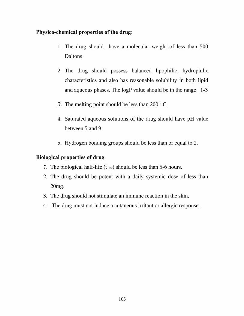
Physico-chemical properties of the drug:
1. The drug should have a molecular weight of less than 500
Daltons
2. The drug should possess balanced lipophilic, hydrophilic
characteristics and also has reasonable solubility in both lipid
and aqueous phases. The logP value should be in the range 1-3
3. The melting point should be less than 200 0 C
4. Saturated aqueous solutions of the drug should have pH value
between 5 and 9.
5. Hydrogen bonding groups should be less than or equal to 2.
Biological properties of drug
1. The biological half-life (t 1/2) should be less than 5-6 hours.
2. The drug should be potent with a daily systemic dose of less than
20mg.
3. The drug should not stimulate an immune reaction in the skin.
4. The drug must not induce a cutaneous irritant or allergic response.
105

2. Literature Review
• J.Y.Park et. al.,71 Studied comparative pharmacokinetic and
pharmacodynamic characteristics of amlodipine besylate and
amlodiple nicotinate in healthy subject and the results shows mean
ratio for AUC0-α and Cmax fell within the predetermined equivalent
range of 80-125% pharmacodynamics profiles including systolic and
diastolic blood pressures and pulses rates exhibited no significant
differences between the two formulations
• Paul W.Stott. et al.,72 Done mechanistic study into the enhanced
transdermal permeation of a model β-blocker, propranolol, by fatty
acid: a melting point depression effect and found that the binary
mixture of propranolol and fatty acid, the addition compounds are
formed by interaction between the carbonyl group of the β-blocker, to
form a salt. The oppositely charged species of the salt have been
shown to permeate the human epidermal membrane by an ion – pair
mechanism. This is in agreement with the work by Green and
Hadgraft (1987) which suggested the formation of ion-pairs between
propranolol and fatty acids. Where the process was driven by a pH
gradient.
• D. Monti et.al.,73 Conducted study on the comparison of the effect of
ultra sound (US) and of chemical enhancers on transdermal
permeation of caffeine(CAF) and morphine through hairless mouse
skin in vitro, and found that CAF confirm greater effect of low-
106

frequency US on skin permeation in vitro. The comparison of US and
chemical enhancement indicates a slight superiority of the
combination oleyl alcohol (OA) / propylene glycol (PG) over low –
frequency US. Concerning morphine (MOR), significantly increased
transdermal fluxes were produced by both low frequency US and by
OA in combination with PG.
• Rajagopal K. et. al.,74 Formulate and evaluate a matrix type
transdermal patches of nimesulide by using different polymers alone
or in combination, dibutyl phthalate as the plasticizer and aluminum
foils as the backing membrane. The studies showed that (2.2)
hydroxyl propyl methyl cellulose (HPMC) and ethyl cellulose (EC)
combination may be the suitable polymer combination for the
development of transdermal drug delivery system of nimesulide
• Jawahar N. et.al.,10 Prepare and evaluate verapamil hydrochloride
transdermal films and study the effect of different formulation
variables. The drug diffusion through the films followed a pattern
close to zero order type. The drug release profile was decreased with
increased polymer concentration and film thickness.
• L.M.A. Nolan et.al.,75 Studies Iontophoretic and chemical
enhancement of drug delivery part I: across artificial membranes and
reported that the delivery of salbutamol from the fatty acid containing
systems was substantially enhanced by iontophoresis and the rates
were shown to be approximately proportional to the assisting currents.
The data clearly indicates the iontophoretic process to be significantly
107
less efficient in the presence by buffer lons but with the iontophoretic
delivery rates being enhanced by the presence of a fatty acid.
• M.B.Blanco.et.al.,76 Transdermal application of bupivacaine – loaded
poly (acrylamide (A)- CO- monomethyl itaconate ) hydrogels found
the skin flux of the drug was between 90 ± 5 and 16 ± 7 mcg/ cm2 /h
depending on the amount of bupivacaine included in the gel and the
gel composition. Skin flux increases with the drug load of the gels.
Furthermore as more MMI in the gel slower skin flux of the drug due
to bupivacaine gel interactions.
• A.Nokhodchi. et. al., 77 Studies the enhancement effect of surfactants
on the penetration of lorazepam through rat skin. And concluded that
the increase in flux at low enhancer concentrations is normally
attributed to the ability of the surfactant molecules to penetrate the
skin and increase its permeatility. Reduction in the rate of transport of
the drug present in enhancer system beyond 1% w/w is attributed to
the ability of the surfactant molecules to form micelles and us
normally observed only if interaction between micelle & the drug
occurs.
• Xiaohong Qi et.. al., 78 Studies convolution method to predict drug
concentration profiles of 2,3,5,6-tetra – methylpyrazine following
transdermal application in rabbit from the in-vitro skin permeation
data and found that in – vitro skin permeation tests could be useful to
108

predict in vivo drug absorption profiles following transdermal
application.
• P.Rama Rao. et.al., 79 Provide comparative in-vivo evaluation of
propranolol hydrochloride after oral and transdermal administration in
rabbits. The PK parameters such as maximum plasma concentration
(Cmax), tmax, MRT Mean residence time) and AUC0-α were
significantly (P< 0.01) different following transdermal administration
compared to oral administration. The t1/2 of transdermally delivered
PPN was found to be similar to that following oral administration. But
sustained activity was observed over a period of 24hrs after
transdermal administration compared to oral. The relative
bioavailability of PPN was increased about fivefold to six fold after
transdermal compare to oral.
• Sandip B. Tiwari. et.al., 80 Done investigation into the potential of
iontophoresis facilitated delivery of ketorolac using rat skin. Results
found that pretreatment of the skin with D-limonene in ethanol or D-
limonene in ethanol + ultra sound significantly enhanced the
iontophoretic flux of the drug in comparision to passive flux with or
without pretreatment. Trimodality treatment comprising of
pretreatment with D-limonene in ethanol+ultrasound in combination
followed by iontophoresis was found to be most potent for enhancing
the rate of permeation of ketorolac
109

• Michal A.Ashburn. et.al., 81 Work on pharmacokinetics of
transdermal Fentanyl delivered with and without controlled heat and
there results suggest controlled heat might be used to significantly
shorten the time needed to reach clinically important fentanyl
concentrations, controlled heat might be useful to produce rapid
increase in serum concentrations for the rapid treatment of
breakthrough pain.
• Jagdish singh.et.al.,82 Done work on electronically facilitated
transdermal delivery of human parathyroid hormone (1-34) using
porcine skin. The flux of hPTH (1-34) with the electroporation pulses
of 100 and 300V followed by lontophoresis at 0.2mA/cm2 was 10 –
and 5 –fold higher respectively, in comparison to the flux with
corresponding pulses alone.
• Franziska Grafe,et.al.,83 Work on carrier mediated transport of
clonidine in human kerationocytes to characterize transport of
clonidine into human keratinocytes to characterize transport of
clonidine into human keratinocytes and conclude that clonidine us
transported into keratinocytes in a pH – dependent manner by a
saturable uptake system different from the keratinocyte choline
transporter
• P.N.Kotiyan. et.al.,84 Studies electron bean irradiation; a novel
technology for the development of transdermal system of lsosorbide
dinitrate (ISDN) dissolving in 2-ethylhexylacrylate (EHA)-acrylic
110

acid (AA) and solution irradiated on a backing membrane at different
doses to get transdermal patches. The ISDN-EHA-AA system
developed at an irradiation dose of 50KGY showed a higher skin
permeation profile as compared to an internationally marketed
transdermal matrix system of ISDN.
• Elvira Escribans. et. al., 85 Done assessment of diclofenaec
permeation with different formulation using human skin(0.4mm
thick)from plastic surgery as a membrane. The results suggest that
topical delivery of sodium diclofenac with an absorption enhancer
such as a mixture of oleic acid and d-limonene may be an effective
medication for both dermal and subdermal Injuries
• Jorg kreuter et.al.,86 Carry studies on crystallization of estradiol
containing TDDS determined by isothermal microcalorimetry X-ray
diffraction and optical microscopy as it is still a problem to achieve a
stable and prolonged constant drug release, to attain high permeation
rates across the skin, the concentrations of the drug dissolved have to
be high and often create supersaturated, thermodynamically
metastable or unstable systems that possess a high tendency to
crystallise.
• M.Jayne Lawrence et.al., 87 Done work on formulation of electrically
conducting micro emulsion – based organogels. (MBG). From work
conclude that MBG not formed with non-ionic surfactant alone, or
when used in combination with another non-ionic surfactant
111
(regardless of oil used). Which is due to inadequate level of water
available for by hydration of the surfactant head group.
• Akirayamanoto et.al.,88 Enhanced transdermal delivery of
phenylalanyl-glycine by chemical modification with various fatty
acids shows the results that the stability and permeability of Phe-Gly
were improved by chemical modification with improved by chemical
modification with fatty acids and this enchanced permeability of Phe-
Gly by the acylation may be attributed to the protection of Phe –Gly
from the enzymatic degradation in the skin and the increase in the
partition of Phe-Gly to the stratum corneum.
• K.C. sung, et.al., 89 assess the effect of electro oration on transdermal
permeation of nalbuphine (NA) and its prodrug. Study demonstrated
that electroporation enhance and control transdermal permeation of
NA and its prodrugs. The results also indicated that the
physicochemical properties of prodrug has significant effects on
kinetics as well as mechanisms of transdermal permeation by
electroporation.
• Ramesh panchagnula et.al.,90 work on transdermal delivery of
zidovudine effect of vehicles on permeation across rat skin and their
mechanism of action, to assess to effect of various solvent systems
containing water, ethanol, propylene glycol (PG). Studies support that
among all the solvent combinations, highest flux and short lag time
were archived with ethanol at 66.6% in water and hence is a suitable
vehicle for transdermal delivery of AZT.
112

3. Aim and Objective
Objective of this study
Cardiovascular drugs have relatively low therapeutic indices, which
place responsibility on the dosage forms for maintaining the drug blood
level within narrow limit .Conventional dosage forms suffer from variations
in the absorption and thus leading to wide fluctuation in the plasma drug
concentration.
Amlodipine Besylate, is an antihypertensive drug, having calcium
channel blocking activity is therapeutically active even at very low doses.
E.g.: 5-10mg/day which is one of the essential requirements of a drug
candidate for use in transdermal delivery systems.
It also has other suitable properties which include.
1. Low molecular weight.
2. High lipid solubility
3. Extensive first pass metabolism etc.
Transdermal film prepared by various polymers can exhibit good
controlled release properties. So, an attempt is done to formulate transdermal
film containing Amlodipine Besylate for achieving following goals.
1. To produce steady state plasma concentration
2. To improve stability of Amlodipine in vivo
3. To reduce adverse effect.
113

4. Plan of Work
1. Identification of drug by spectrophotometric method (IR and UV).
2. Determination of the, partition coefficient and melting point of
Amlodipine.
3. Determination of the drug polymer interaction study between the drug
(Amlodipine Besylate) and the polymer (HPMC) through differential
Scanning calorimetry (DSC)
4. Study on the extent of in vitro permeation of Amlodipine from
Transdermal device in the presence of penetration enhancers.
5. Study on the in vivo release pattern using rabbit.
6. To study the skin irritation of transdermal film using rabbit.
7. Stability Studies
114
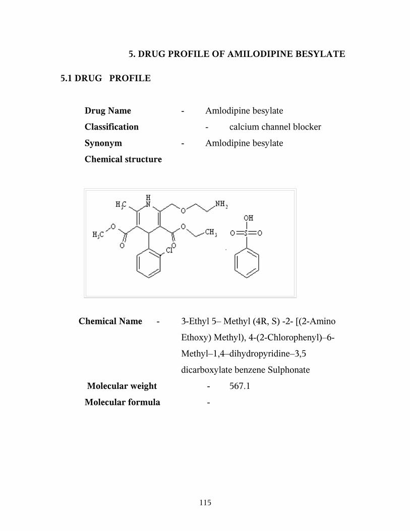
5. DRUG PROFILE OF AMILODIPINE BESYLATE
5.1 DRUG PROFILE
Drug Name - Amlodipine besylate
Classification - calcium channel blocker
Synonym - Amlodipine besylate
Chemical structure
Chemical Name - 3-Ethyl 5– Methyl (4R, S) -2- [(2-Amino
Ethoxy) Methyl), 4-(2-Chlorophenyl)–6-
Methyl–1,4–dihydropyridine–3,5
dicarboxylate benzene Sulphonate
Molecular weight - 567.1
Molecular formula -
115

Description
Colour - white crystalline powder
Partition coefficient(log P) -3.00
Dissociation Constant
(PKa) at 25 0 C - 8.6
Odour - odourless
Solubility - slightly soluble in water
- Sparingly soluble in ethanol
- Freely soluble in methanol
Melting point - 178 -179 0 C
Optical rotation - Racemic mixture
CLINICAL PHARMACOLOGY 12, 16
Amlodipine Besylate is a Dihydro pyridine derivative which is the
most potent Ca2+ Channel Blockers. Bind to specific sites on the alpha1
subunit, all restricting Ca2+ entry
cAMP – phospho diesterase resulting in raised smooth muscle cAMP
showing smooth muscle relaxant action.
Released endothelial nitrous oxide may exert anti atherosclerotic
action.
Amlodipine inhibits the movement of Calcium Ions (Ca2+) across the
cell membrane into vascular smooth and myocytes. Action is greater in the
arterial resistant vessels causing peripheral vasodilatation and reduction in
after load. Action on the myocardium is considerably less. In angina patient
reduction of after load reduces myocardial oxygen requirement.
116

Amlodipine produces vasodilatation resulting in a reduction of supine
and standing blood pressure. Amlodipine does not change sinoatrial (SA)
nodal function or atrioventricular (AV) conduction in intact animals or
humans. The decrease in blood pressure is not accompanied by a significant
change in heart rate or plasma catecholamine levels with chronic dosing.
Vasodilatations also increase the oxygen available to the heart in patients
with coronary artery spasm and blunt coronary vaso constriction.
PHARMACOKINETICS:
Absorption/ Distribution:
Amlodipine is almost completely well absorbed following oral
administration with no effect of food. Bioavailability range is 45 -60% Due
to a significant degree of first pass metabolism. Following oral
administration, . Time to peak plasma levels occur after 6 -12 hours.
Amlodipine, plasma protein binding is 97.5 % having volume of
distribution around 20 L\ Kg. Time to steady state is 7 To 8 days. A normal
serum level of Amlodipine may fall in the range 3-11 ng/ml. Amlodipine
Crosses the Blood Brain Barrier, Placenta & is excreted in Breast Milk.
Metabolism/ Excretion:
Amlodipine is extensively metabolized in the liver, with 10 % of the
parent compound and 60% of the metabolites excreted in the urine. In
patients with hepatic dysfunction, decreased clearance of Amlodipine may
increase the area under the plasma concentration curve by 40 % - 60 % &
dosage reduction may be required. 5% dose of Amoldipine recovered
unchanged through urine.
117

Dosage
Adults: 5 – 10mg once a day
Elderly patient should start with 2-5mg one day & the dose built up as
required.
Therapeutic uses:
Amlodipine is used in the treatment for:
Stable and Prinzmetals angina,
Heart failure,
Hypertension.
5.2 Excipients profiles
Hydroxyl propyl methyl cellulose
Non-proprietary Name Hypromellose
Hydroxyl propyl methylcellulose
Functional category Suspending agent
Coating agents
Tablet binder
Film former
Synonyms Methyl hydroxyl propyl cellulose
Propylene glycol, ether cellulose
Hydroxyl propyl methyl cellulose
Chemical Name Cellulose 2- hydroxyl propyl methyl ether
cellulose, hydroxyl propyl methyl
118

ether.
Typical properties
Density 0.341 gm/cm3
Solubility Soluble in cold water forming viscous colloidal
solution. Insoluble in alcohol, ether and
chloroform.
Grade HPMC K 100 M
HPMC K 4 M
HPMC K 15M
HPMC k100 LVP
Viscosity HPMC K 100 M - 8000 to 120 000 m pas
HPMC K 4 M - 3000 to 5600 m pas
Application of HPMC in Pharmaceutical Formulation
Film former in tablets film coating. Lower viscosity grades are used in
aqueous film coating. Higher viscosity grades may be used to retard the
release of drugs from a matrix a levels of 10 - 80 %w/w in tablet and
capsule. Depending up on the viscosity grade, concentration of 2 - 20% w/w
are used for film forming solution to film coat tablet. Lower viscosity grade
are used in aqueous film coating solution, while higher viscosity grades are
used with organic solvent.
119

6.MATERIALS AND METHODS
6.1 Equipments
UV- Visible spectro photometer
Shimadzu – 1700 model.
Open tubular cell
Magnetic stirrer
PH meter
Differential scanning calorimeter : Perkin Elmer, DSC-7
Fourier Transform Infrared : Perkin Elmer. .
6.2 List of reagents
0.1 N Hydrochloric acid
8.5 ml of Concentrated Hydrochloric acid is dissolved in 1000 ml of
distilled water.
6.3 Materials
Amlodipine Besylate BP- Standard drug Supplied by Orchid
Pharmaceuticals Ltd, Chennai.
Hydroxypropyl methyl Cellulose – Supplied by S.D. Fine chemicals,
Bombay.
120

Methanol – (Analytical grade) supplied by Allied chemicals
corporation, Vadodara.
Dimethyl sulphoxides (DMSO)
supplied by Suvidhinath Laboratories, Baroda.
6.4 Methods
Preformulation Studies
1. IR Determination
IR determination was carried out by Perkin Elmer Infra-Red
Spectrophotometer using KBr Pellet technique 69,70
2. Melting point determination
Melting point of pure Amlodipine Besylate was carried out by open
capillary tube method.
3. Differential scanning calorimetry DSC, 64 70
The excipient induced modification in the thermal behaviour, were
able to correlate long - term stability behaviour simply with the changes in
thermal features characteristics of the drug alone. Compatibility and
incompatibilities in DSC is concluded by elimination of endothermic peaks
121

(s), appearance of new peaks (s), change in peak shape and its onset, peak
temperature / melting point and relative peak area or enthalpy.
Method
The DSC analysis of each sample under the analogues conditions of
temperature range ( 50 – 550C), heating rate (200 min-1), chart speed 1cm-1
under nitrogen atmosphere, with alumina as reference material, were carried
out by using Perkin - Elmer DSC -7 analyser.
In order to avoid the need for normalization of enthalpy change of
account of great variation in sample weight, care should be taken to conduct
DSC analysis accurately. Weight 5 mg or very close to 5 mg portions of the
drug, as well as each excipient and transdermal film (2-8mg) equivalent to
5mg of the drug. This would help not only to evaluate data analysis in terms
of enthalpy change (loss / gain) associated with the peak of interest but also
provide the gross idea about the possibility of interaction simply from visual
comparison of the curves relevant to the individual excipient
122
4. Determination of partition efficient (P) 51,57
Partition coefficient is the ratio of concentration of drug in n-octanol
to the concentration of drug in water. The concentration determined by using
UV-spectrophotometer.
The intrinsic partition coefficient (P1) may be more appropriate
parameter to measure the partition coefficient of the non-ionized
compound. So, P1 was calculated using formula.
P1 = P [antilog [Pk – PH] +1
6.5 Standard Curve for Amlodipine61
Standard curve of Amlodipine were drawn in 0.1NHCl using
UV- Visible spectrophotometer.
Procedure for the Preparation of Standard Curve:-
Amlodipine Besylate 50mg each, were accurately weighed and
dissolved separately in 50ml of methanol. Five ml of the above solutions
diluted separately to standard stock solution of 100 mcg/ml eight dilutions
ranging 5-40 mcg/ml made. The absorbance of each sample was measured
using UV-visible spectrophotometer. 0.1 N HCl as blank.The calibration
curve was drain by plotting concentration Vs and absorbance to obtain
standard calibration curve. The results were shown in table and figure.
123

Table No.2 Standard Curve for Amlodipine
Concentration (mcg/ml) Absorbance (365nm)
5 0.07610 0.15015 0.17920 0.25825 0.28930 0.35335 0.42540 0.468r-0.9968
a-0.0244
b-0.0111
Standard Curve for Amlodipine
124

6.6 procedure for fabrication of transdermal film
Procedure for fabrication of transdermal. Medicated monolithic films:
Transdermal films were prepared by the film casting method of specially
designed glass molds with plastic transparent sheets. Different ratio of
polymer. 1:4(F2), 1:6(F3) and 1:8(F4) (Drug: HPMC) were used for
preparation of films. Varying ratio of polymer dissolved in water. Then drug
which dissolved in small quantity of methanol was incorporated in polymer
solution obtained by stirring with glass rod for 10 min. The penetration
enhancer (Dimethyl sulphoxide) is added to Drug- polymer solution. The
solution is poured on petridish using rod. The rate of evaporation of solvent
was controlled by inverting cup funnel. After 24 hrs, the dried films were out
and stored in desiccator between sheets of paper.
125

Table No.3 Formula for TDDS
Formulation Drug (mg) HPMC
(mg)
DMSO
(ml)
Solvent (water)
(ml)
F1 25 50 2ml Upto 7 ml
F2 25 100 2ml Upto 7 ml
F3 25 150 2ml Upto 7 ml
F4 25 200 2ml Upto 7 ml
126

6.7 EVALUATION OF TRANSDERMAL DRUG DELIVERY
SYSTEM
Permeation Studies 10,49
The in vitro permeation studies were carried out using Dialysis
membrane. The membrane was prepared using following procedure. The
membrane is cut into appropriate size & socked in liquid glycerol for 24
hours for permeable characteristics.
The permeation study was conducted using open tubular cell in the
static mode with an effective diffusion area 2.8cm2. The capacity of receptor
compartment was 200ml and temperature was maintained at 370c+1 by
means of temperature controller of magnetic stirrers. Drug concentration in
donor compartment was 7.473mg/ml Receptor solution was 0.1mHCl which
was continuously stirred at 100 rpm with a Teflon – coated bar movement
placed inside the cell.
Dialysis membrane was mounted between donor and receptor
compartment of the cells and lower side of membrane is in direct contact
with the receptor medium. After fixing of patch on membrane, the 5ml of
solution withdrawn at specific time interval and equal volume of 0.1N HCl
added to receptor solution in an attempt to maintain drug concentration
constant through out the experiment (24 hrs). The amount of drug permeated
from receptor solution at predetermined times were analyzed by UV-visible
spectrophotometer.
127

In vitro diffusion study10,62
The in vitro release profile of Amlodipine from a transdermal device
was determined. An open tubular cell was used for the diffusion studies. The
dialysis membrane was mounted between donor and receptor compartment
of diffusion. The receptor compartment of the diffusion cell was filled with
0.1NHCl. The prepared matrix patch was placed over the membrane. The
temperate of the receptor compartment was maintained at 37±10C. The
receptor solution was stirred with Teflon coated magnet stirrer through out
the experiment. Aliquots of receptor fluid were withdrawn at predetermined
time intervals and replaced immediately with same volume of the fresh fluid,
the samples were analyzed at 365 nm using spectrophotometrically.
In vivo release studies65
In vivo release studies were carried out using both gender rabbits for
transdermal patch containing hydroxy propyl methyl cellulose as polymer
and dimethylsulpoxide (DMSO). Four rabbits weighing 1.5 to 2.0kg were
selected and the dorsal surface was cleaned and hair was removed. Trans
dermal patch of 2.8cm2 having dose equivalent to 7.473mg were placed with
the help of adhesive tape. Blood samples (0.5ml) were with drawn from the
marginal ear (cuboidal vein) into heparinized glass vessels at 0, 0.50, 1, 2, 4,
6, 8, 12, 24 and 48 hrs..The plasma was separated immediately by
centrifugation at 2000 rpm for 10min and stored in refrigerator until
analysis. The absorbance of the solution was measured at 365nm against a
blank
128

Skin irritation test
A primary skin irritation test was performed since skin is vital organ
through which drug is transported. The test was carried out on eight healthy
rabbits weighing 1.5 to 2.0 kg. Drug free polymeric films of diameter
2.8cm2 containing dimethylsulphoxide as a penetrant were used as control.
The dorsal surface of rabbits was cleared well and the hair was removed by
using a fresh blade. The skin was cleared with rectified spirit. Transdermal
patches containing amlodipine (7.47mg equivalent) in HPMC were place
over the skin with the help of adhesive tape. The films and patches were
removed after 48hrs and the skin was examined for erythema / oedema. All
the experimental protocol involving laboratory animals were approved by
the IAEC.
129

6.8 ACCELERATED STABILITY STUDIES 90
Stability
Stability is officially defined as the time lapse during which the
drug product retains the same properties and characteristics that is possessed
at the time of manufacture. This process begins at early devolvement phase.
Instability in modern formulation is often detectable only after
considerable storage period under normal condition. To assess the stability
of a formulated product its usual to expose it to high stress conditions to
enhance its detoriation and therefore the time required for testing is reduce
common high stress like temperature and humidity. This will eliminate
unsatisfactory formulation.
Strategy of stability testing
1. The study of drug decomposition kinetics
2. The development of stability dosage form.
3. Establishment of expiration date for commercially available drug
product is some of the needs of stability testing.
4. Data form stability studies should be provided on atleast three primary
batches of the drug product.
5. The batches should be manufactured to a minimum of pilot scale.
6. Important point of view of the safety of the patient, patient relieves a
uniform dose of drug throughout the shelf life of the product.
130

Table No.4The Stability Storage condition
S.No Study Storage condition Minimum
period
1. Long-term study 25° C ± 2° C
60 % ± 5% RH
12 month
2. Intermediate study 30 ° C ± 2° C
60 % ±5 % RH
6 month
3. Accelerated study 40° C ± 2 ° C
75% ±5 % RH
6 month
ICH (International Conference on Harmonization) Guidelines
Specification
1. 5% potency loss from initial assay of batch
2. Any specification degradation that exceed specification
3. Product failing out of pH limit
4. Dissolution out of specification for 12 minutes
5. Failure to meet specification for appearances and physical properties
131
Any one condition is observed then the stability of the batch is failed.
Procedure
Stability studies were carried out using temperature controlled
environment test chamber according to ICH guidelines by storing the
formulated films (F2) at 400c / 75 + 5% RH for a period of 3 months. The
samples were withdrawn at 30, 60 and 90 days and analysed for drug
content by spectrophotometrically.
132

7. Results and Discussion
Preformulation Studies
Drug Identification
Method - Spectrophotometer method (UV)
Table No.5 UV- Absorbance of Amlodipine Besylate in 0.1 N HCl
S. No
Concentration in mcg/ml
Absorbance at 365nm
1 25 mcg/ml 0.281
Discussion
The UV- absorbances of the Amlodipine besylate were performed at
25 mcg/ml concentration in 0.1 N HCl and their wavelength were
found and compared with monograph.
133

Figure No.7 UV- Absorbance of Amlodipine Besylate in 0.1 N HCl
134

Infra-Red Spectrum
Table No.6 IR Spectrum of Amlodipine Besylate
S.No Frequency cm-1 Functional Group
1 3411 NH Stretching 2 2813 CH-aliphatic 3 1672 C=0 ester 4 561 C - C1 (C1 vibration) 5 1120 - C- 0 - ester 6 1382 – C - CH37 1593 - C = C - aromatic 8 756 Mono substituted aromatic 9 1208 C - 0 Stretching 10 1204 Phenolic Stretching
Discussion
• The structure of Amlodipine Besylate was confirmed by IR spectrum (performed by KBr pellet method).
135
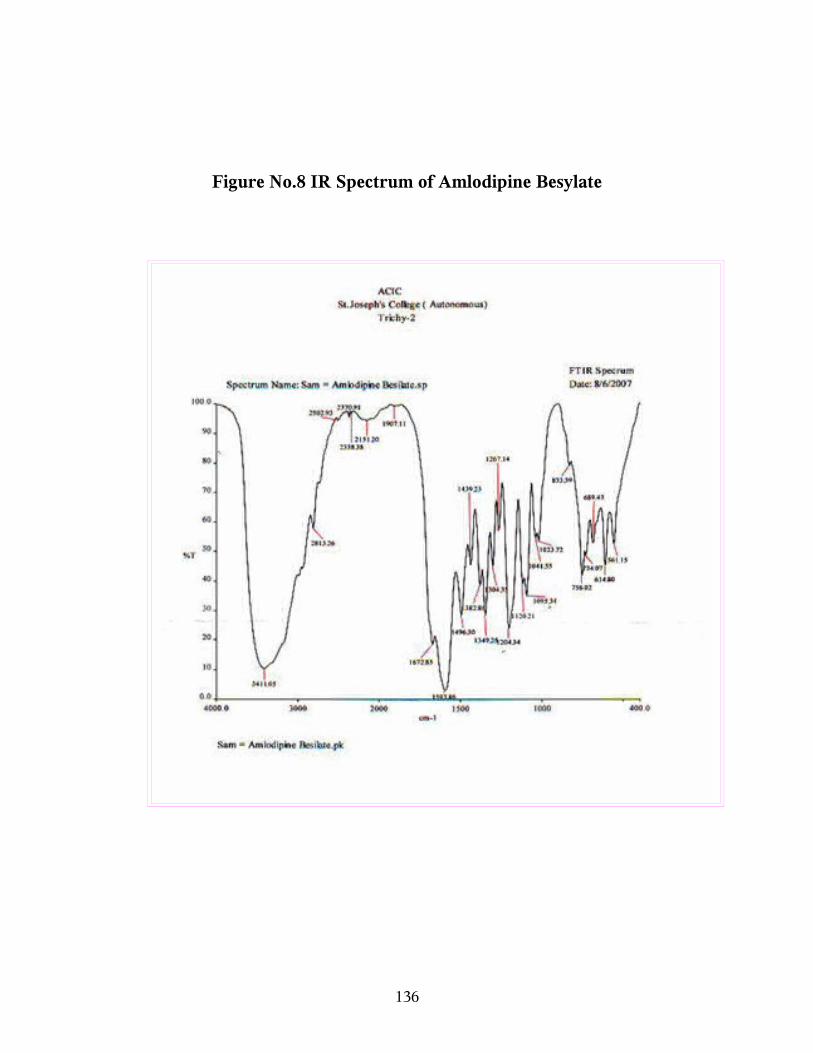
Figure No.8 IR Spectrum of Amlodipine Besylate
136

Compatibility Studies
DSC Graph for Amlodipine Besylate Patch
Discussion
Differential scanning calorimetry (DSC) was used for compatibility
studies between the drug and the excipients. The DSC graph shows that the
drug and the excipients are compatible with each other as no elimination of
endothermic peak occur and the melting point of drug was also seen in the
DSC graph of transdermal film. From above all, the interpretation conclude
that the drug and excipients (HPMC) are compatible with each other when
they are in the film form
137

Figure No.9 DSC Graph for Amlodipine
138

Figure No.10. DSC Graph for HPMC
139
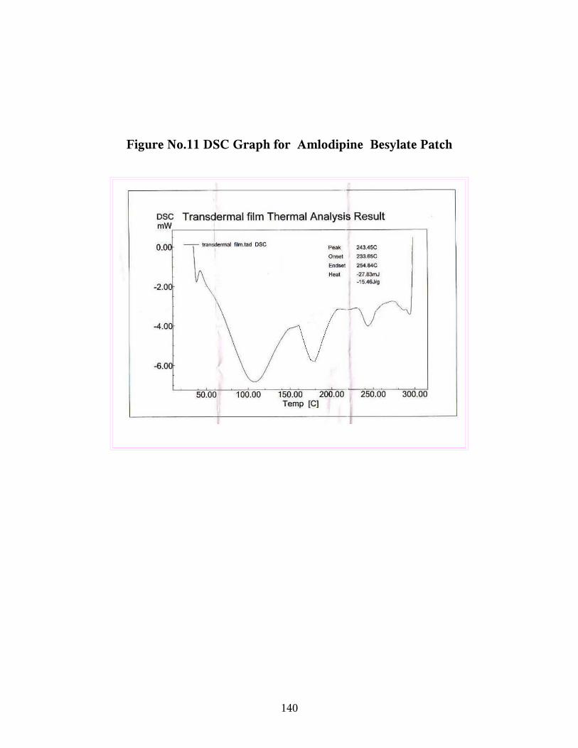
Figure No.11 DSC Graph for Amlodipine Besylate Patch
140
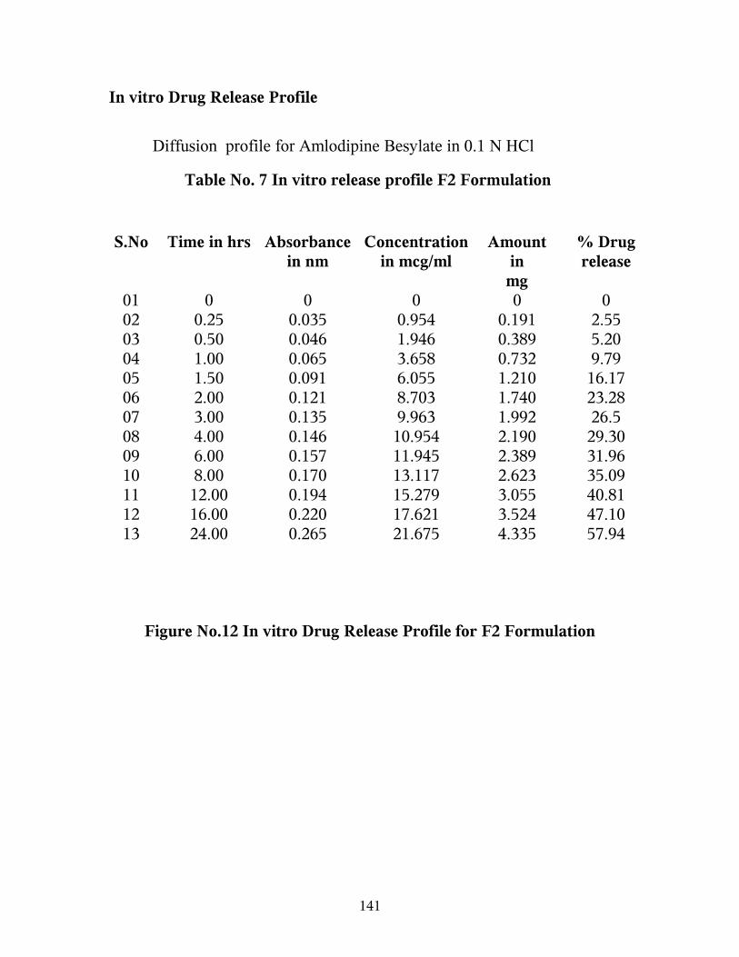
In vitro Drug Release Profile
Diffusion profile for Amlodipine Besylate in 0.1 N HCl
Table No. 7 In vitro release profile F2 Formulation
S.No Time in hrs Absorbance in nm
Concentration in mcg/ml
Amount inmg
% Drug release
01 0 0 0 0 002 0.25 0.035 0.954 0.191 2.5503 0.50 0.046 1.946 0.389 5.2004 1.00 0.065 3.658 0.732 9.7905 1.50 0.091 6.055 1.210 16.1706 2.00 0.121 8.703 1.740 23.2807 3.00 0.135 9.963 1.992 26.508 4.00 0.146 10.954 2.190 29.3009 6.00 0.157 11.945 2.389 31.9610 8.00 0.170 13.117 2.623 35.0911 12.00 0.194 15.279 3.055 40.8112 16.00 0.220 17.621 3.524 47.1013 24.00 0.265 21.675 4.335 57.94
Figure No.12 In vitro Drug Release Profile for F2 Formulation
141

In vitro Drug Release
0
10
20
30
40
50
60
70
0 5 10 15 20 25 30
Time in hours
% D
rug
Re
las
e
F2
142

Table No.8 In Vitro Release Profile F3 Formulation
S.No Time in hrs Absorbance
in nm
Concentration
in mcg/ml
Amount
in
mg
% Drug
release
01 0 - - - -02 0.25 0.008 - - -03 0.50 0.017 - - -04 1.00 0.025 0.054 0.010 0.1305 1.50 0.034 0.872 0.174 2.3206 2.00 0.041 1.495 0.299 4.0007 3.00 0.062 3.387 0.677 9.0508 4.00 0.078 43828 0.965 12.9109 6.00 0.095 6.360 1.272 17.0210 8.00 0.118 8.432 1.686 22.5611 12.00 0.147 11.045 2.209 29.5512 16.00 0.179 13.927 2.785 37.2613 24.00 0.210 16.720 3.344 44.74
Figure No.13 In vitro Drug Release Profile for F3 Formulation
143
In vitro Drug Release
0
5
10
15
20
25
30
35
40
45
50
0 5 10 15 20 25 30
Time in hours
% D
rug
Rel
ease
F3
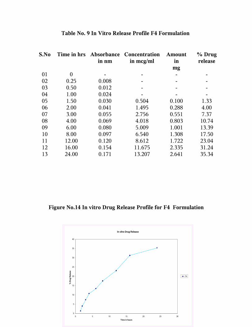
Table No. 9 In Vitro Release Profile F4 Formulation
S.No Time in hrs Absorbance in nm
Concentration in mcg/ml
Amount inmg
% Drug release
01 0 - - - -02 0.25 0.008 - - -03 0.50 0.012 - - -04 1.00 0.024 - - -05 1.50 0.030 0.504 0.100 1.3306 2.00 0.041 1.495 0.288 4.0007 3.00 0.055 2.756 0.551 7.3708 4.00 0.069 4.018 0.803 10.7409 6.00 0.080 5.009 1.001 13.3910 8.00 0.097 6.540 1.308 17.5011 12.00 0.120 8.612 1.722 23.0412 16.00 0.154 11.675 2.335 31.2413 24.00 0.171 13.207 2.641 35.34
Figure No.14 In vitro Drug Release Profile for F4 Formulation
144
In vitro Drug Release
0
5
10
15
20
25
30
35
40
0 5 10 15 20 25 30
Time in hours
% D
rug
Rel
ease
F4

Figure No.15 Comparative In vitro Drug Release Profile
145
In vitro Drug Release Profile
0
10
20
30
40
50
60
70
0 5 10 15 20 25 30
Time in hours
% D
rug
Rel
ease
F2
F3
F4

In vivo Drug Release Profile
Table No.10 In Vivo Release Profile Control Formulation
∑ AUC = 2183.92
∑ AUMC = 53481.1849
Time in hrs (t)
Absorbance in nm
Plasma drug concentration µg / ml
Amount of drug in mg
% Drug release
C T [AUC]t 0
µghr/ml
[AUMC]t 0
µghr2/ml
0 0 - - - - 1.995 0.9976
0.50 0.113 7.981 0.798 7.98 3.990 6.918 5.9208
1.00 0.243 19.693 1.969 19.69 19.693 24.513 39.1765
2.00 0.350 29.333 2.933 29.33 58.666 65.062 201.582
4.00 0.421 35.729 3.572 35.72 142.916 76.143 385.4
6.00 0.473 40.414 4.041 40.41 242.484 83.531 587.42
8.00 0.503 43.117 4.311 43.11 344.936 184.9 1873.864
12.0
0
0.572 49.333 4.933 49.33 591.996 608.754 11058.12
24.0
0
0.603 52.126 5.212 52.12 1251.02
4
1132.104 39328.704
48.0
0
0.493 42.216 4.221 42.21 20.26.3
68
0 0
146

Figure No.16 In vivo Drug Release Profile for control Formulation
147

In vivo Drug Release
0
10
20
30
40
50
60
0 10 20 30 40 50 60
Time in hours
Pla
sma
Dru
g C
onc
entr
atio
n in
mcg
/ml
Control Formulation
148

149

Table No: 11 Pharmacokinetic Parameter for Control
Pharmacokinetic Parameter Values
KE 0.0801 hr-1
t ½ 8.65 hrs
Cmax 52.126 mcg/ml
tmax 24 hrs
[AUMC] 48 0 85375.412 mcg hr2/ml
[AUC] 48 0 2711.62 mcg hr/ml
MRT 31.485 hrs
150

Table No.12 In Vivo Release Profile F2 Formulation
∑ AUC = 1687.0812
∑ AUMC = 44405.0229
Time in
hrs (t)
Absorbance at 365 nm
Plasma drug concentration
µg / ml
Amount of drug in mg
% Drug releas
e
C T [AUC]t 0
µghr/ml[AUMC]t
0
µghr2/ml
0 0 - - - - 0.4187 0.2093
0.50 0.043 1.675 0.167 2.24 0.8375 1.2205 1.0111
1.00 0.060 3.207 0.320 4.29 3.207 4.153 6.7025
2.00 0.081 5.099 0.509 6.82 10.198 14.071 46.086
4.00 0.124 8.972 0.897 12.00 35.888 26.323 139.994
6.00 0.217 17.351 1.735 23.21 104.106 44.161 318.586
8.00 0.322 26.810 2.681 35.87 214.48 135.89 1416.2
12.0
0
0.481 41.135 4.113 55.04 493.62 509.292 9261.288
24.0
0
0.510 43.747 4.374 58.54 1049.92
8
951.552 33075.36
48.0
0
0.419 35.549 3.554 47.57 1706.35
2
0 0
151

Figure No.17 In vivo Drug Release Profile for F2 Formulation
In vivo Drug Release
0
5
10
15
20
25
30
35
40
45
50
0 10 20 30 40 50 60
Time in hours
Pla
sma
Dru
g co
ncen
trat
ion
in m
cg/m
l
F2
152

153

Table No. 13 Pharmacokinetic Parameter for Formulation F2
Pharmacokinetic Parameter
Values
KE 0.1059 hr-1
t ½ 6.54 hrs
Cmax 43.747 mcg/ml
tmax 24 hrs
[AUMC] 48 0 63887.720 mcg hr2/ml
[AUC] 48 0 2022.765 mcg hr/ml
MRT 31.584 hrs
154
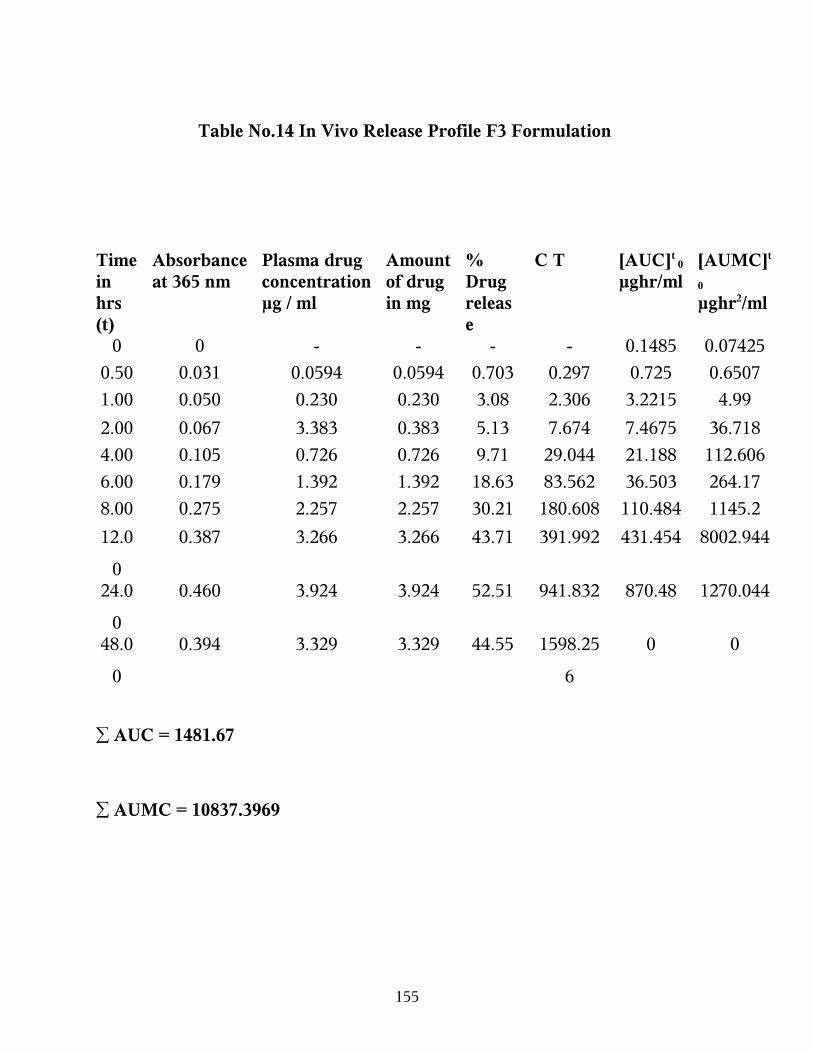
Table No.14 In Vivo Release Profile F3 Formulation
∑ AUC = 1481.67
∑ AUMC = 10837.3969
Time in hrs (t)
Absorbance at 365 nm
Plasma drug concentration µg / ml
Amount of drug in mg
% Drug release
C T [AUC]t 0
µghr/ml[AUMC]t
0
µghr2/ml
0 0 - - - - 0.1485 0.07425
0.50 0.031 0.0594 0.0594 0.703 0.297 0.725 0.6507
1.00 0.050 0.230 0.230 3.08 2.306 3.2215 4.99
2.00 0.067 3.383 0.383 5.13 7.674 7.4675 36.718
4.00 0.105 0.726 0.726 9.71 29.044 21.188 112.606
6.00 0.179 1.392 1.392 18.63 83.562 36.503 264.17
8.00 0.275 2.257 2.257 30.21 180.608 110.484 1145.2
12.0
0
0.387 3.266 3.266 43.71 391.992 431.454 8002.944
24.0
0
0.460 3.924 3.924 52.51 941.832 870.48 1270.044
48.0
0
0.394 3.329 3.329 44.55 1598.25
6
0 0
155

Figure No.18 In vivo Drug Release Profile for F3 Formulation
In vivo Drug Release
0
5
10
15
20
25
30
35
40
45
0 10 20 30 40 50 60Time in hours
Pla
sm
a d
rug
co
nce
ntr
ati
on
in
mc
g/m
l
F3
156

157

Table No. 15 Pharmacokinetic Parameter for Formulation F3
Pharmacokinetic Parameter Values
KE 0.09212 hr-1
t ½ 7.532 hrs
Cmax 39.243 mcg/ml
tmax 24 hrs
[AUMC] 48 0 32119.593 mcg hr2/ml
[AUC] 48 0 1843.200 mcg hr/ml
MRT 17.425 hrs
158

Table No.16 In Vivo Release Profile F4 Formulation
Time In Hrs (T)
Absorbance
at 365 nm
Plasma Drug Concentration µg / Ml
Amount Of Drug In mg
% Drug Release
C T [Auc]t0
µghr/Ml
[Aumc]t0
µghr2/Ml
0 0 - - - - 0.126 0.063
0.50 0.030 0.504 0.050 0.67 0.252 1.565 0.5945
1.00 0.048 2.126 0.212 2.84 2.126 2.8015 4.54
2.00 0.063 3.477 0.347 4.65 6.954 7.585 23.386
4.00 0.070 4.108 0.410 5.49 16.43
2
14.702 79.996
6.00 0.142 10.594 1.059 14.17 63.56
4
26.684 192.284
8.00 0.203 16.090 1.609 21.53 128.7
2
78.052 807.904
12.00 0.279 22.936 2.293 30.69 275.2
32
313.61
4
5875.344
24.00 0.350 29.333 2.933 39.25 703.9
92
629.4 21763.29
648.00 0.281 23.117 2.311 30.93 1109.
616
0 0
∑ AUC =1074.5295∑ AUMC = 28747.4075
159

Figure No.19 In vivo Drug Release Profile for F4 Formulation
in vivo Drug Release
0
5
10
15
20
25
30
35
0 10 20 30 40 50 60
Time in hours
Plas
ma
drug
con
cent
ratio
n in
mcg
/ml
F4
160
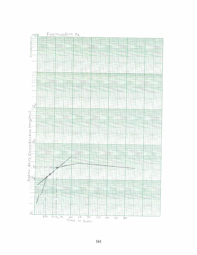
161

Table No. 17 Pharmacokinetic Parameter for F4 Formulation
Pharmacokinetic Parameter Values
KE 0.1287 hr-1
t ½ 5.384 hrs
Cmax 29.333 mcg/ml
tmax 24 hrs
[AUMC] 48 0 38764.775 mcg hr2/ml
[AUC] 48 0 1254.148 mcg hr/ml
MRT 30.90 hrs
162
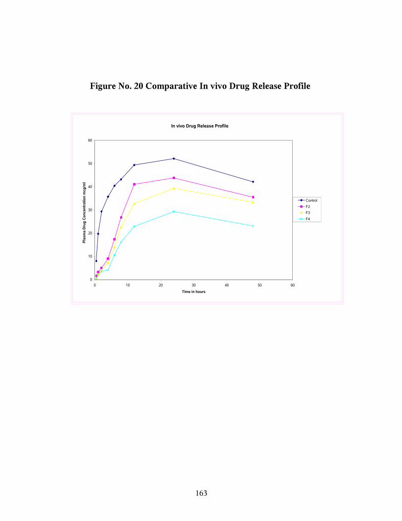
Figure No. 20 Comparative In vivo Drug Release Profile
In vivo Drug Release Profile
0
10
20
30
40
50
60
0 10 20 30 40 50 60
Time in hours
Pla
sma
Dru
g C
on
cen
trat
ion
mcg
/ml
Control
F2
F3
F4
163

Skin Irritation Studies
The results of the skin irritation studies indicate that neither the
adhesive nor Amlodipine have any noticeable irritation on the rabbit skin for
the 48 hours study periods.
Stability Studies
Stability studies revealed that the drug content in formulation F2,
degraded respectively by 0.76%, 1.40% and 1.90% in a period of 90 days.
Table No.14 Stability studies for F2 Formulation
Time period in
month
Drug Content %
Initial 99.24
1st Month 99.14
2nd Month 98.60
3rd Month 98.10
164

165
8. SUMMARY AND CONCLUSION
The formulation of transdermal films were prepared using different ratio
of the same polymers which assigned in increasing order according to
permeation rates F2>F3> F4. The results indicate that the films prepared with
hydroxy proplymethyl cellulose as polymer of 1:4 (Drug: HPMC) F2 was found
to be best during in vitro study. Hence conclude that the increase in the
concentration of polymer and film thickness decreases the drug release profile.
The in vivo studies demonstrate the transdermal drug delivery system
gives sustained plasma concentration and avoids wide fluctuation of plasma
concentration of amlodipine in rabbit.
♦ From the IR interpretation and UV- results the sample confirmed to be of
amlodipine besylate.
♦ Melting point, partition coefficient, and solubility determined
experimentally and molecular weight taken from literature.
♦ All physicochemical character support that the drug amlodipine may be
suitable for transdermal drug delivery system.
♦ DSC studies carried out and it support the drug and excipient was
compatible when they were in film form.
♦ In vitro studies carried out using F2, F3 and F4 formulation as F1
formulation polymer concentration not sufficient to form a film. F2
formulation shows better release than F3 and F4.
166

In vivo studies carried out also using F2, F3 and F4 and better release was
shown only by F2 formulation. The in vivo studies demonstrate the transdermal
drug delivery system gives sustained plasma concentration and avoid wide
fluctuation of plasma concentration of amlodipine in rabbits as diffusion of the
drug was extended over a long period at control rate and it followed a pattern
close to zero order release profile.
The results of the skin irritation studies indicate that neither the adhesive
nor amlodipine have any noticeable irritation on the rabbit skin for the 48 hours
study periods. Skin irritation and / or edema was not observed during skin
irritation test. Hence, amlodipine films can be used in long term management of
cardio vascular diseases, necessitating long term therapy by nullifying the
common adversities of the drug.
Stability studies revealed that the drug content in formulation F2 degraded
respectively by 0.76%, 1.40% and 1.90% in a period of 90 days.
Hence, finally conclude that the study shows the feasibility of
formulating rate controlled transdermal drug delivery system for amlodipine
besylate in order to achieve improved bioavailability and nullifying the
common adversities of the drug.
167
BIBLIOGRAPHY
1. Christopher T. Rhodes, Modern pharmaceutics, second edition, Marcel Dekker ,INC NewYork, P 635-668.
2. Chien, YIE.W. , Novel Drug delivery system, Marcel Dekker , INC , Newyork , P1-6.
3. Joseph R. Robinson and Vincent H.L. Lee, Controlled drug delivery, second edition, Mercel Dekker, INC, Newyork ,P1-6.
4. Michael E.Aulton., Pharmaceutics, The Science of dosage form design, ELBS, P381-400.
5. Remington: The Science and Practice of Pharmacy, 19th edition, Mack publishing company, Pennsylvania P1600-1675.
6. Praveen Tyle., specialized drug delivery system Vol.26, Mercel Dekker, INC P 3-13
7. Dr. P.K Chaudhary., et al., Transdermal drug delivery system: An overview, The Eastern Pharmacist, 45-47, July-97.
8. S.C. Basak., et al, Transdermal drug delivery system: An overview, The Eastern pharmacist, 63-67, Aug’97.
9. Dr.Krishna., et al., Transdermal Drug Delivery System: An overview, The Eastern Pharmacist, 17-22, Oct – 1991.
10. Jawahar N., et al., Evaluation of different Formulation variables on Transdermal films of verapamil hydrochloride: The Indian Pharmacist P.67-69, June –2007.
11. Johnson M.E,et al., A mechanism to enhance transdermal drug delivery. J.Pharm sci., 85, 670-679, 1996.
168
12.www.ahfs drug information .com
13. www.szhaveri.com
14. V.H.Kulkarni et at., Trans dermal delivery of Terbutaline Sulphate through modified chitosan membrane : Indian J.Pharm.Educ.38(4) Oct – Dec 2004 P.189.190
15. D.Monti. et al., Comparison of the effect of ultrasound and of chemical enhancers on trans dermal permeation of caffeine and morphine through hairless mouse skin in vitro: International J. of pharmaceatics:229 (2001) P.131-137
16.www.elsevier.com
17.International Journal of Pharmaceutics 219 (2001) P.161-176
18. N.K. Jain et al., Advances in Transdermal drug delivery system. Pharma Times:Vol.32,5,21-25, May 2000
19. Gerard J.Tortora, Sandra Reynolds Grabowski, Principles of anatomy and physiology, Johan wileyand sons, INC.9th edition, P.140 –143, 2000.
20. Montagna, W.andParakkal, P.F, The structure and function of skin. Academic press New York, 8-10, 1974.
21. Elias, P.M. Lipids and the epidermal permeability barrier, Arch.Dermatol, 95, 1981.
22.International Journal of Pharmaceutics 224 (2001) 1-18.
23. J.Y.Park,et,al., Comparative pharmacokinetic and pharmacodynamic characteristics of amlodipine besylate and amlodipine nicotinate in healthy subject, Int.J.of clinical pharmalogy and Therapeutics, vol.44-No.12/2006 (641 – 647)
169

24. Richard S. Morosco.,et al., Stability of Amlodipine Besylate in two liquid dosage forms; Journal of the American Pharmceutical Association Vol.39, No.3 May/June 1999.
25.www.Pubmed .com
26. http://en.wikipedia.org
27. James swarbrick.,etal., Encyclopedia of Pharmaceutical Technology.Vol.l.Mercel Dekkar, INC, P. 1-30.
28. Guyton C., The text book of medical physiology, 8th Edition, Marcel Dekkar, P888-890
29. Cornwell, P.A & Barry, B.W., J.Pharm. Pharmacol., 43,56,1991
30. Wong O and T Suzuki N, Int.J.Pharm. 52 191 –201, 1989.
31. Kraemer BK., et al., European J.Clin.Pharm. 43, 85-88, 1992.
32. Smith, E.W.Maibach, H.I., Percutaneous Penetration enhancers CRC Press, Boca Ration, 1995.
33. Stanely S.David, Lisbeth illum., Polymers in controlled drug delivery. Wright, Bristol. P1-15 .
34. Dr.N.Udupa., Process in drug delivery research Manipal Experience, P75 –162.
35. Barrie C. Finnin and Timothy M.Morgan Transdermal penetration enhancers. Application, Limitation & Potential. J. pharm.Sci. vol. 88, no 10 955-958-1999
36. Churchill Livingstone, Therapeutic drug delivery vol.1, P 75-79.
37. www.medscape.com
170

38.www.drugs.com
39.www.medicincenet.com
40. White W. B., et al., Journal of Hypertension I (suppl.4): 575-580, 1993.
41. Lahiri A., et al., American Journal of cardiology 56. 769-775.1987.
42. Kaidi Zhao., et al., Mechanism of in vitro percutaneous absorption
43.Enhancement of Tamoxifen by enhancers J. Pharm. Sci, 83, 4,552-555,1994.
44. Cheon Koo Lee, et al., Skin permeability of various drugs with different lipophilicity. J. Pharm.Sci, 83, 4,552-55, 1994.
45. Janicki.S. et al., In vitro penetration of isosorbide dinitrite from a tansdermal device into rat skin pharmazie 48,865 (Nov) 1993
46. S.Narasimha Murthy et al., Formulation and evaluation of transdermal films of Terbutaline sulphate. Indian. J. Pharm .Sci. 5715, 207-209, 1995.
47. N.Udupa., et al,. Evaluation of piroxicam oral and Transdermal preparation: Indian. J. Pharm.Sci.67(5),152-154,1991
48. Chien Y.W et al., Transdermal Controlled administration of indomethacin Pharm Res. 5,103-106, 1998.
49. Ajay Gupta et al., Gelatin based Transdermal therapeutic system. Indian drugs,29(7),309, 1992.
50. P.R.P.Verma and SunilS.Iyer et al., Controlled transdermal delivery of propranolol using HPMC matrices: Design and in vitro and in- vivo evaluation.J. pharm.Pharmacol,52,151-156,2000
171
51. Leon Lachman., et al., The Theory and Practice of Industrial Pharmacy, 3rd edition, Varghese publishing House, Bombay, P.440-460.
52. Martindale’s Extra Pharmacopoeia 30th edition, The Pharmaceutical press, London, 1996, P840.
53. Kusum devi and K.L.K Paranjothy., A mathematical model to determine feasibility of drug delivery through transdermal route. Int .J.Pharm.Sci., 67-69, Jan-Feb-2000.
54. Milo Gibaldi., Biopharmaceutics & clinical pharmacokinetics Lea Febiger, London, 1991. PP 146-176t.
55. Shobha Rani R.Hiremath., Textbook of Biopharmaceutics & Pharmacokinetics. Prism Books Pvt. Ltd; Bangalore, PP 83-100.
56. S.K.Kulkarni., Hand book of Experimental pharmacology 3rd
edition. 164-165,1999..
57.British Pharmacopoeia, Vol. I : 2005, P.no : 126-127
58. Clarke’s Analysis of drug and poison, Third edition, P.no: 629.
59. Identification of drug in Pharmaceutical formulation by Thin layer
chromatography, second edition by P.D. sethi / Dilip charegaonkar;
P.no: 79.
60.Indian Pharmacopoeia – 1996, Vol –II (P-Z), P. A – 144.
61. Drug delivery system, second edition by vasanth V.Ranade and
Mannered A. Hollinger P. 207 – 248, 63-114.
172

62. Sahu R. & Patel V.B. Indian drug 43 (2) Feb 2006, P.no : 160-161
63. Gaud R.S, et.al., Indian drug 43(3) march – 2006 ; P.no : 245-251.
64. Loganathan V, Senthil kumar, Indian drug 40 (11) November –
2003,P.No: 661-664.
65. V. Sankar, D.Benito johnson, Indian J. Pharm. Sci., 2003, 65
(5) : 510-515.
66. Kalia YN, Guy RH. et.al., Interaction between penetration
enhancer and lontophoresis : effect of human skin impedance in - vivo.
Controlled release. 44, 32 -42, 1997.
67. Vanvbever, R, Transdermal delivery of Metoprolol by
electroporation. Pharm. Res. 11, 1657- 1662, 1994.
68. Jeffrey T. Cantrell, et.al., Transdermal Extraction of interstitial
Fluid by low frequency Ultrasound quantified with 3H2O as a tracer
molecule. J.Pharm.Sci., vol 89, 9, 1170 -1178, 2000.
69. Beckett and Stenlack , Text book of Pharmaceutical analysis
Vol. I Pl 08 -112.
173

70. Willard, et.al., Instrumental methods of analysis, sixth edition,
CBS, publication, India, P 177-216.
71. J.Y.Park .et al., International Journal of Clinical Pharmacology
and Therapeutics, Vol.44-No.12/2006 (641 –647).
72. Paul W. stott. et.al.., International Journal of Pharmaceutics 219
(2001)161 – 176.
73. D.Monti. et.al., International Journal of Pharmaceutics 229 (2001)
131 –137.
74. Rajagopal K.et.al., The Indian Pharmacist Vol. IV, NO. 31, Jan –
2005 P.No:77 –80.
75. L.M.A Nolon. et.al., International Journal of Pharmaceutics
257(2003) 41-55.
76. M.D.Blanco.et.al., International Journal of Pharmaceutics
255(2003) 99 -107
77. A.Nokhodchi.et.al., International Journal of Pharmaceutics 250
(2003) 359 –369.
78. Xiaohong Qi et.al., International Journal of Pharmaceutics 259
(2003)39-45.
174
79. P.RamaRao.et.al., European Journal of pharmaceutics and
Biopharmaceutics 56
(2003) 81-85
80. Sandip B.Tiwari – et. al., International Journal of Pharmaceutics
260(2003)93-103.
81. Michael A.Ashburn.et.al., The Journal of pain, Vol.4, No6
(August)2003, PP291-297
82. Jagdish Singh. et. al., International Journal of Pharmaceutics 263
(2003) 25- 33.
83. Franziska Grafe. et.al., European Journal of pharmaceutical
sciences 21 (2004)309-312.
84. P.N.Kotiyan.et.al; International Journal of Pharmaceutics 270
(2004) 47 –54.
85. Elvira Fscribano.et.al., European Journal of Pharmaceutical
Sciences 19 (2003) 203-210
86. Silvia Latsch, Jorg kreuter .et.al., European Journal of
Pharmaceutics and Biopharmaceutics 56 (2003) 43 –52.
175

87. M.Jayne Lawrence .et.al; International journal of pharmaceutics
250 (2003) 65 –83.
88. Akira yamamoto.et.al., International Journal of Pharmaceutical
Sciences 18 (2003) 63 –70
89. Ramesh panchagnula. et.al; European Journal of Pharmaceutical,
sciences 18 (2003) 71-79.
90. David J. Mazzo, International Stability Testing, 14 – 24.
176