forty-five years of marburg virus research€¦ · forty-five years of marburg virus research ......
TRANSCRIPT
Viruses 2012, 4, 1878-1927; doi:10.3390/v4101878
viruses
ISSN 1999-4915 www.mdpi.com/journal/viruses
Review
Forty-Five Years of Marburg Virus Research
Kristina Brauburger †, Adam J. Hume †, Elke Mühlberger †,* and Judith Olejnik †
Department of Microbiology, School of Medicine and National Emerging Infectious Diseases
Laboratories Institute, Boston University, 72 East Concord Street, Boston, MA 02118, USA;
E-Mails: [email protected] (K.B.); [email protected] (A.J.H.); [email protected] (J.O.)
† The authors contributed equally to this work.
* Author to whom correspondence should be addressed; E-Mail: [email protected];
Tel.: +1-617-638-0336; Fax: +1-617-638-4286.
Received: 23 August 2012; in revised form: 19 September 2012 / Accepted: 25 September 2012 /
Published: 1 October 2012
Abstract: In 1967, the first reported filovirus hemorrhagic fever outbreak took place in
Germany and the former Yugoslavia. The causative agent that was identified during this
outbreak, Marburg virus, is one of the most deadly human pathogens. This article provides
a comprehensive overview of our current knowledge about Marburg virus disease ranging
from ecology to pathogenesis and molecular biology.
Keywords: Marburg virus; filoviruses; epidemiology; ecology; clinical manifestations;
virus structure; replication cycle; pathogenesis; animal models; vaccine development
1. Epidemiology
Marburg virus (MARV) first appeared in August 1967, when laboratory workers in Marburg and
Frankfurt, Germany and Belgrade, Yugoslavia (now Serbia) were infected with a previously unknown
infectious agent. The 31 patients (25 primary, six secondary infections) developed severe disease that
progressed to a fatal outcome in seven of the cases. An additional case showing symptoms of disease
was diagnosed retrospectively (reviewed in [1]). The source of infection was traced back to African
green monkeys (Chlorocebus aethiops) that had been imported from Uganda and were shipped to all
three locations. The primary infections ironically occurred when the monkeys were necropsied for the
purpose of obtaining kidney cells to culture poliomyelitis vaccine strains. In the remarkable period of
less than three months the etiologic agent was isolated, characterized, and identified by the joint effort
OPEN ACCESS
Viruses 2012, 4
1879
of scientists in Marburg and Hamburg [2] and was later confirmed by Kunz and colleagues [3] and
Kissling and colleagues [4]. The pathogen was named Marburg virus after the city with the most cases
and represented the first isolation of a filovirus. Erroneously, a study published in ‘The Lancet’
claiming that the mysterious disease was caused by rickettsia or chlamydia has frequently been cited as
the first report on the causative agent of Marburg virus disease (MVD) [5].
It was not until 1976 that the now better-known member of the family, Ebola virus (EBOV), first
emerged in Africa [6,7]. Shortly thereafter marburgviruses and ebolaviruses were classified together in
a newly established family termed Filoviridae, so-named after their distinctive thread-like structure
(filum being Latin for thread).
MARV had not been heard of for eight years, when a young Australian who had traveled
throughout Zimbabwe was admitted to a hospital in Johannesburg, South Africa with symptoms
reminiscent of those observed during the 1967 outbreak in Europe [8]. When he died and the infection
spread to his travel companion and later also to a nurse, Lassa fever was initially suspected resulting in
strict barrier nursing techniques and isolation of the patients and their primary contacts. This lead to a
quick containment of the outbreak, and while the secondary cases recovered, MARV was identified as
the causative agent of the disease. In the following years from 1975 through 1985, only sporadic
outbreaks that affected small numbers of individuals were caused by MARV on the African continent
(Table 1, Figure 1a). As the case fatality rates associated with MVD were also lower than those seen in
the devastating outbreaks associated with EBOV disease that reached up to 90%, MARV was long
thought to be less threatening (Table 1). However, this view had to be revised as MARV reemerged in
two large outbreaks occurring in the Democratic Republic of the Congo (DRC) in 1998–2000 [9] and
then, for the first time also in Western Africa, in Angola, in 2004–2005 [10]. The total number of 406
cases and the high fatality rates (83% in DRC and 90% in Angola) revealed that MARV was as big of
a threat for public health as EBOV [1,11]. The variation observed in disease severity and case fatality
rates between these outbreaks versus the initial one in 1967 may depend on many
complicating/mitigating factors. These include quality and availability of medical care, infectious dose
and route of infection, differences in host population susceptibility (depending on immune and
nutritional status) and genetics, inherent differences in viral variant virulence, and the prevalence of
co-infections (particularly malaria and AIDS in patients from sub-Saharan Africa) [9]. The assumption
that MARV Angola might be inherently more virulent than other MARV variants has been proposed
mainly based on infection studies with nonhuman primates (NHP) [12–14] but is a matter of
debate [15]. The genomes of the Angolan isolates differ about 7% at nucleotide level from the majority
of the East African MARV isolates, including the ones from 1967 [10]. There is no evidence so far that
the observed genetic differences result in higher virulence in humans.
The DRC outbreak was unique, as there were at least nine different virus variants circulating in the
tested patients indicative of several different spillover events from the natural reservoir to the human
population [9]. In contrast, sequence data from the Angolan outbreak suggested a single introduction
of MARV to an unidentified index patient and subsequent spread via person-to-person contact.
The viral genomes showed a remarkably high genetic stability within this outbreak. Identical MARV
genomes were isolated from patients even after two to three human to human transmissions [10].
Viruses 2012, 4
1880
To date there have been in total 452 cases and 368 documented deaths due to MVD. However, there
is reason to assume that the actual numbers might be higher. During investigations of the 1998–2000
MVD outbreak in Durba, DRC, where primary infections were most likely acquired while working in
a gold mine, it became apparent that there had been previous cases of so-called
“hemorrhagic syndrome of Durba” that were associated with the mine but had been unreported since at
least 1987. A nurse, who had survived one of these outbreaks in 1994, was later confirmed to have
antibodies against MARV [9,16].
Compared to an estimated 1.3 million deaths caused by HIV in 2009 in sub-Saharan Africa alone,
MVD remains a rare disease in the endemic areas. Nevertheless, MARV also poses a risk for travelers
to sub-Saharan Africa, and with their return home the chance increases for import and spread of MVD
to other countries. This is underscored by the two most recent cases of MARV infections in 2008 of a
Dutch and an American tourist who both presumably got infected during a visit to Python Cave in
Uganda (Table 1, Figure 2). The Dutch woman succumbed to the infection after her return to the
Netherlands, while the American tourist developed mild symptoms and survived [17,18]. In the nearby
Kitaka mine in the Kamwenge district of Uganda, gold mining activity resulted in four cases of
MARV infection in June–September 2007 [19]. Both locations were closed in response to
the outbreaks.
In addition to these naturally occurring infections, two laboratory infections have been reported of
which one had a fatal outcome [16,20].
Due to the lack of an approved vaccine or treatment, its high lethality and infectivity, and the
potential of aerosol transmission, work with MARV is restricted to high-containment Biosafety
Level 4 (BSL-4) laboratories [21]. Since MARV is considered to have the potential to pose a severe
threat to public health and safety, it has also been classified as Select Agent by the Centers for Disease
Control and Prevention (CDC), as Risk Group 4 agent by the World Health Organization (WHO) and
as Category A Priority Pathogen by the National Institute of Allergy and Infectious Diseases (NIAID).
Viruses 2012, 4
1881
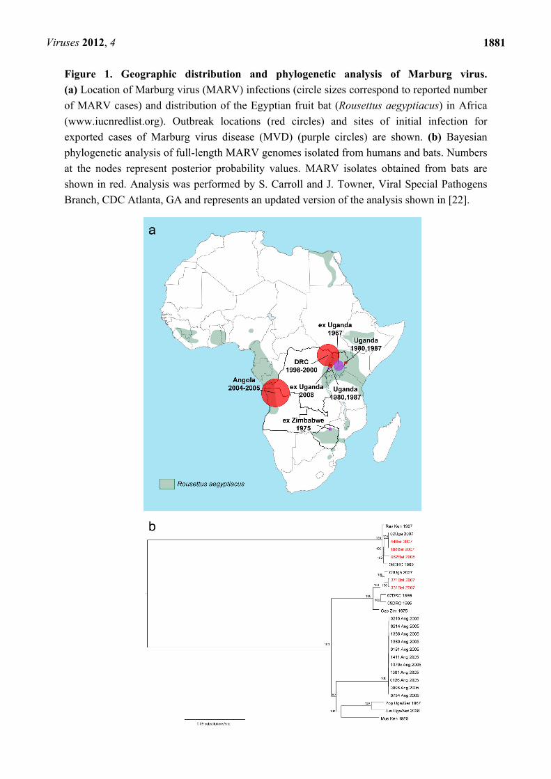
Figure 1. Geographic distribution and phylogenetic analysis of Marburg virus.
(a) Location of Marburg virus (MARV) infections (circle sizes correspond to reported number
of MARV cases) and distribution of the Egyptian fruit bat (Rousettus aegyptiacus) in Africa
(www.iucnredlist.org). Outbreak locations (red circles) and sites of initial infection for
exported cases of Marburg virus disease (MVD) (purple circles) are shown. (b) Bayesian
phylogenetic analysis of full-length MARV genomes isolated from humans and bats. Numbers
at the nodes represent posterior probability values. MARV isolates obtained from bats are
shown in red. Analysis was performed by S. Carroll and J. Towner, Viral Special Pathogens
Branch, CDC Atlanta, GA and represents an updated version of the analysis shown in [22].
Viruses 2012, 4
1882
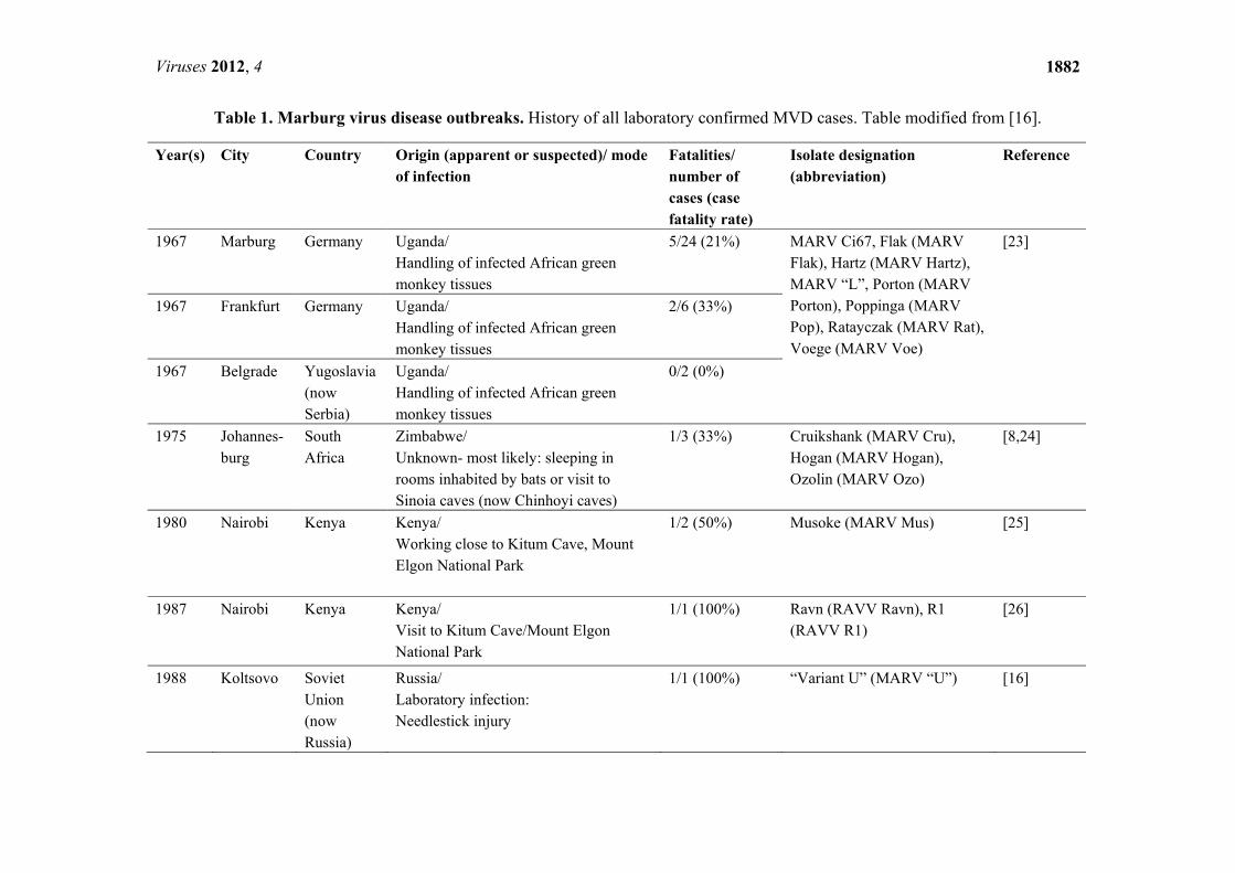
Table 1. Marburg virus disease outbreaks. History of all laboratory confirmed MVD cases. Table modified from [16].
Year(s) City Country Origin (apparent or suspected)/ mode of infection
Fatalities/ number of cases (case fatality rate)
Isolate designation (abbreviation)
Reference
1967 Marburg Germany Uganda/ Handling of infected African green monkey tissues
5/24 (21%) MARV Ci67, Flak (MARV Flak), Hartz (MARV Hartz), MARV “L”, Porton (MARV Porton), Poppinga (MARV Pop), Ratayczak (MARV Rat), Voege (MARV Voe)
[23]
1967 Frankfurt Germany Uganda/ Handling of infected African green monkey tissues
2/6 (33%)
1967 Belgrade Yugoslavia(now Serbia)
Uganda/ Handling of infected African green monkey tissues
0/2 (0%)
1975 Johannes-burg
South Africa
Zimbabwe/ Unknown- most likely: sleeping in rooms inhabited by bats or visit to Sinoia caves (now Chinhoyi caves)
1/3 (33%) Cruikshank (MARV Cru), Hogan (MARV Hogan), Ozolin (MARV Ozo)
[8,24]
1980 Nairobi Kenya Kenya/ Working close to Kitum Cave, Mount Elgon National Park
1/2 (50%) Musoke (MARV Mus) [25]
1987 Nairobi Kenya Kenya/ Visit to Kitum Cave/Mount Elgon National Park
1/1 (100%) Ravn (RAVV Ravn), R1 (RAVV R1)
[26]
1988 Koltsovo Soviet Union (now Russia)
Russia/ Laboratory infection: Needlestick injury
1/1 (100%) “Variant U” (MARV “U”) [16]
Viruses 2012, 4
1883
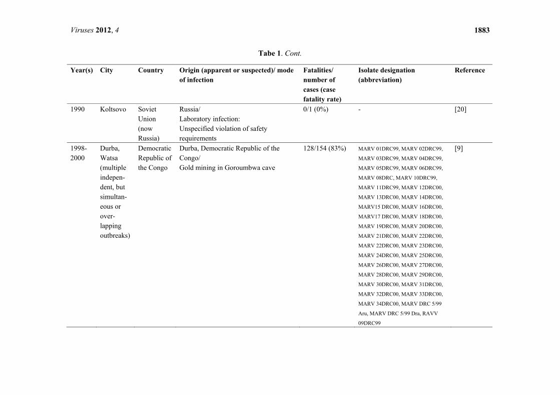
Tabe 1. Cont.
Year(s) City Country Origin (apparent or suspected)/ mode of infection
Fatalities/ number of cases (case fatality rate)
Isolate designation (abbreviation)
Reference
1990 Koltsovo Soviet Union (now Russia)
Russia/ Laboratory infection: Unspecified violation of safety requirements
0/1 (0%) - [20]
1998-2000
Durba, Watsa (multiple indepen-dent, but simultan-eous or over-lapping outbreaks)
Democratic Republic of the Congo
Durba, Democratic Republic of the Congo/ Gold mining in Goroumbwa cave
128/154 (83%)
MARV 01DRC99, MARV 02DRC99,
MARV 03DRC99, MARV 04DRC99,
MARV 05DRC99, MARV 06DRC99,
MARV 08DRC, MARV 10DRC99,
MARV 11DRC99, MARV 12DRC00,
MARV 13DRC00, MARV 14DRC00,
MARV15 DRC00, MARV 16DRC00,
MARV17 DRC00, MARV 18DRC00,
MARV 19DRC00, MARV 20DRC00,
MARV 21DRC00, MARV 22DRC00,
MARV 22DRC00, MARV 23DRC00,
MARV 24DRC00, MARV 25DRC00,
MARV 26DRC00, MARV 27DRC00,
MARV 28DRC00, MARV 29DRC00,
MARV 30DRC00, MARV 31DRC00,
MARV 32DRC00, MARV 33DRC00,
MARV 34DRC00, MARV DRC 5/99
Aru, MARV DRC 5/99 Dra, RAVV
09DRC99
[9]

Viruses 2012, 4
1884
Table 1. Cont.
Year(s) City Country Origin (apparent or suspected)/ mode of infection
Fatalities/ number of cases (case fatality rate)
Isolate designation (abbreviation)
Reference
2004–2005
Uíge Angola Uíge Province, Angola/ unknown
227/252 (90%)
MARV Angola [10,27]
2007 Kam-wenge
Uganda Kamwenge District, Uganda/ Gold mining in Kitaka Cave
1/4 (25%) MARV-01Uga 2007, RAVV- 02Uga 2007
[19]
2008 Colorado, City unreported
USA Uganda/ Visit of Python Cave in Maramagambo Forest
0/1 (0%) - [17]
2008 Leiden Nether-lands
Uganda/ Visit of Python Cave in Maramagambo Forest
1/1 (100%) MARV Leiden [18]
Total: 368/452 (81%)
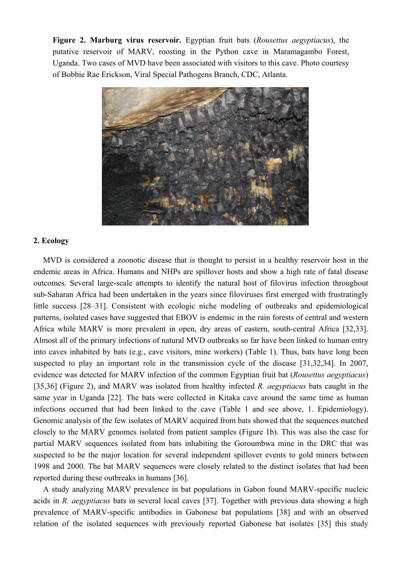
Figure 2. Marburg virus reservoir. Egyptian fruit bats (Rousettus aegyptiacus), the
putative reservoir of MARV, roosting in the Python cave in Maramagambo Forest,
Uganda. Two cases of MVD have been associated with visitors to this cave. Photo courtesy
of Bobbie Rae Erickson, Viral Special Pathogens Branch, CDC, Atlanta.
2. Ecology
MVD is considered a zoonotic disease that is thought to persist in a healthy reservoir host in the
endemic areas in Africa. Humans and NHPs are spillover hosts and show a high rate of fatal disease
outcomes. Several large-scale attempts to identify the natural host of filovirus infection throughout
sub-Saharan Africa had been undertaken in the years since filoviruses first emerged with frustratingly
little success [28–31]. Consistent with ecologic niche modeling of outbreaks and epidemiological
patterns, isolated cases have suggested that EBOV is endemic in the rain forests of central and western
Africa while MARV is more prevalent in open, dry areas of eastern, south-central Africa [32,33].
Almost all of the primary infections of natural MVD outbreaks so far have been linked to human entry
into caves inhabited by bats (e.g., cave visitors, mine workers) (Table 1). Thus, bats have long been
suspected to play an important role in the transmission cycle of the disease [31,32,34]. In 2007,
evidence was detected for MARV infection of the common Egyptian fruit bat (Rousettus aegyptiacus)
[35,36] (Figure 2), and MARV was isolated from healthy infected R. aegyptiacus bats caught in the
same year in Uganda [22]. The bats were collected in Kitaka cave around the same time as human
infections occurred that had been linked to the cave (Table 1 and see above, 1. Epidemiology).
Genomic analysis of the few isolates of MARV acquired from bats showed that the sequences matched
closely to the MARV genomes isolated from patient samples (Figure 1b). This was also the case for
partial MARV sequences isolated from bats inhabiting the Goroumbwa mine in the DRC that was
suspected to be the major location for several independent spillover events to gold miners between
1998 and 2000. The bat MARV sequences were closely related to the distinct isolates that had been
reported during these outbreaks in humans [36].
A study analyzing MARV prevalence in bat populations in Gabon found MARV-specific nucleic
acids in R. aegyptiacus bats in several local caves [37]. Together with previous data showing a high
prevalence of MARV-specific antibodies in Gabonese bat populations [38] and with an observed
relation of the isolated sequences with previously reported Gabonese bat isolates [35] this study
Viruses 2012, 4
1886
suggests that MARV is enzootic in Gabon and raises the concern of further spread of MARV into other
countries. Therefore, close ecological as well as serological surveillance of the bat populations in
sub-Saharan Africa could help to predict and prevent further MVD outbreaks especially in areas where
bats are still used as a food source.
It is not currently clear whether R. aegyptiacus bats are the exclusive reservoir for MARV or if
other bat species reported to be positive for viral antibodies and RNA are also natural reservoirs or
merely intermediate hosts [36].
3. Taxonomy
The genus Marburgvirus includes a single species, Marburg marburgvirus (formerly referred to as
Lake Victoria marburgvirus) [39,40]. Phylogenetic analysis based on genomic sequence data suggests
that the known members of this species can be assigned to at least five different lineages of which four
are very closely related (nucleotide sequences differ up to 7%) while the fifth is divergent (a nucleotide
difference of 21%) (Figure 1b) [10,22,39]. As the genomic divergence between all isolates is below
30%—the cutoff for the classification of the five different ebolaviruses into five different species—the
five marburgvirus lineages were recently reclassified as two viruses. Ravn virus (RAVV) is
represented by the Ravn isolates from 1987, one isolate from the DRC outbreak in 1998–2000, and one
human and several bat isolates from infections that took place in Uganda in 2007. Marburg virus
(MARV) is represented by all other sequenced isolates (Figure 1b, Table 1) [39]. For the sake of
simplicity, in this review the abbreviation “MARV” is used for all marburgviruses and the abbreviation
“EBOV” for all ebolaviruses.
4. Transmission
Initial MVD patients are believed to contract the virus via exposure to an infected animal: either a
reservoir host (several bat species) or a spill-over host such as NHPs as described in the first MVD
outbreak (see above, 1. Epidemiology) [23,41]. Following transmission to humans, spread of the virus
between individuals is the result of direct contact with blood or other body fluids (saliva, sweat, stool,
urine, tears, and breast milk) from infected patients. Typical risks of exposure include administration
of medical care to infected individuals as well as handling of corpses without use of proper
protection [34]. Of particular note, virus has been found in tears, semen, and in a liver biopsy weeks to
months following the onset of symptoms highlighting the importance of monitoring convalescent
patients [20,42–44].
5. Clinical manifestations
Much of what we know about typical MVD symptoms comes primarily from clinical data obtained
during the three largest recorded MVD outbreaks: the 1967 outbreak in Germany and Yugoslavia, the
1998–2000 outbreak in the DRC, and the 2004–2005 outbreak in Angola. Although the case fatality
rates were significantly higher in the latter outbreaks, most of the clinical symptoms observed
were similar.
Viruses 2012, 4
1887
5.1. Incubation Period
Based on the most reliable documented cases of exposure and subsequent illness, MVD has an
incubation period ranging from 3 to 21 days (typically 5 to 10 days), which is likely modulated by
factors such as infectious dose and possibly by route of infection. The course of MVD has
conventionally been broken down into three phases [45]: an initial generalization phase, an early organ
phase, and either a late organ phase or convalescence phase depending upon disease outcome [45].
A summary of MVD symptoms is reviewed below [16,46–49].
5.2. Generalization Phase (Day One-to Four)
The onset of illness begins with generic flu-like symptoms; a characteristic high fever (typically
39–40 oC), severe headache, chills, myalgia, prostration, and malaise. For many patients (50–75%) this
is followed by rapid debilitation characterized by gastrointestinal symptoms including anorexia,
abdominal pain, severe nausea, vomiting, and watery diarrhea. Starting on day four to five patients
commonly develop enanthem, dysphasia, and pharyngitis. Additionally, a characteristic maculopapular
rash is typically the first distinctive feature indicating a filovirus infection versus influenza or malaria.
Other common symptoms include lymphadenopathy, leukopenia, and thrombocytopenia.
5.3. Early Organ Phase (Day Five to Thirteen)
Many of the initial symptoms may persist in the early organ phase, and patients may sustain a high
fever. They may additionally display neurological symptoms including encephalitis, confusion,
delirium, irritability, and aggression. Patients can also develop dyspnea and abnormal vascular
permeability, particularly conjunctival injection and edema. During the latter part of this phase more
than 75% of patients present with some form of clear hemorrhagic manifestation such as petechiae,
mucosal bleeding, melena, bloody diarrhea, hematemesis, and ecchymoses. Due to the unusualness of
hemorrhagic symptoms, diseases caused by filoviruses have sometimes been referred to as
hemorrhagic fevers (Marburg Hemorrhagic Fever (MHF) and Ebola Hemorrhagic Fever (EHF)),
although these terms are currently disfavored since not all patients display hemorrhagic symptoms.
At this stage, multiple organs are affected including the pancreas, kidney, and liver. Elevated serum
activity of a number of liver enzymes including SGOT and SGPT have been observed in most
patients sampled.
5.4. Late Organ/Convalescence Phase (Day Thirteen+)
The late stages of MVD result in one of two potential outcomes: patients either succumb to the
disease or enter a prolonged phase of recuperation. Typical preagonal symptoms include restlessness,
obtundation, confusion, dementia, convulsions, reduced circulation due to severe dehydration,
metabolic disturbances, severe diffuse coagulopathy, multiorgan failure, shock, and coma. Fatalities
typically occur 8–16 days following the onset of symptoms, with death usually the resulting of shock
and multiorgan failure. Non-fatal cases are typified by an extensive convalescent period during which
myalgia, exhaustion, sweating, peeling of the skin at the sites of rash, partial amnesia, and secondary
infections are all common.
Viruses 2012, 4
1888
6. Prevention
Prevention of newly emerging MARV infections and effective containment during ongoing
outbreaks is both essential and challenging, as there is currently no licensed vaccine or treatment
available for general use.
Following the 1967 outbreak of MVD in Europe and cases of infection with ebolavirus Reston in
imported crab-eating macaques (Macaca fascicularis) in the USA in 1989/1990 and 1996 as well as
1992 in Italy (reviewed in [50]), strict quarantine procedures have been put in place that have so far
prevented infections acquired by imported NHPs into non-endemic countries [51,52]. To avoid the
spread of filoviruses by tourists, Python cave was closed to the public following the diagnosis of the
Dutch patient in 2008.
The prevention and control of outbreaks and infections in endemic countries is much more
challenging. In the past, joint efforts of teams from the WHO, Doctors Without Borders, the Red
Cross, the CDC and others in collaboration with the local ministries of health have been undertaken to
cease the spread of MVD. The main focus of outbreak control is the prevention of secondary
transmission and further primary infections.
The first measures in response to a MVD outbreak include setting up isolation wards in hospitals to
assure rapid isolation of MARV-infected patients and prevent person-to-person transmission
(Figure 3b). Proper and fast laboratory diagnosis of suspected cases is key to eliminate further spread.
Nosocomial infections were commonly seen in earlier outbreaks [8,9,23]. However, reinforcement of
barrier nursing techniques and education of health care workers have limited these infections in recent
outbreaks (Figure 3a). Epidemiological surveillance has been crucial in the identification of index
cases as well as the predominant modes of transmission. In endemic areas, secondary infections mainly
occurred while taking care of ill patients and family members or during traditional burial practices
involving close contact to corpses [53]. Therefore, the execution of safe burial and disinfection
techniques and information campaigns to educate the local population are essential in order to contain
the spread of infections in endemic areas (Figure 3a) [27,53,54].
Biosafety and epidemiological efforts alone were not sufficient for efficient outbreak control during
large outbreaks, emphasizing the need for additional psychosocial support of the affected communities
[53]. The fast progression and high lethality rates associated with MVD even—and especially—after
hospital admission resulted in a high level of fear and suspicion by the resident population. The fact
that health care workers wearing recommended personal protective equipment (PPE) were fully
masked and not identifiable further increased anxiety (Figure 3b). This resulted in the hiding of
infected family members and verbal and in some cases physical aggression towards members of aid
organizations [53]. Communicating necessary protective measures while respectfully considering the
affected families’ and communities’ traditions and culture during ongoing outbreaks is therefore
essential for successful outbreak management.
The recent identification of bats as the potential reservoirs for MARV as well as
EBOV [22,35,36,55,56] will help to increase not only the public awareness, but also the effectiveness
of the preventive measures taken in endemic areas to minimize contact with infected animals
(i.e. closing of bat inhabited caves for the public, serosurveillance of bat populations) [18,19]. This is a
challenging task, emphasized by the fact that during the last cluster of MARV infections linked to a

Viruses 2012, 4
1889
gold mine in Uganda, the miner hired to enforce the restricted access to the mine got infected.
The mine had been closed in response to the ongoing outbreak and even though he was aware of the
risk, he had entered the mine without the suggested PPE [19].
Later, the bat population of this mine was eliminated by the owner by means of fumigation [19].
As bats of most species are endangered, this does not seem a viable option and educational campaigns
aimed at villagers living close to bat-inhabited caves as well as tourist groups and tour operators might
prove more sustainable in the future.
Figure 3. Marburg virus disease outbreak control. (a) Signs used to educate the local
population in outbreak areas. Picture taken from [57]. (b) Pictures of the MVD outbreak in
Angola, 2005. Above, nurse being sprayed with chlorine while leaving the isolation ward.
This illustrates the protective clothing worn by nursing staff. Below, view showing a
section of the isolation ward. The ward for confirmed MARV patients is on the left.
The solid plastic sheeting used for the outer wall is shown in the distance. Figure and
legend modified from [54].
7. Virus Structure
7.1. Virion Structure
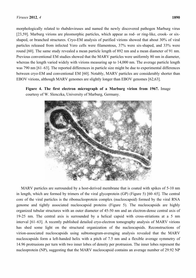
In 1967, during the first reported filovirus disease outbreak in Europe, the identification of the
previously unknown causative agent of the deadly disease was performed by electron microscopy
(EM) (Figure 4). The unusual filamentous structure of the particles led to some confusion and it was
even suggested that the causative agent of the disease might be related to the spiral-shaped Leptospira,
a genus of the spirochaetes bacteria [58]. Others concluded that the observed particles were viruses
Viruses 2012, 4
1890
morphologically related to rhabdoviruses and named the newly discovered pathogen Marburg virus
[23,59]. Marburg virions are pleomorphic particles, which appear as rod- or ring-like, crook- or six-
shaped, or branched structures. Cryo-EM analysis of purified virions showed that about 30% of viral
particles released from infected Vero cells were filamentous, 37% were six-shaped, and 33% were
round [60]. The same study revealed a mean particle length of 892 nm and a mean diameter of 91 nm.
Previous conventional EM studies showed that the MARV particles were uniformly 80 nm in diameter,
whereas the length varied widely with virions measuring up to 14,000 nm. The average particle length
was 790 nm [61–63]. The reported differences in particle size might be due to experimental differences
between cryo-EM and conventional EM [60]. Notably, MARV particles are considerably shorter than
EBOV virions, although MARV genomes are slightly longer than EBOV genomes [62,63].
Figure 4. The first electron micrograph of a Marburg virion from 1967. Image
courtesy of W. Slenczka, University of Marburg, Germany.
MARV particles are surrounded by a host-derived membrane that is coated with spikes of 5-10 nm
in length, which are formed by trimers of the viral glycoprotein (GP) (Figure 5) [60–65]. The central
core of the viral particles is the ribonucleoprotein complex (nucleocapsid) formed by the viral RNA
genome and tightly associated nucleocapsid proteins (Figure 5). The nucleocapsids are highly
organized tubular structures with an outer diameter of 45-50 nm and an electron-dense central axis of
19-25 nm. The central axis is surrounded by a helical capsid with cross-striations at a 5 nm
interval [61–63]. A recently published detailed cryo-electron tomography analysis of MARV virions
has shed some light on the structural organization of the nucleocapsids. Reconstructions of
virion-associated nucleocapsids using subtomogram-averaging analysis revealed that the MARV
nucleocapsids form a left-handed helix with a pitch of 7.5 nm and a flexible average symmetry of
14.96 protrusions per turn with two inner lobes of density per protrusion. The inner lobes represent the
nucleoprotein (NP), suggesting that the MARV nucleocapsid contains an average number of 29.92 NP
Viruses 2012, 4
1891
molecules per turn with each NP molecule packaging six RNA bases [60]. MARV nucleocapsids show
directionality, having a pointed and a barbed tip [60].
7.2. Genome Organization
The nonsegmented negative-sense (NNS) RNA genomes of the various MARV isolates range in
size from 19,111 to 19,114 nts and contain seven monocistronic genes in a linear order
(Figure 5) [66,67]. Each gene is composed of a highly conserved transcription start and stop signal, an
unusually long 3’ and 5’ untranslated region, and the open reading frame (ORF). The genes are either
separated by short intergenic regions that range from 4 to 97 nts, or the transcription stop signal of the
upstream gene and the transcription start signal of the downstream gene overlap, sharing five highly
conserved nts (Figure 5). The structure of this gene overlap is found among all filoviruses and is
unique among members of the order Mononegavirales (for review see [68]). The 3’ and 5’ genome
ends are extracistronic regulatory regions that contain cis-acting signals essential for transcription and
replication, including transcription and replication promoters.
There are generally two types of genomic replication promoters for NNS RNA viruses: a bipartite
promoter found in members of the paramyxovirus subfamily Paramyxovirinae and one continuous
more compact replication promoter for rhabdo- and pneumoviruses [69]. The bipartite promoter
structure of the Paramyxovirinae subfamily is associated with the “rule of six”, i.e., the total genome
length must be a multiple of six [70]. Given that filoviruses do not obey the rule of six, it was
surprising that mapping of the MARV genomic replication promoter revealed a bipartite structure. The
3’ genome end, the leader, comprises 48 nts and contains the first promoter element of the bipartite
genomic replication promoter. The second promoter element consists of a (UNNNNN)3 motif with
three conserved uridine residues separated from each other by five non-conserved nucleotides. The
UN(5) hexamers are located within the 3’ untranslated region of the first MARV gene, the NP gene, and
are separated from the first promoter region by the 12 nts long transcription start signal. Substitutions
in the NP transcription start signal do not affect replication activity but do interfere with transcription
initiation [71]. The 5’ extracistronic region, the trailer, spans the last 75 nts of the MARV genome and
contains the complement of the antigenomic replication promoter (see below, 8.2. Transcription and
Replication). The structure of the MARV antigenomic promoter has not yet been determined.
However, due to the presence of UN(5) hexamers it is likely that it is of bipartite nature, similar to the
genomic promoter. A common feature of the leader and trailer regions of all NNS RNA viruses is a
high degree of complementarity of the 10-15 most terminal 3’ and 5’ nucleotides [72].
Although filoviruses share this feature, both the leader and the trailer also have the capability to form
an internal secondary structure, which is not the case for the leaders and trailers of other NNS RNA
viruses [71,73–75].
Viruses 2012, 4
1892
7.3. Viral Proteins
The MARV genome encodes seven structural proteins listed in Table 2.
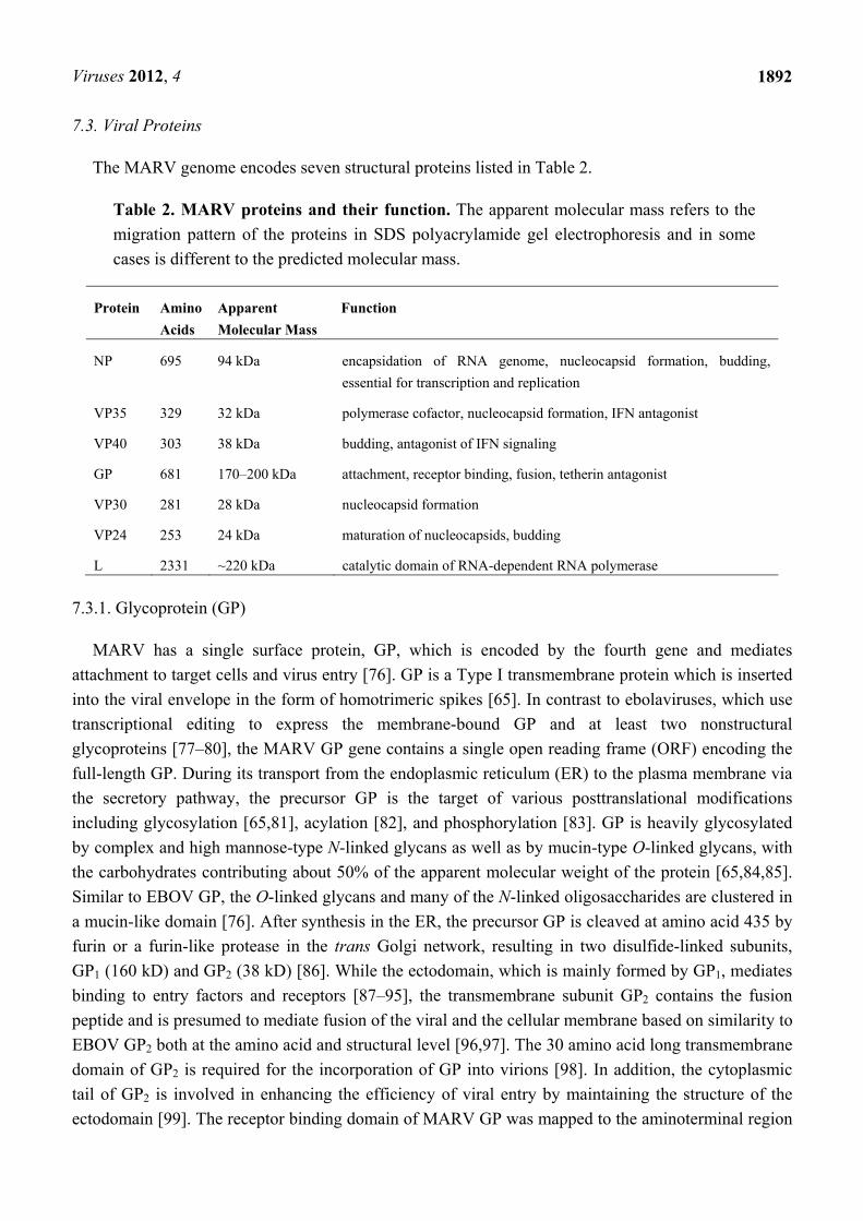
Table 2. MARV proteins and their function. The apparent molecular mass refers to the
migration pattern of the proteins in SDS polyacrylamide gel electrophoresis and in some
cases is different to the predicted molecular mass.
Protein Amino
Acids
Apparent
Molecular Mass
Function
NP 695 94 kDa encapsidation of RNA genome, nucleocapsid formation, budding,
essential for transcription and replication
VP35 329 32 kDa polymerase cofactor, nucleocapsid formation, IFN antagonist
VP40 303 38 kDa budding, antagonist of IFN signaling
GP 681 170–200 kDa attachment, receptor binding, fusion, tetherin antagonist
VP30 281 28 kDa nucleocapsid formation
VP24 253 24 kDa maturation of nucleocapsids, budding
L 2331 ~220 kDa catalytic domain of RNA-dependent RNA polymerase
7.3.1. Glycoprotein (GP)
MARV has a single surface protein, GP, which is encoded by the fourth gene and mediates
attachment to target cells and virus entry [76]. GP is a Type I transmembrane protein which is inserted
into the viral envelope in the form of homotrimeric spikes [65]. In contrast to ebolaviruses, which use
transcriptional editing to express the membrane-bound GP and at least two nonstructural
glycoproteins [77–80], the MARV GP gene contains a single open reading frame (ORF) encoding the
full-length GP. During its transport from the endoplasmic reticulum (ER) to the plasma membrane via
the secretory pathway, the precursor GP is the target of various posttranslational modifications
including glycosylation [65,81], acylation [82], and phosphorylation [83]. GP is heavily glycosylated
by complex and high mannose-type N-linked glycans as well as by mucin-type O-linked glycans, with
the carbohydrates contributing about 50% of the apparent molecular weight of the protein [65,84,85].
Similar to EBOV GP, the O-linked glycans and many of the N-linked oligosaccharides are clustered in
a mucin-like domain [76]. After synthesis in the ER, the precursor GP is cleaved at amino acid 435 by
furin or a furin-like protease in the trans Golgi network, resulting in two disulfide-linked subunits,
GP1 (160 kD) and GP2 (38 kD) [86]. While the ectodomain, which is mainly formed by GP1, mediates
binding to entry factors and receptors [87–95], the transmembrane subunit GP2 contains the fusion
peptide and is presumed to mediate fusion of the viral and the cellular membrane based on similarity to
EBOV GP2 both at the amino acid and structural level [96,97]. The 30 amino acid long transmembrane
domain of GP2 is required for the incorporation of GP into virions [98]. In addition, the cytoplasmic
tail of GP2 is involved in enhancing the efficiency of viral entry by maintaining the structure of the
ectodomain [99]. The receptor binding domain of MARV GP was mapped to the aminoterminal region
Viruses 2012, 4
1893
of GP1 spanning amino acids 38 to 188 [100], whereas the highly glycosylated mucin-like domain is
not essential for virus entry [101]. An important step in MARV entry is the proteolytic activation of
GP1 by endosomal proteases, facilitating binding of the receptor binding region to the endosomal entry
factor Niemann-Pick C1 protein (see below, 8.1. Entry) [94,95].
Besides its function in entry and budding, GP may also play a role in immune evasion.
The IFN-inducible antiviral protein tetherin was shown to block the release of VP40-induced virus-like
MARV and EBOV particles, suggesting that tetherin might act as a restriction factor for filovirus
release [102,103]. However, co-expression of GP was sufficient to counteract the antiviral activity of
tetherin by a yet unknown mechanism [104,105]. It is possible that GP not only subverts innate
immune responses but also suppresses the adaptive immune response. Filoviral GP2 subunits,
including MARV GP2, contain a domain resembling an immunosuppressive motif found in retroviral
envelope proteins [106]. A 17-mer peptide corresponding to the putative immunosuppressive domain
of MARV GP was shown to induce lymphocyte death and suppression of cytokine responses [107].
It is not yet known if this motif plays a role in the induction of lymphocyte apoptosis observed in
MARV infection. Finally, it has been suggested that shedding of the ectodomain of membrane-bound
EBOV GP by tumor necrosis factor α-converting enzyme (TACE) may play a role in blocking the
activity of neutralizing antibodies during infection [108]. It has been reported for MARV that
considerable amounts of GP shed from infected cells, although it is not clear if MARV GP is a target
for TACE cleavage [108,109].
7.3.2. Viral Protein 40 (VP40)
The matrix protein VP40 is encoded by the third MARV gene and is the counterpart of the M
proteins of other NNS RNA viruses. VP40 plays a major role in the formation of virions by
redistributing nucleocapsids from the perinuclear region to the plasma membrane, recruiting GP to the
sites of budding, and mediating particle release [110–112]. Overexpression of VP40 led to reduced
reporter gene expression of MARV minigenomes, suggesting a regulatory role of VP40 in
transcription and/or replication [113].
As a peripheral membrane protein, VP40 coats the inner side of the virion’s membrane
(Figure 5) [114]. Cryo-EM tomography studies suggest that VP40 associates with the nucleocapsid
through flexible interactions [60]. It can be easily removed from the nucleocapsid by salt dissociation,
indicating that it is only loosely connected to the nucleocapsid [115]. After synthesis in the cytoplasm
of the infected cell, VP40 associates rapidly with cellular membranes and accumulates in membranous
structures of the late endosomal compartment, the multivesicular bodies. A minor portion of VP40 is
also found in association with viral nucleocapsids and in inclusions. Additionally, VP40 appears in
patches beneath the plasma membrane where it is transported via the retrograde late endosomal
pathway [62,114,116]. Similar to EBOV VP40, MARV VP40 is the major factor in particle formation
and budding. Expression of VP40 in the absence of other viral proteins leads to the formation and
release of filamentous virus-like particles (VLPs) resembling authentic virions. This process is
enhanced in the presence of GP [113,117–119]. The role of VP40 during budding is described in more
detail below (see 8.3. Budding).
Viruses 2012, 4
1894
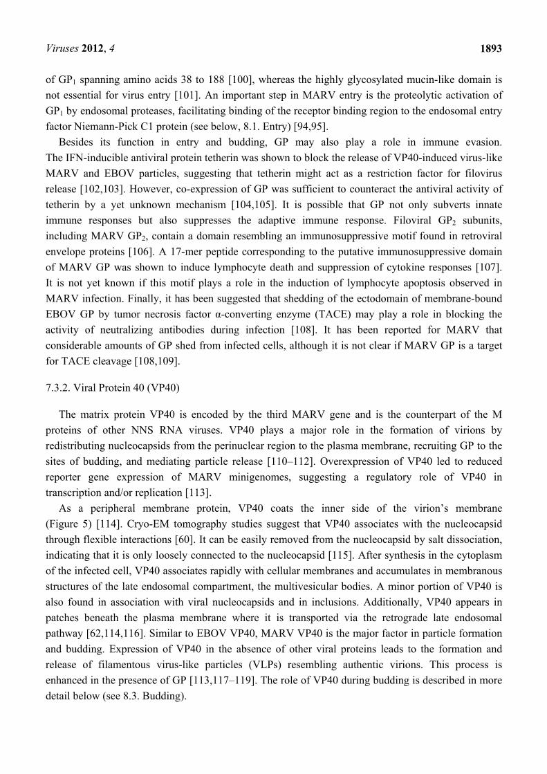
Figure 5. Marburg virion structure and genome organization. Above, schematic of
Marburg virion. Below, structure of the MARV genome with transcription signals. The
colors of the open reading frames correspond to the colors of the viral proteins.
Untranslated regions of the different genes are shown as light grey boxes; intergenic
regions (IR) are shown as dark grey lines and the leader and trailer of the genome are
colored in black. Transcription start signals (Tc start) are represented by green triangles,
while transcription stop signals (Tc stop) are shown as red bars. The sequence of two gene
borders (NP/VP35 and VP30/VP24) is shown in 3’ to 5’ orientation, as it occurs in the
negative sense RNA genome (MARV Musoke, GenBank accession number: NC_001608).
The gene border between VP30 and VP24 contains overlapping transcription signals, with
the start signal of VP24 upstream of the stop signal of VP30.
Compared to EBOV VP40, little is known about the structure of MARV VP40. The N-terminal
domain of MARV VP40 folds into ring-like structures, which have the tendency to polymerize into
rod-like structures. While EBOV VP40 has been shown to form hexamers and octamers, the
stoichiometry of MARV VP40 oligomers is not known [120]. MARV VP40 is phosphorylated at
several tyrosine residues located in the N-terminal region of the protein. A non-phosphorylatable
mutant of VP40 is impaired in its ability to recruit nucleocapsids to the sites of budding, but is still
able to efficiently induce particle release [112]. VP40 also possesses a PPPY late domain motif in its
amino terminus which is important for its interaction with components of the Endosomal Sorting
Complex Required for Transport (ESCRT) machinery in order to mediate budding, including Tumor
susceptibility gene 101 (Tsg101) and the membrane-bound E3 ubiquitin ligase
Nedd4.1 [119,121,122]. Besides the PPPY motif, other motifs and single amino acids have been found
to be important for particle release [123,124].
Besides its role as a classical matrix protein, MARV VP40 also acts as a virulence factor by
counteracting the innate immune response and determining the host tropism for MARV [125,126].
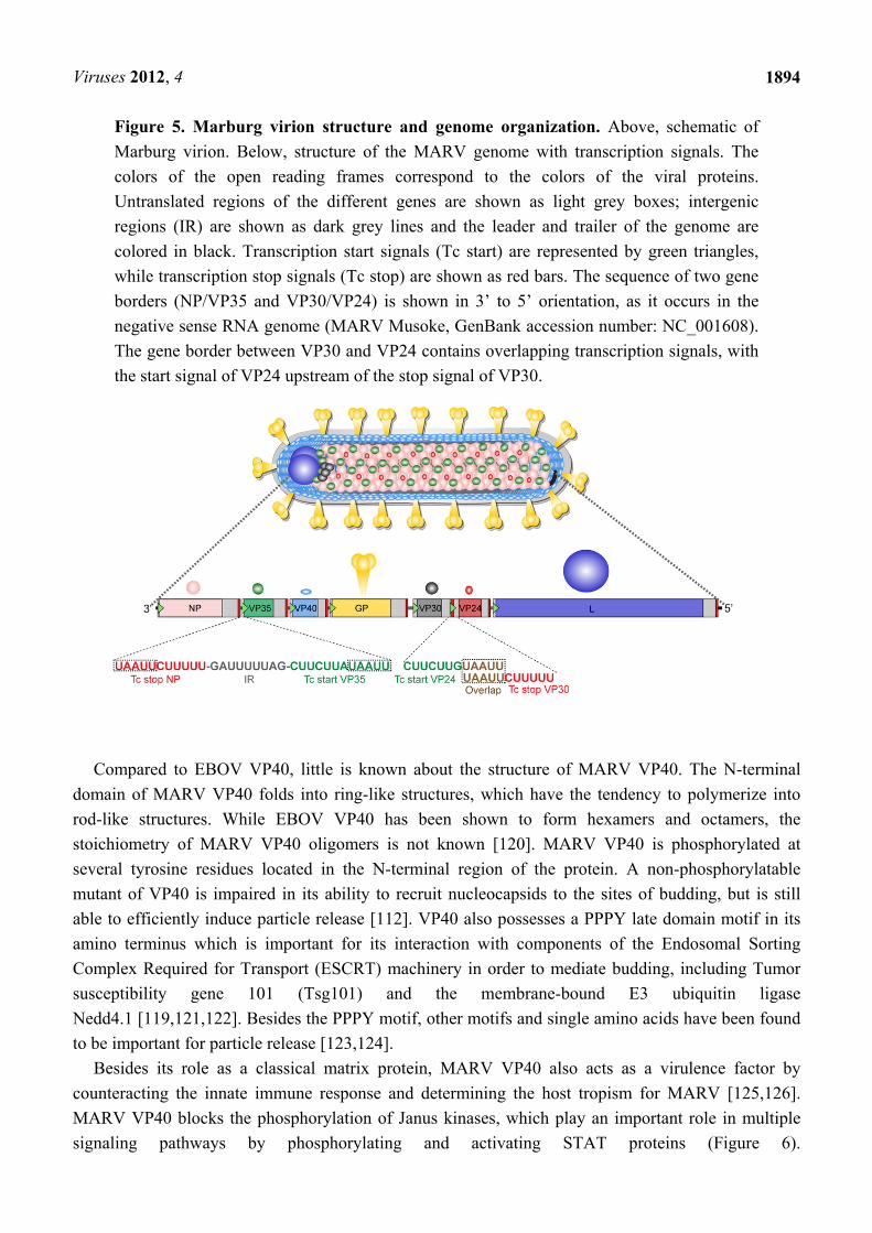
MARV VP40 blocks the phosphorylation of Janus kinases, which play an important role in multiple
signaling pathways by phosphorylating and activating STAT proteins (Figure 6).
Viruses 2012, 4
1895
When MARV-infected cells were treated with various stimuli, including IFNα, IFNγ, and IL6, it was
shown that the STAT proteins were neither phosphorylated nor translocated into the nucleus [126,127].
It was then shown that in MARV-infected cells treated with exogenous stimuli, Janus kinases were
also not phosphorylated and VP40 was identified as the viral protein inhibiting IFN signaling. It is
believed that Jak1 is the target for VP40, however, the mechanism of VP40-induced inhibition is not
completely understood [126]. Intriguingly, EBOV is also able to block IFN signaling by employing a
completely different mechanism. EBOV VP24 blocks the nuclear translocation of phosphorylated
STAT proteins by binding to STAT1 and importins involved in the nuclear transport of specific STAT
proteins (Figure 6) ([128], reviewed in [129]).
When MARV was adapted to non- or less permissive animals, such as mouse and guinea pig, the
adapted viruses showed mutations in VP40. Two of the amino acid changes in the mouse-adapted
MARV VP40 have been shown to be essential for the inhibition of IFN signaling in mouse cells,
underlining the importance of IFN suppression for the virulence and host specificity of
MARV [125,130,131].
Figure 6. Inhibition of JAK-STAT signaling by filoviruses. MARV VP40 inhibits
phosphorylation of Janus kinases and STAT proteins in response to Type I and II IFNs and
IL6, preventing downstream signaling. Phosphorylation of STAT proteins is not inhibited
by Ebola virus (EBOV). EBOV VP24 interacts with STAT1 and members of the nuclear
importin family and prevents nuclear translocation of phosphorylated STAT1.
Viruses 2012, 4
1896
7.3.3. Viral protein 24 (VP24)
The protein product of the sixth gene, VP24, is unique to the filovirus family. VP24 is generally
addressed as a second, minor matrix protein. However, cryo-electron tomography analysis of viral
particles showed that VP24 is located in close proximity to the nucleocapsid proteins, suggesting that it
might be part of the nucleocapsid complex [60]. VP24 can easily be released from virion-associated
nucleocapsids by treatment with increasing salt concentrations, indicating that it is only loosely
connected to the nucleocapsid [115]. Intracellular localization studies of VP24 showed that a minor
part of the protein (approx. 10%) is weakly bound to cellular membranes, including filopodia enriched
with VP40. VP24 is also distributed diffusely in the cytoplasm, relocalizes to
nucleocapsid-containing inclusions, and is found in association with free nucleocapsids. Co-expression
of NP and VP24 is sufficient to direct VP24 to NP inclusions in the cytoplasm [132]. Functional
studies on MARV VP24 suggest that the protein is important for the release of viral particles in the
context of infection, although it influences neither the morphology of VP40-derived VLPs nor the
efficiency of VLP release. In addition, RNAi-mediated knockdown of VP24 in MARV-infected cells
had no impact on viral genome replication, indicating that VP24 is involved in a step after replication
and before budding [132]. According to the model that has been proposed based on these data, VP24 is
involved in the maturation of transport-competent nucleocapsids and/or mediates the interaction
between nucleocapsids and budding sites at the plasma membrane [132]. There is also evidence that
MARV VP24 affects transcription and replication in a transcription and replication competent VLP
system [113].
Structural information about MARV VP24 is very limited. It has been shown that it forms
oligomers, preferentially tetramers [132]. Structure prediction studies have proposed an ancestral link
between VP24 and the Armadillo repeat family [133].
7.3.4. The Nucleocapsid Proteins NP, VP35, VP30 and L
The MARV nucleocapsid complex consists of the genomic RNA and four tightly associated
proteins, NP, VP35, VP30, and L (Figure 5). Encapsidation of the viral RNA by the nucleocapsid
proteins protects it from both RNase degradation and detection by cellular pattern recognition
receptors. Similar to the genomic RNA, the antigenomic RNA, a replicative intermediate, is also
encapsidated by the nucleocapsid proteins (see below, 8.2. Transcription and Replication). In contrast,
the viral mRNAs are not encapsidated [134]. The nucleocapsid rather than naked RNA serves as the
template for viral transcription and replication. In a MARV minigenome system, NP, VP35, and L are
essential for transcription and replication [113,134]. The role of VP30 in MARV transcription and
replication is not well understood and the steps in genome amplification that require, or do not require,
VP30 are not yet defined.
7.3.5. Nucleoprotein (NP)
The nucleoprotein NP enwraps the genomic and antigenomic RNAs. Replication and transcription
activity in a MARV minigenome system depends on the presence of NP [134]. When NP is expressed
in the absence of other nucleocapsid proteins, it self-assembles into highly organized helical tubular
Viruses 2012, 4
1897
structures that resemble the nucleocapsids in infected cells, indicating that it is the driving force for
nucleocapsid formation [60,135,136]. Recently, it has been shown that the conserved 390 N-terminal
residues of MARV NP are sufficient to form the helical structure of the nucleocapsid core [60].
Indeed, NP serves as a viral hub protein. It forms interactions with most of the other viral proteins,
leading to the subcellular redistribution of these proteins. The strong binding of NP to the nucleocapsid
proteins VP35 and VP30 redirects both proteins into NP-derived inclusions [115]. A bipartite
coiled-coil motif in the central part of NP has been shown to play an important role for self-assembly
and NP-VP35 interaction [137]. As mentioned above, there is also a weak interaction between NP and
VP24, leading to the partial relocalization of VP24 into NP-containing inclusions [60,132]. In addition,
NP interacts with VP40, which is important for the transport of newly synthesized nucleocapsids to the
plasma membrane [110,111,138,139]. Interestingly, NP contains a C-terminal late domain motif,
PSAP, which has been shown to be required for budding. NP recruits Tsg101, a component of the
ESCRT I complex, through its late domain motif, leading to enhanced VP40-induced budding [111].
NP is heavily phosphorylated at serine and threonine residues clustered in seven regions in the
C-terminal part of the protein. Only the phosphorylated form of NP is incorporated into
virions [140,141]. Recent studies suggest that the phosphorylation level in Region II modulates
transcription and/or replication activity [142].
7.3.6. Viral Protein 35 (VP35)
VP35 is a polymerase cofactor and essential for transcription and replication. Together with the
catalytic subunit L, VP35 forms the RNA-dependent RNA polymerase complex [134,143]. VP35 is
tightly associated with NP and serves as a bridging protein between the nucleocapsid complex and L.
Without VP35, L is not associated with the nucleocapsids which serve as the templates for viral
transcription and replication [115,134]. VP35 forms homo-oligomers mediated by a coiled-coil motif
located in the N-terminal part of the protein. Homo-oligomerization of VP35 is essential for its
interaction with L but not needed for redistribution of VP35 into NP-derived inclusions [144]. VP35
shares many features with the phospho (P) proteins of other NNS RNA viruses, including its position
as the second gene in the viral genome and its role in transcription and replication. However, in
contrast to the P proteins, VP35 is either not or only very weakly phosphorylated [145].
Besides its function in transcription and replication, MARV VP35 acts as an IFN antagonist. While
the impact of EBOV VP35 on the host’s antiviral response has been intensively investigated (reviewed
in [129]), much less information is available about similar functions of MARV VP35. When we tested
MARV VP35 for its ability to block IFN induction in a reporter gene assay, it blocked reporter gene
expression as efficiently as EBOV VP35 (unpublished data). In addition, Bosio and
colleagues [146] reported that expression of MARV VP35 in the absence of other viral proteins was
sufficient to completely block the induction of IFNα in stimulated human dendritic cells. Besides its
ability to inhibit the induction of Type I IFN, EBOV VP35 has been shown to block the activation of
the antiviral protein PKR and to interfere with RNA silencing pathways. Importantly, EBOV VP35 is a
dsRNA binding protein. The C-terminus of EBOV VP35 contains a domain with patches of basic
amino acids which is important for dsRNA binding and the protein’s inhibitory functions (for review

Viruses 2012, 4
1898
see [129]). This C-terminal region, the so-called IFN inhibitory domain, is conserved in MARV
VP35 [147], suggesting that MARV VP35 possesses similar inhibitory functions.
7.3.7. Viral Protein 30 (VP30)
MARV and EBOV VP30 proteins show many structural similarities. Both MARV and EBOV VP30
proteins are tightly associated with the nucleocapsid via their binding to NP
(Figure 5) [115,148]. Both are highly phosphorylated at N-terminally located serine and threonine
residues, and phosphorylation is crucial for their interaction with NP [148,149]. Both contain an
unusual C3H1 Zn binding domain, which is essential for the function of EBOV VP30 as transcription
initiation factor, but whose functional relevance for MARV VP30 is not known [150]. It has also been
shown that EBOV VP30 forms hexamers [151,152], binds single-stranded RNA [153], and interacts
with L [154]. However, to date, similar data for MARV VP30 are not available.
The role of MARV VP30 in viral transcription and replication is not well understood. In contrast to
EBOV VP30, which plays an important role in regulating transcription initiation [68,143,155–157],
MARV VP30 is not essential for transcription or replication activity in a MARV minigenome system
[113,134]. Nevertheless, it seems to play an important role in viral amplification, since rescue of a
full-length MARV clone is only successful in the presence of VP30 [158]. In addition,
down-regulation of VP30 by RNA interference in MARV-infected cells led to the reduction of both
viral protein synthesis and virion production [159]. Among the NNS RNA viruses, only the members
of the subfamily Pneumovirinae possess a protein similar to VP30, M2-1, which functions as a
transcription processivity factor [160].
7.3.8. Large Protein (L)
The major component of the MARV polymerase complex, L, has an estimated molecular weight of
267 kD [67]. It is essential for transcription and replication and together with VP35 forms the
RNA-dependent RNA polymerase complex (see above, 7.3. Viral Proteins, VP35). L contains the
enzymatic functions of the polymerase. The binding site for VP35 has been mapped to the N-terminal
530 amino acid residues of L [115,134]. The L proteins of the NNS RNA viruses are highly conserved
multifunctional proteins, which are organized in functional domains [161]. Based on this conservation
with other NNS RNA polymerases, MARV L is believed to carry out RNA synthesis, capping, and
polyadenylation of viral mRNAs although these functions have not been shown experimentally.
8. Replication Cycle
To date most of the studies characterizing the MARV replication cycle have utilized recombinant
systems, allowing for these experiments to be performed in a biosafety level 2 (BSL-2) context,
unfettered by the restrictions of a BSL-4 setting. Surrogate systems mimicking specific steps in the
MARV replication cycle include MARV GP-pseudotyped retroviruses or recombinant vesiculoviruses
expressing GP to study entry, VLPs to study budding, and minigenome systems to study replication
and transcription. While such experiments allow for the more facile examination of the MARV
replication cycle, the findings must be recapitulated with infectious MARV since all surrogate systems
Viruses 2012, 4
1899
lack elements of the infectious virus such as the distinct morphological features and virion protein
composition of MARV.
8.1. Entry
Marburg virus entry consists of three distinct phases: cellular attachment, endocytosis, and fusion
(Figure 7). Based on the sequence similarity between EBOV and MARV GPs many investigators have
presumed identical functions and characteristics between the filovirus glycoproteins. This is
presumptuous given the differences in glycosylation and sialic acid linkages [85] and dependence upon
endosomal proteases (see below, 8.1.2. Endocytosis). Despite the existence of a number of detailed
studies and structural analyses of EBOV GP [162–165], relatively few mechanistic studies of MARV
GP have been performed [101], although one recent post-fusion structure of MARV GP2 has been
reported [97]. The structure of MARV GP2 is nearly identical to that of EBOV GP2, indicating that the
mechanisms of fusion between the two viruses is likely conserved [97].
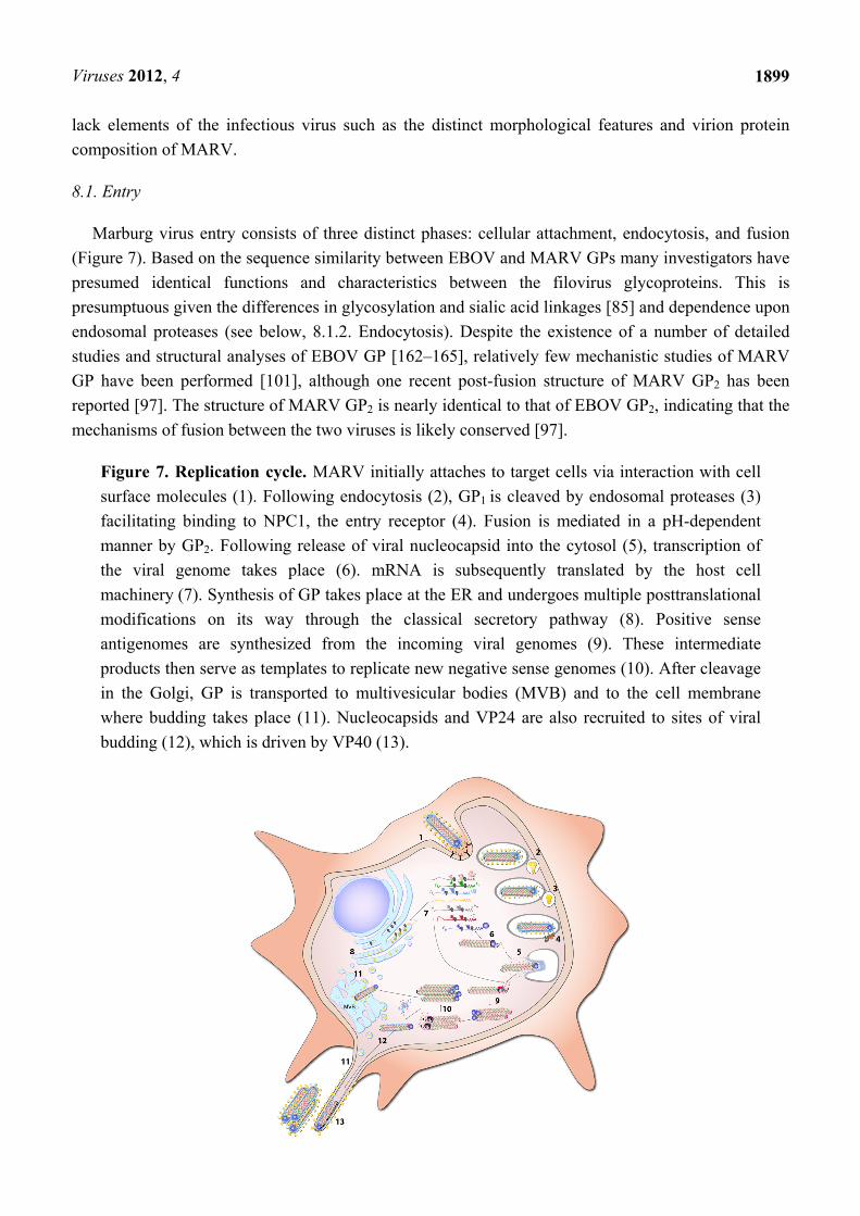
Figure 7. Replication cycle. MARV initially attaches to target cells via interaction with cell
surface molecules (1). Following endocytosis (2), GP1 is cleaved by endosomal proteases (3)
facilitating binding to NPC1, the entry receptor (4). Fusion is mediated in a pH-dependent
manner by GP2. Following release of viral nucleocapsid into the cytosol (5), transcription of
the viral genome takes place (6). mRNA is subsequently translated by the host cell
machinery (7). Synthesis of GP takes place at the ER and undergoes multiple posttranslational
modifications on its way through the classical secretory pathway (8). Positive sense
antigenomes are synthesized from the incoming viral genomes (9). These intermediate
products then serve as templates to replicate new negative sense genomes (10). After cleavage
in the Golgi, GP is transported to multivesicular bodies (MVB) and to the cell membrane
where budding takes place (11). Nucleocapsids and VP24 are also recruited to sites of viral
budding (12), which is driven by VP40 (13).
Viruses 2012, 4
1900
8.1.1. Attachment
MARV GP mediates both cell attachment and fusion of the virus. There is convincing evidence that
initial virus attachment at the cell surface can occur via the binding of GP carbohydrates to various
cellular C-type lectins, including the hepatocyte-specific ASGP-R [87], DC-SIGN and DC-SIGNR
(also known as L-SIGN) [89,92,166], hMGL [91,92], and LSECtin [166,167]. Other cell surface
proteins have also been implicated in facilitating MARV entry including the TAM receptor protein
kinases Ax1, Dtk, and Mer [90], and TIM-1 [93]. However, although these proteins may play a role in
attachment or entry of certain cell types, the ability of MARV to infect cells lacking these receptors
[92,93,168] indicates that there might be redundancy in cellular molecules required for MARV
attachment to cells.
A number of key residues of EBOV GP that are involved in virion incorporation and GP-mediated
entry have been identified [163] and found to play a similar role in MARV GP [101], indicating that
the viruses might utilize similar mechanisms to enter the cell.
8.1.2. Endocytosis
Following attachment, Marburg virions undergo endocytosis mediated through a mechanism that
currently remains undetermined. (Figure 7) [16,62] Initial studies investigating caveolin-mediated
endocytosis showed that depletion of host cell cholesterol reduced viral infectivity but presented no
direct evidence of caveolae involvement [169]. In addition, studies examining the role of caveolae in
EBOV endocytosis are conflicting [169,170]. A major role for clathrin in MARV entry has also been
proposed based upon the ability of chlorpromazine (an inhibitor of both clathrin-mediated endocytosis
and macropinocytosis) as well as RNAi-knockdown of clathrin heavy chain to inhibit MARV
GP-pseudotyped HIV-1 entry [171]. A caveat to these analyses of MARV endocytosis is that they
were performed only in the context of MARV GP-pseudotyped retroviruses which lack the
characteristic filamentous morphology and size of Marburg virions.
While other reports have verified that cholesterol is important for live MARV particle uptake [172],
canonical caveolae- and clathrin-mediated mechanisms are unlikely to be the primary mechanism of
MARV entry due to steric issues. The typical MARV particle size (average 790 nm) is much larger
than canonical caveolae (50–100 nm) or clathrin-coated pits (up to 200 nm) whereas pseudotyped
murine leukemia virus (MLV) (100 × 100 nm) and vesicular stomatitis virus (VSV) (70 × 180 nm) are
not [173]. These findings indicate that involvement of the caveolae- and clathrin-mediated endocytic
pathways for virus entry may therefore be the result of the artificial nature of the pseudotyped virions
and highlights the need to confirm such experiments with live MARV.
Macropinocytosis has been identified as a major entry pathway of EBOV by research using the
morphologically more relevant VLPs and live EBOV [174–177]. Although none of these analyses
examined the role of this pathway during MARV entry, it remains an intriguing possibility given the
cholesterol-dependence and large size of macropinocytotic vesicles (up to 3–5 µm) [178].
Another important process in MARV entry is believed to occur while virions are being trafficked
within endocytic vesicles; the proteolytic cleavage of GP1. Endosomal cleavage of GP has been shown
to be critical for the efficient entry of MARV [179,180]. The current model for MARV entry involves
Viruses 2012, 4
1901
the cleavage of GP1 by host endosomal cysteine proteases. This removal of a large portion of GP1
(including the mucin-like domain) is believed to expose the putative receptor-binding domain based on
studies conducted with EBOV GP [181,182].
Studies examining the roles of endosomal proteases on the entry of MARV and EBOV have
produced mixed results. Experiments analyzing recombinant VSV expressing EBOV GP indicate a
primary role for Cathepsin B (CatB) and minor role for Cathepsin L (CatL) [181]. Entry of
recombinant VSV particles containing MARV GP was inhibited when cells were treated with an
inhibitor of both CatB and CatL [92]. These reports are confounded by a report conducted with
infectious Marburg and Ebola viruses in which CatB and CatL inhibitors greatly reduced EBOV
infection but showed mixed results with MARV [172]. Yet two other, more recent analyses determined
that CatB was not required for MARV entry (although over-expression did enhance infectivity) and
that CatL was required for entry into mouse embryonic fibroblasts but not Vero cells, 293T cells, or
human macrophages [179,180]. These data as well as the ability of other proteases to greatly diminish
MARV infectivity [179,180], indicate that although CatB and CatL likely play a role in cleavage and
activation of GP1 in certain cell types, other endosomal proteases may also be able to facilitate GP1
activation via cleavage.
8.1.3. Fusion
Recently, two independent studies elegantly showed the requirement of the endosomal cholesterol
transporter Niemann-Pick C1 (NPC1) for the entry of both MARV GP-pseudotyped viruses (VSV and
MLV) as well as infectious MARV [94,95]. It was also shown that NPC1 catalytic activity is not
required for EBOV infection indicating that specific binding to NPC1 rather than its role in cholesterol
transport is required, although this was not tested for MARV [95]. In one of the studies identifying
NPC1 as the MARV entry receptor, it was also determined that members of the homotypic fusion and
vacuole protein-sorting (HOPS) complex were important for EBOV entry, although they appeared to
play a less important role in MARV entry [94].
The current model of EBOV and MARV fusion is that GP1 cleavage by endosomal proteases
removes heavily glycosylated domains, exposing the receptor binding domain on GP1 and enabling
binding to NPC1 [95]. The membrane-bound fusogenic GP2 undergoes a low pH-dependent
rearrangement to an extended conformation resulting in the fusion of virion and endo-lysosomal
membranes [96]. In support of the pH-dependence of GP-mediated fusion, pre-treatment of cells with
ammonium chloride prevented entry of a MARV GP-pseudotyped virus [183]. A recent report with
live MARV showed that ammonium chloride inhibited entry and replication, but that Bafilomycin A1,
which specifically inhibits vacuolar-type H(+) ATPase and prevents re-acidification of vesicles of the
central vacuolar system, surprisingly had no effect [172]. Following viral fusion with the endosomal
membrane, the nucleocapsid is released into the cytoplasm (Figure 7).
8.2. Transcription and Replication
After the nucleocapsid is released into the cytoplasm of the infected cell, transcription and
replication of the viral RNA genome takes place (Figure 7). The first morphological sign of viral
replication observed by EM analysis is the appearance of granular material containing RNA and viral
Viruses 2012, 4
1902
proteins in the cytoplasm of the infected cells at 12 h post infection. Later on, tubular structures can be
detected in the granular material representing the newly synthesized nucleocapsids embedded in the
viral inclusions [63]. While experimental data on the sites of MARV replication and transcription are
not available, recent studies on EBOV have shown that viral replication takes place in the inclusions,
while transcription was observed prior to inclusion formation [184].
The encapsidated negative-sense RNA genome is transcribed resulting in seven monocistronic
mRNAs by the viral polymerase. They are co-transcriptionally capped and polyadenylated and
subsequently translated by the cellular machinery (Figure 7). The genomic RNA also serves as the
template for the production of positive-sense antigenomes, which are complementary copies of the
genomes. The antigenomes are encapsidated by the nucleocapsid proteins and are in turn used as
templates for genome synthesis (Figure 7) (for review see [68]). As mentioned above, NP, VP35, L,
and probably VP30 are needed for viral transcription and replication. Analogous to EBOV, it is
conceivable that VP40 and VP24 inhibit transcription and replication [113,185,186]. It is hypothesized
that negative regulators of replication convert the active polymerase complex into an inactive state,
resulting in mature and transport-competent nucleocapsids.
8.3. Budding
Following assembly, newly synthesized nucleocapsids are recruited to the sites of virus budding
(Figure 7). Release of viral particles is mainly mediated by VP40 via recruitment of nucleocapsids
from the inclusions to the plasma membrane, recruiting GP to the sites of budding, and inducing the
formation and release of filamentous VLPs. VP40-induced budding is enhanced by NP, GP, and VP24
[98, 111, 132]. As is the case with many other viruses, MARV exploits the vesicular transport
machinery of the infected cell for viral egress, including the COPII vesicular transport system and the
ESCRT machinery. The COPII vesicular transport system is used by VP40 for its intracellular
trafficking to the multivesicular bodies, where MARV budding takes place [118,187]. Cellular proteins
that promote particle release and are linked to the ESCRT machinery include Tsg101, Vps4A/B, and
Nedd4.1 [111,119,121,122]. MARV budding not only takes place at internal membranes but also at the
plasma membrane [63,118,188]. In cell culture, MARV particles are preferentially released at
filopodia, filamentous cellular protrusions [60,188,189]. Filopodia are used by cells to explore the
extracellular environment, which includes neighboring cells, and it is believed that viral particles can
bud directly into adjacent cells via filopodia-mediated cell-to-cell contact [188,190]. Budding at
filopodia depends on actin and is not sensitive to the depolymerization of microtubules [188].
MARV budding was observed at the basolateral membrane of polarized epithelial cells and
hepatocytes [109,191], whereas viral particles were predominantly released from the apical membrane
of infected endothelial cells [64], suggesting that cell-type specific components determine the sites of
virus release.
Electron tomography studies of MARV-infected cells led to the following model for MARV
particle release: The budding process is initiated when intracellular nucleocapsids associate laterally
with the plasma membrane. Starting from one end, the nucleocapsids are then subsequently wrapped
by the plasma membrane until the viral particles protrude vertically from the cell surface. The release
of infectious filamentous MARV from cultured cells peaked at 1–2 days post infection, when the cells
Viruses 2012, 4
1903
were still intact. At 4 d post infection, when most of the cells were vesiculated, the released virions
were round or bent and infectivity was decreased [189]. Determination of the nucleocapsid orientation
at the sites of budding by 3-D reconstructions revealed that the pointed tip of the budding
nucleocapsids is oriented towards the membrane, indicating that MARV budding is directional [60].
9. Pathogenesis
MARV infections usually occur by direct contact with infected body fluids or direct personal
contact with infected animals or humans. The viruses enter the body through small skin lesions or
mucosal membranes (reviewed in [47]). Cells of the mononuclear phagocyte system, including
monocytes, macrophages and dendritic cells, are early target cells of MARV, as shown in different
experimental animal models [14,192–197]. MARV replication was observed as early as 24 hours post
infection in macrophages of infected guinea pigs [63], and infected monocytes have been found in
cynomolgus macaques at 2 days post infection [193]. Monocytes and macrophages were also identified
as early target cells in human patients [197]. This has been confirmed by cell
culture experiments showing that primary human monocytes and macrophages are highly susceptible
to MARV infection and produce infectious virus [198–200]. In addition, primary human
monocyte-derived dendritic cells (mDCs) and endothelial cells support MARV
replication [64,146,201].
Early sites of virus replication are the lymph nodes, liver, and spleen where the most severe necrotic
lesions are observed [8,13,14,41,130,194,202].
These organs contain high numbers of monocytes and macrophages. Migration of infected
monocytes and macrophages into surrounding tissues or transport of free virus via the lymph- or
bloodstream is believed to facilitate the dissemination to multiple organs, resulting in a systemic
infection [203,204]. Cell-free virus has been observed in the tissue and organs of infected animals, and
high levels of virus have been detected in the blood [12–14,130,194,195,205,206]. Besides monocytes,
macrophages, and dendritic cells, a wide range of cell types including hepatocytes, adrenal cortical and
medullary cells and fibroblasts are permissive to MARV infection [12,14,192–195,197,201,207].
Endothelial cells are late target cells during MARV infection in multiple tissues. Whether or not
replication of MARV in endothelial cells is associated with the observed vascular impairment during
MVD is a matter of debate [64,194]. Only low numbers of infected endothelial cells are observed in
NHP infection and therefore changes in the endothelium are likely caused by paracrine effects of
cytokines [14].
In late stages of infection MARV particles can be isolated from nearly every
organ [12,14,130,194,208]. Despite high viral load and necrotic lesions, only minor
inflammation is observed in infected tissues and organs, indicating a dysregulated immune
response [8,14,41,194]. Strong liver pathology is observed, including increased serum activity of liver
enzymes. This might influence synthesis of clotting factors and contribute to the observed coagulation
defects in MVD [12,41,130,194]. These factors together with systemic virus replication and associated
pathology probably trigger the multiorgan failure associated with fatal cases.
Although lymphocytes are not susceptible to MARV infection [14,193,194,201], massive bystander
lymphocyte apoptosis is a hallmark of MVD [8,14,130,194,195,201]. However, the molecular

Viruses 2012, 4
1904
mechanisms for lymphocyte depletion and the role it may play in the pathogenesis of MVD are far
from being understood.
Cytokine secretion may play a role in the induction of lymphocyte apoptosis, since MARV-infected
cells secrete cytokines known to induce apoptosis, including TNFα [194,198,200,209]. Increased
levels of TNFα have been observed in infected rhesus macaques [210] and mice [130], although no
increase was observed in infected cynomolgus macaques [14]. Elevated TNFα levels may also play a
role in the formation of endothelial gaps in the context of MARV infection [198,199]. In addition,
increased survival of MARV-infected guinea pigs treated with anti-TNFα antibodies suggests that
TNFα indeed plays an important role in MVD pathogenesis [209].
Increased serum levels of additional proinflammatory cytokines and chemokines have been
observed in infected NHPs and in mice, but the reported data are not completely
consistent [12,14,130,193,210]. Cytokine and chemokine secretion has also been observed in infected
primary human monocytes and macrophages [200,211]. However, data about the cytokine levels in the
serum of MARV-infected patients are not available, but high levels of cytokines have been observed in
EBOV-infected patients [212–215].
Upregulation of the proinflammatory cytokines IL6 (mediator of fever and acute inflammatory
response) and IL8 (chemoattraction of neutrophils and macrophages) is consistently found in infected
NHPs, with macrophages and plasmacytoid dendritic cells (pDCs) serving as the main sources of IL6
secretion in the spleen [12,14,193]. Primary human monocytes and macrophages produce both IL6 and
IL8 after infection [200]. Elevated levels of IL6 have also been detected in MARV-infected mice
[130]. Increased levels of IL1β mRNA and secreted protein were detected in primary human cells
[200, 211], but contradictory data have been reported for the NHP model. One study reported elevated
IL1β levels in final disease stages [12], whereas no change was observed in another study [14].
IFNα levels were elevated in infected NHPs and mice [130,193,210]. However, no change in IFNα
levels was detected in another study of infected NHPs [14]. It is unclear whether or not the observed
differences are due to different MARV variants being used for the studies.
Serum levels of several chemokines were also found to be elevated during MARV infection of
NHPs and mice, including macrophage inflammatory proteins (MIP) and monocyte chemotactic
protein 1 (MCP-1) [12,14,130].
The involvement of multiple cell types along with the possible role of non-infected cells in the
secretion of cytokines further complicates the analysis of existing data. Primary human monocytes and
macrophages are activated by MARV infection inducing the secretion of cytokines. Induction of
cytokines has also been described using UV-inactivated MARV, suggesting that viral replication might
not be needed for the observed cytokine increase [200]. In contrast, MARV-infected mDCs show no
upregulation of activation markers, do not secrete cytokines, and fail to stimulate T cells [146].
mDCs treated with VLPs containing MARV VP40 and GP show functional mDC responses including
cytokine secretion indicating that MARV replication is required to inhibit mDC activation [216].
However, infection of mDCs with MARV did not prevent LPS-induced TNFα production whereas
dsRNA-dependent IFNα secretion was inhibited [146], suggesting differential regulation of cytokines
by MARV. Interestingly, pDCs in the spleen were identified as the major source of secreted IFNα in
MARV infected NHPs, but secretion most likely occurs from non-infected cells [193]. It has been
shown for EBOV that pDCs are not productively infected due to impairment of viral entry [217].
Viruses 2012, 4
1905
These results suggest that secretion of cytokines by non-infected bystander cells might play an
important role during MARV pathogenesis. Taken together, MARV infection induces both an increase
in the production of proinflammatory cytokines and high levels of chemokines, but the molecular
mechanisms causing these changes are not well understood.
10. Animal Models
To date four different animal models have been established for MARV infection: NHPs, mice,
guinea pigs, and hamsters. The NHP model best reflects the symptoms and pathology observed
in human cases (described in 5. Clinical Manifestations and reviewed in [46,48]) with uniform
lethality in cynomolgus and rhesus macaques as well as African green
monkeys [12,14,26,193,194, 206,218–220]. The disease symptoms are generally the same for all types
of NHPs. The animals develop febrile illness with high fever, anorexia, weight loss and
unresponsiveness. Death is observed after 6–13 days and thrombocytopenia, lymphopenia, blood
coagulation abnormalities and hemorrhages are observed. Squirrel monkeys have also been
successfully infected with MARV, showing typical disease symptoms [206]. Recently, a small NHP
model using marmosets has been developed recapitulating the features of human infections except for
the typical maculopapular rash development that is observed in other NHPs and humans infected with
MARV [221]. Rodents with an intact immune system do not develop disease after infection with
MARV. MARV variants Musoke and Ci67 and RAVV variant Ravn were adapted to severe-combined
immunodeficiency (scid) mice by serial passaging, reducing the time to death from 50–70 days to 7–10
days for all tested virus variants [195]. Further passaging of the scid mouse-adapted marburgviruses in
immunocompetent mice was used to establish mouse models for RAVV Ravn and MARV
Ci67 [130,131]. Successful adaptation by serial passaging was also used to generate lethal infection
models for both guinea pig [218,222] and hamster [206,208]. Coagulation abnormalities, typical rash
development, and hemorrhagic manifestations (especially in mice) are not as pronounced as in the
NHP model [130]; (reviewed in [223]). Neuropathogenicity, recapitulating the CNS involvement
described during the first human MVD outbreak in Germany [41,224,225], has only been observed in
the hamster model [208]. It is not clear if CNS pathology is developed in other animal models as no
brain pathology has been observed in mice [195] and cynomolgus macaques [14]. Nevertheless, virus
has been isolated from brain from MARV-infected marmosets, showing micro-hemorrhages [221].
Sequence comparison of the rodent-adapted viruses to the human MARV isolates revealed several
mutations. Sixty-one nucleotide changes in the mouse-adapted RAVV Ravn variant were detected in
the ORFs of NP, VP35, VP40, and VP30 (14 amino acid changes in total) or untranslated regions
(VP35, VP40, GP, VP30) [130]. In a second study analyzing genome changes during mouse
adaptation, 75 nucleotide changes during adaptation of RAVV Ravn and 33 changes for MARV Ci67
were described, with most amino acid changes occurring in VP40 [131]. During guinea pig adaptation,
only 11 nucleotide changes were observed, resulting in four amino acid changes. One of these changes
was located in VP40 and the other mutations were detected in the viral polymerase L [222]. The only
amino acid exchange detected in both the mouse- and the guinea pig-adapted MARV is amino acid
184 in VP40 (Asp to Asn). This was also the first mutation detected during mouse adaptation for both
RAVV Ravn and MARV Ci67, as analyzed by sequencing of serial passages [131]. This is of
Viruses 2012, 4
1906
particular interest because VP40 has been shown to function as an inhibitor of IFN signaling (see
above, 7.3. Viral Proteins, VP40) [125,126]. Both IFN receptor- and STAT1-deficient mice develop
disease using non-adapted MARV, highlighting the importance of the IFN pathway for the control of
MVD [226–228].
11. Diagnosis
Virological, serological, and molecular diagnostic methods for the detection for MARV are
available, including virus isolation, ELISA, RT-PCR, EM, and immunohistochemistry (summarized in
[16, 46]). During outbreak settings, mobile laboratories commonly use PCR and/or ELISA analysis for
rapid screening. Sensitive ELISA assays have been developed for detection of viral antigen or
virus-specific antibodies using overexpressed MARV NP or GP [229–232]. Detection of filoviruses by
PCR is the only assay currently available to distinguish between different virus variants for a variety of
tissue and fluid specimens. The use of a combination of virus-specific primer sets for conventional
RT-PCR makes the detection of all know filoviruses in a single assay possible [233]. For more
sensitive and quantitative detection, real-time RT-PCR-based assays have been developed for the
detection of MARV [10,234–238]. Feasibility of real-time RT-PCR in the field was successfully
proven during the MARV outbreak in Uíge, Angola using an improved field laboratory-adapted RNA
isolation protocol [239]. A network of European BSL-4 facilities in collaboration with a company
(QIAGEN) developed the first commercial prototype of a real-time-RT-PCR assay for the detection of
filoviruses [240]. A recently developed assay, RT loop-mediated isothermal amplification (LAMP),
has the potential to significantly improve field diagnosis of MARV infections, by eliminating the need
of PCR machines [241].
12. Vaccine Development
Initial approaches using inactivated virus to develop a vaccine against MARV were unsuccessful or
had contradictory results [16]. In addition, successful protection of rodents did not always translate
into protection of NHPs. For example, inactivated MARV protects guinea pigs from lethal MARV
challenge but only 50% of challenged NHPs survived [210, 242–244]. A panel of different approaches
has been used in order to develop successful vaccines for MARV (reviewed in [245,246]).
Recombinant GP expressed from insect cells or a DNA vaccine based on GP only partially
protected guinea pigs, but use of a combination of both vaccines resulted in 100% survival of guinea
pigs [243]. In another study, complete protection of guinea pigs using a different GP DNA vaccine was
reported, but only four of six vaccinated NHPs survived the challenge with MARV, showing
incomplete protection [247]. Increased doses of a codon-optimized DNA vaccine resulted in 100%
survival of NHPs, although some animals developed symptoms before recovering. In comparison to
other vaccine candidates, a poor induction of virus-specific antibodies was observed using a DNA
vaccine [248]. A codon-optimized DNA vaccine elicited a strong antibody response and resulted in
complete protection of mice with no clinical symptoms observed [249]. Vaccine candidates based on
the Venezuelan equine encephalitis virus (VEEV) replicon system expressing either MARV GP along
with NP or GP alone completely protected guinea pigs and NHPs [250].
Viruses 2012, 4
1907
A vaccine based on VLPs represents an additional candidate for protection against MARV [251].
Complete protection of guinea pigs has been demonstrated with a VLP-based vaccine containing
MARV GP, with induction of virus-specific antibodies. Protection with this vaccine relied on a
functional CD4+ T cell response, whereas depletion of CD8+ T cells did not ablate the protective
response [244]. VLPs containing MARV Musoke GP provided cross-protection in animals challenged
with MARV Ci67 or RAVV Ravn in both guinea pigs and NHPs [252,253].
Another approach to MARV vaccines is the use of viral vectors expressing MARV GP. To date,
two different systems have been established based on replication-defective adenoviral vectors or
recombinant VSV expressing MARV GP. The adenovirus-based vaccine successfully protects guinea
pigs and NHPs, and provides cross-protection. High levels of cross-reactive MARV-specific IgG and T
cell responses are induced, indicating an induction of an immune response [248,254]. Preexisting
immunity against the adenovirus strain Ad5 might pose a problem for its successful use in humans
(reviewed in [245]).
The VSV-based vaccine completely protects NHPs and additionally has proven successful in
post-exposure treatment (reviewed in [255]). A single immunization with recombinant VSV
expressing MARV Musoke GP resulted in 100% protection of cynomolgus macaques challenged by
intramuscular injection or aerosol exposure and protected against RAVV Ravn and MARV Angola
[13,256,257]. Although MARV-specific IgG were produced, only low levels of neutralizing antibodies
were detected [13,257]. Surprisingly, T cell-mediated responses were not observed in NHPs vaccinated
with recombinant VSV expressing MARV GP [13,256]. Safety is a concern for this vaccine, especially
for immunocompromised individuals, as it is a replication-competent VSV vector. However, in all
VSV-based filovirus vaccine studies VSV viremia was observed only shortly after
immunization. Additionally, the VSV-based filovirus GP vaccine was well tolerated and
protective in immunocompromised mice and NHPs and lacked neurovirulence in
NHPs [258–260] (reviewed in [255]).
Cross-protection has not been observed in animals vaccinated with MARV-based vaccines and
subsequently challenged with EBOV, while combined MARV and EBOV vaccines have been
successful in protection against both viruses [252,261,262].
13. Treatment
To date no approved treatment is available for MARV infection. Supportive care (fluids,
anti-microbials, blood transfusions) has been the primary treatment of patients during MVD outbreaks.
In the guinea pig model various treatments had some success as reflected by prolonged survival or
increased survival rates. Applied treatments included cytokine inhibition, IFN treatment, or antibody
transfer. The tested treatments were unsuccessful in the NHP model (reviewed in [16,47]).
A third of EBOV-infected NHPs survived, however, following treatment with recombinant
nematode coagulant protein 2, while only one of six MARV-infected animals survived [12,263].
Treatment using antisense technology to block viral protein expression using phosphorodiamidate
morpholino oligomers (PMO) beginning 30 to 60 minutes after MARV infection completely protected
NHPs [264]. Additionally, a small molecule inhibitor showed complete protection of MARV-infected
mice when administered 24h after infection but has not been tested in NHPs [265].
Viruses 2012, 4
1908
The VSV-based vaccine expressing MARV GP has also been demonstrated to be effective as a
post-exposure treatment. A hundred percent survival of NHPs was observed when the vaccine was
administered 20 to 30 minutes after MARV infection [266]. Delaying the time before treatment results
in incomplete protection, although five of six animals or two of six animals still survived when given
the treatment 1 or 2 days after MARV exposure, respectively [205]. These post-exposure treatments
may be useful to prevent disease after known exposure to MARV, such as a laboratory accident, but
the effective time frame during an outbreak might be too short and alternatives are needed.
Acknowledgments
The authors thank S. Carroll and J. Towner, CDC, Atlanta, GA for providing the phylogenetic tree
analysis shown in Figure 1b, Bobbie Rae Erickson, CDC, Atlanta, GA for providing the photograph
shown in Figure 2, and W. Slenczka, University of Marburg, Germany for providing the electron
micrograph shown in Figure 4. The picture in Figure 3a was taken from [57] the photographs shown in
Figure 3b were taken from [54]. This work was supported by National Institutes of Health (NIH)
grants U01-AI082954 and AI057159 (New England Regional Center of Excellence-Kasper, subaward
149047-0743).
Conflict of Interest
The authors declare no conflict of interest.
References
1. Slenczka, W.; Klenk, H.D. Forty years of marburg virus. J. Infect. Dis. 2007, 196, S131 –135.
2. Siegert, R.; Shu, H.L.; Slenczka, W.; Peters, D.; Müller, G. The aetiology of an unknown human
infection transmitted by monkeys [premilinary communication]. Ger. Med. Mon. 1968, 13, 1–2.
3. Kunz, C.; Hofmann, H.; Kovac W.; Stockinger, L. Biologische und morphologische
charakteristika des virus des in Deutschland aufgetretenen "Haemmorhagischen Fiebers". Wien.
Klin. Wochenschr. 1968, 80, 161–162.
4. Kissling, R.E.; Robinson, R.Q.; Murphy, F.A.; Whitfield, S.G. Agent of disease contracted from
green monkeys. Science 1968, 160, 888–890.
5. Smith, C.E.; Simpson, D.I.; Bowen, E.T.; Zlotnik, I. Fatal human disease from vervet monkeys.
Lancet 1967, 2, 7526, 1119–1121.
6. WHO. Ebola haemorrhagic fever in Sudan, 1976. Bull. World Health Organ. 1978, 56, 247–270.
7. WHO. Ebola haemorrhagic fever in Zaire, 1976. Bull World Health Organ 1978, 56, 271–293.
8. Gear, J.S.; Cassel, G.A.; Gear, A.J.; Trappler, B.; Clausen, L.; Meyers, A.M.; Kew, M.C.;
Bothwell, T.H.; Sher, R.; Miller, G.B.; et al. Outbreak of Marburg virus disease in Johannesburg.
Br. Med. J. 1975, 4, , 489–493.
9. Bausch, D.G.; Nichol, S.T.; Muyembe-Tamfum, J.J.; Borchert, M.; Rollin, P.E.; Sleurs, H.;
Campbell, P.; Tshioko, F.K.; Roth, C.; Colebunders, R.; et al. Marburg hemorrhagic fever
associated with multiple genetic lineages of virus. N. Engl. J. Med. 2006, 355, 909–919.
Viruses 2012, 4
1909
10. Towner, J. S.; Khristova, M. L.; Sealy, T. K.; Vincent, M. J.; Erickson, B. R.; Bawiec, D. A.;
Hartman, A. L.; Comer, J. A.; Zaki, S. R.; Ströher, U.; et al. Marburgvirus genomics and
association with a large hemorrhagic fever outbreak in Angola. J. Virol. 2006, 80, 6497–6516.
11. Feldmann, H. Marburg hemorrhagic fever—the forgotten cousin strikes. N. Engl. J. Med. 2006,
355, 866–869.
12. Geisbert, T.W.; Daddario-DiCaprio, K.M.; Geisbert, J.B.; Young, H.A.; Formenty, P.; Fritz,
E.A.; Larsen, T.; Hensley, L.E. Marburg virus Angola infection of rhesus macaques:
Pathogenesis and treatment with recombinant nematode anticoagulant protein c2. J. Infect. Dis.
2007, 196, S372–S381.
13. Daddario-DiCaprio, K.M.; Geisbert, T.W.; Geisbert, J.B.; Stroher, U.; Hensley, L.E.; Grolla, A.;
Fritz, E.A.; Feldmann, F.; Feldmann, H.; Jones, S.M. Cross-protection against Marburg virus
strains by using a live, attenuated recombinant vaccine. J. Virol. 2006, 80, 9659–9666.
14. Alves, D.A.; Glynn, A.R.; Steele, K.E.; Lackemeyer, M.G.; Garza, N.L.; Buck, J.G.; Mech, C.;
Reed, D.S. Aerosol exposure to the angola strain of marburg virus causes lethal viral
hemorrhagic Fever in cynomolgus macaques. Vet. Pathol. 2010, 47, 831–851.
15. Kuhn, J.H.; Dodd, L.E.; Wahl-Jensen, V.; Radoshitzky, S.R.; Bavari, S.; Jahrling, P.B.
Evaluation of perceived threat differences posed by filovirus variants. Biosecur. Bioterror. 2011,
9, 361–371.
16. Kuhn, J.H. Filoviruses; A Compendium of 40 years of Epidemiological, Clinical, and Laboratory
Studies. Springer Verlag: Vienna, Austria, 2008.
17. From the Centers for Disease Control and Prevention. Imported case of Marburg hemorrhagic
fever - Colorado, 2008. MMWR Morb. Mortal Wkly. Rep. 2009, 58, 1377–1381.
18. Timen, A.; Koopmans, M.P.; Vossen, A.C.; van Doornum, G.J.; Gunther, S.; van den
Berkmortel, F.; Verduin, K.M.; Dittrich, S.; Emmerich, P.; Osterhaus, A.D.; van Dissel, J.T.;
Coutinho, R.A. Response to imported case of Marburg hemorrhagic fever, the Netherlands.
Emerg. Infect. Dis. 2009, 15, 1171–1175.
19. Adjemian, J.; Farnon, E.C.; Tschioko, F.; Wamala, J.F.; Byaruhanga, E.; Bwire, G.S.; Kansiime,
E.; Kagirita, A.; et al. Outbreak of Marburg hemorrhagic fever among miners in Kamwenge and
Ibanda Districts, Uganda, 2007. J. Infect. Dis. 2011, 204, S796–S799.
20. Nikiforov, V.V.; Turovskii Iu, I.; Kalinin, P.P.; Akinfeeva, L.A.; Katkova, L.R.; Barmin, V.S.;
Riabchikova, E.I.; Popkova, N.I.; Shestopalov, A.M.; Nazarov, V.P.; et al. [A case of a
laboratory infection with Marburg fever]. Zh. Mikrobiol. Epidemiol. Immunobiol. 1994, 3,
104–106.
21. U.S. Department of Health and Human Services. Biosafety in Microbiological and Biomedical
Laboratories, 5th ed.; U.S. Department of Health and Human Services: Washington, DC, USA,
2009.
22. Towner, J.S.; Amman, B.R.; Sealy, T.K.; Carroll, S.A.; Comer, J.A.; Kemp, A.; Swanepoel, R.;
Paddock, C.D.; Balinandi, S.; Khristova, M.L.; et al. Isolation of genetically diverse Marburg
viruses from Egyptian fruit bats. PLoS Pathog. 2009, 5, e1000536.
Viruses 2012, 4
1910
23. Martini, G.A.; Siegert, R. Marburg virus disease. Springer: New York, NY, USA, 1971.
24. Conrad, J.L.; Isaacson, M.; Smith, E.B.; Wulff, H.; Crees, M.; Geldenhuys, P.; Johnston, J.
Epidemiologic investigation of Marburg virus disease, Southern Africa, 1975. Am. J. Trop. Med.
Hyg. 1978, 27, 1210–1215.
25. Smith, D.H.; Johnson, B.K.; Isaacson, M.; Swanapoel, R.; Johnson, K.M.; Killey, M.; Bagshawe,
A.; Siongok, T.; Keruga, W.K. Marburg-virus disease in Kenya. Lancet 1982, 1, 816–820.
26. Johnson, E.D.; Johnson, B.K.; Silverstein, D.; Tukei, P.; Geisbert, T.W.; Sanchez, A.N.;
Jahrling, P.B. Characterization of a new Marburg virus isolated from a 1987 fatal case in Kenya.
Arch. Virol. Suppl. 1996, 11, 101–114.
27. Ligon, B.L. Outbreak of Marburg hemorrhagic fever in Angola: A review of the history of the
disease and its biological aspects. Semin. Pediatr. Infect. Dis. 2005, 16, 219–224.
28. Breman, J.G.; Johnson, K.M.; van der Groen, G.; Robbins, C.B.; Szczeniowski, M.V.; Ruti, K.;
Webb, P.A.; Meier, F.; Heymann, D.L. A search for Ebola virus in animals in the Democratic
Republic of the Congo and Cameroon: ecologic, virologic, and serologic surveys, 1979–1980.
Ebola Virus Study Teams. J. Infect. Dis. 1999, 179, S139–S147.
29. Leirs, H.; Mills, J.N.; Krebs, J.W.; Childs, J.E.; Akaibe, D.; Woollen, N.; Ludwig, G.; Peters,
C.J.; Ksiazek, T.G. Search for the Ebola virus reservoir in Kikwit, Democratic Republic of the
Congo: Reflections on a vertebrate collection. J. Infect. Dis. 1999, 179, S155–S163.
30. Reiter, P.; Turell, M.; Coleman, R.; Miller, B.; Maupin, G.; Liz, J.; Kuehne, A.; Barth, J.;
Geisbert, J.; Dohm, D.; et al. Field investigations of an outbreak of Ebola hemorrhagic fever,
Kikwit, Democratic Republic of the Congo, 1995: Arthropod studies. J. Infect. Dis. 1999, 179,
S148–S154.
31. Monath, T.P. Ecology of Marburg and Ebola viruses: Speculations and directions for future
research. J. Infect. Dis. 1999, 179, S127–S138.
32. Peterson, A.T.; Carroll, D.S.; Mills, J.N.; Johnson, K.M. Potential mammalian filovirus
reservoirs. Emerg. Infect. Dis. 2004, 10, 2073–2081.
33. Peterson, A.T.; Lash, R.R.; Carroll, D.S.; Johnson, K.M. Geographic potential for outbreaks of
Marburg hemorrhagic fever. Am. J. Trop. Med. Hyg. 2006, 75, 9–15.
34. Bausch, D.G.; Borchert, M.; Grein, T.; Roth, C.; Swanepoel, R.; Libande, M.L.; Talarmin, A.;
Bertherat, E.; Muyembe-Tamfum, J.J.; Tugume, B.; et al. Risk factors for Marburg hemorrhagic
fever, Democratic Republic of the Congo. Emerg. Infect. Dis. 2003, 9, 1531–1537.
35. Towner, J.S.; Pourrut, X.; Albarino, C.G.; Nkogue, C.N.; Bird, B.H.; Grard, G.; Ksiazek, T.G.;
Gonzalez, J.P.; Nichol, S.T.; Leroy, E.M. Marburg virus infection detected in a common African
bat. PLoS ONE 2007, 2, e764.
36. Swanepoel, R.; Smit, S.B.; Rollin, P.E.; Formenty, P.; Leman, P.A.; Kemp, A.; Burt, F.J.;
Grobbelaar, A.A.; Croft, J.; Bausch, D.G.; Zeller, H.; Leirs, H.; et al. Studies of reservoir hosts
for Marburg virus. Emerg. Infect. Dis. 2007, 13, 1847–1851.
37. Maganga, G.D.; Bourgarel, M.; Ella, G.E.; Drexler, J.F.; Gonzalez, J P.; Drosten, C.; Leroy, E.
M. Is Marburg virus enzootic in Gabon? J. Infect. Dis. 2011, 204, S800–S803.
Viruses 2012, 4
1911
38. Pourrut, X.; Souris, M.; Towner, J.S.; Rollin, P.E.; Nichol, S.T.; Gonzalez, J.P.; Leroy, E. Large
serological survey showing cocirculation of Ebola and Marburg viruses in Gabonese bat
populations, and a high seroprevalence of both viruses in Rousettus aegyptiacus. BMC Infect.
Dis. 2009, 9, 159.
39. Kuhn, J.H.; Becker, S.; Ebihara, H.; Geisbert, T.W.; Jahrling, P.B.; Kawaoka, Y.; Netesov, S. V.;
Nichol, S.T.; Peters, C.J.; Volchkov, V.E.; Ksiazek, T.G. Filoviridae. In Virus Taxonomy:
Classification and Nomenclature of Viruses: Ninth Report of the International Committee on
Taxonomy of Viruses; King, A.M.Q., Adams, M.J., Carstens, E.B., Lefkowitz, E.J., Eds. Elsevier
Academic Press: Amsterdam, Netherlands 2011; volume 9, pp. 665–671.
40. Adams, M.J.; Carstens, E.B. Ratification vote on taxonomic proposals to the International
Committee on Taxonomy of Viruses (2012). Arch. Virol. 2012, 157, 1411–1422.
41. Martini, G.A. Marburg virus disease. Postgrad. Med. J. 1973, 49, 542–546.
42. Martini, G.A. Marburg agent disease: In man. Trans. R. Soc. Trop. Med. Hyg. 1969, 63,
295–302.
43. Slenczka, W.G. The Marburg virus outbreak of 1967 and subsequent episodes. Curr. Top.
Microbiol. Immunol. 1999, 235, 49–75.
44. Gear, J.S.; Cassel, G.A.; Gear, A.J.; Trappler, B.; Clausen, L.; Meyers, A.M.; Kew, M.C.;
Bothwell, T.H.; Sher, R.; Miller, G.B.; et al. Outbreake of Marburg virus disease in
Johannesburg. Br. Med. J. 1975, 4, 489–493.
45. Stille, W.; Böhle, E. Clinical course and prognosis of Marburg virus (green monkey) disease. In
Marburg virus disease; Martini, G.A., Siegert, R. Eds.; Springer: Berlin, Germay, 1971;
pp. 10–18.
46. Hartman, A.L.; Towner, J.S.; Nichol, S.T. Ebola and marburg hemorrhagic fever. Clin. Lab.
Med. 2010, 30, 161–177.
47. Mehedi, M.; Groseth, A.; Feldmann, H.; Ebihara, H. Clinical aspects of Marburg hemorrhagic
fever. Future Virol. 2011, 6, 1091–1106.
48. Kortepeter, M.G.; Bausch, D.G.; Bray, M. Basic clinical and laboratory features of filoviral
hemorrhagic fever. J. Infect. Dis. 2011, 204, S810–6.
49. Bray, M. Epidemiology, pathogenesis, and clinical manifestations of Ebola and Marburg
hemorrhagic fever. Available online: http://www.uptodate.com/contents/epidemiology-
pathogenesis-and-clinical-manifestations-of-ebola-and-marburg-hemorrhagic-fever (accessed on
26 September 2012)
50. Miranda, M.E.; Miranda, N.L. Reston ebolavirus in humans and animals in the Philippines: A
review. J. Infect. Dis. 2011, 204, S757–S760.
51. Roberts, J.A.; Andrews, K. Nonhuman primate quarantine: its evolution and practice. ILAR J.
2008, 49, 145–156.
52. Schou, S.; Hansen, A.K. Marburg and Ebola virus infections in laboratory non–human primates:
A literature review. Comp. Med. 2000, 50, 108–123.
Viruses 2012, 4
1912
53. Roddy, P.; Weatherill, D.; Jeffs, B.; Abaakouk, Z.; Dorion, C.; Rodriguez-Martinez, J.;
Palma, P.P.; de la Rosa, O.; Villa, L.; Grovas, I.; et al. The Medecins Sans Frontieres
intervention in the Marburg hemorrhagic fever epidemic, Uige, Angola, 2005. II. Lessons
learned in the community. J. Infect. Dis. 2007, 196, S162–S167.
54. Jeffs, B.; Roddy, P.; Weatherill, D.; de la Rosa, O.; Dorion, C.; Iscla, M.; Grovas, I.; Palma, P.
P.; Villa, L.; Bernal, O.; et al. The Medecins Sans Frontieres intervention in the Marburg
hemorrhagic fever epidemic, Uige, Angola, 2005. I. Lessons learned in the hospital. J. Infect.
Dis. 2007, 196, S154–S161.
55. Leroy, E.M.; Kumulungui, B.; Pourrut, X.; Rouquet, P.; Hassanin, A.; Yaba, P.; Delicat, A.;
Paweska, J.T.; Gonzalez, J.P.; Swanepoel, R. Fruit bats as reservoirs of Ebola virus. Nature
2005, 438, 575–576.
56. Kuzmin, I.V.; Niezgoda, M.; Franka, R.; Agwanda, B.; Markotter, W.; Breiman, R.F.;
Shieh, W.J.; Zaki, S.R.; Rupprecht, C.E. Marburg virus in fruit bat, Kenya. Emerg. Infect. Dis.
2010, 16, 352–354.
57. Centers for Disease Control and Prevention; World Health Organization. nfection Control for
Viral Haemorrhagic Fevers in the African Health Care Setting: Centers for Disease Control and
Prevention: Atlanta, GA, USA, 1998.
58. Peters, D.; Muller, G. The Marburg agent and structures associated with leptospira. Lancet 1969,
1, 923–925.
59. Siegert, R.; Shu, H.-L.; Slenczka, W.; Peters, D.; Müller, G. Zur Aetiologie einer unbekannten,
von Affen ausgegangenen menschlichen Infektioskrankheit. Dtsch. Med. Wschr. 1967, 51, 2341–
2343.
60. Bharat, T.A.; Riches, J.D.; Kolesnikova, L.; Welsch, S.; Krahling, V.; Davey, N.; Parsy, M.L.;
Becker, S.; Briggs, J.A. Cryo-Electron tomography of Marburg Virus particles and their
Morphogenesis within infected cells. PLoS Biol. 2011, 9, e1001196.
61. Peters, D.; Müller, G.; Slenczka, W. Morphology, development, and classification of the
Marburg virus. In Marburg virus disease; Martini, G.A., Siegert, R. Eds; Springer: Berlin,
Germany, 1971; pp 68–83.
62. Geisbert, T.W.; Jahrling, P.B. Differentiation of filoviruses by electron microscopy. Virus Res.
1995, 39, 129–150.
63. Ryabchikova, E.; Price, B.B.S. Ebola and Marburg Viruses: A View of Infection Using Electron
Microscopy; Battelle Press: Columbus, Ohio, USA, 2004.
64. Schnittler, H.J.; Mahner, F.; Drenckhahn, D.; Klenk, H.D.; Feldmann, H. Replication of Marburg
virus in human endothelial cells. A possible mechanism for the development of viral
hemorrhagic disease. J. Clin. Invest. 1993, 91, 1301–1309.
65. Feldmann, H.; Will, C.; Schikore, M.; Slenczka, W.; Klenk, H.D. Glycosylation and
oligomerization of the spike protein of Marburg virus. Virology 1991, 182, 353–356.

Viruses 2012, 4
1913
66. Feldmann, H.; Mühlberger, E.; Randolf, A.; Will, C.; Kiley, M. P.; Sanchez, A.; Klenk, H. D.
Marburg virus, a filovirus: Messenger RNAs, gene order, and regulatory elements of the
replication cycle. Virus Res. 1992, 24, 1–19.
67. Mühlberger, E.; Sanchez, A.; Randolf, A.; Will, C.; Kiley, M.P.; Klenk, H.D.; Feldmann, H. The
nucleotide sequence of the L gene of Marburg virus, a filovirus: Homologies with
paramyxoviruses and rhabdoviruses. Virology 1992, 187, 534–547.
68. Mühlberger, E. Filovirus replication and transcription. Future Virology 2007, 2, 205–215.
69. Kolakofsky, D.; Roux, L.; Garcin, D.; Ruigrok, R.W. Paramyxovirus mRNA editing, the "rule of
six" and error catastrophe: a hypothesis. J. Gen. Virol. 2005, 86, 1869–1877.
70. Tapparel, C.; Maurice, D.; Roux, L. The activity of Sendai virus genomic and antigenomic
promoters requires a second element past the leader template regions: a motif (GNNNNN)3 is
essential for replication. J. Virol. 1998, 72, 3117–3128.
71. Enterlein, S.; Schmidt, K.M.; Schümann, M.; Conrad, D.; Krahling, V.; Olejnik, J.; Mühlberger,
E. The marburg virus 3' noncoding region structurally and functionally differs from that of ebola
virus. J. Virol. 2009, 83, 4508–4519.
72. Whelan, S.P.; Barr, J.N.; Wertz, G.W. Transcription and replication of nonsegmented negative-
strand RNA viruses. Curr. Top. Microbiol. Immunol. 2004, 283, 61–119.
73. Crary, S.M.; Towner, J.S.; Honig, J.E.; Shoemaker, T.R.; Nichol, S.T. Analysis of the role of
predicted RNA secondary structures in Ebola virus replication. Virology 2003, 306, 210–218.
74. Volchkov, V.E.; Volchkova, V.A.; Chepurnov, A.A.; Blinov, V.M.; Dolnik, O.; Netesov, S.V.;
Feldmann, H. Characterization of the L gene and 5' trailer region of Ebola virus. J. Gen. Virol.
1999, 80, 355–362.
75. Weik, M.; Enterlein, S.; Schlenz, K.; Mühlberger, E. The Ebola virus genomic replication
promoter is bipartite and follows the rule of six. J Virol 2005, 79, 10660–10671.
76. Will, C.; Mühlberger, E.; Linder, D.; Slenczka, W.; Klenk, H.D.; Feldmann, H. Marburg virus
gene 4 encodes the virion membrane protein, a type I transmembrane glycoprotein. J. Virol.
1993, 67, 1203–1210.
77. Volchkov, V.E.; Becker, S.; Volchkova, V.A.; Ternovoj, V.A.; Kotov, A.N.; Netesov, S.V.;
Klenk, H D. GP mRNA of Ebola virus is edited by the Ebola virus polymerase and by T7 and
vaccinia virus polymerases. Virology 1995, 214, 421–430.
78. Sanchez, A.; Trappier, S.G.; Mahy, B.W.; Peters, C.J.; Nichol, S.T. The virion glycoproteins of
Ebola viruses are encoded in two reading frames and are expressed through transcriptional
editing. Proc. Natl. Acad. Sci. U.S.A. 1996, 93, 3602–3607.
79. Volchkova, V.A.; Feldmann, H.; Klenk, H.D.; Volchkov, V.E. The nonstructural small
glycoprotein sGP of Ebola virus is secreted as an antiparallel-orientated homodimer. Virology
1998, 250, 408–414.
80. Mehedi, M.; Falzarano, D.; Seebach, J.; Hu, X.; Carpenter, M.S.; Schnittler, H.J.; Feldmann, H.
A new Ebola Virus nonstructural glycoprotein expressed through RNA editing. J. Virol. 2011,
pages.
Viruses 2012, 4
1914
81. Becker, S.; Klenk, H.-D.; Mühlberger, E. Intracellular transport and processing of the Marburg
virus surface protein in vertebrate and insect cells. Virology 1996, 225, 145–155.
82. Funke, C.; Becker, S.; Dartsch, H.; Klenk, H.D.; Mühlberger, E. Acylation of the Marburg virus
glycoprotein. Virology 1995, 208, 289–297.
83. Sänger, C.; Mühlberger, E.; Lötfering, B.; Klenk, H.D.; Becker, S. The Marburg virus surface
protein GP is phosphorylated at its ectodomain. Virology 2002, 295, 20–29.
84. Geyer, H.; Will, C.; Feldmann, H.; Klenk, H.D.; Geyer, R. Carbohydrate structure of Marburg
virus glycoprotein. Glycobiology 1992, 2, 299–312.
85. Feldmann, H.; Nichol, S.T.; Klenk, H.D.; Peters, C.J.; Sanchez, A. Characterization of
filoviruses based on differences in structure and antigenicity of the virion glycoprotein. Virology
1994, 199, 469–473.
86. Volchkov, V.E.; Volchkova, V.A.; Stroher, U.; Becker, S.; Dolnik, O.; Cieplik, M.; Garten, W.;
Klenk, H.D.; Feldmann, H. Proteolytic processing of Marburg virus glycoprotein. Virology 2000,
268, 1–6.
87. Becker, S.; Spiess, M.; Klenk, H.D. The asialoglycoprotein receptor is a potential liver-specific
receptor for Marburg virus. J. Gen. Virol. 1995, 76, 393–399.
88. Chan, S.Y.; Empig, C.J.; Welte, F.J.; Speck, R.F.; Schmaljohn, A.; Kreisberg, J.F.; Goldsmith,
M.A. Folate receptor-alpha is a cofactor for cellular entry by Marburg and Ebola viruses. Cell
2001, 106, 117–126.
89. Marzi, A.; Gramberg, T.; Simmons, G.; Möller, P.; Rennekamp, A.J.; Krumbiegel, M.; Geier,
M.; Eisemann, J.; Turza, N.; Saunier, B.; et al. DC-SIGN and DC-SIGNR interact with the
glycoprotein of Marburg virus and the S protein of severe acute respiratory syndrome
coronavirus. J. Virol. 2004, 78, 12090–12095.
90. Shimojima, M.; Takada, A.; Ebihara, H.; Neumann, G.; Fujioka, K.; Irimura, T.; Jones, S.;
Feldmann, H.; Kawaoka, Y. Tyro3 family-mediated cell entry of Ebola and Marburg viruses. J.
Virol. 2006, 80, 10109–10116.
91. Takada, A.; Fujioka, K.; Tsuiji, M.; Morikawa, A.; Higashi, N.; Ebihara, H.; Kobasa, D.;
Feldmann, H.; Irimura, T.; Kawaoka, Y. Human macrophage C–type lectin specific for galactose
and N-acetylgalactosamine promotes filovirus entry. J. Viro.l 2004, 78, 2943–2947.
92. Matsuno, K.; Kishida, N.; Usami, K.; Igarashi, M.; Yoshida, R.; Nakayama, E.; Shimojima, M.;
Feldmann, H.; Irimura, T.; Kawaoka, Y.; Takada, A. Different potential of C-type lectin-
mediated entry between Marburg virus strains. J. Virol. 2010, 84, 5140–5147.
93. Kondratowicz, A.S.; Lennemann, N.J.; Sinn, P.L.; Davey, R.A.; Hunt, C.L.; Moller-Tank, S.;
Meyerholz, D.K.; Rennert, P.; Mullins, R.F.; Brindley, M.; Sandersfeld, L.M.; Quinn, K.; et al.
T-cell immunoglobulin and mucin domain 1 (TIM-1) is a receptor for Zaire Ebolavirus and Lake
Victoria Marburgvirus. Proc. Natl. Acad. Sci. U.S.A. 2011, 108, 8426–8431.
94. Carette, J.E.; Raaben, M.; Wong, A.C.; Herbert, A.S.; Obernosterer, G.; Mulherkar, N.; Kuehne,
A.I.; Kranzusch, P.J.; Griffin, A.M.; Ruthel, G.; et al. Ebola virus entry requires the cholesterol
transporter Niemann-Pick C1. Nature 2011, 477 , 340–343.
Viruses 2012, 4
1915
95. Cote, M.; Misasi, J.; Ren, T.; Bruchez, A.; Lee, K.; Filone, C.M.; Hensley, L.; Li, Q.; Ory, D.;
Chandran, K.; Cunningham, J. Small molecule inhibitors reveal Niemann-Pick C1 is essential for
Ebola virus infection. Nature 2011, 477 , 344–348.
96. Weissenhorn, W.; Carfi, A.; Lee, K.H.; Skehel, J.J.; Wiley, D.C. Crystal structure of the Ebola
virus membrane fusion subunit, GP2, from the envelope glycoprotein ectodomain. Mol. Cell.
1998, 2, 605–616.
97. Koellhoffer, J.F.; Malashkevich, V.N.; Harrison, J.S.; Toro, R.; Bhosle, R.C.; Chandran, K.;
Almo, S.C.; Lai, J.R. Crystal Structure of the Marburg Virus GP2 core domain in its
post-fusion conformation. Biochemistry 2012, doi.10.1021/bi300976m.
98. Mittler, E.; Kolesnikova, L.; Strecker, T.; Garten, W.; Becker, S. Role of the transmembrane
domain of marburg virus surface protein GP in assembly of the viral envelope. J. Virol. 2007, 81,
3942–3948.
99. Mittler, E.; Kolesnikova, L.; Hartlieb, B.; Davey, R.; Becker, S. The cytoplasmic domain of
Marburg virus GP modulates early steps of viral infection. J. Virol. 2011, 85, 8188–8196.
100. Kuhn, J.H.; Radoshitzky, S.R.; Guth, A.C.; Warfield, K.L.; Li, W.; Vincent, M.J.; Towner, J.S.;
Nichol, S.T.; Bavari, S.; Choe, H.; et al. Conserved receptor-binding domains of Lake Victoria
marburgvirus and Zaire ebolavirus bind a common receptor. J. Biol. Chem. 2006, 281,
15951–15958.
101. Manicassamy, B.; Wang, J.; Rumschlag, E.; Tymen, S.; Volchkova, V.; Volchkov, V.; Rong, L.
Characterization of Marburg virus glycoprotein in viral entry. Virology 2007, 358, 79–88.
102. Neil, S.J.; Sandrin, V.; Sundquist, W.I.; Bieniasz, P.D. An interferon-alpha-induced tethering
mechanism inhibits HIV-1 and Ebola virus particle release but is counteracted by the HIV-1 Vpu
protein. Cell Host Microbe. 2007, 2, 193–203.
103. Jouvenet, N.; Neil, S.J.; Zhadina, M.; Zang, T.; Kratovac, Z.; Lee, Y.; McNatt, M.;
Hatziioannou, T.; Bieniasz, P.D. Broad-spectrum inhibition of retroviral and filoviral particle
release by tetherin. J. Virol. 2009, 83, 1837–1844.
104. Kaletsky, R.L.; Francica, J.R.; Agrawal-Gamse, C.; Bates, P. Tetherin-mediated restriction of
filovirus budding is antagonized by the Ebola glycoprotein. Proc. Natl. Acad. Sci. U.S.A. 2009,
106, 2886–2891.
105. Kuhl, A.; Banning, C.; Marzi, A.; Votteler, J.; Steffen, I.; Bertram, S.; Glowacka, I.; Konrad, A.;
Sturzl, M.; Guo, J.T.; et al. The Ebola virus glycoprotein and HIV-1 Vpu employ different
strategies to counteract the antiviral factor tetherin. J. Infect. Dis. 2011, 204, S850–S860.
106. Bukreyev, A.; Volchkov, V.E.; Blinov, V.M.; Netesov, S.V. The GP-protein of Marburg virus
contains the region similar to the 'immunosuppressive domain' of oncogenic retrovirus P15E
proteins. FEBS Lett. 1993, 323 , 183–187.
107. Yaddanapudi, K.; Palacios, G.; Towner, J.S.; Chen, I.; Sariol, C.A.; Nichol, S.T.; Lipkin, W.I.
Implication of a retrovirus-like glycoprotein peptide in the immunopathogenesis of Ebola and
Marburg viruses. Faseb. J. 2006, 20, 2519–2530.
Viruses 2012, 4
1916
108. Dolnik, O.; Volchkova, V.; Garten, W.; Carbonnelle, C.; Becker, S.; Kahnt, J.; Stroher, U.;
Klenk, H.D.; Volchkov, V. Ectodomain shedding of the glycoprotein GP of Ebola virus. Embo.
J. 2004, 23, 2175–2184.
109. Sänger, C.; Mühlberger, E.; Ryabchikova, E.; Kolesnikova, L.; Klenk, H.D.; Becker, S. Sorting
of Marburg virus surface protein and virus release take place at opposite surfaces of infected
polarized epithelial cells. J. Virol. 2001, 75, 1274–1283.
110. Dolnik, O.; Kolesnikova, L.; Becker, S. Filoviruses: Interactions with the host cell. Cell. Mol.
Life Sci. 2008, 65, 756–776.
111. Dolnik, O.; Kolesnikova, L.; Stevermann, L.; Becker, S. Tsg101 is recruited by a late domain of
the nucleocapsid protein to support budding of Marburg virus-like particles. J. Virol. 2010, 84,
7847–7856.
112. Kolesnikova, L.; Mittler, E.; Schudt, G.; Shams-Eldin, H.; Becker, S. Phosphorylation of
Marburg virus matrix protein VP40 triggers assembly of nucleocapsids with the viral envelope at
the plasma membrane. Cell Microbiol. 2012, 14, 182–197.
113. Wenigenrath, J.; Kolesnikova, L.; Hoenen, T.; Mittler, E.; Becker, S. Establishment and
application of an infectious virus-like particle system for Marburg virus. J. Gen. Virol. 2010, 91,
1325–1334.
114. Kolesnikova, L.; Bugany, H.; Klenk, H.D.; Becker, S. VP40, the matrix protein of Marburg
virus, is associated with membranes of the late endosomal compartment. J. Virol. 2002, 76,
1825–1838.
115. Becker, S.; Rinne, C.; Hofsäss, U.; Klenk, H.-D.; Mühlberger, E. Interactions of Marburg virus
nucleocapsid proteins. Virology 1998, 249, 406–417.
116. Kolesnikova, L.; Bamberg, S.; Berghofer, B.; Becker, S. The matrix protein of Marburg virus is
transported to the plasma membrane along cellular membranes: exploiting the retrograde late
endosomal pathway. J. Virol. 2004, 78, 2382–2393.
117. Swenson, D.L.; Warfield, K.L.; Kuehl, K.; Larsen, T.; Hevey, M. C.; Schmaljohn, A.; Bavari, S.;
Aman, M.J. Generation of Marburg virus-like particles by co-expression of glycoprotein and
matrix protein. FEMS Immunol. Med. Microbiol. 2004, 40, 27–31.
118. Kolesnikova, L.; Berghofer, B.; Bamberg, S.; Becker, S. Multivesicular bodies as a platform for
formation of the Marburg virus envelope. J. Virol. 2004, 78, 12277–12287.
119. Kolesnikova, L.; Strecker, T.; Morita, E.; Zielecki, F.; Mittler, E.; Crump, C.; Becker, S.
Vacuolar protein sorting pathway contributes to the release of Marburg virus. J. Virol. 2009, 83,
2327–2337.
120. Timmins, J.; Schoehn, G.; Kohlhaas, C.; Klenk, H.D.; Ruigrok, R.W.; Weissenhorn, W.
Oligomerization and polymerization of the filovirus matrix protein VP40. Virology 2003, 312,
359–368.
121. Urata, S.; Noda, T.; Kawaoka, Y.; Morikawa, S.; Yokosawa, H.; Yasuda, J. Interaction of
Tsg101 with Marburg virus VP40 depends on the PPPY motif, but not the PT/SAP motif as in
Viruses 2012, 4
1917
the case of Ebola virus, and Tsg101 plays a critical role in the budding of Marburg virus-like
particles induced by VP40, NP, and GP. J. Virol. 2007, 81, 4895–4899.
122. Urata, S.; Yasuda, J. Regulation of Marburg virus (MARV) budding by Nedd4.1: a different
WW domain of Nedd4.1 is critical for binding to MARV and Ebola virus VP40. J. Gen. Virol.
2010, 91, 228–234.
123. Liu, Y.; Cocka, L.; Okumura, A.; Zhang, Y. A.; Sunyer, J. O.; Harty, R. N. Conserved motifs
within Ebola and Marburg virus VP40 proteins are important for stability, localization, and
subsequent budding of virus-like particles. J. Virol. 2010, 84, 2294–2303.
124. Makino, A.; Yamayoshi, S.; Shinya, K.; Noda, T.; Kawaoka, Y. Identification of amino acids in
Marburg virus VP40 that are important for virus-like particle budding. J. Infect. Dis. 2011, 204,
S871–S877.
125. Valmas, C.; Basler, C.F. Marburg Virus VP40 Antagonizes Interferon Signaling in a Species-
Specific Manner. J. Virol. 2011, 85, 4309–4317.
126. Valmas, C.; Grosch, M.N.; Schümann, M.; Olejnik, J.; Martinez, O.; Best, S.M.; Krähling, V.;
Basler, C.F.; Mühlberger, E. Marburg virus evades interferon responses by a mechanism distinct
from ebola virus. PLoS Pathog. 2010, 6, e1000721.
127. Kash, J.C.; Mühlberger, E.; Carter, V.; Grosch, M.; Perwitasari, O.; Proll, S.C.; Thomas, M.J.;
Weber, F.; Klenk, H.D.; Katze, M.G. Global suppression of the host antiviral response by Ebola-
and Marburgviruses: increased antagonism of the type I interferon response is associated with
enhanced virulence. J. Virol. 2006, 80, 3009–3020.
128. Zhang, A.P.; Bornholdt, Z.A.; Liu, T.; Abelson, D.M.; Lee, D.E.; Li, S.; Woods, V.L., Jr.;
Saphire, E.O. The ebola virus interferon antagonist VP24 directly binds STAT1 and has a novel,
pyramidal fold. PLoS Pathog. 2012, 8, e1002550.
129. Ramanan, P.; Shabman, R.S.; Brown, C.S.; Amarasinghe, G.K.; Basler, C.F.; Leung, D.W.
Filoviral immune evasion mechanisms. Viruses 2011, 3, 1634–1649.
130. Warfield, K.L.; Bradfute, S.B.; Wells, J.; Lofts, L.; Cooper, M.T.; Alves, D.A.; Reed, D.K.;
VanTongeren, S.A.; Mech, C. A.; Bavari, S. Development and characterization of a mouse
model for Marburg hemorrhagic fever. J. Viro.l 2009, 83, 6404–6415.
131. Lofts, L.L.; Wells, J.B.; Bavari, S.; Warfield, K.L. Key genomic changes necessary for an in vivo
lethal mouse marburgvirus variant selection process. J. Virol. 2011, 85, 3905–3917.
132. Bamberg, S.; Kolesnikova, L.; Möller, P.; Klenk, H.D.; Becker, S. VP24 of Marburg virus
influences formation of infectious particles. J. Virol. 2005, 79, 13421–13433.
133. Lee, M.S.; Lebeda, F.J.; Olson, M.A. Fold prediction of VP24 protein of Ebola and Marburg
viruses using de novo fragment assembly. J. Struct. Biol. 2009, 167, 136–144.
134. Mühlberger, E.; Lötfering, B.; Klenk, H.-D.; Becker, S. Three of the four nucleocapsid proteins
of Marburg virus, NP, VP35, and L, are sufficient to mediate replication and transcription of
Marburg virus-specific monocistronic minigenomes. J. Virol. 1998, 72, 8756–8764.
Viruses 2012, 4
1918
135. Kolesnikova, L.; Mühlberger, E.; Ryabchikova, E.; Becker, S. Ultrastructural organization of
recombinant Marburg virus nucleoprotein: comparison with Marburg virus inclusions. J. Virol.
2000, 74, 3899–3904.
136. Mavrakis, M.; Kolesnikova, L.; Schoehn, G.; Becker, S.; Ruigrok, R.W. Morphology of Marburg
virus NP-RNA. Virology 2002, 296, 300–307.
137. DiCarlo, A.; Möller, P.; Lander, A.; Kolesnikova, L.; Becker, S. Nucleocapsid formation and
RNA synthesis of Marburg virus is dependent on two coiled coil motifs in the nucleoprotein.
Virol. J. 2007, 4, 105.
138. Liu, Y.; Stone, S.; Harty, R.N. Characterization of filovirus protein-protein interactions in
mammalian cells using bimolecular complementation. J. Infect. Dis. 2011, 204, S817–S824.
139. Spiegelberg, L.; Wahl-Jensen, V.; Kolesnikova, L.; Feldmann, H.; Becker, S.; Hoenen, T.
Genus-specific recruitment of filovirus ribonucleoprotein complexes into budding particles.
J. Gen. Virol. 2011, 92, 2900–2905.
140. Becker, S.; Huppertz, S.; Klenk, H.D.; Feldmann, H. The nucleoprotein of Marburg virus is
phosphorylated. J. Gen. Virol. 1994, 75, 809–818.
141. Lötfering, B.; Mühlberger, E.; Tamura, T.; Klenk, H.D.; Becker, S. The nucleoprotein of
Marburg virus is target for multiple cellular kinases. Virology 1999, 255, 50–62.
142. DiCarlo, A.; Biedenkopf, N.; Hartlieb, B.; Klussmeier, A.; Becker, S. Phosphorylation of
Marburg virus NP region II modulates viral RNA synthesis. J. Infect. Dis. 2011, 204, S927–
S933.
143. Mühlberger, E.; Weik, M.; Volchkov, V.E.; Klenk, H.-D.; Becker, S. Comparison of the
transcription and replication strategies of marburg virus and Ebola virus by using artificial
replication systems. J. Viro.l 1999, 73, 2333–2342.
144. Möller, P.; Pariente, N.; Klenk, H.D.; Becker, S. Homo-oligomerization of Marburgvirus VP35
is essential for its function in replication and transcription. J. Virol. 2005, 79, 14876–14886.
145. Becker, S.; Mühlberger, E. Co- and posttranslational modifications and functions of Marburg
virus proteins. Curr. Top Microbiol. Immunol. 1999, 235, 23–34.
146. Bosio, C.M.; Aman, M.J.; Grogan, C.; Hogan, R.; Ruthel, G.; Negley, D.; Mohamadzadeh, M.;
Bavari, S.; Schmaljohn, A. Ebola and Marburg viruses replicate in monocyte-derived dendritic
cells without inducing the production of cytokines and full maturation. J. Infect. Dis. 2003, 188,
1630–1638.
147. Hartman, A.L.; Towner, J.S.; Nichol, S.T. A C-terminal basic amino acid motif of Zaire
ebolavirus VP35 is essential for type I interferon antagonism and displays high identity with the
RNA-binding domain of another interferon antagonist, the NS1 protein of influenza A virus.
Virology 2004, 328, 177–184.
148. Modrof, J.; Mühlberger, E.; Klenk, H. D.; Becker, S. Phosphorylation of VP30 Impairs Ebola
Virus transcription. J. Biol. Chem. 2002, 277, 33099-33104.
Viruses 2012, 4
1919
149. Modrof, J.; Moritz, C.; Kolesnikova, L.; Konakova, T.; Hartlieb, B.; Randolf, A.; Mühlberger,
E.; Becker, S. Phosphorylation of Marburg virus VP30 at serines 40 and 42 is critical for its
interaction with NP inclusions. Virology 2001, 287, 171–182.
150. Modrof, J.; Becker, S.; Mühlberger, E. Ebola virus transcription activator VP30 is a zinc-binding
protein. J Virol 2003, 77, 3334–3338.
151. Hartlieb, B.; Modrof, J.; Mühlberger, E.; Klenk, H.D.; Becker, S. Oligomerization of Ebola virus
VP30 is essential for viral transcription and can be inhibited by a synthetic peptide. J. Biol.
Chem. 2003, 278, 41830–41836.
152. Hartlieb, B.; Muziol, T.; Weissenhorn, W.; Becker, S. Crystal structure of the C-terminal domain
of Ebola virus VP30 reveals a role in transcription and nucleocapsid association. Proc. Natl.
Acad. Sci. U.S.A. 2007, 104, 624–629.
153. John, S.P.; Wang, T.; Steffan, S.; Longhi, S.; Schmaljohn, C.S.; Jonsson, C.B. The Ebola Virus
VP30 is an RNA Binding Protein. J. Virol. 2007, 81, 8967–8976.
154. Groseth, A.; Charton, J.E.; Sauerborn, M.; Feldmann, F.; Jones, S.M.; Hoenen, T.; Feldmann, H.
The Ebola virus ribonucleoprotein complex: a novel VP30-L interaction identified. Virus Res.
2009, 140, 8–14.
155. Weik, M.; Modrof, J.; Klenk, H.D.; Becker, S.; Mühlberger, E. Ebola Virus VP30-Mediated
Transcription Is Regulated by RNA Secondary Structure Formation. J. Virol. 2002, 76,
8532–8539.
156. Martinez, M.J.; Biedenkopf, N.; Volchkova, V.; Hartlieb, B.; Alazard-Dany, N.; Reynard, O.;
Becker, S.; Volchkov, V. Role of Ebola virus VP30 in transcription reinitiation. J. Virol. 2008,
82, 12569–12573.
157. Martinez, M.J.; Volchkova, V.A.; Raoul, H.; Alazard-Dany, N.; Reynard, O.; Volchkov, V.E.
Role of VP30 phosphorylation in the Ebola virus replication cycle. J. Infect. Dis. 2011, 204,
S934–S940.
158. Enterlein, S.; Volchkov, V.; Weik, M.; Kolesnikova, L.; Volchkova, V.; Klenk, H.D.;
Mühlberger, E. Rescue of recombinant Marburg virus from cDNA is dependent on nucleocapsid
protein VP30. J. Virol. 2006, 80, 1038–1043.
159. Fowler, T.; Bamberg, S.; Möller, P.; Klenk, H.D.; Meyer, T.F.; Becker, S.; Rudel, T. Inhibition
of Marburg virus protein expression and viral release by RNA interference. J. Gen. Virol. 2005,
86, 1181–1188.
160. Collins, P.L.; Hill, M.G.; Cristina, J.; Grosfeld, H. Transcription elongation factor of respiratory
syncytial virus, a nonsegmented negative-strand RNA virus. Proc. Natl. Acad. Sci. U.S.A. 1996,
93, 81–85.
161. Poch, O.; Blumberg, B.M.; Bougueleret, L.; Tordo, N. Sequence comparison of five polymerases
(L proteins) of unsegmented negative-strand RNA viruses: theoretical assignment of functional
domains. J. Gen. Virol. 1990, 71, 1153–1162.
162. Wang, J.; Manicassamy, B.; Caffrey, M.; Rong, L. Characterization of the receptor-binding
domain of Ebola glycoprotein in viral entry. Virol. Sin. 2011, 26, 156–170.
Viruses 2012, 4
1920
163. Manicassamy, B.; Wang, J.; Jiang, H.; Rong, L. Comprehensive analysis of ebola virus GP1 in
viral entry. J. Virol. 2005, 79, 4793–4805.
164. Weissenhorn, W.; Dessen, A.; Calder, L.J.; Harrison, S.C.; Skehel, J.J.; Wiley, D.C. Structural
basis for membrane fusion by enveloped viruses. Mol. Membr. Biol. 1999, 16, 3–9.
165. Jeffers, S.A.; Sanders, D.A.; Sanchez, A. Covalent modifications of the ebola virus glycoprotein.
J. Virol. 2002, 76, 12463–12472.
166. Gramberg, T.; Hofmann, H.; Möller, P.; Lalor, P.F.; Marzi, A.; Geier, M.; Krumbiegel, M.;
Winkler, T.; Kirchhoff, F.; Adams, D.H.; Becker, S.; Munch, J.; Pohlmann, S. LSECtin interacts
with filovirus glycoproteins and the spike protein of SARS coronavirus. Virology 2005, 340,
224–236.
167. Dominguez-Soto, A.; Aragoneses-Fenoll, L.; Martin-Gayo, E.; Martinez-Prats, L.; Colmenares,
M.; Naranjo-Gomez, M.; Borras, F. E.; Munoz, P.; Zubiaur, M.; Toribio, M. L.; et al. The DC-
SIGN-related lectin LSECtin mediates antigen capture and pathogen binding by human myeloid
cells. Blood 2007, 109, 5337–5345.
168. Sinn, P.L.; Hickey, M.A.; Staber, P.D.; Dylla, D.E.; Jeffers, S.A.; Davidson, B.L.; Sanders, D.
A.; McCray, P.B., Jr., Lentivirus vectors pseudotyped with filoviral envelope glycoproteins
transduce airway epithelia from the apical surface independently of folate receptor alpha.
J. Virol. 2003, 77, 5902–5910.
169. Empig, C.J.; Goldsmith, M.A. Association of the caveola vesicular system with cellular entry by
filoviruses. J. Virol. 2002, 76, 5266–5270.
170. Simmons, G.; Rennekamp, A.J.; Chai, N.; Vandenberghe, L.H.; Riley, J.L.; Bates, P. Folate
receptor alpha and caveolae are not required for Ebola virus glycoprotein-mediated viral
infection. J. Virol. 2003, 77, 13433–13438.
171. Bhattacharyya, S.; Hope, T.J.; Young, J.A. Differential requirements for clathrin endocytic
pathway components in cellular entry by Ebola and Marburg glycoprotein pseudovirions.
Virology 2011, 419, 1–9.
172. Sanchez, A. Analysis of filovirus entry into vero e6 cells, using inhibitors of endocytosis,
endosomal acidification, structural integrity, and cathepsin (B and L) activity. J. Infect. Dis.
2007, 196, S251–S258.
173. Aleksandrowicz, P.; Marzi, A.; Biedenkopf, N.; Beimforde, N.; Becker, S.; Hoenen, T.;
Feldmann, H.; Schnittler, H.J. Ebola virus enters host cells by macropinocytosis and clathrin-
mediated endocytosis. J. Infect. Dis. 2011, 204, S957–S967.
174. Hunt, C.L.; Kolokoltsov, A.A.; Davey, R.A.; Maury, W. The Tyro3 receptor kinase Axl
enhances macropinocytosis of Zaire ebolavirus. J. Virol. 2011, 85, 334–347.
175. Mulherkar, N.; Raaben, M.; de la Torre, J.C.; Whelan, S.P.; Chandran, K. The Ebola virus
glycoprotein mediates entry via a non–classical dynamin-dependent macropinocytic pathway.
Virology 2011, 419, 72–83.
Viruses 2012, 4
1921
176. Nanbo, A.; Imai, M.; Watanabe, S.; Noda, T.; Takahashi, K.; Neumann, G.; Halfmann, P.;
Kawaoka, Y. Ebolavirus is internalized into host cells via macropinocytosis in a viral
glycoprotein-dependent manner. PLoS Pathog. 2010, 6, e1001121.
177. Saeed, M.F.; Kolokoltsov, A.A.; Albrecht, T.; Davey, R.A. Cellular entry of Ebola Virus
involves uptake by a Macropinocytosis-Like mechanism and Subsequent trafficking through
early and aate endosomes. PLoS Pathog. 2010, 6, e1001110.
178. Mercer, J.; Helenius, A. Virus entry by macropinocytosis. Nat. Cell. Biol. 2009, 11, 510–20.
179. Misasi, J.; Chandran, K.; Yang, J.Y.; Considine, B.; Filone, C.M.; Cote, M.; Sullivan, N.;
Fabozzi, G.; Hensley, L.; Cunningham, J. Filoviruses require endosomal cysteine proteases for
entry but exhibit distinct protease preferences. J. Virol. 2012, 86, 3284–3292.
180. Gnirss, K.; Kuhl, A.; Karsten, C.; Glowacka, I.; Bertram, S.; Kaup, F.; Hofmann, H.; Pohlmann,
S. Cathepsins B and L activate Ebola but not Marburg virus glycoproteins for efficient entry into
cell lines and macrophages independent of TMPRSS2 expression. Virology 2012, 424, 3–10.
181. Chandran, K.; Sullivan, N. J.; Felbor, U.; Whelan, S. P.; Cunningham, J. M. Endosomal
Proteolysis of the Ebola Virus Glycoprotein Is Necessary for Infection. Science 2005, 308,
1643–1645.
182. Schornberg, K.; Matsuyama, S.; Kabsch, K.; Delos, S.; Bouton, A.; White, J. Role of endosomal
cathepsins in entry mediated by the Ebola virus glycoprotein. J. Virol. 2006, 80, 4174–4178.
183. Chan, S.Y.; Speck, R.F.; Ma, M.C.; Goldsmith, M.A. Distinct mechanisms of entry by envelope
glycoproteins of Marburg and Ebola (Zaire) viruses. J. Virol. 2000, 74, 4933–4937.
184. Hoenen, T.; Shabman, R.S.; Groseth, A.; Herwig, A.; Weber, M.; Schudt, G.; Dolnik, O.; Basler,
C.F.; Becker, S.; Feldmann, H. Inclusion bodies are a site of Ebola virus replication. J. Virol.
2012, doi: 10.1128/JVI.01525-12.
185. Hoenen, T.; Jung, S.; Herwig, A.; Groseth, A.; Becker, S. Both matrix proteins of Ebola virus
contribute to the regulation of viral genome replication and transcription. Virology 2010, 403,
56–66.
186. Watanabe, S.; Noda, T.; Halfmann, P.; Jasenosky, L.; Kawaoka, Y. Ebola virus (EBOV) VP24
inhibits transcription and replication of the EBOV genome. J. Infect. Dis. 2007, 196, S284–S290.
187. Yamayoshi, S.; Noda, T.; Ebihara, H.; Goto, H.; Morikawa, Y.; Lukashevich, I.S.; Neumann, G.;
Feldmann, H.; Kawaoka, Y. Ebola virus matrix protein VP40 uses the COPII transport system
for its intracellular transport. Cell Host Microbe. 2008, 3, 168–177.
188. Kolesnikova, L.; Bohil, A.B.; Cheney, R.E.; Becker, S. Budding of Marburgvirus is associated
with filopodia. Cell Microbiol. 2007, 9, 939–951.
189. Welsch, S.; Kolesnikova, L.; Krahling, V.; Riches, J.D.; Becker, S.; Briggs, J. A. Electron
tomography reveals the steps in filovirus budding. PLoS Pathog. 2010, 6, e1000875.
190. Schmidt, K.M.; Schümann, M.; Olejnik, J.; Krahling, V.; Mühlberger, E. Recombinant Marburg
virus expressing EGFP allows rapid screening of virus growth and real-time visualization of
virus spread. J. Infect. Dis. 2011, 204, S861–S870.
Viruses 2012, 4
1922
191. Kolesnikova, L.; Ryabchikova, E.; Shestopalov, A.; Becker, S. Basolateral budding of Marburg
virus: VP40 retargets viral glycoprotein GP to the basolateral surface. J. Infect. Dis. 2007, 196,
S232–S236.
192. Skripchenko, A.A.; Riabchikova, E.I.; Vorontsova, L.A.; Shestopalov, A.M.; Viazunov, S.A.
(Marburg virus and mononuclear phagocytes: study of interactions). Vopr. Virusol. 1994, 39,
214–218.
193. Fritz, E.A.; Geisbert, J.B.; Geisbert, T.W.; Hensley, L.E.; Reed, D.S. Cellular immune response
to Marburg virus infection in cynomolgus macaques. Viral Immunol. 2008, 21, 355–363.
194. Hensley, L.E.; Alves, D.A.; Geisbert, J.B.; Fritz, E.A.; Reed, C.; Larsen, T.; Geisbert, T.W.
Pathogenesis of Marburg hemorrhagic fever in cynomolgus macaques. J. Infect. Dis. 2011, 204,
S1021–S1031.
195. Warfield, K.L.; Alves, D.A.; Bradfute, S.B.; Reed, D.K.; VanTongeren, S.; Kalina, W.V.;
Olinger, G.G.; Bavari, S. Development of a model for marburgvirus based on severe-combined
immunodeficiency mice. Virol. J. 2007, 4, 108.
196. Ryabchikova, E.; Kolesnikova, L.; Smolina, M.; Tkachev, V.; Pereboeva, L.; Baranova, S.;
Grazhdantseva, A.; Rassadkin, Y. Ebola virus infection in guinea pigs: Presumable role of
granulomatous inflammation in pathogenesis. Arch. Virol. 1996, 141, 909–921.
197. Geisbert, T.W.; Jaax, N.K. Marburg hemorrhagic fever: report of a case studied by
immunohistochemistry and electron microscopy. Ultrastruct. Pathol. 1998, 22, 3–17.
198. Feldmann, H.; Bugany, H.; Mahner, F.; Klenk, H.D.; Drenckhahn, D.; Schnittler, H.J. Filovirus-
induced endothelial leakage triggered by infected monocytes/macrophages. J. Virol. 1996, 70,
2208–2214.
199. Bockeler, M.; Stroher, U.; Seebach, J.; Afanasieva, T.; Suttorp, N.; Feldmann, H.; Schnittler,
H.J. Breakdown of paraendothelial barrier function during Marburg virus infection is associated
with early tyrosine phosphorylation of platelet endothelial cell adhesion molecule-1. J. Infect.
Dis. 2007, 196, S337–S346.
200. Ströher, U.; West, E.; Bugany, H.; Klenk, H.D.; Schnittler, H.J.; Feldmann, H. Infection and
activation of monocytes by Marburg and Ebola viruses. J. Virol. 2001, 75, 11025–11033.
201. Geisbert, T.W.; Hensley, L.E.; Gibb, T.R.; Steele, K.E.; Jaax, N.K.; Jahrling, P.B. Apoptosis
induced in vitro and in vivo during infection by Ebola and Marburg viruses. Lab. Invest. 2000,
80, 171–186.
202. Gedigk, P.; Bechtelsheimer, H.; Korb, G. The morbid anatomy of Marburg virus disease. Ger.
Med. Mon. 1969, 14, 68–77.
203. Schnittler, H.J.; Feldmann, H. Marburg and Ebola hemorrhagic fevers: does the primary course
of infection depend on the accessibility of organ-specific macrophages? Clin. Infect. Dis. 1998,
27, 404–406.
204. Schnittler, H.J.; Feldmann, H. Viral hemorrhagic fever-a vascular disease? Thromb. Haemost.
2003, 89, 967–972.
Viruses 2012, 4
1923
205. Geisbert, T.W.; Hensley, L.E.; Geisbert, J.B.; Leung, A.; Johnson, J.C.; Grolla, A.; Feldmann, H.
Postexposure treatment of Marburg virus infection. Emerg. Infect. Dis. 2010, 16, 1119–1122.
206. Simpson, D.I. Marburg agent disease: In monkeys. Trans. R. Soc. Trop. Med. Hyg. 1969, 63,
303–309.
207. Ryabchikova, E.; Strelets, L.; Kolesnikova, L.; Pyankov, O.; Sergeev, A. Respiratory Marburg
virus infection in guinea pigs. Arch. Virol. 1996, 141, 2177–2190.
208. Zlotnik, I. Marburg agent disease: pathology. Trans R Soc Trop Med Hyg 1969, 63, (3), 310–
327.
209. Ignat'ev, G. M.; Strel'tsova, M. A.; Kashentseva, E. A.; Patrushev, N. A. [Effects of tumor
necrosis factor antiserum of the course of Marburg hemorrhagic fever]. Vestn Ross Akad Med
Nauk 1998, 3, 35–38.
210. Ignatyev, G. M.; Agafonov, A. P.; Streltsova, M. A.; Kashentseva, E. A. Inactivated Marburg
virus elicits a nonprotective immune response in Rhesus monkeys. J Biotechnol 1996, 44, (1–3),
111–118.
211. Ignat'ev, G.M.; Strel'tsova, M.A.; Kashentseva, E.A. Induction of immune mediators in human
cultured mononuclear cells by Marburg virus. Vopr. Virusol. 1998, 43, 169–173.
212. Villinger, F.; Rollin, P.E.; Brar, S.S.; Chikkala, N.F.; Winter, J.; Sundstrom, J.B.; Zaki, S.R.;
Swanepoel, R.; Ansari, A.A.; Peters, C.J. Markedly elevated levels of interferon (IFN)-gamma,
IFN-alpha, interleukin (IL)-2, IL-10, and tumor necrosis factor-alpha associated with fatal Ebola
virus infection. J. Infect. Dis. 1999, 179, S188–S191.
213. Wauquier, N.; Becquart, P.; Padilla, C.; Baize, S.; Leroy, E.M. Human fatal zaire ebola virus
infection is associated with an aberrant innate immunity and with massive lymphocyte apoptosis.
PLoS Negl. Trop. Dis. 2010, 4, e837.
214. Gupta, M.; Mahanty, S.; Ahmed, R.; Rollin, P.E. Monocyte-derived human macrophages and
peripheral blood mononuclear cells infected with ebola virus secrete MIP-1alpha and TNF-alpha
and inhibit poly-IC-induced IFN-alpha in vitro. Virology 2001, 284, 20–25.
215. Gupta, M.; MacNeil, A.; Reed, Z.D.; Rollin, P.E.; Spiropoulou, C.F. Serology and cytokine
profiles in patients infected with the newly discovered Bundibugyo ebolavirus. Virology 2012,
423, 119–124.
216. Bosio, C.M.; Moore, B.D.; Warfield, K.L.; Ruthel, G.; Mohamadzadeh, M.; Aman, M.J.; Bavari,
S. Ebola and Marburg virus-like particles activate human myeloid dendritic cells. Virology 2004,
326, 280–287.
217. Leung, L.W.; Martinez, O.; Reynard, O.; Volchkov, V.E.; Basler, C.F. Ebola virus failure to
stimulate plasmacytoid dendritic cell interferon responses correlates with impaired cellular entry.
J. Infect. Dis. 2011, 204, S973–S977.
218. Simpson, D.I.; Zlotnik, I.; Rutter, D.A. Vervet monkey disease. Experiment infection of guinea
pigs and monkeys with the causative agent. Br. J. Exp .Pathol. 1968, 49, 458–464.
Viruses 2012, 4
1924
219. Lub, M.; Sergeev, A.N.; P'Iankov O.V.; P'Iankova O.G.; Petrishchenko, V.A.; Kotliarov, L.A.
Certain pathogenetic characteristics of a disease in monkeys in infected with the Marburg virus
by an airborne route. Vopr. Virusol. 1995, 40, 158–161.
220. Gonchar, N.I.; Pshenichnov, V.A.; Pokhodiaev, V.A.; Lopatov, K.L.; Firsova, I.V. The
sensitivity of different experimental animals to the Marburg virus. Vopr. Virusol. 1991, 36, 435–
437.
221. Carrion, R., Jr.; Ro, Y.; Hoosien, K.; Ticer, A.; Brasky, K.; de la Garza, M.; Mansfield, K.;
Patterson, J.L. A small nonhuman primate model for filovirus-induced disease. Virology 2011,
420, 117–124.
222. Lofts, L.L.; Ibrahim, M.S.; Negley, D.L.; Hevey, M.C.; Schmaljohn, A.L. Genomic differences
between guinea pig lethal and nonlethal Marburg virus variants. J. Infect. Dis. 2007, 196,
S305–S312.
223. Bente, D.; Gren, J.; Strong, J. E.; Feldmann, H. Disease modeling for Ebola and Marburg
viruses. Dis. Model .Mech. 2009, 2, 12–17.
224. Bechtelsheimer, H.; Jacob, H.; Solcher, H. On the neuropathology of the green monkey
(Cercopithecus aethiops) transmitted infectious diseases in Marburg. Dtsch. Med. Wochenschr.
1968, 93, 602–604.
225. Kissling, R.E.; Murphy, F.A.; Henderson, B.E. Marburg virus. Ann. N. Y. Acad. Sci. 1970, 174,
932–945.
226. Bray, M. The role of the Type I interferon response in the resistance of mice to filovirus
infection. J. Gen. Virol. 2001, 82, 1365–1373.
227. Lever, M.S.; Piercy, T.J.; Steward, J.A.; Eastaugh, L.; Smither, S.J.; Taylor, C.; Salguero, F.J.;
Phillpotts, R.J. Lethality and pathogenesis of airborne infection with filoviruses in A129
alpha/beta -/- interferon receptor-deficient mice. J. Med. Microbiol. 2011, 61, 8–15.
228. Raymond, J.; Bradfute, S.; Bray, M. Filovirus infection of STAT-1 knockout mice. J. Infect. Dis.
2011, 204, S986–S990.
229. Saijo, M.; Niikura, M.; Morikawa, S.; Ksiazek, T.G.; Meyer, R.F.; Peters, C.J.; Kurane, I.
Enzyme-linked immunosorbent assays for detection of antibodies to Ebola and Marburg viruses
using recombinant nucleoproteins. J. Clin. Microbiol. 2001, 39, 1–7.
230. Saijo, M.; Georges-Courbot, M.C.; Fukushi, S.; Mizutani, T.; Philippe, M.; Georges, A.J.;
Kurane, I.; Morikawa, S. Marburgvirus nucleoprotein-capture enzyme-linked immunosorbent
assay using monoclonal antibodies to recombinant nucleoprotein: Detection of authentic
Marburgvirus. Jpn. J. Infect. Dis. 2006, 59, 323–325.
231. Sherwood, L.J.; Osborn, L.E.; Carrion, R., Jr.; Patterson, J.L.; Hayhurst, A. Rapid assembly of
sensitive antigen-capture assays for Marburg virus, using in vitro selection of llama single-
domain antibodies, at biosafety level 4. J. Infect. Dis. 2007, 196, S213–S219.
232. Nakayama, E.; Yokoyama, A.; Miyamoto, H.; Igarashi, M.; Kishida, N.; Matsuno, K.; Marzi, A.;
Feldmann, H.; Ito, K.; Saijo, M.; et al. Enzyme-linked immunosorbent assay for detection of
filovirus species-specific antibodies. Clin. Vaccine Immunol. 2010, 17, 1723–1728.
Viruses 2012, 4
1925
233. Ogawa, H.; Miyamoto, H.; Ebihara, H.; Ito, K.; Morikawa, S.; Feldmann, H.; Takada, A.
Detection of all known filovirus species by reverse transcription-polymerase chain reaction using
a primer set specific for the viral nucleoprotein gene. J. Virol. Methods. 2010, 171,
310–313.
234. Drosten, C.; Gottig, S.; Schilling, S.; Asper, M.; Panning, M.; Schmitz, H.; Gunther, S. Rapid
detection and quantification of RNA of Ebola and Marburg viruses, Lassa virus, Crimean-Congo
hemorrhagic fever virus, Rift Valley fever virus, dengue virus, and yellow fever virus by real-
time reverse transcription-PCR. J. Clin. Microbiol .2002, 40, 2323–2330.
235. Gibb, T.R.; Norwood, D.A., Jr.; Woollen, N.; Henchal, E.A. Development and evaluation of a
fluorogenic 5'-nuclease assay to identify Marburg virus. Mol. Cell. Probes. 2001, 15, 259–266.
236. Trombley, A.R.; Wachter, L.; Garrison, J.; Buckley-Beason, V.A.; Jahrling, J.; Hensley, L.E.;
Schoepp, R.J.; Norwood, D.A.; Goba, A.; Fair, J.N.; Kulesh, D.A. Comprehensive panel of real-
time TaqMan polymerase chain reaction assays for detection and absolute quantification of
filoviruses, arenaviruses, and New World hantaviruses. Am. J. Trop. Med. Hyg. 2010, 82,
954–960.
237. Weidmann, M.; Mühlberger, E.; Hufert, F.T. Rapid detection protocol for filoviruses. J. Clin.
Virol. 2004, 30, 94–99.
238. Weidmann, M.; Hufert, F.T.; Sall, A.A. Viral load among patients infected with Marburgvirus in
Angola. J. Clin .Virol .2007, 39, 65–66.
239. Towner, J.S.; Sealy, T.K.; Ksiazek, T.G.; Nichol, S.T. High-throughput molecular detection of
hemorrhagic fever virus threats with applications for outbreak settings. J. Infect. Dis. 2007, 196,
S205–S212.
240. Panning, M.; Laue, T.; Olschlager, S.; Eickmann, M.; Becker, S.; Raith, S.; Courbot, M.C.;
Nilsson, M.; Gopal, R.; Lundkvist, A.; et al. Diagnostic reverse-transcription polymerase chain
reaction kit for filoviruses based on the strain collections of all European biosafety level 4
laboratories. J. Infect. Dis. 2007, 196, S199–S204.
241. Kurosaki, Y.; Grolla, A.; Fukuma, A.; Feldmann, H.; Yasuda, J. Development and evaluation of
a simple assay for Marburg virus detection using a reverse transcription-loop-mediated
isothermal amplification method. J. Clin. Microbiol. 2010, 48, 2330–2336.
242. Hevey, M.; Negley, D.; Geisbert, J.; Jahrling, P.; Schmaljohn, A. Antigenicity and vaccine
potential of Marburg virus glycoprotein expressed by baculovirus recombinants. Virology 1997,
239, 206–216.
243. Hevey, M.; Negley, D.; VanderZanden, L.; Tammariello, R.F.; Geisbert, J.; Schmaljohn, C.;
Smith, J.F.; Jahrling, P.B.; Schmaljohn, A.L. Marburg virus vaccines: comparing classical and
new approaches. Vaccine 2001, 20, 586–593.
244. Warfield, K.L.; Swenson, D.L.; Negley, D.L.; Schmaljohn, A.L.; Aman, M.J.; Bavari, S.
Marburg virus-like particles protect guinea pigs from lethal Marburg virus infection. Vaccine
2004, 22, 3495–3502.
Viruses 2012, 4
1926
245. Geisbert, T.W.; Bausch, D.G.; Feldmann, H. Prospects for immunisation against Marburg and
Ebola viruses. Rev. Med. Viro.l 2010, 20, 344–357.
246. Grant–Klein, R.J.; Altamura, L.A.; Schmaljohn, C.S. Progress in recombinant DNA-derived
vaccines for Lassa virus and filoviruses. Virus Res. 2011, 162, 148–161.
247. Riemenschneider, J.; Garrison, A.; Geisbert, J.; Jahrling, P.; Hevey, M.; Negley, D.; Schmaljohn,
A.; Lee, J.; Hart, M.K.; Vanderzanden, L.; et al. Comparison of individual and combination
DNA vaccines for B. anthracis, Ebola virus, Marburg virus and Venezuelan equine encephalitis
virus. Vaccine 2003, 21, 4071–4080.
248. Geisbert, T.W.; Bailey, M.; Geisbert, J.B.; Asiedu, C.; Roederer, M.; Grazia-Pau, M.; Custers, J.;
Jahrling, P.; Goudsmit, J.; Koup, R.; Sullivan, N.J. Vector choice determines immunogenicity
and potency of genetic vaccines against Angola Marburg virus in nonhuman primates. J. Virol.
2010, 84, 10386–10394.
249. Grant-Klein, R.J.; Van Deusen, N.M.; Badger, C.V.; Hannaman, D.; Dupuy, L.C.; Schmaljohn,
C.S. A multiagent filovirus DNA vaccine delivered by intramuscular electroporation completely
protects mice from ebola and Marburg virus challenge. Hum. Vaccin. Immunother. 2012, 8,
in press.
250. Hevey, M.; Negley, D.; Pushko, P.; Smith, J.; Schmaljohn, A. Marburg virus vaccines based
upon alphavirus replicons protect guinea pigs and nonhuman primates. Virology 1998, 251,
28–37.
251. Warfield, K.L.; Aman, M.J. Advances in virus-like particle vaccines for filoviruses. J. Infect.
Dis. 2011, 204, S1053–S1059.
252. Swenson, D.L.; Warfield, K.L.; Negley, D.L.; Schmaljohn, A.; Aman, M.J.; Bavari, S.
Virus-like particles exhibit potential as a pan-filovirus vaccine for both Ebola and Marburg viral
infections. Vaccine 2005, 23, 3033–3042.
253. Swenson, D.L.; Warfield, K.L.; Larsen, T.; Alves, D.A.; Coberley, S.S.; Bavari, S. Monovalent
virus-like particle vaccine protects guinea pigs and nonhuman primates against infection with
multiple Marburg viruses. Expert Rev. Vaccines 2008, 7, 417–429.
254. Wang, D.; Schmaljohn, A.L.; Raja, N.U.; Trubey, C.M.; Juompan, L.Y.; Luo, M.; Deitz, S.B.;
Yu, H.; Woraratanadharm, J.; Holman, D.H.; et al. De novo syntheses of Marburg virus antigens
from adenovirus vectors induce potent humoral and cellular immune responses. Vaccine 2006,
24, 2975–2986.
255. Geisbert, T.W.; Feldmann, H. Recombinant vesicular stomatitis virus-based vaccines against
Ebola and Marburg virus infections. J. Infect. Dis. 2011, 204, S1075–1081.
256. Geisbert, T.W.; Daddario-Dicaprio, K.M.; Geisbert, J.B.; Reed, D.S.; Feldmann, F.; Grolla, A.;
Stroher, U.; Fritz, E.A.; Hensley, L.E.; Jones, S.M.; Feldmann, H. Vesicular stomatitis virus-
based vaccines protect nonhuman primates against aerosol challenge with Ebola and Marburg
viruses. Vaccine 2008, 26, 6894–6900.
Viruses 2012, 4
1927
257. Jones, S.M.; Feldmann, H.; Stroher, U.; Geisbert, J.B.; Fernando, L.; Grolla, A.; Klenk, H.D.;
Sullivan, N.J.; Volchkov, V. E.; Fritz, E.A.; et al. Live attenuated recombinant vaccine protects
nonhuman primates against Ebola and Marburg viruses. Nat. Med. 2005, 11, 786–790.
258. Jones, S.M.; Stroher, U.; Fernando, L.; Qiu, X.; Alimonti, J.; Melito, P.; Bray, M.; Klenk, H.D.;
Feldmann, H. Assessment of a vesicular stomatitis virus-based vaccine by use of the mouse
model of Ebola virus hemorrhagic fever. J. Infect. Dis. 2007, 196, S404–S412.
259. Geisbert, T.W.; Daddario-Dicaprio, K.M.; Lewis, M.G.; Geisbert, J.B.; Grolla, A.; Leung, A.;
Paragas, J.; Matthias, L.; Smith, M.A.; Jones, S.M.; et al. Vesicular stomatitis virus-based ebola
vaccine is well-tolerated and protects immunocompromised nonhuman primates. PLoS Pathog.
2008, 4, e1000225.
260. Mire, C.E.; Miller, A.D.; Carville, A.; Westmoreland, S.V.; Geisbert, J.B.; Mansfield, K.G.;
Feldmann, H.; Hensley, L.E.; Geisbert, T.W. Recombinant vesicular stomatitis virus vaccine
vectors expressing filovirus glycoproteins lack neurovirulence in nonhuman primates. PLoS
Negl. Trop. Dis. 2012, 6, e1567.
261. Geisbert, T.W.; Geisbert, J.B.; Leung, A.; Daddario-DiCaprio, K.M.; Hensley, L.E.; Grolla, A.;
Feldmann, H. Single-injection vaccine protects nonhuman primates against infection with
marburg virus and three species of ebola virus. J. Virol. 2009, 83, 7296–7304.
262. Swenson, D.L.; Wang, D.; Luo, M.; Warfield, K.L.; Woraratanadharm, J.; Holman, D.H.; Dong,
J.Y.; Pratt, W.D. Vaccine to confer to nonhuman primates complete protection against
multistrain Ebola and Marburg virus infections. Clin. Vaccine Immunol. 2008, 15, 460–467.
263. Geisbert, T.W.; Hensley, L.E.; Jahrling, P.B.; Larsen, T.; Geisbert, J.B.; Paragas, J.; Young, H.
A.; Fredeking, T.M.; Rote, W.E.; et al. Treatment of Ebola virus infection with a recombinant
inhibitor of factor VIIa/tissue factor: a study in rhesus monkeys. Lancet 2003, 362, 1953–1958.
264. Warren, T.K.; Warfield, K.L.; Wells, J.; Swenson, D.L.; Donner, K.S.; Van Tongeren, S.A.;
Garza, N.L.; Dong, L.; Mourich, D.V.; Crumley, S.; et al. Advanced antisense therapies for
postexposure protection against lethal filovirus infections. Nat. Med. 2010, 16, 991–994.
265. Warren, T.K.; Warfield, K.L.; Wells, J.; Enterlein, S.; Smith, M.; Ruthel, G.; Yunus, A.S.;
Kinch, M.S.; Goldblatt, M.; Aman, M.J.; Bavari, S. Antiviral activity of a small-molecule
inhibitor of filovirus infection. Antimicrob. Agents Chemother. 2010, 54, 2152–2159.
266. Daddario-DiCaprio, K.M.; Geisbert, T.W.; Stroher, U.; Geisbert, J.B.; Grolla, A.; Fritz, E.A.;
Fernando, L.; Kagan, E.; Jahrling, P.B.; Hensley, L.E.; et al. Postexposure protection against
Marburg haemorrhagic fever with recombinant vesicular stomatitis virus vectors in non-human
primates: an efficacy assessment. Lancet 2006, 367, 1399–1404.
© 2012 by the authors; licensee MDPI, Basel, Switzerland. This article is an open access article
distributed under the terms and conditions of the Creative Commons Attribution license
(http://creativecommons.org/licenses/by/3.0/).