fourier transform infrared spectra of calf thymus dna and its reactions with the anticancer drug...
TRANSCRIPT

submitted papers
Fourier Transform Infrared Spectra of Calf Thymus DNA and its Reactions with the Anticancer Drug Cisplatin*
THEOPHILE T H E O P H A N I D E S Department of Chemistry, Universitd de Montrdal, C.P. 6210, Succ. A. Montreal, Quebec, Canada H3C 3VI
Fourier transform i n f r a r e d (FT-IR) spectra of deoxyribonucleic acid (DNA) and its reaction products with the anticancer drug cis-Pt(NHa)2Cl~, c / s - d i a m m i n o d i c h l o r o p l a t i n u m ( a b b r e v i a t e d e ispla t in) in water and h e a v y w a t e r solutions or gels h a v e b e e n o b t a i n e d and studied. The mid-IR spectra show considerable changes in the carbonyl (C-~--O) region and weaker, but signifi- c an t perturbations in the sugar-phosphate absorption r eg ion (PO2-, OPC and COP). The carbonyl bands a t 1710 a n d 1686 cm-1 of the control DNA in H20 and D20, respectively, disappear and shift to lower frequencies in the spectra of cisplatin + DNA w i t h a r ffi P t / P ffi 0.2. F u r t h e r m o r e , the sugar-phosphate band a t 1054 cm -1 of the C--O stretching vibration of the ribose- phosphate bond is reduced in intensity and slightly shifted to h i g h e r f requencies , i n d i c a t i n g a r e o r i e n t a t i o n of the phosphate group. In addition, the P--O stretching v i b r a t i o n f r e q u e n c y a t 970 cm -1 is reduced in intensity on interaction with the drug. These changes depend to some extent on the water content in the control DNA and in the cisplatin + DNA complex. The water molecu les in DNA in the presence of cisplatin reorganize them- selves. The a t t a c k of the platinum d r u g a t the N7 si te of the g u a n i n e molecu le in DNA exer t s a n effect on the DNA structure through a modification of the w a t e r a v e r a g e configuration and its stabilizing effect on the secondary structure. Index Headings: I n f r a r e d ; I n s t r u m e n t a t i o n , FT-IR.
INTRODUCTION
Studies on the understanding of DNA's architecture are important, in particular, with the recently renewed view of DNA by geneticists as being a malleable and active molecule rather than a kind of rigidly constructed model. Fourier transform infrared (FT-IR) interferome- try is a powerful technique with excellent reproducibility and much higher spectral sensitivity and resolution than the classical prism or grating spectrometers. The rapid computer controlled scanning is ideal for studying weak interactions such as the effects of metal ions on nucleic acids. The chemically bound cisplatin at the guanine bases causing also weak interactions at the backbone of DNA and disrupting the hydrogen bonding in the sec- ondary or tertiary structure could be studied by FT-IR spectroscopy. The advantages of FT-IR compared to the classical infrared spectrometers have been described in detail in a recent FT-IR study. 1
Conventional infrared spectroscopy of nucleic acids
Received 18 April 1981. * Presented in part at the Canadian Spectroscopy Society Symposium,
Toronto, 6-8 October 1980.
has been excellently reviewed. 2 However, recent devel- opments with FT-IR spectroscopy brought up a renais- sance in this field. Biopolymers can be studied readily with this technique as water solutions or gels using the application of solvent substraction with a fast computer. This is very important for the understanding of weak interactions and possible conformational changes caused by the drugs on biological systems. In a recent review 3 the experimental details of this method have been out- lined.
Classical infrared studies with nucleic acids as solids, films, and aqueous or nonaqueous solutions have been reported 2 with particular emphasis on the vibrations of the sugar-phosphate groups in DNA. These studies were performed on dry films of calf thumus DNA. 4 Spectral changes due to the absorbed water have been observed in the region of H20 absorptions. It was found that uptake of water by DNA produces slight changes in the antisymmetric stretching frequency of the P02- group. ~ Small shifts to lower frequencies of the same group vibrations on dehydration of hydrated molecules have been also observed. 6 The hydrophobic region of DNA comprising the base residues forming the planes inside the double helix seems to be also affected on dehydration of DNA. The carbonyl and amide bands of the purine and pyrimidine bases in the 1720 to 1500 cm -1 region of the mid-IR spectrum are affected on hydration (by changing the water content of DNA). In this region are observed the guanine-cytosine (G-C) and the adenine- thymine (A-T) carbonyl and amide vibrations. These absorption bands are modified on drying the DNA sam- p ies .
In this work the mid-infrared FT-IR spectra of DNA control and DNA + cisplatin (antitumor drug 7) in H20 or D20 gels have been studied as thin films on CaF2 or KRS-5 windows. We have carried out a study of the DNA control gels in H20 and D20 and DNA + platinum gels and compared the spectra taken under the same conditions. Subtraction techniques have also been used to determine-the induced frequency shifts of the DNA bases and phosphate or sugar-phosphate groups and the results are reported herein.
I. EXPERIMENTAL
A. Chemicals . Calf thymus DNA was obtained as Na salt from Sigma Chemical Company. The pH of the solutions or gels was neutral (~7.3). The drug cisplatin,
Volume 35, Number 5, 1981 APPLIED SPECTROSCOPY 461

was prepared in our laboratory and its purity was checked. The reactions of the platinum salt with DNA have been described earlier, s The platinum to phospho- rus ratio was varied and the following concentrations have been used (= P t /P = 0.02, 0.03, 0.05, and 0.2). The amount of DNA calf thymus used for the spectra was approximately 2 mg. The DNA gels or gummy materials of DNA + platinum salt were smeared on CaF2, BaF2, or KRS-5 windows to obtain the infrared spectra as thin films.
The infrared spectra have been recorded on a DIGI- LAB FTS-15/C Fourier Transform Michelson infrared interferometer equipped with a high-sensitivity HgCdTe detector (Infrared Associates, New Brunswick, NJ) and a KBr beam splitter. Some spectra have been taken earlier on a Nicolet Spectrometer.
Normally, 32 to 500 interferograms were recorded with an optical velocity of 1.2 cm sec -~, co-added and Fourier transformed with a spectral resolution of 2 to 4 cm -1.
Frequencies were determined by computing the center of area top segment of a band. The frequency scale of the instrument is reproducible to within ~ ± 0.05 cm -~ and therefore the frequencies are accurate and reproduc- ible to better than ±1 cm -1. Bandwidths were determined by subtracting a base line interpolated between 900 and 1300 cm -~ or 1800 and 1300 cm -1 from the spectrum and computing the width at half-height of the band.
Deuteration of DNA was obtained by dissolving the calf thymus DNA in D20. Exchange of deuterium by hydrogen was observed during the experiments since the spectra were not recorded in sealed cells. However, the water bands were not strongly absorbing and the two regions mentioned above were still sufficiently clear in order to observe modifications of the bands.
II. RESULTS AND DISCUSSION
Fig. 1 shows survey IR spectra (2000 to 800 cm -1) of DNA control and DNA + cisplatin in D20 gels weakly exchanged with H20. The difference spectra of DNA + cisplatin minus DNA control in D20 gels are shown in Fig. 2. Clearly, there are considerable changes in the carbonyl and sugar-phosphate regions of the spectra as the concentration of the drug is increased. The difference spectra show a substantial shift of the highest band in the carbonyl region.
Nucleic acids normally give strong absorptions in the 1800 to 1500 cm -1 infrared region. The positions and the relative intensities of the various bands observed in this region change slightly depending on the humidity and deuteration of the DNA samples. 9 The bands in this region are assigned 9 to the in-plane stretching vibrations of the nucleic bases, adenine, thymine, guanine, and cytosine, with their characteristic vibrations due to the multiple bonds: C=O, C-~C, C - N , and NH2 groups and the deformation vibrations of the water molecules (H20) in DNA. Furthermore, the nucleic acids give strong in- frared absorptions in the 1300 to 800 cm -1 region. These vibrations are assigned 9 to the stretching (-PO~) vibra- tions of the phosphate unit and the sugar-phosphate bonds, C--O and P--O. In addition, there are weaker absorptions in the region 800 to 400 cm -~, which have been tentatively assigned to aromatic and skeletal ring
462 Volume 35, Number 5, 1981
I.L} O Z <
0 d3
1,~0
1,55
1,20
1,05
0,90
0,75
0,60
0,45
0,50
0,15
0,00
/ J " ~,r'k i ; \ / \ . . \ ;i
I zooo J~oo ~doo ~4oo
i
t
~4 r - 0.03
J~oo I000 60o WAVENUMBERS
FIG. 1. FT-infrared spectra of calf thumus DNA and DNA + the drug cisplatin (r = 0.03 and 0.2 Pt /P) in D20. Spectra were taken as smeared films between BaF2 windows.
and sugar-phosphate backbone deformation vibrations. 2 Assignments of all these bands are given in Table I for DNA control and the reaction mixtures of DNA + cis- platin obtained in D20 and H20 solutions.
The region below 400 cm -1 (400 to 40 cm -1) is domi- nated by water absorptions and it is difficult to obtain good spectra in this region. This region is under study and we will later report our findings.
In this report are discussed some interesting features of the DNA mid-infrared spectra and the spectral changes caused by the antitumor drug cisplatin. The discussion will be centered around the carbonyl region bands (1800 to 1500 cm -~) and the strong sugar-phos- phate absorptions in the region (1300 to 800 cm-]).
A. The carbonyl region (1800 to 1500 cm-1). Mid- IR spectra of DNA control and DNA + cisplatin (two concentrations, r = 0.03 and 0.2, respectively) are shown in Fig. 3 for comparison purposes in the above region.
It has been observed previously that the strong band near 1710 cm -1 becomes weaker on drying or heating to 100°C for 10 min or treating with formamide at 30°C or by increasing the pH to 11 and then neutralize. This behavior has been explained 9 by a breakdown of the secondary structure of DNA. The band at 1710 cm -1 shifts to 1685 cm -1 in D20 solutions and weakens in
- , 0 5 c,
- ,051
-,065
- , 0 7 5
LIJ -,087.
0
~ - ,099.
-,111
-,12,3
-,155
-, 14T 2
O.O~ CIS PLATIN - 0N.A CONTROL
1 I I 19CX3 WOO 1500 i5100 11~30
WAVENUMBER5
30
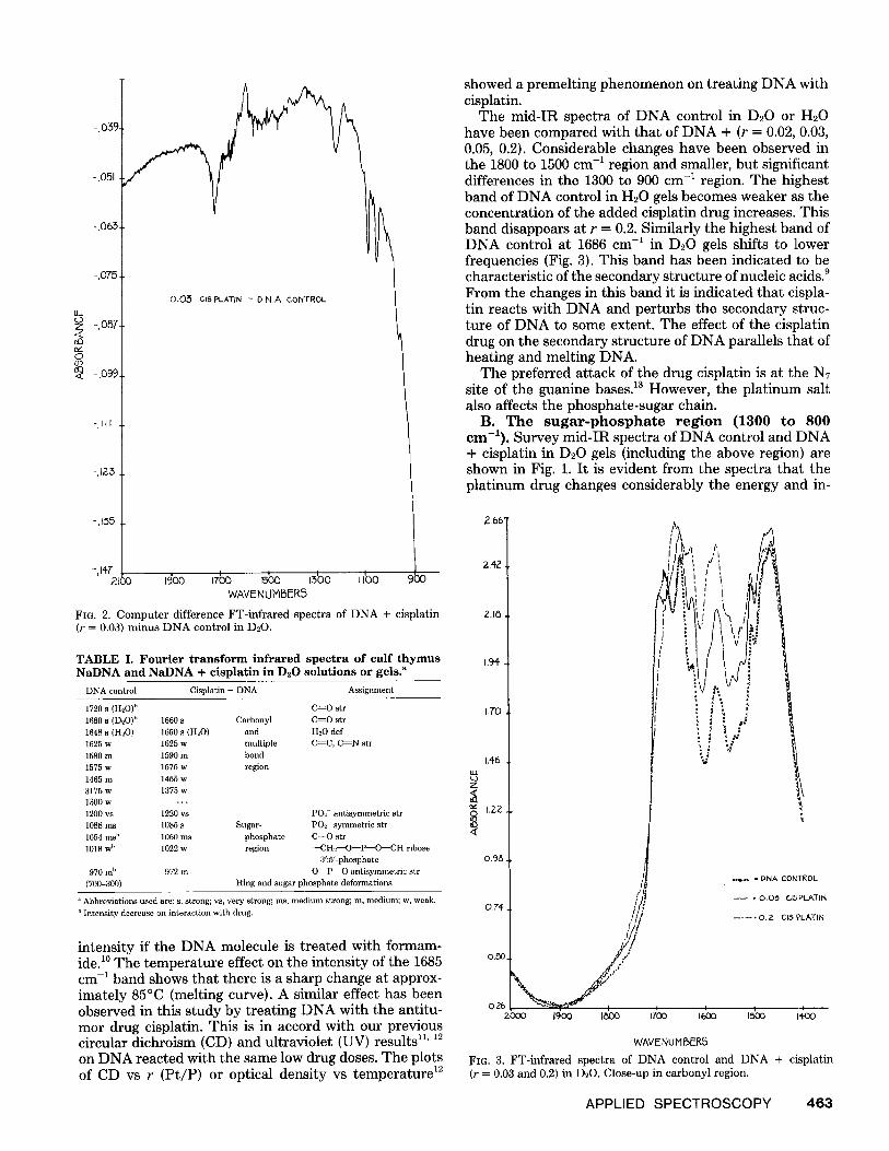
FIG. 2. C o m p u t e r difference FT- infrared spectra of D N A + cisplat in ( r = 0 .03) m i n u s D N A control in D 2 0 .
T A B L E I. F o u r i e r t r a n s f o r m i n f r a r e d s p e c t r a o f c a l f t h y m u s N a D N A a n d N a D N A + c i s p l a t i n i n D 2 0 s o l u t i o n s o r ge l s ."
DNA control Cisplatin + DNA Assignment
1720s(H20)" 1680 s (D20) b 1660 s 1648 s (H20) 1650 s (H20) 1625 w 1625 w 1580m 1590m 1575 w 1575 w 1465m 1455 w 3175w 1375w 1300w , . . 1200 vs 1220 vs 1088ms 1085 s 1054ms h 1060ms 1018 w b 1022 w
970m" 972m (700-300)
C ~ O str Carbonyl C=O str
and H~O def multiple C~C, C ~ N str bond region
PO2- antisymmetric str Sugar- POx symmetric str
phosphate C--O str r e g i o n --CH2--O--P--O--CH ribose
3':5'-phosphate O--P--O antlsymmetric str
Ring and sugar phosphate deformations
a Abbreviations used are: s, strong; vs, very strong; ms, medium strong; m, medium; w, weak. I' Intensity decrease on interaction with drug.
intensity if the DNA molecule is treated with formam- ide. 1° The temperature effect on the intensity of the 1685 cm -1 band shows that there is a sharp change at approx- imately 85°C (melting curve). A similar effect has been observed in this study by treating DNA with the antitu- mor drug cisplatin. This is in accord with our previous circular dichroism (CD) and ultraviolet (UV) results TM 12 on DNA reacted with the same low drug doses. The plots of CD vs r (Pt/P) or optical density vs temperature m
showed a premelting phenomenon on treating DNA with cisplatin.
The mid-IR spectra of DNA control in D20 or H20 have been compared with that of DNA + (r = 0.02, 0.03, 0.05, 0.2). Considerable changes have been observed in the 1800 to 1500 cm -1 region and smaller, but significant differences in the 1300 to 900 cm -1 region. The highest band of DNA control in H20 gels becomes weaker as the concentration of the added cisplatin drug increases. This band disappears at r = 0.2. Similarly the highest band of DNA control at 1686 cm -1 in D20 gels shifts to lower frequencies (Fig. 3). This band has been indicated to be characteristic of the secondary structure of nucleic acids. 9 From the changes in this band it is indicated that cispla- tin reacts with DNA and perturbs the secondary struc- ture of DNA to some extent. The effect of the cisplatin drug on the secondary structure of DNA parallels that of heating and melting DNA.
The preferred attack of the drug cisplatin is at the N7 site of the guanine bases, m However, the platinum salt also affects the phosphate-sugar chain.
B. The sugar-phosphate region (1300 to 800 cm-1). Survey mid-IR spectra of DNA control and DNA + cisplatin in D20 gels (including the above region) are shown in Fig. 1. It is evident from the spectra that the platinum drug changes considerably the energy and in-
2.66"
242
2.1~
1.94
I.TO
1,46
Z
~ 1.22
O.g&
O.~-.
O.26 zcx~o i~oo goo
z,l'!
i V
V
x ~ - O N A CONTKOL
= 0.05 CISPI..ATIN
. . . . 0 .2 C l S P L A T I N
,~o ,g0o ,5oo i4oo
W'AVENU MfSERS
F I G . 3. FT- infrared spectra of D N A contro l and D N A + c isplat in ( r = 0 .03 a n d 0.2) i n D,20. Close-up in carbony l region.
APPLIED S P E C T R O S C O P Y 4 6 3

tensity of some of the bands of the sugar-phosphate linkage, The phosphate PO2- bands (asymmetric and symmetric) are not perturbed, whereas the intensity of the OPO vibration at 970 and 1054 cm -~ is decreased. Both bands are slightly shifted (see Table I). The band at 1054 cm --~ was originally assigned to the PO2- sym- metric stretching vibration. TM However, later 9 the same group assigned the 1088 cm -~ band to the PO2- group. The results in this study are in agreement with this latter assignment. If the band at 1054 cm -1 was assigned to the symmetric stretching vibration of the PO2- then we would also expect a change in the antisymmetric stretch- ing vibration of the same group near 1220 cm -~ on treat- ment of DNA in H20 with the platinum drug. The fact that only the band at 1054 cm -~ is affected indicates that this band cannot be assigned to the P02- symmetric stretching vibration. The assignment of this band to the C-~O stretching vibration of the ester linkage is valid. The reaction of cisplatin with DNA perturbs only slighly the energy of the infrared stretching vibration of the ribose-phosphate band at 970 cm -1 (ribosyl-3'-O-(PO2-)- O-5'-CH2-), however its intensity is reduced consider- ably.
A close-up of the phosphate-ribosyl absorption region of control DNA and DNA + drug is shown in Fig. 4. The antisymmetric stretching vibration of PO2- absorbing near 1200 cm -~ is masked by the D20 deformation vibra- tion in D20 gels. It has been reported previously 9 that increased relative humidity of DNA had a slight effect on the PO2- symmetric and antisymmetric vibrations and their intensities. Hydrogen bonding with water was given as an explanation. 9 In our experiments we have not observed any changes in the absorption of the PO2- vibrations in DNA control and DNA + cisplatin at 1220 (in H20) and 1188 cm-' and the spectra were compared
under approximately similar conditions as far as water content was concerned. The only band that was signifi- cantly shifted and reduced in intensity was that at 1054 cm -1. To a much lesser degree were also perturbed the bands at 1022 and 970 cm -1 assigned to the vibrations involving the chain mode of the ribose-phosphate diester linkage 15. is (see Table I).
The platinum drug that is covalently bound to DNA may be at two adjacent guanine bases along the chain 12 and most likely may cause localized unwinding of the double helix affecting also the ribose-phosphate diester linkage. The cisplatin with its two adjacent ammonia molecules and carrying two positive charges cis- Pt(NH~)2(NT)22+--DNA may disrupt even at low doses the hydrogen bonding localized between the G--C bases and change the orientation of the phosphate group at 5'-CH2- and/or 3 '--CH--. The charged complex species (NH~)2Pt(NT)2 -2+ may also turn toward the negatively charged phosphate oxygen atoms of the 5 '~CH2--O--PO2---O--3 ' - -CH-- backbone in NaDNA perturbing the C--O ribose-phosphate linkage and the CH2 hydrogens as it was indicated from the changes of the bands 1054 cm -1 and 970 cm -~ (see Figs. 1 and 4).
Computer subtraction spectra of DNA + cisplatin mi- nus DNA control in D20 gels do confirm the shifts observed in the spectra. The following Scheme I shows the shapes expected for weak perturbations caused by a solvent or reactant to an absorption band. An example of a computer subtraction spectrum in the 1800 to 1500 cm -~ region is shown in Fig. 2 showing a shift of the highest band in D20 gels by approximately 90 cm -~. The computer subtractions are difficult to perform for such complicated systems, however they definitely show that there is a substantial shift of the highest carbonyl band to lower frequencies on treatment of DNA with the drug
lOOT.
ILl Z <
0 <
0,1~
0,1'4"
O,ID.
O, IZ.
0,11.
O, lO.
]' /
/ I
/ / / /
\,\
J059.9
I0~.1
. . . . O..Z CISPLATIN - - • PNA CONTROL
\ , \ ,
/ /
" \ /
tlSO II10 1090 IOTO I0,~0 1050 I010 990 970
971,2
\ \
\ \,
\ \
WAVENuPllSER$
F1o. 4. FT-infrared spectra of DNA control and DNA + cisplatin (r = 0.2). Close-up of sugar-phosphate region.
464 Volume 35, Number 5, 1981

SHIFT TO THE FRIGHT
SHIffT to THE LEFT
iSROADENIN61NARROWlN~ _~ INTENSITY
CH -I SCHEME I
cisplatin. A similar shift was also observed with subtrac- tion techniques in the Raman spectra in these very systems which were prepared under the same conditions. 8 This additional IR evidence of the carbonyl perturbation on treatment of DNA with cisplatin supports the car- bonyl implication in the complex with platinum 17-2° either through (G)NTN7 or N706 chelation or bridging with the metal. TM However, the mere disruption of hydrogen bond- ing in DNA can shift the highest carbonyl band of NaDNA to lower frequencies. Consequently, it seems that the perturbation by cisplatin of both the highest carbonyl band as well as the ribose-phosphate bands at 1054, 1022, and 970 cm -1 may be responsible for the antitumor mechanism of cisplatin. The effect of the drug on the ribose-phosphate band seems to be crucial to conformational changes of the sugar-phosphate chain.
More studies are needed particularly on the absorptiv- ity of the water molecules in DNA and DNA + cisplatin gels or solutions in order to understand the role of water molecules in DNA and their arrangement around the double helix. 21 From these FT-IR studies it is suggested that denaturation of DNA by cisplatin may be a result of the parallel arrangement of the planes of the bases fol- lowed by conformational changes. Departure from any given arragement reflects disruption or local melting of the ordered DNA structure and some sort of collapse of the DNA structure may result. The denaturing complex cations [(cis-Pt(NHs)2)(N7)2] ++ may exert their effect on DNA through their modification of the structure of water (hydrophobic agent). The structural details of these ef- fects are not at present established. The interpretation of the infrared spectral changes which has been given above is only qualitative. In fact, until recently 2~ the f t has been little information about the structure of v~ater in DNA on the basis of which to give a precise structural definition. In addition, the implication of H20 molecules in the double helix of DNA and the effects on them of low doses of the drug, cisplatin are not well understood.
The function of water and other alkaline or alkaline- earth salts in stabilizing the secondary structure provide the basis for more fruitful model building to understand the role of hydrophobic and hydrophylic forces in DNA. However, base stacking in addition to hydrogen bonding is also a major cause of DNA stability and reagents that attack the bases, as in the case of cisplatin, may decrease the stability of the bases and reduce the stacking tend- ency. Furthermore, if a chemical agent decreases the solubility of the sugar-phosphate chain in DNA, it also reduces the stacking tendency of the bases. The drug interaction with DNA may destabilize the DNA struc- ture. 12 The results here suggest that the attack of the drug cisplatin at the guanine bases even at low doses (r = 0.02) locally changes to some extent the secondary structure of DNA involving the sugar-phosphate back- bone.
ACKNOWLEDGMENTS
I am grateful to the National Sciences and Engineering Research Council of Canada, and the "Minist~re de l'Education du Quebec" for financial support and to Johnson Matthey & Mallory Ltd. for the loan of platinum salts. I also wish to thank the late Dr. P. K. Ganguli for the preparation of the DNA samples.
1. R. J. Bell, Introductory Fourier Transform Spectroscopy (Academic Press, New York, 1972, pp. 19-25.
2. M. Tsuboi, "Infrared and Raman Spectroscopy," in Basic Principles in Nucleic Acids, P. O. P. Tso, Ed. (Academic Press, New York, 1974), Vol. 1, pp. 399-452.
3. L. D. Esposito and J. L. Koenig, Fourier Transform IR Spectroscopy, (Aca- demic Press, New York, 1978), vol. 1.
4. M. C. Tobin, Spectrochim, Acta Part A25, 1855 (1969). 5. M. Tsuboi, Progr. Theor. Phys. Suppl. 17, 99 (1961). 6. M. Falk, K. A. Harman, Jr., and R. C. Lord, J. Amer. Chem. Soc. 85, 387, 391
(1963). 7. B. Rosenberg, L. Van Camp, and T. Krigas, Nature 205, 698 (1965). 8. A. J. P. Alix, L. Bernard, M. Manfait, P. K. Ganguli, and T. Theophanides,
Inorg. Chim. Acta 55, 147 {1981). 9. T. Shimanouchi, M. Tsuboi, and Y. Kyozoku, in Advances in Chemical
Physics, J. Duchesne, Ed. (Interscience, New York, 1964), Vol. VII, Chap. 12, p. 435.
1O. Y. Kyogoku, M. Tsuboi, T. Shimanouchi, and I. Watanabe, J. Mol. Biol. 3, 741 (1961).
11. P. K. Ganguli and T. Theophanides, J. Eur. Biochem. 101, 377 (1979}. 12. P. K. Ganguli and T. Theophanides, Inorg. Chim. Acta 55, L43, (1981). 13. B. Rosenberg, The Proceedings of Symposium on Coordination Chemistry
and Cancer Chemotherapy (Platinum Complexes and Cytochrome), P-450 Enzymes Biochimie, 60, 859 (1978).
14. G. B. B. M. Sutherland and M. Tsuboi, Proc. Roy. Soc. (London) A239, 446 (1957).
15. W. L. Peticolas and M. Tsuboi, "The Raman Spectroscopy of Nucleic Acids" in Infrared and Raman Spectroscopy of Biological Molecules T. Theophan- ides, Ed. (D. Reidel Publishing Co, Dordrecht, Holland, 1979), pp 153-165.
16. T. Theophanides, "Vibrational Spectroscopy of Metal Nucleic Systems" in Infrared and Raman Spectroscopy of Biological Molecules T. Theophanides, Ed. (D. Reidel Publishing Co, Dordrecht, Holland, 1979), pp 205-223.
17. M. M. Millard, J.-P. Macquet, and T. Theophanides, Biochim. Biophys. Acta 402, 166 {1975).
18. J.-P. Macquet and T. Theophanides, Bioinorg. Chem. 5, 59 (1975); and Inorg. Chim. Acta 18, 189 {1976).
19. B. Rosenberg, The Proceedings of the Third International Symposium on Platinum Coordination Complexes in Cancer Chemotherapy {Dallas, TX, 1976); and Biochimie, 60, 864 (1978).
20. G. Y. H. Chu, S. Mansy, E. E. Duncan, and R. S. Tobias, J. Amer. Chem. Soc. 100, 607 (1978).
21. E. Clementi, "Computational Aspects for Large Chemical Systems" in Lecture Notes in Chemistry, G. Berthier et al., Eds. (Springer-Verlag, New York, 1980), p. 131.
APPLIED SPECTROSCOPY 465