fourier transform infrared spectroscopy (ftir ) spectral ... · and, ftir spectral analysis of the...
TRANSCRIPT
Research Journal of Chemical
Vol. 6(2), 29-36, February (201
International Science Community Association
Fourier Transform Infrared Spectroscopy
Nanoparticles (BSA NPs) and Egg Albumin Nanoparticles (EA NPs)
Amity Institute of Biotechnology, Amity
Available online at: Received 28th January
Abstract
Advance protein based nanobiotechnology are now, known for its advanced approach to synthesize various nanomaterials
using various newly developed chemical and green technologies to carry out their applications in various fields of life
sciences. Now these days, Bovine serum albumin nanoparticles (BSA NPs) are very popular as non
vehicle systems for drug delivery used in number of therapeutic strategies to be considered in combating cancers, tumors,
hormone associated and neurodegenerative disease. The albumin nanoparticles e.g. bovine serum albumin nanoparticles
(BSA NPs) and egg albumin nanopartic
desolvation process to get desired particle size at nanosacle e.g. range between 100 to 300 nm with minimal size distribution
The size controlled BSA and egg albumin nanoparticles ca
and non-viral biocompatible nanodevice and nanovehicle carrier for loading of desired drug/ biological active ingredients/
hormones molecules/ chimeric-DNA with efficient target delivery sys
using emulsified and desolvation method are done via glutaraldehyde coupling for loading of
found to be better eco-friendly and cost
Sometimes, the metalonanoparticles or nanomaterials are found to be very costly and toxic because of their slow natural
degradation in host and their chemically driven synthesis. Hence, the synthesis of bio
might be used for their further consideration for their safe and non
technology, biomedical approaches and pharmaceutical industry for the treatment of various diseases. Hence, this d
Fourier Transform Infrared Spectroscopy
albumin nanoparticles (BSA NPs) and egg albumin nanoparticles (EA NPs) and their active functional groups and chemical
interactions for loading desired biological components or drugs that can might be further considered to increase their safe
and cost effective therapeutic viability.
Keywords: Bovine serum albumin nanoparticles, Egg albumin nanoparticles, BSA NPs, EA NPs,
Emulsification method, Cicer arietinum amylase, Drug delivery, FTIR.
Introduction
Fine particles are considered to be known which are sized inbetween 100 and 2,500 nanometers and ultrafine particles are purely called nanoparticles which were found to be in the range of 1-100 nanometers in size
1,2. These days, protein based
nanotechnology are very much considered to the research and
technological developments at atomic, molecular and macromolecular scales to prepare synthetic polymers together with the absorbability and low toxicity of the degradation end
products3,4
. Silver-poly(methylmethacrylate) (Ag/PMMA) nanocomposites were prepared via in-situ polymerization technique using N,N’-dimethylformamide (DMF) and
characterized by Fourier transform infrared spectroscopy (FTIR)
to know the details of active functional groups present in the synthesized PMMA
5. FTIR spectra of silver nanoparticles was
also found to be observed to have exhibited prominent peaks at
2,927, 1,631, and 1,383 cm−1
which showed sharp and strong absorption band at 1,631 cm
−1 assigned to the stretching
Chemical Sciences _______________________________________
(2016)
Association
Fourier Transform Infrared Spectroscopy (FTIR) Spectral Analysis of BSA
Nanoparticles (BSA NPs) and Egg Albumin Nanoparticles (EA NPs)
Kirti Rani Amity Institute of Biotechnology, Amity University Uttar Pradesh, Noida, Sec-125, Noida-201303,
Available online at: www.isca.in, www.isca.me, www.isca.me January 2016, revised 2nd February 2016, accepted 16th February 201
Advance protein based nanobiotechnology are now, known for its advanced approach to synthesize various nanomaterials
using various newly developed chemical and green technologies to carry out their applications in various fields of life
sciences. Now these days, Bovine serum albumin nanoparticles (BSA NPs) are very popular as non
r drug delivery used in number of therapeutic strategies to be considered in combating cancers, tumors,
hormone associated and neurodegenerative disease. The albumin nanoparticles e.g. bovine serum albumin nanoparticles
(BSA NPs) and egg albumin nanoparticles (EA NPs) are easily prepared by number of established emulsified and
desolvation process to get desired particle size at nanosacle e.g. range between 100 to 300 nm with minimal size distribution
The size controlled BSA and egg albumin nanoparticles can be used as the standard cost effective, non
viral biocompatible nanodevice and nanovehicle carrier for loading of desired drug/ biological active ingredients/
DNA with efficient target delivery system. As well as, fabrication of BSA NPs and EA NPs by
using emulsified and desolvation method are done via glutaraldehyde coupling for loading of Cicer arietinum
friendly and cost-effective choice as compared to synthesis of other metal based nanoparticles.
Sometimes, the metalonanoparticles or nanomaterials are found to be very costly and toxic because of their slow natural
degradation in host and their chemically driven synthesis. Hence, the synthesis of bio-compatible
might be used for their further consideration for their safe and non-toxic therapeutic applications in fields of biosensor
technology, biomedical approaches and pharmaceutical industry for the treatment of various diseases. Hence, this d
Fourier Transform Infrared Spectroscopy (FTIR) analysis was used to determine the purity of the prepared bovine serum
albumin nanoparticles (BSA NPs) and egg albumin nanoparticles (EA NPs) and their active functional groups and chemical
s for loading desired biological components or drugs that can might be further considered to increase their safe
Bovine serum albumin nanoparticles, Egg albumin nanoparticles, BSA NPs, EA NPs,
amylase, Drug delivery, FTIR.
Fine particles are considered to be known which are sized in-between 100 and 2,500 nanometers and ultrafine particles are purely called nanoparticles which were found to be in the range
. These days, protein based
gy are very much considered to the research and
technological developments at atomic, molecular and macromolecular scales to prepare synthetic polymers together with the absorbability and low toxicity of the degradation end
ethacrylate) (Ag/PMMA) situ polymerization
dimethylformamide (DMF) and
characterized by Fourier transform infrared spectroscopy (FTIR)
to know the details of active functional groups present in the FTIR spectra of silver nanoparticles was
also found to be observed to have exhibited prominent peaks at
which showed sharp and strong assigned to the stretching
vibration of (NH) C=O group; developed band 1,383 for Cand C–N stretching; observed sharp peak at 2,927
assigned to C–H and C–H (methoxy compounds) stretching vibration
6. FTIR spectra was also used for characterization of
ZnO nanoparticles. Infrared studies wer
ascertain the purity and nature of the metal nanoparticles that had observed peak at 3452.30 and 1119.15 cm
present due to O-H stretching and deformation. Other observed peaks at 1634.00 and 620.93 cm
correspond the Zn-O stretching and deformation vibration, respectively
7.
And, FTIR spectral analysis of the iron oxide nanoparticles and microgel magnetic particles were also reported to characteristic
the absorption band of Fe–O bond of bulk Fecm−1 to reduced the size of prepared nanoparticles to nanoscale dimensions
8. The molecular characteristics of the resulting BSA
NPs were also characterized by FTIR to have sharp FTIR peaks at 3385 cm-1 , 3113 cm-1, 1707 cm
___________E-ISSN 2231-606X
Res. J. Chem. Sci.
29
) Spectral Analysis of BSA
Nanoparticles (BSA NPs) and Egg Albumin Nanoparticles (EA NPs)
201303, UP, India
2016
Advance protein based nanobiotechnology are now, known for its advanced approach to synthesize various nanomaterials by
using various newly developed chemical and green technologies to carry out their applications in various fields of life-
sciences. Now these days, Bovine serum albumin nanoparticles (BSA NPs) are very popular as non-toxic and nonviral
r drug delivery used in number of therapeutic strategies to be considered in combating cancers, tumors,
hormone associated and neurodegenerative disease. The albumin nanoparticles e.g. bovine serum albumin nanoparticles
les (EA NPs) are easily prepared by number of established emulsified and
desolvation process to get desired particle size at nanosacle e.g. range between 100 to 300 nm with minimal size distribution.
n be used as the standard cost effective, non-toxic, non-allergic
viral biocompatible nanodevice and nanovehicle carrier for loading of desired drug/ biological active ingredients/
tem. As well as, fabrication of BSA NPs and EA NPs by
Cicer arietinum amylase that
s of other metal based nanoparticles.
Sometimes, the metalonanoparticles or nanomaterials are found to be very costly and toxic because of their slow natural
compatible BSA NPs and EA NPs
toxic therapeutic applications in fields of biosensor
technology, biomedical approaches and pharmaceutical industry for the treatment of various diseases. Hence, this designed
(FTIR) analysis was used to determine the purity of the prepared bovine serum
albumin nanoparticles (BSA NPs) and egg albumin nanoparticles (EA NPs) and their active functional groups and chemical
s for loading desired biological components or drugs that can might be further considered to increase their safe
Bovine serum albumin nanoparticles, Egg albumin nanoparticles, BSA NPs, EA NPs, Desolvation method,
C=O group; developed band 1,383 for C–C N stretching; observed sharp peak at 2,927 cm
−1 that
H (methoxy compounds) stretching . FTIR spectra was also used for characterization of
ZnO nanoparticles. Infrared studies were carried out in order to
ascertain the purity and nature of the metal nanoparticles that had observed peak at 3452.30 and 1119.15 cm-1 which may be
H stretching and deformation. Other observed peaks at 1634.00 and 620.93 cm-1 were also reported to
O stretching and deformation vibration,
And, FTIR spectral analysis of the iron oxide nanoparticles and microgel magnetic particles were also reported to characteristic
O bond of bulk Fe3O4 at 570 and 375 −1 to reduced the size of prepared nanoparticles to nanoscale
. The molecular characteristics of the resulting BSA NPs were also characterized by FTIR to have sharp FTIR peaks
1, 1707 cm-1 , 1533 cm-1 and 1242
Research Journal of Chemical Sciences __________________________________________________________E-ISSN 2231-606X
Vol. 6(2), 29-36, February (2016) Res. J. Chem. Sci.
International Science Community Association 30
cm-1 that are assigned to the stretching vibration of OH, amide A (mainly NH stretching vibration), amide I (mainly C=O
stretching vibrations), amide II (the coupling out phase of
bending vibration of N-H and stretching vibration of C-N
bands) and amide III (is in the phase combination of N-H in plane bending and C-N stretching)
9. Revioulsy, various
techniques e.g. desolvation, emulsification, thermal gelation, nanospary drying, nab-technology and self-assembly techniques
were used for the preparation of albumin nanoparticles which do
not require high temperature and therefore may be useful when
heat-sensitive bioactive compounds are designed to be
immobilized into fabricated albumin nanoparticles10
.
Hence, protein nanoparticles are observed to be an attractive
biomolecule which have their unique characteristic including
biodegradability, nontoxicity, metabolized in vivo to produce
innocuous degradation products, water solubility, simplicity in
purification and nonimmunogenic nature that allow their ease of controlled and targeted delivery as an ideal nanopreparation
11.
And, desolvation methods are found to be reported more advantageous over emulsification method to prepare albumin nanoparticles due to avoidance of organic solvents to remove the used oily residues and surfactants which are required for the emulsion stabilization. Desolvation technique was found to be more cost effective to prepare more small sized, thermo-stabilized, storage-stabilized nanoparticles with their reported significant alkaline protease mediated controlled biodegradation and increased shelf life period over upto 12-14 months
11,12.
Previously, the enzyme immobilization was also reported into those prepared BSA NPs
13 and EA NPs
14,15 nanoparticles which
were more cost effective, eco-friendly biocompatible, non-toxic, non-allergic and non-corrosive enzyme loading carrier by using desolvation method
11 and emulsification method
12 to make them
more industrially viable11-15
. Hence, this concrete Fourier Transform Infrared Spectroscopy (FTIR) spectral analysis was used for the interpretation of prepared purity of the prepared bovine serum albumin nanoparticles (BSA NPs)
11 and egg albumin nanoparticles (EA
NPs) by desolvation method13
; bovine serum nanoparticles (BSA NPs)
12 and egg albumin nanoparticles (EA NPs) by
emulsification method14.
As well as, their active functional groups and molecular interactions were also assigned to get to
know the altered chemical interactions in prepared BSA NPs11,12
and EA NPs13-15
before and after the glutaraldehyde activation
that were further used for desired Cicer arietinum amylase immobilization to be used for their controlled alkaline protease mediated bioproteolysis and effective washing study. So, FTIR analysis was used to carried for the functional chemical interactions of prepared BSA NPs and EA NPs which can might
be exploited for their further inevitable use to load the desired drug/ biological active ingredients/ hormones molecules/
chimeric-DNA with efficient target delivery system as cost effective, non-toxic, non-allergic and non-viral biocompatible nanodevice and nanovehicle carriers. That may become a
landmark in non-toxic therapeutic applications especially in fields of biosensor technology, biomedical approaches and
pharmaceutical industry for the treatment of various diseases (cancer, tumors, hormone-associated and neurodegenerative
disorders) as green clinical therapeutic technology.
Materials and Methods
Preparation of BSA NPs and Cicer arietinum amylase loaded
BSA nanoparticle (Amylase-BSA NPs) by Deslovation
method: BSA NPs and Amyalse-BSA NPs were prepared by
desolvation method by using n-butanol given 2012 and Rani K
& Chauhan C, 201511
.
Preparation of BSA NPs and Cicer arietinum amylase loaded
BSA nanoparticle (Amylase-BSA NPs) by Emulsification
method: BSA NPs and Amyalse-BSA NPs were prepared by
Emulsification method by using coconut oil as natural
occurring emulsifier given Rani K & Chauhan C, 201512
.
Preparation of EA NPs and Cicer arietinum amylase loaded
Egg Albumin nanoparticle (Amylase-BSA NPs) by
Deslovation method: EA NPs and Amyalse-EA NPs were
prepared by desolvation method by using toluene given by Rani
K, 201515
.
Preparation of EA NPs and Cicer arietinum amylase loaded
Egg Albumin nanoparticle (Amylase-BSA NPs) by
Emulsification method: EA NPs and Amyalse-EA NPs were
prepared by emulsification method by using using coconut oil as
natural occurring emulsifier with toluene and n-butanol, given
by 2012 and Rani K, 201513
.
Alkaline Protease Mediated Bioproteolysis of Prepared
Amylase-BSA NPs and Amylase-EA NPs11-15
: Amylase loaded
BSA NPs and EA NPs were also subjected to proteolysis with
standardized 35 U of alkaline protease to confirmed loading of
desired amylase into prepared active BSA NPs and EA NPs.
Characterization: Fourier Transform Infrared
Spectroscopy (FTIR) analysis of prepared BSA NPs and EA
NPs: The prepared BSA NPs and EA NPs; Amylase-BSA NPs
and Amylase-EA NPs were subjected for characterization to
Fourier Transform Infrared Spectroscopy (FTIR) analysis to
determine the purity of fabricated BSA NPs and EA NPs before
and after the immobilization done by glutaraldehyde coupling.
Designed FTIR spectroscopy was carried out to be scanned at
the range of 500-4000cm-19,16
.
Results and Discussion
Previously reported data of DLS and SEM for confirmed
BSA NPs and EA NPs by Rani K & Chauhan C, 2015; Rani, K, 2015
11-14:
Dynamic Light scattering (DLS) and Scanning
Electron Microscopy (SEM) of prepared BSA NPs and
Amylase-BSA NPs by desolvation method and emulsification
method were already reported by Rani K & Chauhan C, 2015, to
confirm the size of nanoparticles in the approximate range of

Research Journal of Chemical Sciences __________________________________________________________E-ISSN 2231-606X
Vol. 6(2), 29-36, February (2016) Res. J. Chem. Sci.
International Science Community Association 31
2nm to 11nm and 56nm to 107.4nm respectively11,12
. Dynamic
Light scattering (DLS) and Scanning Electron Microscopy
(SEM) results of prepared EA NPs and Amylase-EA NPs by
desolvation method and emulsification method were observed
by Rani K, 2015, to confirm the size of nanoparticles in the
approximate range of 5nm to upto 100nm and 101.2nm to
209nm respectively13,14
. The observed size of BSA NPs and
Amylase-NPs were found to be less than of EA NPs and
Amylase-EA NPs11-14
Those observation were also comparable
similar to other previous reports9,16-18
.
Fourier Transform Infrared Spectroscopy (FTIR) analysis
of prepared BSA NPs and Amylase-BSA NPs & EA NPs and Amylase-EA NPs: FTIR spectroscopy was used to determine
the purity of fabricated BSA NPs and EA NPs before and after
the immobilization of desired enzyme done by glutaraldehyde
coupling11-15
. It was scanned at the range, 500-4000 cm-1
9,16.
Fourier Transform Infrared Spectroscopy (FTIR) analysis
of prepared BSA NPs and Amylase-BSA NPs: The FTIR
peaks of prepared BSA NPs by using desolvation method11
before immobilization of amylase were observed at 3381 cm-1
that assigned to the stretching vibration of OH [amide A, mainly
NH stretching vibration]; at 2958 cm-1, at 2932 cm-1 and at
2873 cm-1 that approximate assigned C–H and C–H (methoxy
compounds) stretching vibration; at 1642 cm-1 and at 1292 cm-
1 that assigned to having approximate vibration of (NH) C=O
group and for C–C and C–N stretching respectively (Figure-1).
The FTIR peaks of BSA NPs after immobilization of amylase
were observed at 3388 cm-1 that assigned to the stretching
vibration of OH [amide A, mainly NH stretching vibration]; at
1649 cm-1 that assigned approximate the stretching vibration of
(NH) C=O group (Figure-2). Observed FTIR peaks at 2958 cm-
1, at 2932 cm-1 and at 2873 cm-1 were only observed in BSA
NPs before immobilization only (Figure-1) which confirmed
that immobilization of enzyme was done at these assigned
activated functional groups (Figure-2). These observations were
found to be comparably similar with previous FTIR analysis of
nanoparticles6,9,16-19
.
The FTIR peaks of BSA NPs by using emulsification method12
before immobilization of desired enzyme were observed at 3478
cm-1 that assigned to approximate stretching vibration of OH
[amide A, mainly NH stretching vibration]; at 2923 cm-1 and at
2854 cm-1 that assigned approximate C–H and C–H (methoxy
compounds) stretching vibration; at 1697 cm-1 that assigned to
having approximate vibration of (NH) C=O group and for C–C
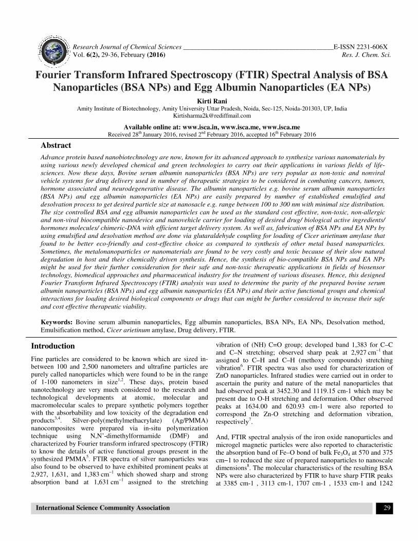
and C–N stretching respectively (Figure-3). The FTIR peaks of
BSA NPs after immobilization of amylase were observed at
3566 cm-1 that assigned to have approximate possible stretching
vibration of OH [amide A, mainly NH stretching vibration]; at
1647 cm-1 that assigned approximate the stretching vibration of
(NH) C=O group; at 1369 cm-1 tha assigned to have possible
C–C and C–N stretching (Figure-4). Observed FTIR peaks at
2958 cm-1 and at 2854 cm-1 were only observed in BSA NPs
before immobilization only (Figure-3) which confirmed that
immobilization of enzyme was done at these assigned activated
functional groups (Figure-4). These reported were found to be
comparably similar with previous FTIR interpretation of
nanoparticles6,9,16,20
.
Figure-1
FTIR spectra of prepared activated BSA NPs by desolvation method before the immobilization of Cicer arietinum amylase

Research Journal of Chemical Sciences __________________________________________________________E-ISSN 2231-606X
Vol. 6(2), 29-36, February (2016) Res. J. Chem. Sci.
International Science Community Association 32
Figure-2
FTIR spectra of prepared activated BSA NPs by desolvation method after the immobilization having loaded Cicer arietinum
amylase (immobilization done by glutaraldehyde coupling)
Figure-3
FTIR spectra of prepared activated BSA NPs by emulsification method before the immobilization of Cicer arietinum
amylase
Research Journal of Chemical Sciences __________________________________________________________E-ISSN 2231-606X
Vol. 6(2), 29-36, February (2016) Res. J. Chem. Sci.
International Science Community Association 33
Figure-4
FTIR spectra of prepared activated BSA NPs by emulsification method after the immobilization having loaded Cicer
arietinum amylase (immobilization done by glutaraldehyde coupling)
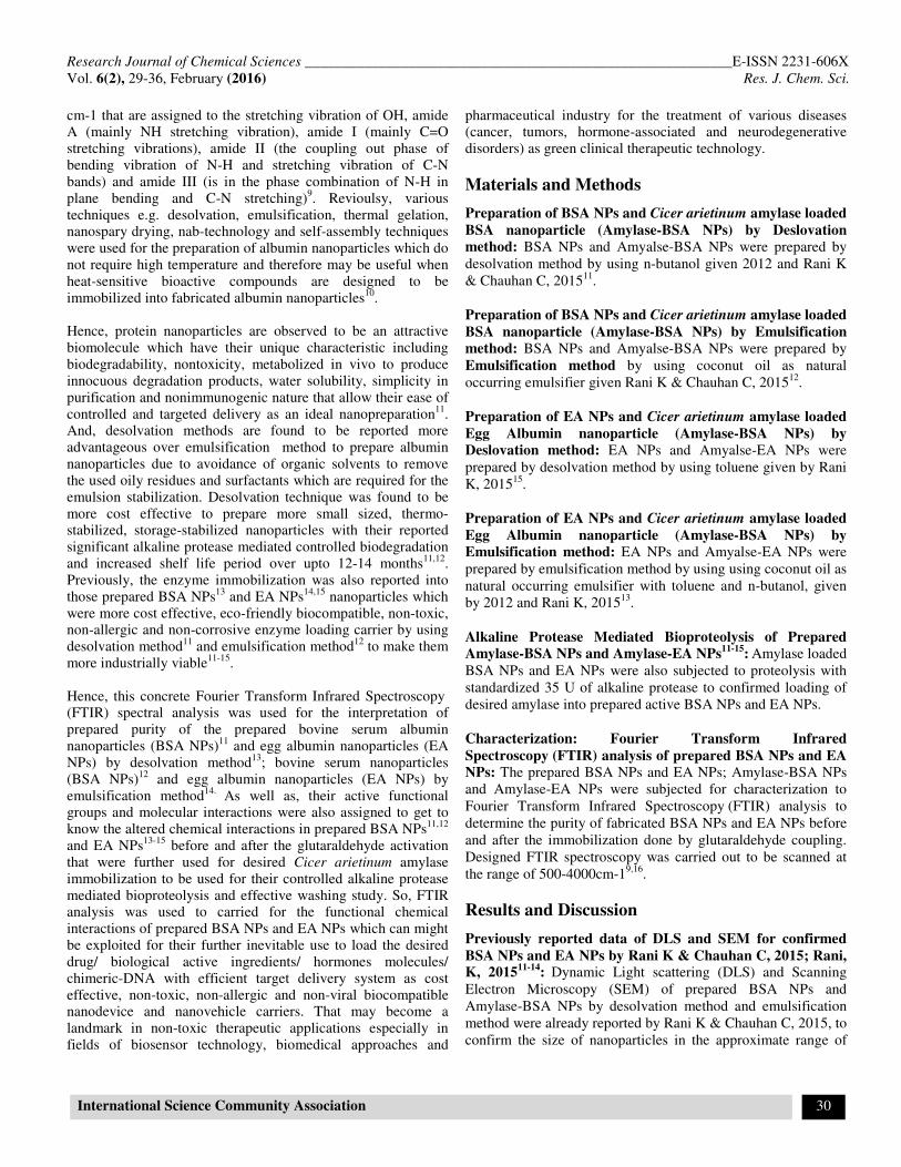
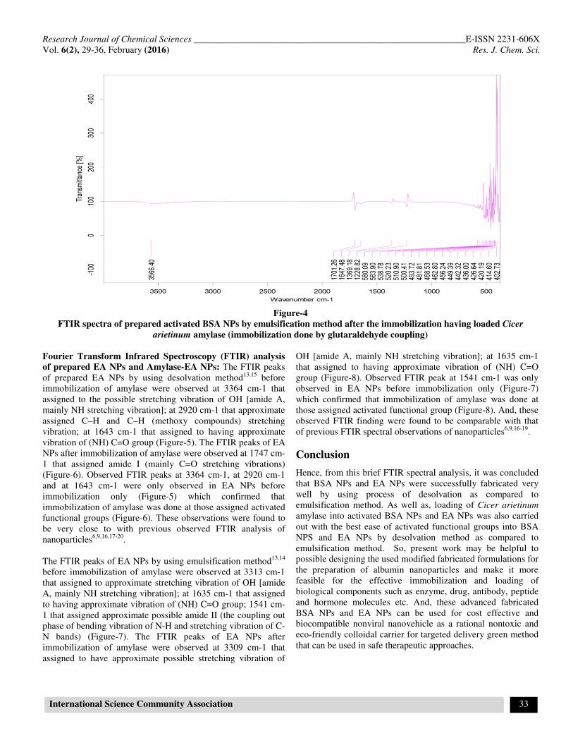
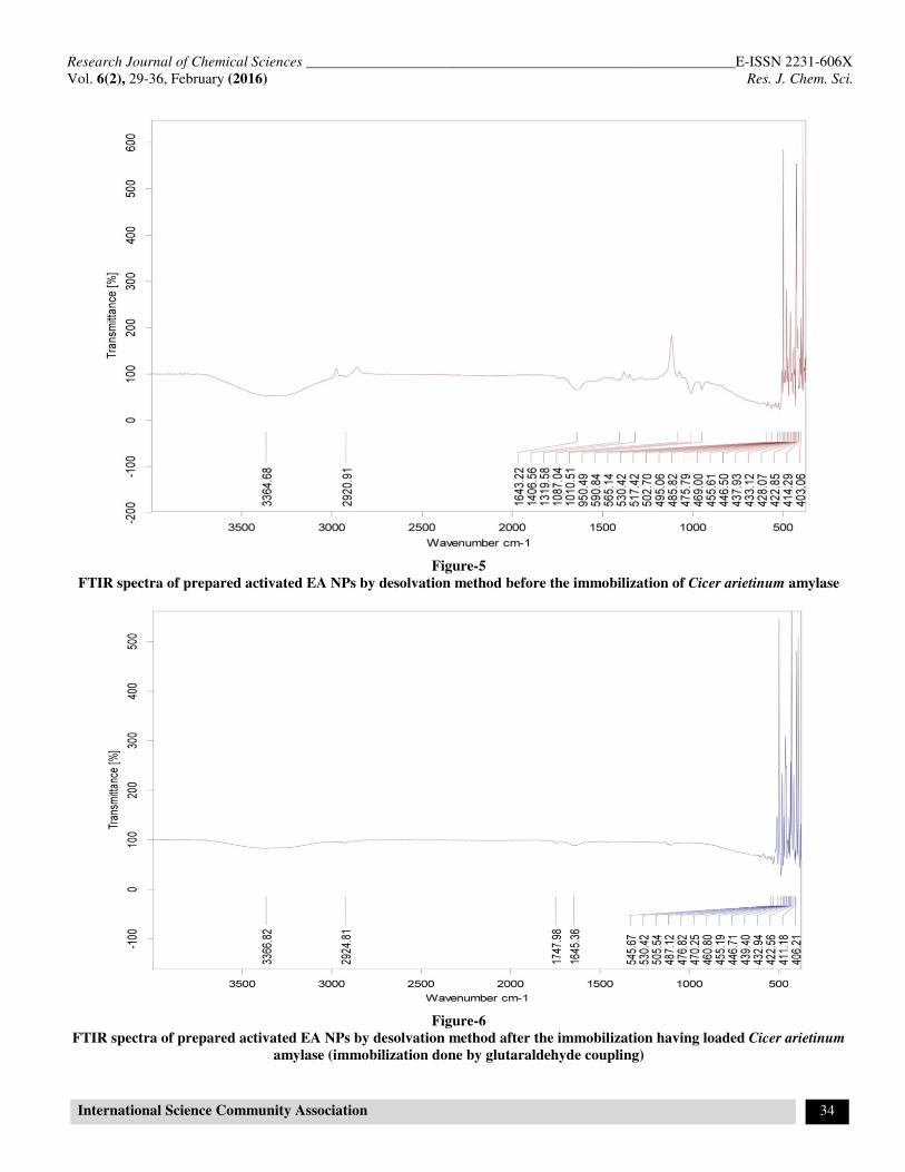
Fourier Transform Infrared Spectroscopy (FTIR) analysis
of prepared EA NPs and Amylase-EA NPs: The FTIR peaks
of prepared EA NPs by using desolvation method13,15
before
immobilization of amylase were observed at 3364 cm-1 that
assigned to the possible stretching vibration of OH [amide A,
mainly NH stretching vibration]; at 2920 cm-1 that approximate
assigned C–H and C–H (methoxy compounds) stretching
vibration; at 1643 cm-1 that assigned to having approximate
vibration of (NH) C=O group (Figure-5). The FTIR peaks of EA
NPs after immobilization of amylase were observed at 1747 cm-
1 that assigned amide I (mainly C=O stretching vibrations)
(Figure-6). Observed FTIR peaks at 3364 cm-1, at 2920 cm-1
and at 1643 cm-1 were only observed in EA NPs before
immobilization only (Figure-5) which confirmed that
immobilization of amylase was done at those assigned activated
functional groups (Figure-6). These observations were found to
be very close to with previous observed FTIR analysis of
nanoparticles6,9,16,17-20
.
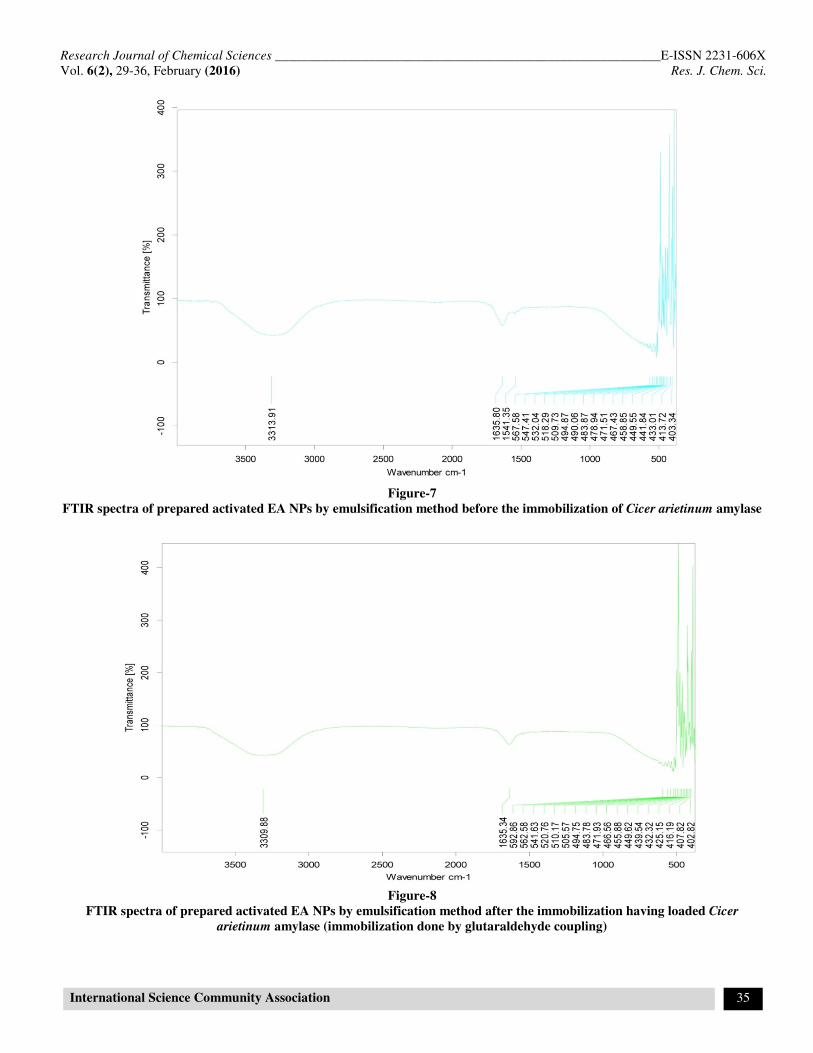
The FTIR peaks of EA NPs by using emulsification method13,14
before immobilization of amylase were observed at 3313 cm-1
that assigned to approximate stretching vibration of OH [amide
A, mainly NH stretching vibration]; at 1635 cm-1 that assigned
to having approximate vibration of (NH) C=O group; 1541 cm-
1 that assigned approximate possible amide II (the coupling out
phase of bending vibration of N-H and stretching vibration of C-
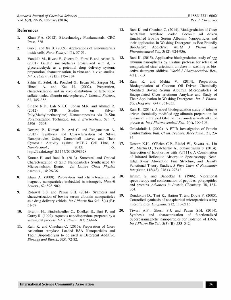
N bands) (Figure-7). The FTIR peaks of EA NPs after
immobilization of amylase were observed at 3309 cm-1 that
assigned to have approximate possible stretching vibration of
OH [amide A, mainly NH stretching vibration]; at 1635 cm-1
that assigned to having approximate vibration of (NH) C=O
group (Figure-8). Observed FTIR peak at 1541 cm-1 was only
observed in EA NPs before immobilization only (Figure-7)
which confirmed that immobilization of amylase was done at
those assigned activated functional group (Figure-8). And, these
observed FTIR finding were found to be comparable with that
of previous FTIR spectral observations of nanoparticles6,9,16-19
.
Conclusion
Hence, from this brief FTIR spectral analysis, it was concluded
that BSA NPs and EA NPs were successfully fabricated very
well by using process of desolvation as compared to
emulsification method. As well as, loading of Cicer arietinum
amylase into activated BSA NPs and EA NPs was also carried
out with the best ease of activated functional groups into BSA
NPS and EA NPs by desolvation method as compared to
emulsification method. So, present work may be helpful to
possible designing the used modified fabricated formulations for
the preparation of albumin nanoparticles and make it more
feasible for the effective immobilization and loading of
biological components such as enzyme, drug, antibody, peptide
and hormone molecules etc. And, these advanced fabricated
BSA NPs and EA NPs can be used for cost effective and
biocompatible nonviral nanovehicle as a rational nontoxic and
eco-friendly colloidal carrier for targeted delivery green method
that can be used in safe therapeutic approaches.
Research Journal of Chemical Sciences __________________________________________________________E-ISSN 2231-606X
Vol. 6(2), 29-36, February (2016) Res. J. Chem. Sci.
International Science Community Association 34
Figure-5
FTIR spectra of prepared activated EA NPs by desolvation method before the immobilization of Cicer arietinum amylase
Figure-6
FTIR spectra of prepared activated EA NPs by desolvation method after the immobilization having loaded Cicer arietinum
amylase (immobilization done by glutaraldehyde coupling)
Research Journal of Chemical Sciences __________________________________________________________E-ISSN 2231-606X
Vol. 6(2), 29-36, February (2016) Res. J. Chem. Sci.
International Science Community Association 35
Figure-7
FTIR spectra of prepared activated EA NPs by emulsification method before the immobilization of Cicer arietinum amylase
Figure-8
FTIR spectra of prepared activated EA NPs by emulsification method after the immobilization having loaded Cicer
arietinum amylase (immobilization done by glutaraldehyde coupling)
Research Journal of Chemical Sciences __________________________________________________________E-ISSN 2231-606X
Vol. 6(2), 29-36, February (2016) Res. J. Chem. Sci.
International Science Community Association 36
References
1. Khan F.A. (2012). Biotechnology Fundamentals, CRC
Press, 328.
2. Gao J. and Xu B. (2009). Applications of nanomaterials
inside cells, Nano Today, 4 (1), 37-51.
3. Vandelli M., Rivasi F., Guerra P., Forni F. and Arletti R.
(2001). Gelatin microspheres crosslinked with d, l-
glyceraldehyde as a potential drug delivery system:
preparation, characterization, in vitro and in vivo studies.
Int. J. Pharm., (215), 175– 184.
4. Sahin S., Selek H., Ponchel G., Ercan M., Sargon M.,
Hincal A. and Kas H. (2002). Preparation,
characterization and in vivo distribution of terbutaline
sulfate loaded albumin microspheres. J. Control. Release,
82, 345–358.
5. Singho N.D., Lah N.K.C., Johan M.R. and Ahmad R.
(2012). FTIR Studies on Silver-
Poly(Methylmethacrylate) Nanocomposites via In-Situ
Polymerization Technique. Int. J. Electrochem. Sci., 7,
5596 – 5603.
6. Devaraj P., Kumari P., Arti C. and Renganathan A.
(2013). Synthesis and Characterization of Silver
Nanoparticles Using Cannonball Leaves and Their
Cytotoxic Activity against MCF-7 Cell Line, J.
Nanotechnol., 2013: 1-5.
http://dx.doi.org/10.1155/2013/598328
7. Kumar H. and Rani R. (2013). Structural and Optical
Characterization of ZnO Nanoparticles Synthesized by
Microemulsion Route, Int Letters Chem Physics
Astronm., 14: 26-36.
8. Khan A. (2008). Preparation and characterization of
magnetic nanoparticles embedded in microgels. Materil
Letters., 62: 898–902.
9. Rohiwal S.S. and Pawar S.H. (2014). Synthesis and
characterization of bovine serum albumin nanoparticles
as a drug delivery vehicle. Int J Pharm Bio Sci., 5(4) (B):
51-57.
10. Ibrahim H., Bindschaedler C., Doelker E., Buri P. and
Gurny R. (1992). Aqueous nanodispersions prepared by a
salting out process. Int. J. Pharm., 87: 239-46.
11. Rani K. and Chauhan C. (2015). Preparation of Cicer
Artienium Amylase Loaded BSA Nanoparticles and
Their Bioproteolysis to be used as Detergent Additive.
Bioengg and Biosci., 3(5): 72-82.
12. Rani K. and Chauhan C. (2014). Biodegradation of Cicer
Arietinum Amylase loaded Coconut oil driven
Emulsified Bovine Serum Albumin Nanoparticles and
their application in Washing Detergents as Eco-Friendly
Bio-Active Addictive. World J Pharm and
Pharmaceutical Sci., 3(12): 924-936.
13. Rani K. (2015). Applicative biodegradation study of egg
albumin nanospheres by alkaline protease for release of
encapsulated cicer arietinum amylase in washing as bio-
active detergent additive. World J Pharmaceutical Res.,
4(1): 1-13.
14. Rani K. and Mehta V. (2014). Preparation,
Biodegradation of Coconut Oil Driven Chemically
Modified Bovine Serum Albumin Microparticles of
Encapsulated Cicer arietinum Amylase and Study of
Their Application in Washing Detergents. Int. J. Pharm.
Sci. Drug Res., 6(4): 351-355.
15. Rani K. (2014). A novel biodegradation study of toluene
driven chemically modified egg albumin preparation for
release of entrapped Glycine max amylase with alkaline
proteases. Int J Pharmaceutical Res., 6(4), 100-103.
16. Grdadolnik J. (2002). A FTIR Investigation of Protein
Conformation. Bull. Chem. Technol. Macedonia., 21, 23-
34
17. Dostert K.H., O’Brien C.P., Riedel W., Savara A., Liu
W., Martin O., Tkatchenko A., Schauermann S. (2014).
Interaction of Isophorone with Pd(111): A Combination
of Infrared Reflection–Absorption Spectroscopy, Near-
Edge X-ray Absorption Fine Structure, and Density
Functional Theory Studies. J Phys Chem C Nanomater
Interfaces, 118(48), 27833–27842.
18. Krimm S. and Bandekar J. (1986). Vibrational
spectroscopy and conformation of peptides, polypeptides
and proteins. Advances in Protein Chemistry, 38, 181–
364.
19. Dendukuri D., Tsoi K., Hatton T. and Doyle P. (2005).
Controlled synthesis of nonspherical microparticles using
microfluidics. Langmuir, 212, 113-2116.
20. Tiwari A.P., Ghosh S.J. and Pawar S.H. (2014).
Synthesis and characterization of functionalized
Superparamagnetic nanoparticles for isolation of DNA.
Int J Pharm Bio Sci., 5(3) (B), 533–542.