fractal dimension analysis of swarms of gold nano/micro
TRANSCRIPT

ISSN 2348-1218 (print)
International Journal of Interdisciplinary Research and Innovations ISSN 2348-1226 (online) Vol. 7, Issue 1, pp: (416-425), Month: January - March 2019, Available at: www.researchpublish.com
Page | 416 Research Publish Journals
Fractal Dimension Analysis of Swarms of Gold
Nano/Micro Particles Produced By Wild
Rhizobium Cultures Using A Novel Slide Based
System
Sujata Dabolkar1, Nandkumar Kamat
2
12Mycological Laboratory, Department of Botany, Goa University, Taleigao, Goa, 403206, India
Abstract: There is a lot of interest in autonomous movement and collective behavior of synthetic nanomaterials
which have important applications in nanomedicine, nanobiotechnology and nanosensors. Present work using
viable cultures and cell free extracts of Rhizobium sp. to produce and analyze swarms of monodisperse Au
Nanoparticles (Au NPs) and polydisperse Au microparticles (Au MPs) was inspired by previously reported work
on chemically triggered swarming of commercially available Au MPs. We developed a simple, glass slide based
technique for rapid production, microscopic visualization, morphological analysis, study of swarming behavior
and monitoring of effects of heat on Au NPs and Au MPs forms and assemblages. We designed cell based and cell
free microbial system such as Rhizobium sp. to test the hypothesis that unidentified proteinaceous factors could be
involved in producing simple monodisperse and complex polydisperse geometric forms of Au NPs and Au MPs.
We used pure and wild type Rhizobium cell suspensions prepared from surface sterilized root nodules of Mimosa
pudica. Cell free extracts from wild type and pure cultures were prepared by warming cell suspensions for 15, 30,
and 45 sec. Cultures and extracts were separately mixed with HAuCl4 in equal proportions in each treatment on
slides and monitored microscopically. For studying effect of heat slides were warmed for 15, 30, 45 seconds. Mixed,
dense swarms of monodisperse Au NPs were obtained with polydisperse Au MPs using wild type and pure cultures
as well as cell free extracts. Heat treatment yielded interesting forms and complex assemblages exhibiting fractal
properties. We postulate that Rhizobium sp. specific unidentified cell bound heat responsive proteins may trigger
monodisperse Au NPs swarms and simple and complex assemblages or acidic conditions may release amino acids
from protein hydrolysis which may act as capping and linking agents. Some of these assemblages or acidic
conditions may release amino acids from protein hydrolysis which may act as capping and linking agents. Some of
these assemblages such as open and closed rings, interlocked rings are unique. Using our simple slide based
technique a lot of scope exists for applying a combination of microbial cell free extracts and timed heat treatment
to obtain defined monodisperse GNP swarms. This study showed the potential of fractal dimension analysis to
study biotechnologically useful forms of Au NPs and polydisperse Au MPs.
Keywords: Mimosa pudica, Rhizobium, cell free extracts, Au Nanoparticles, Au microparticles, Au NPs swarms
I. INTRODUCTION
Gold nanoparticles (GNPs) are of special interest due to their remarkable optical properties such as biocompatibility and
ease of surface functionalization. Development of novel profitable and environmentally-friendly methods of obtaining
nanoparticles are of high interest and bacteria, fungi, plants and algae have been used for their production [1] [2] [3].
Bacterial cultures such as Rhizobium are heavy metal tolerant [4] [5] [6], show swarming migration behavior and produce
extracellular polymeric substances (EPS) which contain polysaccharides [7] and proteinaceous factors [8]. Present work
using viable cultures and cell free extracts of Rhizobium sp. to produce and analyze swarms of monodisperse Au NPs and

ISSN 2348-1218 (print)
International Journal of Interdisciplinary Research and Innovations ISSN 2348-1226 (online) Vol. 7, Issue 1, pp: (416-425), Month: January - March 2019, Available at: www.researchpublish.com
Page | 417 Research Publish Journals
polydisperse Au MPs was inspired by previously reported work [9] on chemically triggered swarming of commercially
available Au MPs. This work was aimed at developing a simple, glass slide based technique for rapid production,
microscopic visualization, morphological analysis, study of swarming behavior and monitoring of effect of heat on
AuNPs and AuMPs forms and assemblages using cell free microbial system such as Rhizobium sp. to test the hypothesis
that unidentified proteinaceous factors could be involved in producing simple monodisperse and complex polydisperse
geometric forms of Au NPs and Au MPs at different heat flux. Metal nanoparticle aggregates formed under different
conditions show a symmetry that can be described by fractal analysis [10]. The properties of nanoparticles and its
aggregation as well as convective heat transfer of nano fluids have received great attentions over the last few decades. It is
well certified that nanoparticles and its aggregation can be successfully described by fractal theory and technology [11].
Fractal analysis of nanoparticles can provide extra information about primary particle size distributions. The fractal theory
developed by Mandelbrot proposed new approach to define the geometry of those systems which have no definite
geometry. The most important numerical parameter to calculate the fractal of any mass is fractal dimension. Fractal
dimension is defined simply as the number of independent quantity needed to specify the position or arrangement of point
on the object [12]. Many concepts have been proposed for the calculation of fractal dimension of any fractal mass. Low
fractal dimension shows formation of aggregates due to cluster to cluster collision. Fractal dimension of nanoparticles
have been determined using two-dimensional images and particle size distributions from either electron microscopy or
dynamic light scattering measurements [13]. There are reports on fractal analysis of inter-particle interaction forces in
gold nanoparticle aggregates [14]. Recently fractal analysis has been employed for characterizing the membrane protein
aggregation using fluorescence resonance energy transfer [15] as well as applied to illustrate various behaviors of
biological molecules by using data extracted from plentiful techniques, such as light scattering, small angle X-ray
scattering, and conductivity measurements [16] [17] [18]. The surface-enhanced Raman scattering (SERS) activity of
multi-branched gold nanostars with fractal structure has been investigated for trace detection of pesticide thiram [19].
II. MATERIALS AND METHODS
A. Sample collection
Mimosa pudica roots were collected from different parts of mining and non-mining areas of Goa (table 1 and 2) from
which root nodules were obtained. The root nodules were further used for isolation of bacteria.
TABLE I: COLLECTION OF MIMOSA PUDICA ROOTS WITH NODULES FROM MINING AREAS
Sr.no location Designation Latitude Longitude Number of samples
1. Shirgao RNM-01 15‘36‘19.54‖N 73‘53‘39.45‖E 4
2. Bicholim RNM-02 15‘35‘11.15‖N 73‘56‘35.96‖E 5
3. Pissurlem RNM-03 15‘31‘57.39‖N 74‘03‘56.31‖E 5
4. Velgeum RNM-04 15‘30‘04.26‖N 73‘03‘22.28‖E 10
TABLE II: COLLECTION OF MIMOSA PUDICA ROOTS FROM NON-MINING AREAS
Sr.no Location Designation Latitude Longitude Number of samples
1. Taleigao RNNM-01 15‘27‘33.36‖N 73‘49‘55.59‖E 4
2. Taleigao RNNM-02 15‘27‘42.03‖N 73‘49‘53.34‖E 5
3. Taleigao RNNM-03 15‘27‘26.47‖N 73‘50‘05.58‖E 5
4. Chapora RNNM-04 15‘36‘08.77‖N 73‘44‘14.08‖E 10
5. Moira RNNM-05 15‘36‘05.87‖N 73‘50‘07.70‖E 3
6. Siolim RNNM-06 15‘37‘12.89‖N 73‘46‘02.85‖E 2
7. Sancoale RNNM-07 15‘23‘47.01‖N 73‘52‘16.39‖E 4
B. Isolation of cultures of Rhizobium sp.
Rhizobium, Cupriavidus metallidurans, necator are some bacteria found associated with the root nodules of leguminous
species such as Mimosa pudica. C. metallidurans metalogenic bacteria forms biofilms on gold and is involved in the gold
biomineralization [20]. Standard technique [21] was used to isolate Rhizobium on Congo red Yeast Extract Mannitol
(YEM) agar (HIMEDIA). Morphological studies and preparation of cell suspension. The culture obtained were further

ISSN 2348-1218 (print)
International Journal of Interdisciplinary Research and Innovations ISSN 2348-1226 (online) Vol. 7, Issue 1, pp: (416-425), Month: January - March 2019, Available at: www.researchpublish.com
Page | 418 Research Publish Journals
sub cultured on plates of YEM agar and maintained on slants of same medium at Goa University Fungal Culture
Collection (GUFCC 3050-3078) unit. The isolates were assigned numbers GUFCC 3050 to 3078. The cell morphology
was recorded using monochrome staining, were checked for Gram‘s nature, and also checked for motility using cavity
slide.
C. Preparation of extracts and detection of proteinaceous factor
To test the possibility of a proteinaceous factor involved in swarm formation, preparation of hot water extract [22] was
carried out using the Rhizobium cell suspension. The extracts (10ml) were passed through membrane filter with pore size
of 0.22 μm to prepare cell free extracts (CFE). The absorption of CFE at 280nm was measured using UV-1800
spectrophotometer (Shimadzu corp) to check for presence of proteinaceous factor using sterile D/W as a control.
D. Trial Gold nanoparticle (GNP) synthesis and Effect of thermal treatment of GNP assemblies
Extracts and HAuCl4 (0.01275,0.0255,0.051,0.102,0.254,0.508,1.27,2.54,5.08,10.15,20.3,25.375mM) were mixed in
equal proportion (50:50 v/v) on a clean microscope slide with plain ground edges (76x26x1.25mm-Borosil). Slides were
heated for 15sec, 30sec, and 45sec on a spirit lamp (using methylated Spirit and methanol). The slide were mounted with
DPX mountant, and further microscopic characterization was done using Nikon Eclipse E200 microscope. Images were
captured at different magnifications using NIS elements microscope imaging software.
E. Fractal Dimension analysis
The obtained images were subjected to digital image analysis using two software Mountains Map Premium 7.2 which is
surface imaging, analysis and metrology software (Digital surf) (http://cme.msu.edu/cmeias/fracAnalysis.shtml). For
fractal dimension analysis JFrad (Center For Microbial Ecology Image Analysis System-CMEIAS) was used
(http://cme.msu.edu/cmeias/fracAnalysis.shtml). We obtain fractal dimension which is a measure of fractality or a shape
being ‗fractal‘ using different techniques, at least 11 are used by the software JFRAD. Most of these computational
techniques like JFRAD produce scores with fractions with several decimal places. Instead of this we simplify the fractal
dimension by multiplying by 1000 so we get a whole number. We designate it as Fractality Index (FI) which we defined
as the four digit number obtained by multiplying the score produced by the fractal analysis software like JFRAD by 1000
with the last number being rounded up.
III. RESULTS AND DISCUSSION
A. Isolation of bacteria from root nodules of Mimosa pudica plants
The isolation efforts resulted in 25 different Rhizobium cultures (table 3) among these 15 isolates were from mining areas
(GUFCC 3050 to GUFCC 3064) and 10 were from non-mining areas (GUFCC 3065 to GUFCC 3074). Root nodules
separated from roots of Mimosa pudica as shown in fig 1a. Viable bacterial cultures were isolated from 90% of root
nodules, representative pure culture of Rhizobium obtained on Congo red YEM agar is shown in fig 1c. Mining areas of
Goa were found to be most fertile for obtaining Rhizobium from root nodules. Rhizobium cultures were Gram negative,
rod shaped, and motile(fig1d).
TABLE III: DESIGNATION OF STRAINS
Sr.no Designation of strain
1. GUFCC 3050
2. GUFCC 3051
3. GUFCC 3052
4. GUFCC 3053
5. GUFCC 3054
6. GUFCC 3055
7. GUFCC 3056
8. GUFCC 3057
9. GUFCC 3058
10. GUFCC 3059
11. GUFCC 3060

ISSN 2348-1218 (print)
International Journal of Interdisciplinary Research and Innovations ISSN 2348-1226 (online) Vol. 7, Issue 1, pp: (416-425), Month: January - March 2019, Available at: www.researchpublish.com
Page | 419 Research Publish Journals
12. GUFCC 3061
13. GUFCC 3062
14. GUFCC 3063
15. GUFCC 3064
16. GUFCC 3065
17. GUFCC 3066
18. GUFCC 3067
19. GUFCC 3068
20. GUFCC 3069
21. GUFCC 3070
22. GUFCC 3071
23. GUFCC 3072
24. GUFCC 3073
25. GUFCC 3074
Fig. 1a Fig. 1b
Fig. 1c Fig. 1d
Fig. 1(a-d) a-Root nodules separated from roots of Mimosa pudica; b-Suspension of Rhizobium cells in Congo red YEM
agar; c-Pure cultures of Rhizobium obtained on Congo red YEM agar; d-Rhizobium cell morphology in crystal violet stain
B. GNP synthesis and Effect of thermal treatment of GNP assemblies
CFE showed absorption at 280nm indicating presence of unidentified proteinaceous factor having optical density 2.3.
After testing all cultures with Only one promising culture (GUFCC 3064) showing more swarm formation was used for
the study of AuNPs. 45 sec thermal treatment showed most promising results with 20.3 and 25.375mM concentration of
Gold. Cell Free Extract (CFE) showed good results compared to cell suspension. The variation in the formation of AuNPs
complexes was noticed, at lowest concentration (0.01275mM) the formation of monodispersed particles swarming
occurred. As the concentration increased, dense monodisperse building blocks, globus particles transiting into
assemblages was observed (Fig 2a-b). At concentration of 0.102 mM and 0.254mM, formation of dendritic pattern
occurred, and further binding of the patterns to form assemblages (fig 2d-f). Floret formation was noticed at the
concentration of 2.54mM (Fig 2h). Complex floret forms were seen at concentration 5.08 mM and 10.15mM (Fig 2i-j).
10μm
10μm

ISSN 2348-1218 (print)
International Journal of Interdisciplinary Research and Innovations ISSN 2348-1226 (online) Vol. 7, Issue 1, pp: (416-425), Month: January - March 2019, Available at: www.researchpublish.com
Page | 420 Research Publish Journals
At concentration of 20.3mM Complexity increased forming 3D stacking [23] and production of thick crust, or plate of
gold nanoparticle occurred on the surface of the slide at concentration of 25.375mM (Fig 2k-l).
Fig. 2a Fig. 2b F ig. 2c
Fig. 2d Fig. 2e Fig. 2f
Fig. 2g Fig. 2h Fig. 2i
Fig. 2j Fig. 2k Fig. 2l
Fig. 2a-l Indicate the fractal nature of the GNP swarms and their complexity with increasing concentration of Gold
(yellow colour denotes the concentration of Gold in mM)
C. Fractal Dimension analysis
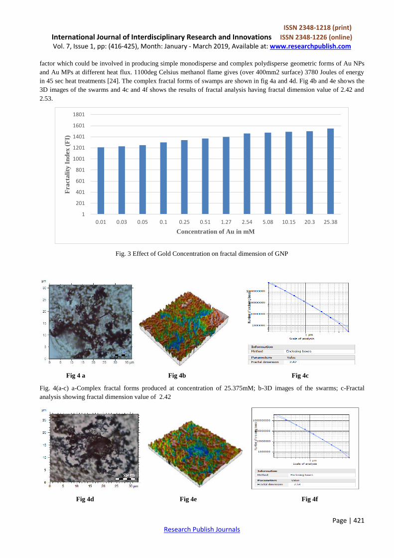
The variation in the fractal Index (FI) values by Euclidean distance methods is shown in Fig 3. As the concentration of
Gold in mM increased, the fractality Index also increased from 1201 to 1550. CFE produces unidentified proteinaceous
0.01275 0.0255 0.051
0.102 0.254 0.508
1.27 2.54 5.08
20.3 25.375
10μm
10μm 10μm 10μm
10μm 10μm
10μm 10μm 10μm
10μm 10μm
10μm
10.15
ISSN 2348-1218 (print)
International Journal of Interdisciplinary Research and Innovations ISSN 2348-1226 (online) Vol. 7, Issue 1, pp: (416-425), Month: January - March 2019, Available at: www.researchpublish.com
Page | 421 Research Publish Journals
factor which could be involved in producing simple monodisperse and complex polydisperse geometric forms of Au NPs
and Au MPs at different heat flux. 1100deg Celsius methanol flame gives (over 400mm2 surface) 3780 Joules of energy
in 45 sec heat treatments [24]. The complex fractal forms of swamps are shown in fig 4a and 4d. Fig 4b and 4e shows the
3D images of the swarms and 4c and 4f shows the results of fractal analysis having fractal dimension value of 2.42 and
2.53.
Fig. 3 Effect of Gold Concentration on fractal dimension of GNP
Fig 4 a Fig 4b Fig 4c
Fig. 4(a-c) a-Complex fractal forms produced at concentration of 25.375mM; b-3D images of the swarms; c-Fractal
analysis showing fractal dimension value of 2.42
Fig 4d Fig 4e Fig 4f
1
201
401
601
801
1001
1201
1401
1601
1801
0.01 0.03 0.05 0.1 0.25 0.51 1.27 2.54 5.08 10.15 20.3 25.38
Fra
cta
lity
In
dex
(F
I)
Concentration of Au in mM

ISSN 2348-1218 (print)
International Journal of Interdisciplinary Research and Innovations ISSN 2348-1226 (online) Vol. 7, Issue 1, pp: (416-425), Month: January - March 2019, Available at: www.researchpublish.com
Page | 422 Research Publish Journals
Fig. 4(d-f) d-Complex fractal forms produced at concentration of 25.375mM; e- 3D images of the swarms; f- Fractal
analysis showing fractal dimension value of 2.53
It was found that when concentration at (25.375mM) leading to steady concentration and variation of thermal treatment
was from 60 to 300 sec produced complexed 3D stacking and thick crustose assemblies indicating that heat resistant
proteins may be implicated, which can withstand prolonged heat (Fig 5).
Fig. 5 Effect of heat on fractality of AuNP swarms
It was possible to obtain GNP swarms. The results shows that a simple, glass slide based technique can be successfully
used for rapid production, microscopic visualization, morphological analysis, study of swarming behavior and monitoring
of effect of heat on Au NPs and Au MPs forms and assemblages using cell free microbial system such as Rhizobium sp. In
this study we used CFE of Rhizobium culture which are reported to be heavy metal tolerant [4] [5] [6]. CFE produces
unidentified proteinaceous factor which could be involved in producing simple monodisperse and complex polydisperse
geometric forms of Au NPs and Au MPs at different heat flux. Earlier reports were on chemically triggered swarming of
Au microparticles [9]. 1100deg Celsius flame gives 2.09 Mega Joule energy but heating slide over 400mm2 surface
produces energy which means 3780 Joules of energy is involved in 45 sec heat treatment [Farrier law of heat conduction
heat release rate is 84Joule/second, 1100deg Celsius flame=2.09 Mega Joule energy but heating slide over 400mm2
surface produces energy which means 3780 Joules of energy is involved in 45 sec heat treatment]. As the concentration
increased from 1.21 to 1.51mM the complexity of GNP increased, concentration from 1.21 to 0.051mM produced
swarms, as the concentration crosses 0.051mM nanoparticle alignment occurs forming dendritic assemblages, florets
forms, finally leading to complex forms, similar finding has been reported where amino acid induced fractal aggregations
of AuNPs occurs [25]. Earlier studies were on the synthesis of star-like gold nanoparticles (SGNs) in a temperature-
controlled environment which allows for temperature modulation and facilitates the growth of highly branched
nanoparticles [26]. There are reports on dendritic structures of AuNPs [27] [28]. It was noticed that swarms were
appearing in low concentration treatment ie 0.021mM concentration and this concentration was found to be best for
monodispersed GNP production. There are studies on a single large assembly with dynamically fluctuating swarms of
gold nanoparticles formed by trapping laser [29]. Previous report shows aggregates formed more quickly at higher
concentrations and temperatures. It was found that when concentration was kept (25.375mM) constant and thermal
treatment is increased from 60 to 300 sec, the complexity increasing forming 3D stacking and producing thick crust
surface indicating that heat resistant proteins are implicated, which can withstand prolonged heat. Scheme has been
summarized in the figure 6 and 7. Previous reports has also shown that aggregates formed more quickly at higher
concentrations and temperatures [30]. When CFE containing the proteinaceous factor is added to highly acidic solution of
HAuCl4 (pH 1.5), hydrolysis of protein occurs giving rise to mixture of amino acid, thus AuNPs which are formed may be
amino acid capped AuNPs, thus forming a crossed bridging between AuNPs giving rise to complexity of AuNPs. It is
possible to analyze the fractal forms by using Jfrad and mountains Map software. Earlier reports were on fractal analysis
of inter-particle interaction forces in gold nanoparticle aggregates [31]. Using our simple slide based technique a lot of
scope exists for applying a combination of microbial CFE and timed heat treatment to obtain defined monodisperse GNP
swarms and study postulated heat responsive protein mediated genesis and architecture of biotechnologically useful forms
of Au NPs and polydisperse Au MPs.
0
500
1000
1500
2000
2500
3000
3500
60 120 180 240 300

ISSN 2348-1218 (print)
International Journal of Interdisciplinary Research and Innovations ISSN 2348-1226 (online) Vol. 7, Issue 1, pp: (416-425), Month: January - March 2019, Available at: www.researchpublish.com
Page | 423 Research Publish Journals
Fig. 6 Change in complexity of AuNPs with increase in the concentration from 0.01275 to 25.375mM
Fig. 7 Increase of complexity with increase in thermal treatment and constant concentration
IV. CONCLUSIONS
Rhizobium sp. cultures were successfully isolated from root nodules of Mimosa Pudica plant sp. and used for the
production of AuNPs. It was possible to obtain GNP swarms. It was found that CFE showed good results at 45sec thermal
treatment forming complex structures at 25.75mM concentration. This is the first study of use of rhizobium cultures for
the production of AuNPs in Goa and India. This is the one among the few report on study of fractality Index of AuNPs.
Using our simple slide based technique a lot of scope exists for applying a combination of CFE and timed heat treatment
to obtain defined monodisperse GNP swarms. As the concentration crosses 0.051mM nanoparticle alignment occurs
forming dendritic assemblages, florets forms, finally leading to complex forms, similar finding has been reported where
amino acid induced fractal aggregations of AuNPs occurs [25]. In our studies we also observed that when concentration is
increased to 25.375mM and thermal treatment is increased from 60 to 300 sec, the complexity increases forming 3D
stacking and producing thick crust surface. We postulate that Rhizobium sp. specific unidentified cell bound heat
responsive proteins may trigger monodisperse Au NPs swarms and simple, complex assemblages or acidic conditions
may release amino acids from protein hydrolysis which may act as capping and linking agents. Some of these
assemblages or acidic conditions may release amino acids from protein hydrolysis which may act as capping and linking
agents. Some of these assemblages such as open and closed rings, interlocked rings are unique. This heat responsive
protein mediated genesis and architecture of biotechnologically useful forms of Au NPs and polydisperse Au MPs. This
indicated that heat resistant proteins are implicated, which can withstand prolonged heat. If it is possible to produce
AuNPs swarms with use of Rhizobium cultures than it is possible to use other microorganisms to produce NPs. It is
possible to analyze the fractal forms by using Jfrad and mountains Map software. There is a lot of interest in autonomous
movement and collective behavior of synthetic nanomaterials which have important applications in nanomedicine,
nanobiotechnology and nanosensors. Further work is required to study and characterize the proteinaceous factor. Further
modified microfluidic technique will be designed and used for AuNPs synthesis.
ACKNOWLEDGEMENTS
This work was supported by UGC-SAP Phase II – Biodiversity, Bioprospecting programme and Goa University Fungus
Culture Collection (GUFCC). First author also acknowledges UGC, NF-OBC Junior Research fellowship.

ISSN 2348-1218 (print)
International Journal of Interdisciplinary Research and Innovations ISSN 2348-1226 (online) Vol. 7, Issue 1, pp: (416-425), Month: January - March 2019, Available at: www.researchpublish.com
Page | 424 Research Publish Journals
REFERENCES
[1] Radtsig, M. A., Koksharova, O. A., and Nadtochenko, V. A. (2016). ―Production of gold nanoparticles by
biogenesis using bacteria‖. Microbiology, 85(1): 63-70.
[2] Molnár, Z., Bódai, V., Szakacs, G., Erdélyi, B., Fogarassy, Z., Sáfrán, G., and Lagzi, I. (2018). Green synthesis of
gold nanoparticles by thermophilic filamentous fungi. Scientific reports, 8(1), 3943.
[3] Iravani, S. (2014). Bacteria in nanoparticle synthesis: current status and future prospects. International scholarly
research notices, 2014.
[4] Grison, C. M., Jackson, S., Merlot, S., Dobson, A., and Grison, C. (2015). Rhizobium metallidurans sp. nov., a
symbiotic heavy metal resistant bacterium isolated from the Anthyllis vulneraria Zn-hyperaccumulator. International
journal of systematic and evolutionary microbiology, 65(5): 1525-1530.
[5] Lebrazi, S., and Fikri-Benbrahim, K. (2018). Rhizobium-Legume Symbioses: Heavy metal effects and principal
approaches for bioremediation of contaminated soil. In Legumes for Soil Health and Sustainable Management (pp.
205-233). Springer, Singapore.
[6] Singh, A. K., and Singh, G. (2015). A study of multiple heavy metal tolerance in root nodulating bacteria. Int J Res
Dev Pharm Life Sci, 4, 1713-1721.
[7] Kopycińska, M., Lipa, P., Cieśla, J., Kozieł, M., and Janczarek, M. (2018). Extracellular polysaccharide protects
Rhizobium leguminosarum cells against zinc stress in vitro and during symbiosis with clover. Environmental
microbiology reports, 10(3), 355-368.
[8] Maan, P. K., and Garcha, S. (2018). Bacteriocins from Gram-negative Rhizobium spp. Advances in
Bioresearch, 9(1).
[9] Kagan, D., Balasubramanian, S., and Wang, J. (2011). Chemically triggered swarming of gold microparticles.
Angewandte Chemie International Edition, 50(2): 503-506.
[10] M.K. Bera, M.K. Sanyal, L. Yang, K. Biswas, A. Gibaud, C.N.R. Rao, Small-anglex-ray scattering study of the
aggregation of gold nanoparticles during formation at the toluene-water interface, Phys. Rev. B 81 (March (11))
(2010)115415.
[11] Cai, J., Hu, X., Xiao, B., Zhou, Y., & Wei, W. (2017). Recent developments on fractal-based approaches to
nanofluids and nanoparticle aggregation. International Journal of Heat and Mass Transfer, 105, 623-637.
[12] Shirali, S. A. (2014). Fractal dimension and the Cantor set. Resonance, 19(11), 1000-1004.
[13] D. Brune, H. Ernst, H. Grunwald, A. Grünwald, W. Hofmann, H. Krug, H. Janich,P. Mayor, M. Rathgeber, W.
Schmid, G. Simon, U. Vogel, V. Wyrwa,Nanotechnology, Assessment and Perspectives, 1st ed., Springer
BerlinHeidelberg, Berlin, Heidelberg, 2006.
[14] T.G. Dewey, Fractals in Molecular Biophysics, Oxford University Press, 1997.
[15] M. Tirado-Miranda, A. Schmitt, J. Callejas-Fernández, A. Fernández-Barbero,The aggregation behaviour of protein-
coated particles: a light scatteringstudy, Eur. Biophys. J. 32 (May (2)) (2003) 128–136.
[16] A.C. Castellano, M. Barteri, A. Bianconi, E. Borghi, L. Cassiano, M. Castagnola, S.Della Longa, X-ray small angle
scattering of the human transferrin proteinaggregates. A fractal study, Biophys. J. 64 (February (2)) (1993) 520–524.
[17] Zhu, J., Liu, M. J., Li, J. J., Li, X., & Zhao, J. W. (2018). Multi-branched gold nanostars with fractal structure for
SERS detection of the pesticide thiram. Spectrochimica Acta Part A: Molecular and Biomolecular Spectroscopy,
189, 586-593.
[18] Kanniah, V., Wu, P., Mandzy, N., and Grulke, E. A. (2012). Fractal analysis as a complimentary technique for
characterizing nanoparticle size distributions. Powder technology, 226, 189-198.
ISSN 2348-1218 (print)
International Journal of Interdisciplinary Research and Innovations ISSN 2348-1226 (online) Vol. 7, Issue 1, pp: (416-425), Month: January - March 2019, Available at: www.researchpublish.com
Page | 425 Research Publish Journals
[19] Abdellatif, M. H., Abdelrasoul, G. N., Salerno, M., Liakos, I., Scarpellini, A., Marras, S., & Diaspro, A. (2016).
Fractal analysis of inter-particle interaction forces in gold nanoparticle aggregates. Colloids and Surfaces A:
Physicochemical and Engineering Aspects, 497, 225-232.
[20] Rea, M. A., Zammit, C. M., and Reith, F. (2016). ―Bacterial biofilms on gold grains—implications for geomicrobial
transformations of gold‖. FEMS microbiology ecology, 92(6): fiw082.
[21] Rajendran, G., Sing, F., Desai, A. J., and Archana, G. (2008). Enhanced growth and nodulation of pigeon pea by co-
inoculation of Bacillus strains with Rhizobium spp. Bioresource Technology, 99(11), 4544-4550.
[22] Micheli, L., Uccelletti, D., Palleschi, C., and Crescenzi, V. (1999). Isolation and characterisation of a ropy
Lactobacillus strain producing the exopolysaccharide kefiran. Applied Microbiology and Biotechnology, 53(1): 69-
74.
[23] Wang, X., Zhu, X., Shi, H., Chen, Y., Chen, Z., Zeng, Y., ... & Duan, H. (2018). Three-Dimensional-Stacked Gold
Nanoparticles with Sub-5 nm Gaps on Vertically Aligned TiO2 Nanosheets for Surface-Enhanced Raman Scattering
Detection Down to 10 fM Scale. ACS applied materials & interfaces, 10(41), 35607-35614.
[24] Lienhard, J. H. (2013). A heat transfer textbook. Courier Corporation. Dover Publication.
[25] Doyen, M., Goole, J., Bartik, K., and Bruylants, G. (2016). Amino acid induced fractal aggregation of gold
nanoparticles: Why and how. Journal of colloid and interface science, 464, 160-166.
[26] Darienzo, R., Mironava, T., and Tannenbaum, R. (2018). Raman Signal Enhancement by Quasi-Fractal Geometries
of Gold Nanoparticles.
[27] Abdellatif, M. H., Abdelrasoul, G. N., Scarpellini, A., Marras, S., & Diaspro, A. (2015). Induced growth of dendrite
gold nanostructure by controlling self-assembly aggregation dynamics. Journal of colloid and interface science,
458, 266-272.
[28] Agrawal, V. V., Kulkarni, G. U., & Rao, C. N. R. (2008). Surfactant-promoted formation of fractal and dendritic
nanostructures of gold and silver at the organic–aqueous interface. Journal of colloid and interface science, 318(2),
501-506.
[29] Kudo, T., Yang, S. J., and Masuhara, H. (2018). A single large assembly with dynamically fluctuating swarms of
gold nanoparticles formed by trapping laser. Nano letters, 18(9), 5846-5853.
[30] Gharagozloo, P. E., & Goodson, K. E. (2010). Aggregate fractal dimensions and thermal conduction in nanofluids.
Journal of Applied Physics, 108(7), 074309.
[31] Abdellatif, M. H., Abdelrasoul, G. N., Salerno, M., Liakos, I., Scarpellini, A., Marras, S., and Diaspro, A. (2016).
Fractal analysis of inter-particle interaction forces in gold nanoparticle aggregates. Colloids and Surfaces A:
Physicochemical and Engineering Aspects, 497, 225-232.
[32] http://cme.msu.edu/cmeias/fracAnalysis.shtml
[33] https://www.digitalsurf.com