fractionation and characterization of a cyclic
TRANSCRIPT

FRACTIONATION AND CHARACTERIZATION OF A CYCLIC ADENINE RIBONUCLEOTIDE FORMED
BY TISSUE PARTICLES*
BY EARL W. SUTHERLAND AND T. W. RALL
(From the Department of Pharmacology, School of Medicine, Western Reserve University, Cleveland, Ohio)
(Received for publication, October 23, 1957)
The isolation of an unusual adenine ribonucleotide was noted in a pre- vious report (1) ; this compound was formed by particulate fractions of liver homogenates in the presence of adenosine triphosphate, magnesium ions, and epinephrine or glucagon. The adenine ribonucleotide was found to be identical (2) to a product isolated from a barium hydroxide digest of adenosine triphosphate by Cook, Lipkin, and Markham (3).’ These authors originally termed the compound a cyclic dianhydrodiadenylic acid with phosphate esterification to ribose at positions 3 and 5. However, more recent evidence indicates that the compound is the mononucleotide, adenosine-3’) 5’-phosphoric acid.2 In this report the compound will be termed 3,5-AMP3 for convenience. The formation of this compound in various tissue preparations has been described (4); the experiments re- ported in this paper describe the purification and certain properties of this ribonucleotide.
Methods
Materials-A highly purified prostatic phosphatase and a commercial sample of Russell’s viper venom were supplied by Dr. Henry Sable; a highly purified spleen phosphodiesterase was supplied by Dr. Leon Heppel.
* This investigation was supported in part by a research grant (No. H-2745) from the National Heart Institute of the United States Public Health Service, and a grant- in-aid from the Lilly Research Laboratories.
1 Dr. Cook, Dr. Lipkin, and Dr. Markham furnished samples which had been iso- lated by paper chromatography. The criteria of identity have been summarized in a previous note (2).
2 In a personal communication, Professor David Lipkin has informed us that a molecular weight determination and other data prove that the compound is aden- osine-3’,5’-phosphoric acid. The complete proof of structure of the nucleotide will be presented in a forthcoming publication by D. Lipkin, R. Markham, and W. H. Cook.
3 The following abbreviations are used: cyclic 3,5-AMP, adenosine-3’,5’-phos- phoric acid; 5’-AMP, adenosine 5’-phosphate; 2’-AMP, adenosine 2’-phosphate; 3’- AMP, adenosine 3’-phosphate; ATP, adenosine triphosphate; ADP, adenosine di- phosphate; Tris, tris(hydroxymethyl)aminomethane; D = optical density.
1077

1078 PROPERTIES OF CYCLIC ADENINE NUCLEOTIDE
The intestinal phosphatase was purchased from the Nutritional Biochemi- cals Corporation and was dialyzed versus Hz0 before use. Crystalline ribonuclease was obtained from the Worthington Biochemical Corpora- tion. Yeast adenylic acid was purchased from the Schwarz Laboratories, Inc., and ribose 5-phosphate was purchased from the Nutritional Biochemi- cals Corporation.
Assays-The assays of cyclic 3,5-AMP were performed as described (4). Assay of the cyclic 3,5-AMP-inactivating enzyme (phosphodiesterase) involved incubation of 1 X 1O-4 M cyclic 3,5-AMP with the enzyme prepara- tion for 10 minutes at 30” in 0.04 M Tris buffer (pH 7.45) and 2 X 1O-3 M
MgC12. After heating in a boiling bath for 2 minutes, the reaction mixture was cooled and diluted suitably for assay of residual cyclic 3,5-AMP.
Ion Exchange Resin Chromatography-Analytical grade resins AG 2-X8 chloride and AG 50-X8 hydrogen of 200 to 400 mesh were purchased from the Bio-Bad Laboratories; these had been processed from Dowex 2-X8 and Dowex 50-X8. The resins were washed twice with 5 volumes of 2 N HCl, then twice with distilled water, 2 N NaOH, distilled water, 2 N
HCI, and then with glass-distilled water until chloride-free. Such resins were used for the first two steps; resins for later steps were washed further with 0.1 N NaOH, distilled water, 0.1 N HCl, and glass-distilled water.
The pressure of gravity was utilized in all cases for absorption and elu- tion procedures and averaged about 30 cm. for absorption of cyclic 3,5- AMP on Dowex 2 resins at neutral pH, while an average pressure of 15 cm. of fluid was used for elution from Dowex 2 columns and for all steps with Dowex 50 resins. The temperature of the air-conditioned room varied from about 22-28”.
EXPERIMENTAL
Isolation of Cyclic 3,5-AMP
Preparation of Heated Extracts-Heated extracts containing cyclic 3,5- AMP were prepared by the procedure described previously (4), adapted to a large scale. Washed particles derived from one dog liver, treated in a blendor in 0.25 M sucrose, were incubated in a medium containing at final concentrations 0.04 M Tris (pH 7.5), 2.5 X 1O-3 M MgS04, 2 X 1O-3 M
ATP, 6.67 X 1O-3 M caffeine, 0.01 M NaF, 5.5 y per ml. of epinephrine, 1 y per ml. of glucagon, and approximately 0.2 M sucrose. On the average, each liter of incubation mixture contained washed particles derived from about 280 gm. of liver. The mixtures were incubated at 30” for 30 min- utes with mechanical stirring in an atmosphere of 100 per cent 02 and then were heated for 5 to 7 minutes in boiling water. After chilling, the insol- uble material was removed by centrifugation and the supernatant fluid (heated or boiled extract) was stored at -20”.
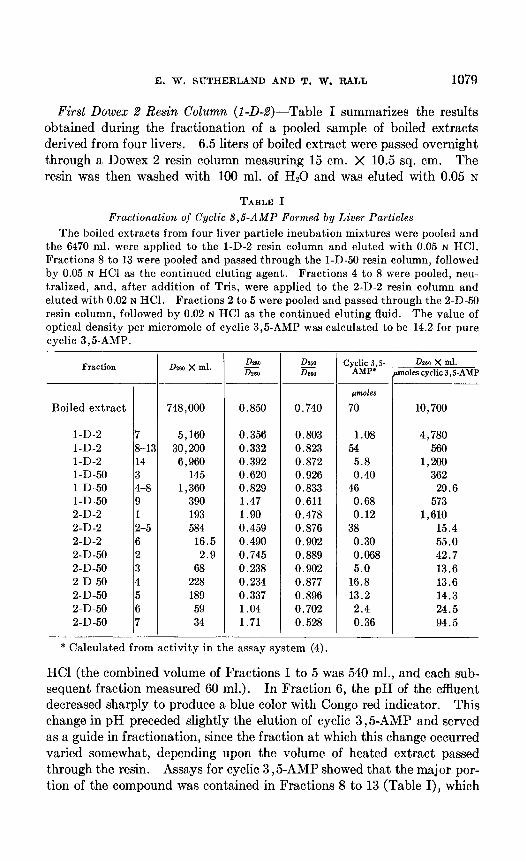
E. W. SUTHERLAND AND T. W. RALL 1079
First Dowex 2 Resin Column (1-D-2)-Table I summarizes the results obtained during the fractionation of a pooled sample of boiled extracts derived from four livers. 6.5 liters of boiled extract were passed overnight through a Dowex 2 resin column measuring 15 cm. X 10.5 sq. cm. The resin was then washed with 100 ml. of Hz0 and was eluted with 0.05 N
TABLE I
Fractionation of Cyclic $,& Formed by Liver Particles
The boiled extracts from four liver particle incubation mixtures were pooled and the 6470 ml. were applied to the l-D-2 resin column and eluted with 0.05 N HCI. Fractions 8 to 13 were pooled and passed through the l-D-50 resin column, followed by 0.05 N HCl as the continued eluting agent. Fractions 4 to 8 were pooled, neu- tralized, and, after addition of Tris, were applied to the 2-D-2 resin column and eluted with 0.02 N HCI. Fractions 2 to 5 were pooled and passed through the 2-D-50 resin column, followed by 0.02 N HCI as the continued eluting fluid. The value of optical density per micromole of cyclic 3,5-AMP was calculated to be 14.2 for pure cyclic 3,5-AMP.
Fraction
Boiled extract
l-D-2 7 l-D-2 8-C l-D-2 14 1-D-50 3 1-D-50 4-8 1-D-50 9 2-D-2 1 2-D-2 2-5 2-D-2 6 2-D-50 2 2-D-50 3 2-D-50 4 2-D-50 5 2-D-50 6 2-D-50 7
Dmo x ml. DZSO x0
DlW DIaa
CY~di& .s- DPSO X ml. moles cyclic 3, S-AMP
748,000 0.850 0.740
~tnmoles
70 10,700
5,160 0.356 0.803 1.08 4,780 30,200 0.332 0.823 54 560
6,960 0.392 0.872 5.8 1,200 145 0.620 0.926 0.40 362
1,360 0.829 0.833 46 29.6 390 1.47 0.611 0.68 573 193 1.90 0.478 0.12 1,610 584 0.459 0.876 38 15.4
16.5 0.490 0.902 0.30 55.0 2.9 0.745 0.889 0.068 42.7
68 0.238 0.902 5.0 13.6 228 0.234 0.877 16.8 13.6 189 0.337 0.896 13.2 14.3 59 1.04 0.702 2.4 24.5 34 1.71 0.528 0.36 94.5
* Calculated from activity in the assay system (4).
HCl (the combined volume of Fractions 1 to 5 was 540 ml., and each sub- sequent fraction measured 60 ml.). In Fraction 6, the pH of the effluent decreased sharply to produce a blue color with Congo red indicator. This change in pH preceded slightly the elution of cyclic 3,5-AMP and served as a guide in fractionation, since the fraction at which this change occurred varied somewhat, depending upon the volume of heated extract passed through the resin. Assays for cyclic 3,5-AMP showed that the major por- tion of the compound was contained in Fractions 8 to 13 (Table I), which

1080 PROPERTIES OF CYCLIC ADENINE NUCLEOTIDE
also encompassed the last two-thirds of the 5’-AMP peak and the first one-third of the ADP peak. The l-D-2 column served mainly to concen- trate cyclic 3,5-AMP and to remove large amounts of various impurities.
First Dowex 50 Resin Column (1-D-50)-Fractions 8 to 13 from the l-D-2 column were pooled and passed through a Dowex 50 resin column measuring 15 cm. X 7.0 sq. cm. previously washed with 0.05 N HCI. For continued elution, 0.05 N HCl was added. The combined volume of Fractions 1 and 2 was 450 ml., while each subsequent fraction was 134 ml. Practically all the ADP appeared in Fractions 1 and 2, and almost all the AMP was still on the resin after Fraction 9 was collected. These nucleotides had been present in large excess over the cyclic 3,5-AMP, which, in this case, was confined mainly to Fractions 4 to 8 (Table I). This step also removed a considerable amount of buffering material which appeared to be primarily inorganic phosphate.
Second Dowex 2 Resin Column (2-D-2)-Fractions 4 to 8 from the 1-D-50 column were pooled and neutralized, and, after the addition of 0.2 per cent volume of 1.0 M Tris (pH 7.45), were passed overnight through a Dowex 2 resin column measuring 15 cm. X 0.79 sq. cm. The next morning the resin was washed successively with 10 ml. of Hz0 and 50 ml. of 0.005 N
HCI; 0.02 N HCI was then added, and 10 ml. fractions were collected. In general, this step separated almost all other components from cyclic 3,5-AMP, at least in selected fractions. However, some difficulty was encountered in freeing the Dowex 2 resin of elutable ultraviolet-absorbing material which then contaminated presumably all fractions. Fortunately, this material appeared to be retained on Dowex 50 resins at acid pH. In addition, a number of the fractions containing a considerable portion of the cyclic 3,5-AMP frequently contained small amounts of other nucle- otides, as was obviously the case in the example shown in Table I.
Second Dowex 50 Resin Column (d-D-50)-Fractions 2 to 5 from the 2-D-2 column were pooled and passed through a Dowex 50 resin column measuring 15 cm. X 1.13 sq. cm. previously washed with 0.02 N HCl; for continued elution, 0.02 N HCl was added. The volume of Fraction 1 was 40 ml., and each subsequent fraction was 20 ml. The impurities re- moved by this step appear to be compounds containing cytosine and gua- nine, as judged by the ultraviolet spectrum (Table I). The theoretical limit of 14.2 for DzgO X ml. per micromole (millimolar extinction coeffi- cient) was occasionally exceeded slightly, as shown in Table I, last column. Whenever highly active fractions have been reassayed and refractionated, all results have indicated that a value of about 14.2 represents maximal activity. The over-all recovery of cyclic 3,5-AMP in Fractions 3, 4, and 5 from the 2-D-50 column was 50 per cent.
Crystallization of Cyclic S,5-AMP-Several preparations from the 2-D-2

E. W. SUTHERLAND AND T. W. RALL 1081
fractionation, similar to Fractions 2 to 5 in Table I, were pooled, neutral- ized, and fractionated again on a Dowex 2 chloride resin column. The most active fractions were pooled and fractionated further on a Dowex 50 resin column, and the peak fractious yielded 27 ml. of a 1.2 X 1OW M solu- tion in 0.05 N HCl with high activity (OpeO per micromole = theory). This sample (Special D-50) was lyophilized without neutralization. To one-half of the resultant powder in a small flask was added 1.0 ml. of glass- distilled water, and the flask and contents were warmed to 50”. The sample was transferred to a centrifuge tube and, on chilling, crystals ap- peared with a pronounced sheen. In general, small rods often in rosettes were most common; the ends were generally pointed when the rods were larger. At times, flat rods or plates with pointed tips were formed; oc- casionally these crystals were large and readily visible with 100 X , or less magnification. The warmed solution was 3.6 X 1OF M with a pH of about 2, while the supernatant fluid above the crystals at 2” was 0.9 X l@-2 M.
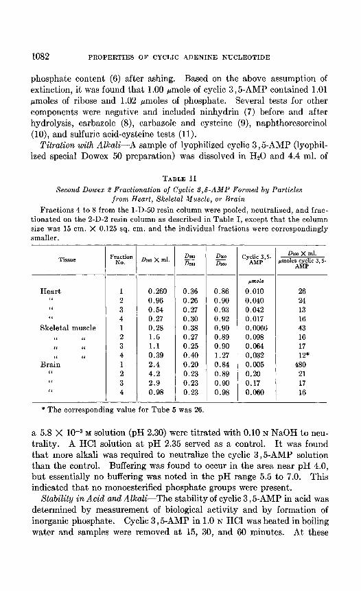
Isolation of Cyclic 3,5-AMP from Extrahepatic Tissues-Boiled extracts were prepared from incubation mixtures in which particles from dog heart, skeletal muscle, and brain were substituted for liver particles (4). These boiled extracts were fractionated by ion exchange resins as described above, except that smaller volumes of extracts were used and the resin columns were proportionately of smaller diameter. The results of fractionation on the 2-D-2 resin column are summarized in Table II. Over-all recoveries in Fractions 1 to 4 were 53 per cent for heart and skeletal muscle and 45 per cent for brain. It may be noted that the values 0260 X ml. per micro- mole of cyclic 3 ,& for Fractions 2, 3, and 4 do not differ greatly from the value obtained with the fractionation described in Table I. Samples 2, 3, and 4 were pooled, passed through Dowex 50, and appropriate frac- tions were lyophilized and characterized further as reported below.
Properties of Cyclic 3,5-AMP
Spectrum and Components-The ultraviolet spectrum of highly purified samples was similar to that of 5’-AMP. The li:,,, in 0.05 N HCl was at 257 rnp with essentially maximal absorption at 256 rnp. In acid, the D280,260 was 0.23 to 0.24, and the D 250,260 was 0.88 to 0.89; the corresponding values for 5’-AMP were smaller. The spectrum in alkali was almost identical to the spectrum of 5’-AMP.
The assumption was made that the molar extinction coefficient for cyclic 3,5-AMP in acid at 260 rnp was 14.2 X 10³. The validity of this assump- tion was established by experiments reported below in which cyclic 3,5- AMP was converted quantitatively to 5’-AMP without detectable change in optical density at 260 rnp. Ribose content was determined (5), and
1082 PROPERTIES OF CYCLIC ADENINE NUCLEOTIDE
phosphate content (6) after ashing. Based on the above assumption of extinction, it was found that 1.00 pmole of cyclic 3,5-AMP contained 1.01 pmoles of ribose and 1.02 Bmoles of phosphate. Several tests for other components were negative and included ninhydrin (7) before and after hydrolysis, carbazole (8), carbazole and cysteine (9), naphthoresorcinol (lo), and sulfuric acid-cysteine tests (11).
Titration with Alkali-A sample of lyophilized cyclic 3,5-AMP (lyophil- ized special Dowex 50 preparation) was dissolved in Hz0 and 4.4 ml. of
TABLE II
Second Dowex 2 Fractionation of Cyclic 3,6-AMP Formed by Particles from Heart, Skeletal Muscle, or Brain
Fractions 4 to 8 from the l-D-50 resin column were pooled, neutralized, and frac- tionated on the 2-D-2 resin column as described in Table I, except that the column size was 15 cm. X 0.125 sq. cm. and the individual fractions were correspondingly smaller.
Tissue
Heart “ “ “
Skeletal muscle “ “ ‘I I‘
BraYn “
I‘ “ I‘
Fraction NO. 0260 x ml. DvlO
izz DlsO iG
1 0.260 0.36 0.86 2 0.96 0.26 0.90 3 0.54 0.27 0.93 4 0.27 0.30 0.92 1 0.28 0.38 0.90 2 1.6 0.27 0.89 3 1.1 0.25 0.90 4 0.39 0.40 1.27 1 2.4 0.20 0.84 2 4.2 0.23 0.89 3 2.9 0.23 0.90 4 0.98 0.23 0.98
jmole
0.010 0.040 0.042 0.017 0.0066 0.098 0.064 0.032 0.005 0.20 0.17 0.060
I
-
Dwo X ml. rmoles cyclic 3, S-
AMP
26 24 13 16 43 16 17 12*
480 21 17 16
* The corresponding value for Tube 5 was 26.
a 5.8 X 1O-3 M solution (pH 2.30) were titrated with 0.10 N NaOH to neu- trality. A HCl solution at pH 2.35 served as a control. It was found that more alkali was required to neutralize the cyclic 3,5-AMP solution than the control. Buffering was found to occur in the area near pH 4.0, but essentially no buffering was noted in the pH range 5.5 to 7.0. This indicated that no monoesterified phosphate groups were present.
Stability in Acid and Alkali-The stability of cyclic 3,5-AMP in acid was determined by measurement of biological activity and by formation of inorganic phosphate. Cyclic 3,5-AMP in 1.0 N HCl was heated in boiling water and samples were removed at 15, 30, and 60 minutes. At these

E. W. SUTHERLAND AND T. W. RALL 1083
times, the losses of biological activity were 19, 54, and 78 per cent, respec- tively, and the amounts of inorganic phosphate formed were 17, 41, and 63 per cent of the total. Cyclic 3,5-AMP in 1.0 N NaOH was also heated in boiling water and samples were removed at 7, 15, and 40 minutes. At these times, the losses of biological activity were 31, 64, and 90 per cent. These and other experiments showed that the nucleotide was very resistant to inactivation by acid or alkali.
Hydrolysis with Dowex 50 and IdentiJication of Products
The hydrogen form of Dowex 50 has been used as a catalyst for hydroly- sis of the glycosidic link of adenine ribonucleotides. With use of this catalyst, the hydrolysis was so rapid that ribose 2-phosphate and ribose 3-phosphate were formed from 2’-AMP and 3’-AMP, respectively, without significant isomerization (12). However, in preliminary experiments cyclic 3,5-AMP was hydrolyzed only about 35 per cent after 15 minutes heating in the presence of Dowex 50 and 0.05 N HCl. Under these condi- tions, isomerization of phosphate attached to the 2 or 3 position of ribose might be expected. In these experiments the ribose phosphate or phos- phates formed from cyclic 3,5-AMP differed from ribose 5-phosphate in having a slower rate of color development with orcinol reagent, and a faster rate with the sulfuric acid-cysteine reagents. The ribose phosphate was more acid-labile than ribose 5-phosphate and was eluted from Dowex 2 by NH&l in the presence of borate.
A larger scale experiment was performed to produce amounts of ribose phosphate sufficient for characterization by chromatography. A Dowex 50 resin sample was washed three times with 0.05 N HCl and upon centrifu- gation at 1200 X g packed to a volume of 2.9 ml. To this were added 10 ml. of cyclic 3,5-AMP in 0.05 N HCl containing the equivalent of 600 y of ribose. The mixture was stirred for 20 minutes at room temperature, then heated in a boiling bath for 25 minutes with stirring, cooled to room temperature, and centrifuged for 7 minutes at 1200 X g. The supernatant fluid was removed, and to the precipitate were added 10 ml. of 0.05 N HCl. The suspension was heated in the boiling water bath for 25 minutes with stirring, cooled, and, after centrifugation, the supernatant fluid was re- moved and pooled with the previous supernatant fluid. The pooled super- natant fluids contained ribose or ribose derivatives equivalent to 490 y of ribose. After neutralization and addition of 2 per cent by volume of 0.2 M Tris, pH 7.4, the sample was passed over a Dowex 2 chloride resin column 12.2 cm. X 0.28 sq. cm. The resin was washed with Hz0 and eluted with 0.015 N HCI. (Preliminary experiments showed that ribose phosphates were separated from cyclic 3,5-AMP and ribose by this fractionation.) The eluted fractions contained 93 per cent of the applied ribose or ribose

1084 PROPERTIES OF CYCLIC ADENINE NUCLEOTIDE
derivatives and it was estimated that 72 per cent of the eluted material was present as ribose phosphate, 14 per cent as free ribose, and about 14 per cent as nucleotide.
The ribose phosphates were pooled and the major portion was fraction- ated on an anion exchange resin as described in Fig. 1, a modification of the methods of Khym and Cohn being used (12, 13). A mixture of ribose 3-phosphate and ribose 2-phosphate was prepared from yeast adenylic
Om YEAST AMP+ DOWEX-50+RIBOSE-5-PO4
I a..**...@ 3,5-AMP+ DOWEX- 50 NH,CI
IO 50 100 150 200 250 ml OF EFFLUENT
FIG. 1. Fractionation of ribose phosphates from Dowex 50-catalyzed hydrolysis of cyclic 3,5-AMP. The control sample contained (as ribose) 425 y of ribose phosphates from yeast adenylic acid and 165 y of ribose 5-phosphate in 10.9 ml. of 0.013 M KCl, containing0.0035 M Tris (pH 7.4). The ribose phosphates from cyclic 3,5-AMP were pooled samples from the Dowex 2 fractionation described in the text. The sample contained (as ribose) 220 y of ribose phosphates in 11.4 ml. of 0.013 M KCI, containing 0.0035 M Tris (pH 7.4). Each sample was passed through a Dowex 2 chloride resin column 5 cm. X 0.125 sq. cm. and the resin was washed with 10 ml. of HzO, and then eluted. The eluting agent was 240 ml. of 0.04 M NHaCI, containing 0.002 M Na2B107; then 0.04 M NH&l alone was added, as indicated by arrow.
acid in a similar fashion and was fractionated by the same methods. Pilot experiments showed that the ribose phosphates were separated by relatively short resin columns. The results of the experiment summarized in Fig. 1 indicated that the ribose phosphates formed from cyclic 3,5-AMP in the presence of. the hydrogen form of Dowex 50 were ribose 3-phosphate and ribose 2-phosphate. Only about 5 per cent of the total ribose was found in the fractions in which ribose 5-phosphate was eluted after addition of NH&l without borate. All of the ribose content applied to the columns was accounted for in the eluted fractions shown in Fig. 1.

E. W. SUTHERLAND AND T. W. RALL 1085
The Dowex 50 resin present after hydrolysis of the cyclic 3,5-AMP was washed with 0.05 N HCl and then eluted. To a control resin were added 3 pmoles of adenine in 0.05 N HCl, and after stirring for 30 minutes at room temperature, the control resin was treated exactly as the experimental resin. The resins were placed in glass columns forming a bed 3.6 X 0.79 sq. cm. and, after washing with 70 ml. of 0.05 N HCl, to each was added 0.5 N KOH until the pH of the effluent became suddenly alkaline, at which time 0.05 N KOH was used to elute the resins. From these alkaline frac- tions were recovered, on the basis of spectrum, 3.1 pmoles of adenine from the control and 2.3 pmoles from the resin incubated with cyclic 3,5-AMP. The peak fractions were passed over Dowex 2 and the spectra of fractions were identical in acid and alkali. The absorption maximum of both sam- ples was 263 rnl.c in 0.1 N HCl and 269 rnp in 0.1 N KOH.
Enzymatic Hydrolysis of Cyclic 3,5-AMP and IdentiJication of Product
Preparation ojIZnzymes---Fresh beef hearts were chilled and the ventricles were ground in a meat grinder. The ground muscle was homogenized with 2 volumes of cold 0.33 M sucrose in a Waring blendor for about 1 min- ute until the tissue was well dispersed. The homogenate was centrifuged at 4100 X g for 15 minutes and the supernatant fluid (sucrose extract) was decanted.
Solid ammonium sulfate was added to the sucrose extract in amount sufficient to produce a 45 per cent saturated solution (31.5 gm. of am- monium sulfate plus 1.37 ml. of 1 N KOH per 100 ml.). After standing for 20 minutes in an ice bath, the precipitate was collected by centrifuga- tion at 7000 X g for 15 minutes, and the supernatant fluid was discarded. The precipitate was suspended or dissolved with glass-distilled water, the volume added being 10 per cent of the volume of the sucrose extract. A neutralized solution of ammonium sulfate (pretreated with Versene (14) and saturated at room temperature) was added in volume equal to the previous water addition. After standing for 10 minutes in an ice bath, the precipitate was collected by centrifugation at 7000 X g for 15 minutes, and the supernatant fluid was discarded. The precipitate (0.5 precipitate) was suspended or dissolved in glass-distilled water, the volume added being 4 per cent that of the sucrose extract, and the preparation was then frozen at -20’ for overnight or longer.
The 0.5 precipitate was thawed, mixed well, and, after centrifugation at 10,000 X g for 15 minutes, the precipitate was discarded. The superna- tant fluid was then centrifuged at 70,000 X g for 40 minutes, and the re- sulting supernatant fluid was decanted and frozen at -20” or fractionated further without freezing. The ammonium sulfate concentration was es- timated, and additional saturated ammonium sulfate solution was added

1086 PROPERTIES OF CYCLIC ADENINE NUCLEOTIDE
to produce a final concentration of 50 per cent saturation. After standing 10 minutes in an ice bath, the precipitate was collected by centrifugation at 10,000 X g for 15 minutes, was dissolved in glass-distilled water (the volume being about 1.5 per cent of the sucrose extract), and the prepara- tion was dialyzed against 8 X 1O-4 N KOH for 2 hours, 4 X 1O-4 N KOH for 1 hour, and glass-distilled water for 20 hours in the cold. The dialyzed preparation was diluted IO-fold with cold glass-distilled water and 0.1 volume of calcium phosphate gel (15) was added. After 20 minutes of stirring, the gel was collected and washed once with water and twice with 0.1 M Tris (pH 7.45). The gel was dissolved by the addition of 0.1 M
citrate, pH 6.5 (4 per cent of sucrose extract volume). This hazy solution was dialyzed for 2 hours versus 0.05 M citrate, pH 6.5, then the precipitate, collected by centrifugation at 10,000 X g for 15 minutes, was discarded. Ammonium sulfate was added to the supernatant fluid to 14 per cent saturation and the resulting precipitate was again discarded. At times, formation of this precipitate was aided by freezing. The 14 to 40 per cent saturated ammonium sulfate fraction was collected, was dissolved in water (0.5 per cent of sucrose extract volume), and was dialyzed versus dilute KOH and water as above. The slightly hazy solution was clarified by centrifugation at 100,000 X g and the resulting supernatant fluid served as the partially purified enzyme from heart. This enzyme was activated by magnesium ions.
Extracts from brain were more active than extracts from heart and were fractionated by a similar procedure; however, traces of interfering enzyme, or enzymes, remained in the final brain preparation. Extracts from liver had relatively less activity and have not been fractionated. The enzymatic activity of extracts of heart, brain, and liver was inhibited by caffeine.
Hydrolysis and Identijication of Products-The cyclic 3,5-AMP was not inactivated when incubated with prostatic phosphatase, crude intestinal phosphatase, Russell’s viper venom (1)) nor by incubation with ribonuclease or highly purified spleen phosphodiesterase. However, as reported (l), cyclic 3,5-AMP was rapidly inactivated by extracts from brain and heart, and less rapidly by extracts from liver. In the following experiments the partially purified enzyme from heart was used and was found to release no inorganic phosphate when incubated with 5’-AMP, St-AMP, or 2’-AMP, and contained no adenosinetriphosphatase activity. In one experiment the enzyme was incubated with ribonucleic acid core (16) and did not attack this substrate under the conditions employed.
Preliminary experiments showed that the enzymatic inactivation of cyclic 3,5-AMP, catalyzed by the heart enzyme, proceeded without forma- tion of inorganic phosphate and with spectral change limited to a slight lowering of the Dz8~p~ and 0260,260 ratios. Experiments such as the one summarized in Table III established that cyclic 3,5-AMP was quantitatively

E. W. SUTHERLAND AND T. W. RALL 1087
converted to 5’-AMP by the heart enzyme as judged by enzymatic de- phosphorylation of the product by the 5’-nucleotidase contained in Russell’s viper venom and by paper chromatography of the reaction mixture. Since the reaction mixtures were not deproteinized before application to the paper, 5’-AMP was also incubated with the heart enzyme to serve as a
TABLE III
Characterization of Product Formed on Enzymatic Inactivation of Cyclic 3’,6-AMP
The reaction mixture (3.6 ml.) contained 4.8 X lo+ M cyclic 3,5-AMP, 2 X 10-S M MgSOa, 4 X lo-* M Tris (pH 7.45), and an amount of partially purified heart enzyme capable of inactivating the cyclic 3,5-AMP completely in less than 60 minutes at 30”. A control reaction mixture contained 4.6 X lo+ M 5’-AMP. At zero time and after 60 minutes of incubation at 30”, samples were removed for determination of activity, ultraviolet spectrum, migration on paper, and for incubation with several phospha- tases. Chromatography was carried out at 22” for 17 hours with No. 1 paper. Pros- tatic phosphatase was incubated at pH 5.6, and Russell’s viper venom at pH 7.4; an excess of enzyme was used in both cases.
2’-AMP. 3’-AMP. 5’-AMP. Cyclic 3,5-AMP
“ “ + heart enzyme. 5’-AMP + heart enzyme..
Paper chromatography
Solvent 1’ Solvent 2t Russell’s Prostatic viper venom phosphatase
RF RF @noles pt?wk?s
0.37 0.29 0.0 1.15 0.22 0.28 0.0 1.0 0.42 0.26 0.98 1.05 0.15 0.33 0.0 0.0 0.44 0.25 1.09 0.97 0.44 0.25 1.05 1.02
Inorganic phos hate released per JUUO P e corn-
pound by
* (NHI)zS04, isopropanol, acetate (pH 6.0); ascending (17). t 95 per cent ethanol, ammonium citrate (pH 4.4); descending (18).
control. Small amounts of protein were sometimes observed to alter the rate of migration, especially in the ascending system used.
Cyclic 3,5-AMP from Heart, Brain, and Skeletal Muscle
Samples from 2-D-50 columns were lyophilized to obtain sufficient con- centration for comparison with cyclic 3,5-AMP from liver particles. Sam- ples obtained from heart, brain, and skeletal muscle sources were identical to cyclic 3,5-AMP from liver by the following criteria: (a) ultraviolet spec- trum, (b) biological activity, (c) paper chromatography, (d) loss of biologi- cal activity when incubated with partially purified enzyme from heart, and (e) quantitative conversion to 5’-AMP on incubation with the partially purified enzyme from heart.

1088 PROPERTIES OF CYCLIC ADENINE NUCLEOTIDE
Cyclic 3,6-AMP from Ba(OH)z Digest of ATP
Cook, Lipkin, and Markham have isolated a cyclic adenylic acid which was produced during the barium hydroxide hydrolysis of ATP (3). By a number of criteria this compound was found to be identical to the adenine ribonucleotide (cyclic 3,5-AMP) produced in the presence of cellular particles (2). Additional supporting evidence for identity includes the fractionation of the synthetic compound on Dowex 50 resin columns, and subsequent crystallization of the compound. The cyclic nucleotide was synthesized by the method of Cook, Lipkin, and Markham by incubating 5.0 gm. of ATP in 60 ml. of saturated Ba(OH), solution in a boiling bath for 35 minutes with shaking. 5 N J&S04 was added in amounts sufficient to precipitate all barium and to bring the pH to about 2. The supernatant fluid was passed through a Dowex 50 column 15 cm. X 7.1 sq. cm. and 0.05 N HCI was added as in the first Dowex 50 step in Table I. By this pro- cedure the cyclic 3,5-AMP was separated from the other adenine nucleo- tides. The fractions containing cyclic 3,5-AMP were lyophilized, and a concentrated solution was prepared by adding 10 ml. of Hz0 to one-half of the powder, and warming to 42”. After brief centrifugation, the super- natant fluid was chilled in a centrifuge tube and crystals appeared as the temperature approached 30”. After chilling to 2”, the crystals were sep- arated by centrifugation, dissolved in Hz0 at 42”, and recrystallized by chilling. When the resultant crystals were obtained by centrifugation and diluted to a 3.8 X lop2 M solution, chilling in a cold room or ice bath was required for crystal production. At 2”, the supernatant fluid above the crystals contained cyclic 3,5-AMP at a concentration of 0.90 X 1OF M.
In later experiments it was found that the disodium salt was much more soluble in water than the free acid form. Lyophilized cyclic 3,5- AMP could be readily dissolved in dilute NaOH at room temperature to make a solution at pH 5.0 which was several times as concentrated as that achieved when water at 42” was used. Upon acidification with HCl, crystals were obtained; recrystallization from water was performed as before. With this procedure, 490 mg. of crystals were obtained from 15 gm. of disodium ATP, which is about 6 per cent of the theoretical yield.
DISCUSSION
The adenine ribonucleotide isolated from animal tissues has been shown t? be identical (2) with a product of the alkaline degradation of ATP isolated by Cook, Lipkin, and Markham (3). The pooled information on the properties of the compound isolated from both sources is compatible with the formulation of the compound as adenosine3’,5’-phosphoric acid. This structure accounts for the value of 1 for the molar ratio of adenine to ribose to phosphate as well as for the absence of monoesterified phos-

E. W. SUTHERLAND AND T. W. RALL 1089
phate. That the compound contains only diesterified phosphate is sup- ported by a large body of data, including titration with alkali (2), resistance to attack by phosphomonoesterases (l-3), and electrophoretic mobility on paper (3), and is suggested by its behavior during paper and ion exchange chromatogra,phy. The sole product of hydrolysis catalyzed by the par- tially purified enzyme from heart was 5’-AMP, and acid hydrolysis, with Dowex 50 as catalyst, led to the formation of an equilibrium mixture of ribose 2-phosphate and ribose S-phosphate, indicating the existence of 3’, 5’- (or 2’) 5’-) phosphodiester bonds.2
Cyclic 3,5-AMP was not attacked by a number of phosphodiesterases, including crude intestinal phosphatase, spleen phosphodiesterase, and pancreatic ribonuclease (l), and was hydrolyzed only very slowly by high concentrations of crude Crotalus adamanteus venom (2, 3). However, it was apparent from early experiments on the biological production of cyclic 3,5-AMP that, once formed, the compound was subject to rapid destruction while in contact with tissue preparations. Various tissue extracts were fractionated in a search for an enzyme inactivating the compound to give products which would yield information regarding structure. It was possible to purify an enzyme from extracts of heart muscle which quanti- tatively converted cyclic 3,5-AMP to 5’-AMP. Although extracts of brain were more active in destroying cyclic 3,5-AMP than were extracts of heart muscle, purified brain preparations hydrolyzed the compound to give dephosphorylated and deaminated derivatives of 5’-AMP as well as 5’- AMP itself. Although neither one of these partially purified enzymes has been adequately characterized, it seems probable that they are phospho- diesterases. Extracts of liver were much less active in destroying cyclic 3,5-AMP than those of either heart or brain; however, there was enough ac- tivity in preparations of liver tissue to require addition of caffeine for max- imal accumulation of the compound (4). The distribution and properties of enzymes inactivating cyclic 3,5-AMP have not been studied in detail; it is possible that a given tissue may contain more than one enzyme catalyzing the formation of 5’-AMP, or perhaps other products, from cyclic 3,5-AMP. Future studies on the mechanism of action of epinephrine and glucagon, as well as of other chemical agents (e.g. caffeine), will include consideration of the possible participation of these enzymes. Also, it would be expected that the pharmacological effects of exogenous cyclic 3,5-AMP on various tissues would be influenced, not only by the rate of entry of the compound, but also by its enzymatic destruction.4
4 Rabbit liver slices were found to have increased phosphorylase concentration and decreased glycogen content, accompanied by increased glucose output when in- cubated in the presence of 2 X 10-S M cyclic 3,5-AMP. (Smith, L., neuter, S., Sutherland, E., and Rail, T., unpublished observations.)

1090 PROPERTIES OF CYCLIC ADENINE NUCLEOTIDE
SUMMARY
1. An adenine ribonucleotide (formed by particulate fractions of liver homogenates in the presence of adenosine triphosphate, magnesium ions, and epinephrine or glucagon) was isolated in good yield by use of ion ex- change resins and was crystallized.
2. An adenine ribonucleotide, produced in the presence of particulate fractions from heart, skeletal muscle, and brain was isolated and found to be identical to the one formed by particulate fractions from liver.
3. The adenine ribonucleotide contained no monoesterified phosphate groups and was quantitatively converted to adenosine 5’-phosphate when incubated with a partially purified enzyme from heart. When hydrolysis of the ribonucleotide was catalyzed by the hydrogen form of Dowex 50, the products were identified as adenine and a mixture of ribose 3-phosphate and ribose 2-phosphate. The evidence indicated that the compound was a cyclic adenylic acid.
4. The cyclic adenylic acid was found to be identical to the cyclic adenylic acid isolated by Cook, Lipkin, and Markham from barium hydrox- ide digests of adenosine triphosphate and recently determined by these authors to be adenosine-3’) 5’-phosphoric acid (cyclic 3,5-AMP).
5. An enzyme capable of inactivating cyclic 3,5-AMP was found in several tissues. The enzyme, probably a phosphodiesterase, was especially active in brain extracts and was partially purified from extracts of brain and heart. The enzyme was activated by magnesium ions and was in- hibited by caffeine.
The authors wish to thank Mr. James W. Davis, Miss Arleen M. Max- well, and Mr. Robert H. Sharpley for technical assistance in these studies.
BIBLIOGRAPHY
1. Rall, T. W., Sutherland, E. W., and Berthet, J., J. Bid. Chem., 224, 463 (1957). 2. Sutherland, E. W., and Rail, T. W., J. Am. Chem. Sot., 79, 3608 (1957). 3. Cook, W. H., Lipkin, D., and Markham, R., J. Am. Chem. Sot., 79, 3607 (1957). 4. Rall, T. W., and Sutherland, E. W., J. Biol. Chem., 333, 1065 (1958). 5. Umbreit, W. W., Burris, R. H., and Stauffer, J. F., Manometric techniques and
tissue metabolism, Minneapolis, 191 (1949). 6. Fiske, C. H., and Subbarow, Y., J. Biol. Chem., 66, 375 (1925). 7. Troll, W., and Cannan, R. K., J. Biol. Chem., 200, 803 (1953). 8. Dische, Z., J. Biol. Chem., 167, 189 (1947). 9. Dische, Z., and Borenfreund, E., J. BioZ. Chem., 192, 583 (1951).
10. Maughan, G. B., Evelyn, K. A., and Browne, J. S. L., J. BioZ. Chem., 126, 567 (1938).
11. Dische, Z., in Chargaff, E., and Davison, J. N., The nucleic acids, New York, 1, 294 (1955).
12. Khym, J. X., and Cohn, W. E., J. Am. Chem. Sot., 76, 1818 (1954). 13. Khym, J. X., and Cohn, W. E., J. Am. Chem. Sot., 76, 1163 (1953).

E. W. SUTHERLAND AND T. W. RALL 1091
14. Sutherland, E. W., and Wosilait, W. D., .I. Biol. Chem., 218, 459 (1956). 15. Keilin, D., and Hartree, E. F., Proc. Roy. Sot. London, Series B, 124, 397 (1937-
38). 16. Schmidt, G., Cubiles, R., Zellner, N., Hecht, L., Strickler, N., Seraidarian, K.,
Seraidarian, M., and Thannhauser, S. J., J. Biol. Chem., 192, 715 (1951). 17. Markham, R., and Smith, J. D., Biochem. J., 49, 401 (1951). 18. Greenberg, G. R., J. Biol. Chem., 219, 423 (1956).