francardo et al, 2011 - lu
TRANSCRIPT

LUPLund University Publications
Institutional Repository of Lund University
This is an author produced version of a paperpublished in Neurobiology of Disease. This paper has
been peer-reviewed but does not include the finalpublisher proof-corrections or journal pagination.
Citation for the published paper:Veronica Francardo, Alessandra Recchia,
Nataljia Popovic, Daniel Andersson, Hans Nissbrandt, Angela Cenci Nilsson
"Impact of the lesion procedure on the profiles ofmotor impairment and molecular responsiveness toL-DOPA in the 6-hydroxydopamine mouse model of
Parkinson's disease."
Neurobiology of Disease2011 Feb 19
http://dx.doi.org/10.1016/j.nbd.2011.01.024
Access to the published version may require journalsubscription.
Published with permission from: Elsevier
1
Impact of the lesion procedure on the profiles of motor
impairment and molecular responsiveness to L-DOPA in the
6-hydroxydopamine mouse model of Parkinson’s disease.
Veronica Francardo1, Alessandra Recchia1, Nataljia Popovic1, Daniel
Andersson2, Hans Nissbrandt2, and M. Angela Cenci1 *
1Basal Ganglia Pathophysiology Unit, Department of Experimental Medical Science,
Lund University, BMC F11, Sölvegatan 19, 221 84 Lund, Sweden; 2Dept. Pharmacology, The Sahlgrenska Academy at the University of Gothenburg,
Göteborg (Sweden)
* Corresponding author
Phone: +46-46-2221431
Fax: +46-46-2224546

2
ABSTRACT
6-hydroxydopamine (6-OHDA) lesions are being used in the mouse for basic research
on Parkinson´s disease and L-DOPA-induced dyskinesia. We set out to compare
unilateral lesion models produced by intrastriatal or intramesencephalic injections of a
fixed 6-OHDA concentration (3.2 µg/µl) in C57BL/6 mice. In the first experiment,
toxin injections were performed either at two striatal coordinates (1 or 2 µl per site,
termed “striatum2x1µl” and “striatum2x2µl” models), in the medial forebrain bundle
(MFB), or in the substantia nigra pars compacta (SN) (1 µl per site). All the four
lesion models produced significant forelimb use asymmetry, but spontaneous turning
asymmetry only occurred in the MFB and striatum2x2µl models. After the behavioral
studies, the induction of phosphorylated extracellular signal-regulated kinases 1 and 2
(pERK1/2) by acute L-DOPA (30 mg/kg) was used as a marker of post-synaptic
supersensitivity. Striatal pERK1/2 expression was sparse in the SN and striatum2x1µl
groups, but pronounced in the striatum2x2µl and MFB-lesioned mice. In further
experiments, mice with MFB and striatal2x2µl lesions were used to compare behavioral
and molecular responses to chronic L-DOPA treatment (12 days at 3 and 6
mg/kg/day). Maximally severe abnormal involuntary movements (AIMs) occurred in
all MFB-lesioned mice, whereas only 35% of the mice with striatal lesions developed
dyskinesia. Striatal tissue levels of dopamine were significantly lower in the
dyskinetic animals (both MFB and striatum2x2µl groups) in comparison with the non-
dyskinetic ones. Noradrenaline levels were significantly reduced only in MFB
lesioned animals and did not differ among the dyskinetic and non-dyskinetic cases
with striatal lesions. In all groups, the L-DOPA-induced AIM scores correlated
closely with the number of cells immunoreactive for tyrosine hydroxylase or
FosB/∆FosB in the striatum. In conclusion, among the four lesion procedures
examined here, only the MFB and striatum2x2µl models yielded a degree of dopamine
denervation sufficient to produce spontaneous postural asymmetry and molecular
supersensitivity to L-DOPA. Both lesion models are suitable to reproduce L-DOPA-
induced dyskinesia, although only MFB lesions yield a pronounced and widespread
expression of post-synaptic supersensitivity markers in the striatum.
Keywords

3
Motor complications, neurotoxin, 6-hydroxydopamine, rotation, nigrostriatal
pathway, monoamine.
4
INTRODUCTION
Parkinson’s disease (PD) is a progressive neurodegenerative disorder characterized by
a loss of dopamine (DA) neurons in the substantia nigra pars compacta (SNpc)
projecting to the striatum, leading to bradykinesia, tremor, rigidity and postural
abnormalities (Fahn, 2003). The mechanisms underlying this neuronal degeneration
are still unclear and no current treatment can inhibit the progression of the disease.
The signs and symptoms of PD are dramatically ameliorated by the DA precursor, L-
DOPA. However, the vast majority of PD patients treated with L-DOPA develop
dyskinesia (abnormal involuntary movement, AIMs) and motor fluctuations (Ahlskog
and Muenter, 2001). L-DOPA-induced dyskinesia is believed to result from
fluctuations in central levels of DA, causing aberrant plasticity in dopaminoceptive
brain structures, among which the striatum plays a crucial role (reviewed in Bezard et
al., 2001; Carta et al., 2008; Cenci and Konradi, 2010; Chase, 1998). Animal models
of PD and L-DOPA-induced dyskinesia are essential to investigate pathophysiological
mechanisms and identify new potential therapies. Parkinsonian-like motor features
and L-DOPA-induced dyskinesia can be reproduced in mammalian species sustaining
neurotoxic lesions of nigral DA neurons (reviewed in (Cenci et al., 2002; Fox and
Brotchie, 2010). Unilateral injections of 6-hydroxydopamine (6-OHDA) in the
nigrostriatal pathway have been widely used and extensively characterized in rats
(reviewed in (Blandini et al., 2008; Cenci et al., 2002; Schwarting and Huston, 1996).
In the past few years, this lesion procedure has been increasingly used also in mice to
obtain stable and pronounced striatal DA denervation, which is required for the
animals to exhibit dyskinesia on L-DOPA treatment (Cenci et al., 2002; Darmopil et
al., 2008; Lundblad et al., 2004; Lundblad et al., 2005; Santini et al., 2007). The first
description of 6-OHDA-lesioned mice exhibiting L-DOPA-responsive motor deficits
and developing AIMs with chronic L-DOPA treatment was provided by Lundblad et
al. (Lundblad et al., 2004). In this study, unilateral DA-denervating lesions were
performed by injecting 6-OHDA either in the striatum or in the MFB, and animals
were sacrificed at 7-8 weeks post lesion following treatment with escalating doses of
L-DOPA. Both striatal and MFB lesions were found to produce a significant deficit in
spontaneous forelimb use, and to confer susceptibility to dyskinesia, although the
sensitivity to L-DOPA differed between the two models, and striatally lesioned mice
required a 4.5 fold larger dose to reach a severity of AIMs comparable to MFB-
5
lesioned animals (Lundblad et al., 2004). Because MFB lesions were reported to
entail a post-operatve mortality rate of 82% (Lundblad et al., 2004), most of the
subsequent studies using mice opted for the intrastriatal 6-OHDA lesion procedure
(Kachroo et al., 2005; Lundblad et al., 2005; Pavon et al., 2006; Santini et al., 2007).
More recently, the injection of 6-OHDA in the SNpc was proposed as a good model
of partial nigrostriatal DA denervation, with negligible post-operative mortality and
stable motor deficits (Grealish et al., 2010).
The increasing interest in the use of mice with 6-OHDA lesions, the
description of alternative procedures of toxin injection, and our recent refinement of
animal nursing protocols, which have greatly reduced the post-operative mortality
following MFB lesions (Cenci and Lundblad, 2007) have prompted us to carry out a
new and more extensive comparison between different 6-OHDA lesion models in the
mouse. The purpose of this study was to provide a complete set of data on the
behavioral and histomolecular consequences of intrastriatal and intramesencephalic 6-
OHDA injections, which would inform the selection of experimental models for
future applications. In a first experiment, toxin injections were performed in the
striatum at two different doses, in the MFB, or in the SN. The two models yielding the
most reliable outcome were then selected for a second experiment aimed at
characterizing the behavioral and molecular effects of repeated L-DOPA exposure.
MATERIALS and METHODS
Subjects
The study was performed in C57BL/6 mice (Charles River Laboratories, Germany)
aged approx. 8 weeks when purchased. Mice were housed under a 12-h light/dark
cycle with free access to food and water. Housing conditions and experimental
treatments had been approved by the Malmö-Lund Ethical Committee on Animal
Research. A total of 57, 53 and 41 mice were used in experiments 1, 2 and 3,
respectively (see Table 1).
Treatment groups and experimental design
The study consists of three different experiments. Experiment 1 aimed at comparing

6
four 6-OHDA lesion procedures, namely, a two-site striatal lesion with two different
toxin volumes (termed “striatum2x1µl”, “striatum2x2µl”), versus a lesion in the medial
forebrain bundle (MFB) or in the substantia nigra (SN). Behavioral tests were carried
out at 3 and 12 weeks post-lesion to evaluate spontaneous turning behavior, horizontal
activity, and forelimb use asymmetry. At the end of the behavioral studies mice
received an acute challenge injection of L-DOPA (30 mg/kg i.p. complemented with 12
mg/kg benserazide) or saline, and were perfusion-fixed 20 min later for phospho-
ERK1/2 (pERK1/2) and tyrosine hydroxylase (TH) immunohistochemistry.
Experiment 2 compared the behavioral and molecular effects of chronic L-
DOPA treatment (12 days of single daily injections, ascending doses from 3 to 6
mg/kg/day methyl L-DOPA combined with 12 mg/kg/day benserazide).
In experiment 3, the same experimental groups were prepared for a biochemical
determination of dopamine (DA), serotonin (5-HT) and noradrenaline (NA) levels in
striatal tissue.
The study design is summarized in Fig. 1 and the total number of animals used
in the different experimental phases of experiments 1, 2 and 3 is shown in Table 1.
6-OHDA lesion and post-operative care
Mice were anesthetized with a mixture 4% Isofluorane in air (Isoba®vet.,
Apoteksbolaget, Sweden) (400 ml/min for 5 min), placed in a stereotaxic frame with a
mouse-adaptor (Kopf Instruments, Tujunga, USA), and kept anesthetized using 2%
Isofluorane (200 ml/min). 6-OHDA hydrochloride (Sigma Aldrich AB, Sweden) was
dissolved at a fixed concentration of 3.2 µg/µl free-base in 0.02% ice-cold
ascorbate/saline and used within 2 h. Immediately after surgery, the analgesic
Marcain (Bupivacain, 2.5 mg/ml, AstraZeneca, Sweden) was injected subcutaneously
(s.c; 10 μl/10 g body weight) in 3-4 sites around the wound.
Injections of 6-OHDA were made at the following coordinates (in mm relative
to bregma, sagittal suture and dural surface, cf. (Paxinos and Franklin, 2001): (a)
striatum2x1µl lesion, 2 injections of 1 µl each at (i) AP = + 1.0, L = - 2.1, DV = - 2.9;
and (ii) AP = + 0.3, L = - 2.3, DV = - 2.9; (b) striatum2x2µl lesion, 2 injections of 2 µl
each of the same coordinates as above; (c) MFB lesion, 1 injection of 1 µl at AP = -
1.2, L = - 1.3, DV = -4.75; (d) SN lesion, 1 injection of 1 µl at AP= - 3.6, L= - 1.1,

7
DV= - 3.75; (e) sham lesion was carried out by 1 µl injection of 0.02% ascorbic acid-
saline at the two striatal coordinates.
To minimize unspecific tissue damage, the injections were performed at a rate
of 0.5 μl/min using a glass capillary with an outer tip diameter of 50 μm attached to a
10-μl Hamilton syringe. The capillary was left in place for 2 min before and 2 min
after the injection.
In experiment 1, MFB and striatum2x2µl lesions resulted in 20% mortality during
the first 2 post-operative weeks, while the milder lesions (striatum2x1µl and SN) caused
less than 10% mortality. In experiments 2 and 3 no animal died thanks to the improved
nursing protocol that is described below.
To prevent dehydration mice received sterile glucose-saline solution (50
mg/ml, Baxter Medical AB, Sweden, 0.1 ml/10 g body weight, s.c) immediately after
surgery and once a day during the first post-operative week. Where necessary, this
treatment was given on alternate days for up to 3 weeks post lesion. In addition, for 2
weeks post-surgery, food pellets soaked in 15% sugar/water solution were placed in a
shallow vessel on the floor of the cages twice a day (in the morning and in the
evening). Mice that showed difficulties in eating due to severe postural asymmetry
were hand-fed (i.e. they were presented with the food while being held by the hands
of the investigator). In order to avoid competition for the food, weaker mice were
placed in cages other than those containing unimpaired mice. After implementing this
protocol (experiments 2 and 3), the post-operative survival rate was 100% in all the
groups.
L-DOPA treatment
L-DOPA methyl ester and the peripheral DOPA decarboxylase inhibitor benserazide-
hydrochloride (Sigma Aldrich AB, Sweden) were dissolved in physiological saline
immediately prior to use. The drugs were injected at the volume of 0.1 ml/10 g body
weight in a single intraperitoneal (i.p) injection per day. Throughout the study, each
L-DOPA dose was combined with a fixed dose of 12 mg/kg benserazide. In
experiment 1, animals received an acute injection of 30 mg/kg L-DOPA, a dose above
the threshold for induction of severe dyskinetic behaviors also in mice with
intrastriatal 6-OHDA lesions (Lundblad et al., 2004; Lundblad et al., 2005). In

8
experiments 2 and 3, chronic daily treatment with L-DOPA started 21 days post-
lesion and continued for 12 days. The L-DOPA dosage was 3 mg/kg/day for the first
4 days and 6 mg/kg/day for the last 8 days of treatment.
Open field test
In experiment 1, the open field test was performed in a cardboard box (64 x 64 cm),
where the floor was divided into squares (8 x 8 cm) by a grid of black lines (Lundblad
et al., 2004). The mouse was placed in the center of the box and videotaped for 10 min
(corresponding to the period of maximal activity in the open field). The number of 180°
turns (scored as one turn) and 360° turns (scored as two turns) ipsilateral and
contralateral to the lesion was counted manually, then summed and expressed as
absolute number of ipsilateral and contralateral turns. The number of line crossings
during the monitoring period was also counted.
In experiments 2 and 3, the open field activity was monitored using a video
tracking system with customized software (Viewer2, Biobserve GmbH, Bonn, Germany)
which can detect and track the position of the animal’s head, body and tail and provides
measures of both rotational and horizontal activity. Turning movements were detected
based on the rotation of the mouse body axis and expressed as number of 360° turns.
Horizontal activity was quantified by a parameter called “rated zone crossing”
(RZC), which rates the total number of line crossing events with an algorithm that
emphasizes linear movements and discards oscillations at each zone’s margin.
Cylinder test
To examine side bias in spontaneous forelimb use, mice were placed individually
inside a glass cylinder (10 cm diameter, 14 cm height), which was located in front of
two vertical mirrors in order to be able to view the mice from all angles. Mice were
immediately videorecorded for 3 min using a digital video camera (Sony Handcam,
DCR-HC90E PAL) (Lundblad et al., 2002). No habituation of the animals to the
testing cylinder was allowed before videorecording them. Videorecordings were then
examined to count the number of supporting wall touches (contacts with fully
extended digits) executed independently with the forelimb ipsilateral and contralateral
to the lesion. All the animals performed a minimum of 9 supporting wall contacts per

9
testing session. A measure of forelimb use asymmetry was obtained by expressing the
touches performed by the paw contralateral to the lesion (left paw) as a percentage of
the total number of touches in each session.
Behavioral testing during chronic L-DOPA treatment
In experiments 2 and 3, all mice (both saline and L-DOPA-treated) underwent 3 types
of behavioral tests during the L-DOPA treatment period (i.e. open field, cylinder test
and rating of AIMs). Open field recordings and AIMs ratings were performed
alternately every other day for a total of 5 and 6 sessions, respectively. The cylinder
test on L-DOPA was performed only once (day 9, L-DOPA dose 6 mg/kg) in order to
avoid habituation of the animals to the test. Open field recordings were carried out
using a video tracking system and Viewer2 software. The animals were placed in glass
bowls (9.5 cm diameter at their base) and videotaped for 20 min starting 20 min after
L-DOPA injection, in order to cover the period of maximal drug effect. Horizontal
activity was estimated by the RZC parameter.
To avoid motor interferences due to dyskinesias (Lundblad et al., 2002;
Picconi et al., 2003), animals were put in the cylinder during the rapidly declining
phase of the AIMs curve, corresponding to 120 and 100 min post-injection for MFB-
and striatal lesioned mice, respectively.
AIMs rating
Abnormal involuntary movements (AIMs) were rated as previously described
(Lundblad et al., 2004). Each mouse was observed individually for 1 min every 20
min for 3 h, starting 20 min after L-DOPA/benserazide (or saline) administration.
Only hyperkinetic and dystonic movements that could be clearly distinguished from
naturally occurring behaviors (i.e. grooming, sniffing, rearing and gnawing) were
considered in the ratings. The AIMs were classified into three different subtypes
based on their topographic distribution: (i) axial AIMs, i.e. twisting of the neck and
upper body towards the side contralateral to the lesion; (ii) orolingual AIMs, i.e. jaw
movements and contralateral tongue protrusion; (iii) forelimb AIMs, i.e. purposeless
movements of the contralateral forelimb, sometimes combined with grabbing
movement of the paw. Examples of the three subtypes of dyskinetic behavior are
10
shown in Supplemental material, movies 1-3. Each AIM subtype was scored on a
severity scale from 0 to 4: 0, absent; 1, present during less than half of the observation
time; 2, present during more than half of the observation time; 3, present all the time
but suppressible by sensory stimuli; 4, continuous, severe and not suppressible. Each
mouse could thus reach a theoretical maximum score of 108 in one session (maximum
score per monitoring period = 12; number of monitoring periods per session = 9). The
mice were evaluated on this test a total of 6 times during the chronic L-DOPA
treatment period.
Immunohistochemistry
Mice were killed by transcardial perfusion 20 min (experiment 1) or 24 h (experiment
2) after the last L-DOPA injection. Animals were deeply anesthetized with sodium
pentobarbital (240 mg/kg, 10 ml/kg of body weight, i.p; Apoteksbolaget, AB, Sweden)
and perfusion-fixed with 0,9% saline, followed by 4% ice-cold, buffered (pH 7.4)
paraformaldehyde (PFA) (Merck via VWR, Stockholm, Sweden) (dissolved in 0.1 M
phosphate buffer, PB, pH 7.4). The fixative solution was delivered by a peristaltic pump
at the speed of 20 ml/min during 5 min. Brains were rapidly extracted and post-fixed in
the same fixative solution for 2 h, and then cryoprotected in ice-cold 25% phosphate-
buffered sucrose (in 0.1 M PB) over night. Coronal sections of 30 µm thickness were
cut on a freezing microtome and stored in a non-freezing solution (30% ethylene glycol
and 30% glycerol in 0.1 M PB) at -20°C until being used for the immunohistochemical
staining.
Bright-field immunohistochemistry for TH and FosB/∆FosB-related proteins
was performed according to our standard protocols. Because the FosB primary
antibody recognizes both full-length FosB and ΔFosB-related proteins, the
immunostaining obtained with this antibody will be referred to as FosB/ΔFosB
positivity. Sections were rinsed three times in 0.02 M potassium phosphate buffered
saline (KPBS) pH 7.4 and pretreated with 3% hydrogen peroxide (H2O2) in 10%
methanol/water to quench endogenous peroxidase activity (this step was omitted for
FosB/ΔFosB). Sections were then preicubated for 1h in blocking buffer, consisting of
5% normal serum in KPBS containing 0.25% Triton-X (KPBS/T). This was followed
by overnight incubation at 4 °C with one of the following primary antibodies: rabbit

11
anti-tyrosine hydroxylase (TH) (1:1000; Pel-Freez, Rogers, AR), mouse anti-
FosB/ΔFosB (1:15000, preadsorbed on normal mouse brain tissue for 6 h; Santa Cruz
Inc, USA). After incubation with the primary antibody, sections were rinsed and
incubated with the biotinylated goat anti-rabbit (BA1000) or horse anti-mouse
(BA2001) secondary antibodies (1:200; Vector Laboratories, Burlingame, CA). This
was followed by incubation in an avidin-biotin-peroxidase solution (Vectastain Elite
ABC; Vector Laboratories) for 1 h at room temperature. The immunocomplexes were
visualized using 3,3-diaminobenzidine (DAB) and H2O2 (both from Sigma-Aldrich).
Sections were then rinsed in KPBS/T, mounted onto chromalum-coated slides, and
coverslipped using DPX mounting medium (Sigma-Aldrich).
For pERK1/2 immunohistochemistry, the sections were kept on ice through all
the steps preceding the incubation with secondary antibodies, as in Westin et al. (Westin
et al., 2007). After a quenching step in 3% H2O2 and 10% methanol sections were
preincubated for 1 h with 5% normal goat serum (NGS) in Tris-HCl buffered saline
(TBS)/0.1% Triton. The primary antibody recognized ERK1/2 when phosphorylated on
the threonine 202 and tyrosine 204 residues (polyclonal rabbit anti phospho-p44/42
MAP-Kinase; Cell Signaling Technology, Inc., USA; 1:200). Incubation with this
antibody was carried out overnight at 4°C. On the following day the sections were
incubated for 2 h with a goat anti-rabbit antibody (Vector Labs, Burlingame, USA,
dilution 1:200). Detection of the bound antibodies was carried out using a standard
peroxidase-based method (ABC-kit, Vectastain, Vector Labs, USA; 2 h incubation), and
diaminobenzidine (DAB) as a chromogen.
Image analysis and expression of the data
Densitometric measurements of TH immunostaining and counts of immunoreactive
cells on regions of interest (experiments 1 and 2) were performed using the freeware,
NIH Image J 1.43 (National Institute of Health, Bethesda, MD, 2007; downloadable
from http://rsbweb.nih.gov/). Sample areas were digitized through a videocamera
(Nikon DMX 1200F) connected to a Nikon Eclipse 80i microscope using a 10x
objective. In experiment 1, counts of pERK1/2-immunoreactive cells and
measurements of TH optical density (O.D.) were carried out separately in the medial
and lateral parts of the striatum in two sections per animal, corresponding to the mid-

12
caudal level of the caudate-putamen (bregma +0.74 to +0.14). Data were expressed as
number of pERK1/2-positive cells per mm2 or percentage of residual TH O.D. on the
lesion side relative to the contralateral intact side in each section.
In experiment 2, TH O.D. was analyzed in the striatum, SN and VTA using
the same software (see Supplement 1 for a definition of rostrocaudal levels and
sample areas). The same four rostrocaudal levels through the striatum used for TH
O.D. analysis (I, bregma +1.34/+1.18; II, bregma +0.74/+0.50; III, bregma +0.14; IV,
bregma -0.34/-0.50) (Paxinos and Franklin, 2001) were used to count the number of
TH- and FosB/ΔFosB-positive cells with NIH Image J software. FosB/ΔFosB cell
counts were carried out separately in the medial and lateral parts of the striatum,
whereas the number of TH-positive cells was determined in the whole striatum.
Images were acquired using a 4x objective and digitized through the Nikon DMX
1200F videocamera. Data were expressed as number of cells/mm2.
Stereological cell counts
In experiment 2, the number of TH-positive cell bodies in the SNpc and VTA was
determined by unbiased stereology according to the optical fractionator method
(West, 1999). Following TH-immunostaining and mounting, sections through the
midbrain were counterstained with Cresyl Violet (Nissl staining). Stereological
analysis was carried out on 18 animals treated with L-DOPA: 5 MFB-lesioned
dyskinetic mice (MFB-LDdys), 5 striatally lesioned animals that developed
dyskinesia (ST-LDdys), 8 non-dyskinetic mice with striatal lesions (ST-LDnd), and 5
sham-lesioned mice (sham). Eight 30 µm thick coronal sections throughout the
rostrocaudal extent of SNpc and seven throughout the VTA were sampled from each
animal. The SNpc was delineated from -2.7 to -3.88 mm posterior to bregma and the
VTA from -2.92 to -3.88 mm posterior to bregma, according with the mouse atlas
(Paxinos and Franklin, 2001). Analysis was performed using a Nikon 80i microscope
with an x-y motor stage controlled by the NewCAST software (Visiopharm,
Hoersholm, Denmark, 2008). The SNpc (excluding the SN pars lateralis) and VTA
were delineated at low magnification (4x objective). Cell counting was performed
using a 100x oil immersion objective with a numerical aperture of 1.4. The border
between SNpc (A9 cell group) and VTA (A10 cell group) was defined using a 4x
13
objective by drawing a vertical line passing through the medial tip of the cerebral
peduncle and the medial terminal nucleus of the accessory optic tract. The counting
frame area was 1066.2 µm2 and guard volumes of 5 µm from the top and from the
bottom of the section were excluded from both the surfaces. The sampling fraction
was varied to generate at least 100 sampled neurons in each structure analyzed per
animal and side. The criterion for counting a TH-positive neuron was the presence of
its nucleus in the focal plane either within the counting frame or touching the right or
top frame lines, but not touching the left or bottom lines. The total number of TH-
labeled cells in the SNpc and VTA was estimated according to the optical fractionator
formula (West, 1999).
Biochemical analysis
In experiment 3 a total of 13 MFB-lesioned mice (n=7 L-DOPA, n=6 saline), 17 mice
with striatum2x2µl lesions (n=4 L-DOPA-dyskinetic, n=7 L-DOPA-non-dyskinetic and
n=6 saline) and 11 sham-lesioned controls were killed 24 h after the last injection of
L-DOPA or saline and used for biochemical measurements of dopamine (DA), 5-
hydroxytryptamine (5-HT) and noradrenaline (NA) tissue levels in the striatum. After
decapitation, the brains were rapidly extracted, frozen at -80ºC and mounted onto a
cryostat chuck. Tissue punches (2 mm diameter) were taken from the dorsolateral
striatum and part of the medial region on both hemispheres, spanning rostrocaudal
levels +1.18 to -0.34 mm relative to bregma. The samples were kept frozen at -80º C
until analysis. After ultrasound homogenization (Sonifier Cell Disruptor B30;
Branson Sonic Power Co.) in 0.1 M perchloric acid, 2.5 mM of Na2EDTA and
subsequent centrifugation, the supernatant was used for high pressure liquid
chromatography (HPLC) followed by electrochemical detection as described in
(Lindgren et al., 2010).
Statistical analysis
Data obtained from a single time point (behavioral data in experiment 1 and all
histomolecular and biochemical data) were compared using one-factor analysis of
variance (ANOVA) and post-hoc Student-Newman-Keuls test. AIMs scores and other
behavioral data recorded on multiple testing sessions during the chronic L-DOPA

14
treatment period were compared using repeated measures ANOVA and Tukey´s
honestly significant difference (HSD) test. Relations between variables (AIMs,
FosB/ΔFosB, TH cell counts, DA and NA striatal contents) were examined by linear
regression (log-transformed values were used for DA and NA tissue concentrations).
The alpha level of statistical significance was set at p < 0.05. All data are expressed as
group means ± standard error of the mean (SEM).
Results
Comparison of motor deficits in four lesion models
In the first experiment, mice sustaining four types of lesion (MFB, striatum2x2µl,
striatum2x1µl, SN) and sham-lesioned controls were evaluated in an open field test of
rotational and horizontal activity, and in the cylinder test of forelimb use asymmetry at
3 and 12 weeks post-surgery. For the analysis of rotational behavior, only turns in the
direction ipsilateral to the lesion were considered because contralateral turns were very
few (below 0.5 turn/min in all cases). Significant group differences in ipsilateral
rotation were found at both 3 weeks (Fig. 2A; F(4,52) = 6.04, p < 0.001) and 12 weeks
(Fig. 2A’, F(4, 51) = 4.85, p = 0.002). On the 3-weeks test, both MFB- and striatum2x2µl–
lesioned mice showed a significant rotational asymmetry, whereas the striatum2x1µl and
SN groups did not differ from sham-lesion controls. The MFB-lesioned mice, but not
the striatum2x2µl ones, displayed a significant rotational asymmetry also at 12 weeks
post-lesion (Fig. 2A’). The number of line crossings in the open field revealed
significant group differences at 3 weeks (Fig 2B; F(4,52) = 9.07, p < 0.001) but not 12
weeks post-lesion (Fig. 2B’, F(4,51) = 1.63, p = 0.179). On the 3-weeks test, the number
of line crossings was significantly reduced below sham-lesion values in all the 6-OHDA
models but the SN one, and the reduction was greatest in mice with striatum2x2µl, and
MFB lesions (-45% and -56% vs sham, respectively). The lack of significant group
differences at 12 weeks post-surgery depended on the low levels of horizontal activity
displayed by the sham-lesioned controls (in fact, the number of line crossings was
similar to the 3-weeks values in all the other groups, Fig. 2B’). In the cylinder test,
highly significant group differences occurred at both 3 weeks (Fig. 2C, F(4, 52) = 6.75, p
< 0.001) and 12 weeks (Fig. 2C’; F(4,51) = 4,05, p = 0.006). On the 3 weeks test,
15
striatum2x1µl, striatum2x2µl, and MFB groups showed a similar degree of forelimb use
asymmetry, performing approximately 30% wall contacts with the paw contralateral to
the lesion (p < 0.05 vs sham-lesion controls in each group). In contrast, SN-lesioned
mice did not differ significantly from controls (42% contralateral paw use). At 12 weeks
post-surgery, all the four lesion types differed significantly from sham-lesion controls
(< 40% contralateral paw use; Fig. 2C’).
Post-synaptic supersensitivity to L-DOPA in the four lesion models
Following the behavioral studies, animals were given one acute injection of a relatively
high dose of L-DOPA (30 mg/kg) and transcardially perfused 20 min later for an
immunohistochemical analysis of active ERK1/2 (pERK1/2) in the striatum. Cells
immunoreactive for pERK1/2 were counted in two regions, a lateral one showing the
largest extent of TH loss in all groups (Fig. 3A; Fig. 4 A-E) and a medial one showing a
significant (≥ 60%) residual TH-innervation in the striatal and SN lesion models (Fig.
3C). In the medial striatal region, only MFB-lesioned mice showed a significant
upregulation of pERK1/2 (Fig. 3D, F (4,25) = 67.96, p < 0.001; p < 0.05 for MFB vs all
the groups in the post-hoc comparisons). In the same region, the levels of TH
immunoreactivity were greatly reduced in MFB-lesioned animals compared to all the
other groups (Fig. 3C). In the lateral region, all lesion models but the SN one showed a
significant upregulation of pERK1/2, although marked group differences occurred (F(4,
25) = 27.67, p < 0.001; Fig. 3B, Fig. 4 F-J). The largest number of pERK1/2 positive
cells was found in the MFB mice (Fig. 4I, p < 0.05 vs all the other groups), followed by
the striatum2x2µl group (Fig. 4H, p < 0.05 vs striatum2x1µl, SN and sham lesions).
Interestingly, despite clear differences in pERK1/2 immunoreactivity, the amount of
residual TH innervation in this lateral region did not differ significantly between MFB,
striatum2x2µl, and striatum2x1µl groups (Fig. 3A; p < 0.05 for each of these groups vs
sham lesions and SN group).
Taken together, the behavioral and immunohistochemical data from experiment
1 showed that the striatum2x2µl and MFB lesion models had produced comparable levels
of short-term motor impairment and a robust post-synaptic supersensitivity to L-DOPA
in the lateral striatum. These two lesion models were therefore selected to examine the
effects of chronic L-DOPA treatment.

16
L-DOPA-induced abnormal involuntary movements
In experiment 2, mice with striatum2x2µl and MFB lesions received a chronic course of
L-DOPA treatment (3 and 6 mg/kg/day L-DOPA for 4 and 8 consecutive days,
respectively). A comparison of the axial, limb and orolingual AIM scores between
groups and testing sessions showed significant overall differences (Fig. 5A; repeated
measures ANOVA, group effect: F(3,130) = 89.01, p < 0.001; time effect: F(5,130) =
34.75, p < 0.001; interaction: F(15,130) = 19.64, p < 0.001). While all the MFB-lesioned
animals developed marked dyskinesia, mice with striatal lesions clustered in two
groups, one of which showed very low AIM scores (severity grade 0-1) and did not
differ significantly from saline-injected 6-OHDA-lesioned mice and sham-lesioned
control animals. This group (consisting of about 60% of all the striatal lesioned mice
treated with L-DOPA in both experiment 2 and 3) will be named ST-LDnd (Striatum-
LDOPA-non-dyskinetic) in all the following descriptions. The other group of
striatally lesioned mice displayed consistent dyskinesia from treatment day 5 onwards
(ST-LDdys, Fig. 5A). However, AIM scores in these animals were significantly lower
than those recorded from MFB lesioned mice. Indeed, while the ST-LDdys group
reached AIM severity grade 2 (indicating alternation between dyskinetic movements
and normal activity), mice with MFB lesions exhibited continuous dyskinetic
behaviors, reaching the highest severity grade (3-4) on the rodent AIM scale (p < 0.05
for MFB-LDdys vs all the other groups). Moreover, the temporal profile of the AIMs
post L-DOPA dosing differed between MFB and striatal lesions (Fig. 5B; repeated
measures ANOVA: group effect: F(3,156) = 109.19, p < 0.001; time effect: F(6,156) =
37.64, p < 0.001; interaction: F(18,156) = 15.57, p < 0.001). In both groups, the peak
AIM severity occurred between 40 and 60 min post injection, but AIMs had a longer
duration in the MFB-LDdys group vs the ST-LDdys one (160 min vs 100 min,
respectively; p < 0.05).
L-DOPA-induced rotational asymmetry, horizontal activity and cylinder test
In experiment 2, a comparison of L-DOPA-induced rotational counts between groups
and testing sessions revealed significant overall differences (Fig. 6A; repeated
measures ANOVA group effect: F(5,160) = 87.30, p < 0.001; time effect: F(4,160) = 1.35,
p > 0.05; interaction: F(20,160) = 2.39, p = 0.001). All lesioned groups treated with L-
17
DOPA (tested 20-40 min after drug administration) showed increased rotational
activity compared with saline-treated groups. Dyskinetic animals with MFB or striatal
lesions (MFB-LDdys and ST-LDdys groups) showed the highest levels of rotational
activity (p < 0.05 vs ST-LDnd), and did not differ significantly from each other up to
the fourth day of treatment (3 mg/kg L-DOPA dose). From day 6 to 10, when the L-
DOPA dose was increased to 6 mg/kg, the rotational counts gradually increased in
MFB-LDdys mice while remaining stable in ST-LDdys group (p < 0.05 MFB-LDdys
vs ST-LDdys). Striatally lesioned mice with no or low AIM scores (ST-LDnd group)
showed rotational counts intermediate between the saline-treated animals (MFB-sal
and ST-sal) and the L-DOPA-treated, dyskinetic ones (p < 0.05 for ST-LDnd vs
MFB-sal and ST-sal on days 2, 6, 8 and 10; p < 0.05 vs MFB-LDdys and ST-LDdys
mice on days 2, 4, 6, 8 and 10). Sham-lesioned animals treated with L-DOPA did not
show any rotational activity.
On the counts of horizontal activity (rated zone crossings), all 6-OHDA
lesioned groups treated with L-DOPA showed a significant motor activation
compared with saline-injected 6-OHDA-lesioned mice (MFB-sal and ST-sal) and L-
DOPA-treated sham-lesioned controls (Fig. 6B; repeated measures ANOVA, group
effect: F(5,160) = 34.88, p < 0.001; time effect: F(4,160) = 0.52, p > 0.05; interaction:
F(20,160) = 1.67, p = 0.042). The two dyskinetic groups (MFB-LDdys and ST-LDdys)
showed comparable levels of horizontal activity on all the sessions. The L-DOPA-
treated non-dyskinetic mice (ST-LDnd) exhibited levels of horizontal activity
intermediate between the two above groups and saline-treated controls up to the
fourth day of treatment (p < 0.05 for ST-LDnd vs MFB-LDdys, ST-LDdys, MFB-sal,
ST-sal, sham). When the L-DOPA dose was increased to 6 mg/kg, rated zone
crossings scores from ST-LDnd mice did not differ significantly from those of the
dyskinetic groups (cf. days 8 and 10 in Fig. 6B).
The percentage use of the contralateral forepaw in the cylinder test showed
significant overall differences between groups and testing sessions (Fig. 6C; repeated
measures ANOVA group effect: F(5,47) = 24.89, p < 0.001; time effect: F(1,47) = 14.57,
p < 0.001; interaction: F(5,47) = 9.22, p < 0.001). All the lesioned groups exhibited a
significant forelimb use asymmetry when examined off L-DOPA (Fig. 6C, left panel).
Following the administration of 6 mg/kg L-DOPA (100-120 min interval, cf.
Methods), all lesion groups showed a similar reversal of forelimb akinesia, regardless
18
of the presence or absence of AIMs (Fig. 6C, right panel, p < 0.05 for MFB-LDdys,
ST-LDdys, and ST-LDnd vs MFB-sal and ST-sal).
Extent of the dopaminergic lesions and their relationship with dyskinesia
In experiment 2, the extent of dopaminergic lesion was examined by densitometric
analyses of TH in the striatum, VTA and SNpc (see Fig. S1 A-F and Table S1 in
Supplement 1). In summary, TH levels were generally more depleted in MFB-
lesioned mice compared to those with striatum2x2µl lesions, but within the latter group
there was no significant difference between mice that developed AIMs (ST-LDdys
group) and those that remained free from dyskinesia (ST-LDnd group) in any region.
To better compare the extent of DA cell loss between dyskinetic and non-
dyskinetic animals, stereological counts of TH-positive cells in the SNpc and VTA
were performed in all the L-DOPA-treated mice. This analysis revealed a significant
difference between the lesion groups on the side ipsilateral to the lesion (Table 2;
VTA, F(2,16) = 7.49, p < 0.001; SNpc, F(2,16) = 17.42, p < 0.001). MFB-lesioned mice
showed the lowest number of TH-positive cells (approx. 1% and 39% of sham control
values in the SNpc and VTA, respectively), differing significantly from striatally
lesioned animals (approx. 24-30% and 56-62% of controls in the SNpc and VTA,
respectively; Table 2). Among the mice with striatal lesions, no significant difference
was observed between dyskinetic and non-dyskinetic cases (ST-LDdys and ST-LDnd
groups), which is quite in agreement with the TH O.D. data from the same regions (cf.
Table S1). No significant differences between groups were found on the side
contralateral to the lesion (Table 2; intact side; one-factor ANOVA, VTA F(3,19) =
0.27, p > 0.05; SNpc F(3,19) = 1.85, p > 0.05).
FosB/ΔFosB positive cells in the striatum
Because of its gradual increase during chronic L-DOPA treatment (Andersson et al.,
2001; Cenci et al., 1999; Valastro et al., 2007) and persistent expression (Andersson
et al., 2003), FosB/∆FosB immunoreactivity was used as a marker of striatal post-
synaptic supersensitivity in experiment 2. The number of FosB/∆FosB-positive cells
differed significantly among the groups in both medial and lateral striatum (Table 3,

19
medial striatum, F(5,47) = 13.57, p < 0.001; lateral striatum, F(5,47) = 82.72, p < 0.001).
In the medial striatum, MFB-LDdys mice showed a several-fold larger number of
FosB/ΔFosB positive cells than did L-DOPA-treated mice with striatal lesions and
saline-injected animals (Table 3). Among striatally lesioned mice, FosB/∆FosB cell
counts in the medial striatum did not differ between dyskinetic and non-dyskinetic
cases, and neither of these groups differed significantly from saline-injected lesioned
animals (Table 3). In the lateral striatum, the number of FosB/∆FosB-immunoreactive
cells was largest in MFB-LDdys mice (Fig. 7A; p < 0.05 for MFB-LDdys vs ST-
LDdys), but a significant elevation of FosB/∆FosB immunoreactivity was found also
in striatally lesioned animals that had developed dyskinesia (p < 0.05 for ST-LDdys
animals vs ST-LDnd, MFB-sal, ST-sal and sham; cf. Fig. 7B with Figs. 7C-F).
In agreement with our previous study in the mouse (Lundblad et al., 2004), the
number of FosB/ΔFosB-like positive cells was positively correlated with the L-
DOPA-induced AIMs scores (Fig. 8A; R2 = 0.824, p < 0.001).
TH-positive cells in the striatum and their correlations with markers of dyskinesia
A previous study reported the occurrence of TH-immunoreactive cells in the mouse
striatum following 6-OHDA lesion, and this cell population appeared to be modulated
by L-DOPA treatment (Darmopil et al., 2008). We therefore counted the number of
TH-immunoreactive neurons in the DA-denervated striata in all the animals from
experiment 2. The number of TH-positive cells/mm2 differed significantly between
groups (Figs. 7 G-L; Table 3; F(4,33) = 71.29, p < 0.001). A clear increase in TH-
positive cell numbers above saline controls values occurred in both MFB-lesioned
mice and striatally lesioned mice that had developed AIMs (Figs. 7 G-H; p < 0.05 vs
all the other groups). Among the L-DOPA-treated mice with striatal lesions, the
number of TH-positive cells was significantly larger in dyskinetic cases (ST-LDdys)
compared to the non-dyskinetic ones (ST-LDnd; Table 3). A very low number of TH-
positive cells were counted in saline-treated animals (Fig. 7 J-K), whereas cell
counting was not possible in the sham-lesioned mice because of the very high TH
fiber density (Fig. 7L).
Interestingly, the number of TH-immunoreactive cells showed tight linear
correlation with the number of FosB/ΔFosB positive cells (Fig. 8B, R2 = 0.779, p <

20
0.001), with the L-DOPA-induced AIM scores (Fig. 8C, R2 = 0.819, p < 0.001) and
with the number of L-DOPA-induced contralateral rotations (Fig. 8D, R2 = 0.684, p <
0.001). These results raised the question, whether a striatal induction of dopaminergic
neurons by L-DOPA may be involved in the generation of dyskinesia by providing an
aberrant source of DA. Such question prompted the biochemical analyses described
below.
Striatal tissue concentration of DA, 5-HT and NA
An additional set of animals with identical lesions, treatments and group allocations
were prepared for a biochemical determination of monoamine levels (DA, NA and 5-
HT) in striatal tissue during the “off L-DOPA” condition (experiment 3). Striatal DA
concentrations differed significantly among the groups on the side ipsilateral to the
lesion (Table 4; F(6,34) = 514.659, p < 0.001), being reduced by > 99.5% in MFB-
lesioned mice, and by 90-97% in mice with the striatum2x2µl lesion (p < 0.05 for each
6-OHDA-lesioned group vs sham-lesioned controls). Among the L-DOPA-treated
groups, striatal DA levels were significantly lower in dyskinetic mice (both MFB- and
striatally lesioned ones) than in non-dyskinetic cases (ST-LDnd group). Moreover, a
significant, inverse logarithmic relationship (y = b1 – b0*log[x]) was found between
DA contents and L-DOPA-induced AIM scores (R2 = 0.451; p < 0.001; Fig. 9A). No
significant group difference in DA levels was found on the intact sides (Table 4; one-
factor ANOVA F(6,34) = 1.94, p = 0.102).
Striatal NA tissue contents showed a significant overall group difference on
the DA-denervated side (Table 4; F(6,31) = 3.70, p = 0.006), where the MFB lesions
had reduced NA levels by approximately 60% (p < 0.05 for both saline-treated and L-
DOPA-treated MFB–lesioned animals vs sham). A trend towards reduced NA levels
also occurred in the mice with striatal lesions, although no post-hoc comparison
involving any of these groups reached statistical significance. Striatal NA contents
showed a weak, inverse logarithmic relationship with the L-DOPA-induced AIM
scores (R2 = 0.259; p = 0.015; Fig. 9B). No significant group difference in NA levels
was found on the intact side (Table 4; F(6,34) = 0.61, p=0.720).

21
The tissue concentrations of 5-HT did not show any significant group
differences or either side of the striatum (Table 4; lesion side F(6,34) = 1.62, p = 1.692;
intact side F(6,33) = 1.780, p = 0.1338).
Discussion
Following its introduction by Ungerstedt (Ungerstedt, 1968), unilateral 6-OHDA
lesions of the nigrostriatal pathway in the rat became the most widely used animal
model of PD (reviewed in Cenci et al., 2002; Schwarting and Huston, 1996). Even
today, intracerebral injections of 6-OHDA provide the most reliable procedure to
induce stable and reproducible damage to the nigrostriatal system, and can be
virtually applied to any species. One of the advantages of this procedure lies in its
versatility, because the degree and the temporal course of nigrostriatal DA
degeneration can be controlled and standardized by choosing appropriate toxin
concentrations and injection sites (Blandini et al., 2008).
Thanks to a high level of genomic homology with humans (Bradley, 2002)
and to a vast availability of genetically engineered strains, the mouse currently
represents the preferred mammalian species for detailed molecular investigations on
disease mechanisms. In the past few years, an increasing number of laboratories have
applied 6-OHDA lesions to mice in order to obtain models of PD-like motor deficits
and L-DOPA-induced dyskinesia. Our laboratory was the first to report that mice with
6-OHDA injections in the striatum or the MFB, treated chronically with L-DOPA,
developed dyskinetic movements that exhibited marked phenomenological and
molecular similarities to those of more complex animal models (Lundblad et al.,
2004). In this previous study we had identified some problems associated with the use
of 6-OHDA in mice. In particular, we had reported a high post-operative mortality
(82% and 30% following MFB and striatum2x2µl lesions, respectively), which also was
encountered by other authors using these types of lesion (Grealish et al., 2010). We
had therefore pointed to the need for further studies evaluating alternative injection
sites and/or toxin concentrations. In addition to behavioral data, further studies should
also provide a biochemical characterization of the lesions, aimed at defining the
precise extent of DA depletion and the possible damage produced to other
monoaminergic systems.
22
This study represents the largest systematic evaluation of different 6-OHDA
lesion models in the mouse. Such a large study, in which 151 mice reached all the
predetermined behavioral and biochemical endpoints, would not have been possible
without a significant improvement in our surgical and post-operative routines. At least
80% of the mice in experiment 1 and 100% of the animals in experiment 2 and 3
survived the critical 3-weeks post-operative period, even after the most severe lesions.
This survival rate is strikingly higher than that previously reported (Lundblad et al.,
2004) and depends on two main technical improvements, (i) the use of gaseous
instead of injectable anesthesia, yielding a fast post-surgical awakening; (ii) a much
improved protocol of post-operative care, including daily subcutaneous injections of
glucose-saline solution, placing sweetened smashed food inside the home cages, and
hand-feeding the mice, where necessary, during the first post-operative days. The first
post-operative week is particularly critical for the mice, because the severe and
sudden unilateral DA loss seems to cause transient dystonic postures that are
evidently disabling. During this week, daily animal care is needed in order to prevent
a fast loss of body weight and dehydration. Thanks to the excellent post-operative
recovery of the mice and large number of animals, we were able to perform extensive
comparisons between different 6-OHDA procedures using not only partial lesion
models (as the striatal or the nigral one), but also MFB lesions.
In the first experiment, we set out to find a manageable lesion procedure
yielding a degree of DA denervation sufficient to mimick a symptomatic state of PD,
associated with molecular supersensitivity to L-DOPA. Of the four procedures
examined, only the striatum2x2µl and the MFB one offered these features. We thus
performed additional experiments aimed at determining the behavioral, molecular and
biochemical responses to chronic L-DOPA treatment in these two models. Following
the administration of a therapeutic dose of L-DOPA (6 mg/kg), all mice with
striatum2x2µl or MFB lesions showed similar reversal of forelimb akinesia and
increases in horizontal activity, although severe axial, limb and orolingual AIMs only
occurred in the MFB group. Among the mice with striatum2x2µl lesions, 65% did not
develop dyskinesia, whereas 35% exhibited AIMs of mild severity. These two groups
differed significantly with respect to their striatal DA tissue contents and their
expression of post-synaptic supersensitivity markers in striatal neurons, as will be
discussed below.
23
Comparison between four lesion models
The results obtained in the first experiment showed that the MFB lesion had yielded
severe and uniform DA depletion in both the lateral and medial striatum, whereas the
striatal lesions (striatum2x2µl and striatum2x1µl) had caused comparable DA denervation
only in the lateral striatum. The SN lesions had resulted in a depletion of TH not
exceeding 30% even in the most affected striatal regions. The MFB lesion produced
the most stable behavioral deficits, while a recovery of spontaneous rotational
asymmetry occurred in mice with striatal lesions by 12 weeks post lesion. These
results suggest that striatally lesioned mice are more prone to undergo spontaneous
behavioral recovery than MFB-lesioned ones. This conclusion is in line with
observations from previous studies performed in both rats and mice. Stanic et al.
(Stanic et al., 2003a; Stanic et al., 2003b) described a long-term (16 weeks)
compensatory axonal sprouting and behavioral recovery in rats that underwent partial
6-OHDA nigrostriatal lesions not exceeding 70%. Moreover, mice treated with MPTP
can exhibit spontaneous histochemical, behavioral and biochemical recovery that
seems to depend on a compensatory sprouting of catecholamine nerve terminals (Date
et al., 1990; Hallman et al., 1985; Mitsumoto et al., 1998). A partial recovery of
behavioral parameters also has been reported in a study using mice with striatal 6-
OHDA lesions, where the animals showed an improvement in rotarod performance
over a 2 months period (Alvarez-Fischer et al., 2008). The occurrence of
dopaminergic axonal sprouting in the striatum may be a positive feature of the striatal
6-OHDA model in studies assessing neurorestorative and neuroprotective treatments
(Biju et al., 2010; Schwarting and Huston, 1996). The same phenomenon may,
however, represent a confounding variable in studies assessing the effects of chronic
symptomatic treatments.
Several studies in animal models of PD have led to the identification of
plastic changes occurring in the basal ganglia during the development of L-DOPA-
induced dyskinesia (reviewed in (Cenci and Konradi, 2010; Cenci et al., 2009; Jenner,
2008). The induction of pERK1/2 in striatal neurons provides an early marker of
aberrant neuroplasticity and post-synaptic D1 receptor supersensitivity in dyskinetic
animals (Pavon et al., 2006; Santini et al., 2007; Westin et al., 2007). The striatal
expression of pERK1/2 is transiently induced by each L-DOPA dose, and subsides
completely within 24 hours (Westin et al., 2007). In the present work, the expression
pattern of pERK1/2 was examined in the four lesion models following acute
24
administration of a high dose of L-DOPA (30 mg/kg). Within each lesion group, the
distribution of pERK1/2 immunoreactivity was found to provide a mirror image of
that of TH. Animals with striatal 6-OHDA lesions showed a significant number of
pERK-positive cells only in the most denervated lateral striatal regions, whereas areas
with more than 60% residual TH staining (medial striatum) did not show any ERK
activation. In contrast, MFB-lesioned mice exhibited a widespread upregulation of
pERK1/2 throughout the striatum, matching their pronounced and uniform loss of TH.
Interestingly, although the amount of residual DA innervation in the lateral striatum
did not differ significantly between MFB- and striatally lesioned animals, the levels of
pERK immunoreactivity were significantly higher in the former compared to the latter
model. We interpret these results as indicating that, in striatal 6-OHDA models,
denervation-induced DA receptor supersensitivity in the lateral striatum is mitigated
by volume transmission of DA from residual fibers located in the medial striatum.
The SN lesion model showed low levels of ERK activation also in the most affected
region (the lateral striatum), indicating that this type of lesion is not suitable for
studies aimed at reproducing L-DOPA-induced dyskinesia in the mouse.
Effects of chronic L-DOPA treatment in mice with striatum2x2µl or MFB lesions
In further experiments, we selected the MFB and striatum2x2µl lesion model to
compare the behavioral and molecular effects of chronic L-DOPA treatment. During
the course of the treatment, the animals underwent AIM ratings, cylinder test, and
measurements of horizontal and rotational activity using a new videotracking system.
Interestingly, all the MFB-lesioned mice developed pronounced dyskinesia, whereas
the severity of axial, limb and orolingual AIMs varied markedly among the striatally
lesioned animals, many of which remained totally free from AIMs. The absence of
dyskinesia was not due to a lack of response to L-DOPA, because the non-dyskinetic
mice showed levels of horizontal activity and cylinder test performance on L-DOPA
that were comparable with those of the dyskinetic groups.
Following the behavioral studies, we investigated the expression pattern of
ΔFosB-related proteins, stable transcription factors that accumulate in the brain in
response to chronic perturbations (Hope et al., 1994). The striatal expression of
FosB/ΔFosB immunoreactivity provides a robust correlate of dyskinesia in rats
(Andersson et al., 1999), mice (Fasano et al., 2010; Lundblad et al., 2004; Pavon et
25
al., 2006) and non-human primate models of PD (Berton et al., 2009; Fasano et al.,
2010). As expected, the number of FosB/ΔFosB-immunoreactive cells showed a
strong positive correlation with the L-DOPA-induced AIM scores, being significantly
higher in dyskinetic mice than in non-dyskinetic cases. Remarkably, we also found a
strong correlation between AIM scores and number of TH-positive cell bodies in the
striatum. Previous studies have demonstrated the presence of dopaminergic neurons in
the striatum both in parkinsonian patients (Huot and Parent, 2007) and in mice with
striatal 6-OHDA lesions (Darmopil et al., 2008). In the latter study, these neurons
were seen between 3 days and 1 week after the lesion, and then progressively
declined, but their number increased again following L-DOPA treatment (Darmopil et
al., 2008). This is the first study reporting a correlation between striatal TH-positive
neurons, AIM scores and a well-established molecular markers of dyskinesia such as
FosB/ΔFosB. The distribution of these cells resembled those of pERK and
FosB/ΔFosB, occurring throughout the striatum in the MFB-lesioned dyskinetic mice,
but mostly in the lateral regions in mice with striatal lesions. Importantly, dyskinetic
mice with striatal lesions showed a significantly higher number of striatal TH-positive
cell bodies than did non-dyskinetic cases within the same lesion model. Since these
cells don’t express the dopamine transporter (Darmopil et al., 2008), we initially
hypothesized that they could play a role in the development of dyskinesia by
providing an unregulated source of DA production and release. This hypothesis was
however contradicted by the results of the biochemical analyses, which did not
evidence any increase in striatal DA tissue contents “off L-DOPA” in the dyskinetic
groups. On the contrary, DA levels were significantly reduced in the dyskinetic
animals compared to non-dyskinetic ones.
Biochemical analysis of NA striatal concentrations revealed a significant
decrease below sham-control values only in MFB-lesioned mice, but a trend towards
reduction was observed also in striatally lesioned animals. Although an inverse
logarithmic relationship was found between striatal NA contents and AIM scores, no
difference in NA levels occurred between dyskinetic and non-dyskinetic cases within
the striatal lesion group, nor between either of these groups and the MFB one. These
results suggest that variations in striatal NA concentrations due to the lesion
procedure are unlikely to have a major impact on the susceptibility to L-DOPA-
induced dyskinesia, at least under the present experimental conditions. Such a

26
conclusion would be supported by a study in MFB-lesioned rats, where the behavioral
response to L-DOPA was not affected by a concomitant lesion of the locus coeruleus
(Marin et al., 2008).
Since striatal 5-HT contents did not differ significantly between the groups,
we conclude that neither the MFB lesion nor the striatum2x2µl one had affected the
serotonin system. This situation is at variance with our results from the rat model of
L-DOPA-induced dyskinesia. In this model, the MFB lesion causes partial damage to
the ascending serotonin projections, and chronic L-DOPA treatment causes sprouting
of 5-HT axon terminals, associated with an increase in striatal 5-HT levels in animals
that develop dyskinesia (Lindgren et al., 2010; Rylander et al., 2010). The lower 6-
OHDA concentration used in mice compared to rats (~ 3 vs 7 μg/injection), along
with a shorter duration of L-DOPA treatment, provide a likely explanation to the lack
of biochemical changes in striatal 5-HT projections in the mouse model.
Summary and concluding remarks
Taken together, our results indicate that the MFB lesion should be viewed as the
procedure of choice to obtain a mouse model of severe and evenly distributed DA
denervation, stable behavioral deficits, and maximal supersensitivity to L-DOPA. In
contrast, striatal lesions yield a heterogeneous distribution of DA denervation and
post-synaptic supersensitivity to L-DOPA. At the behavioral level, these features are
associated with some degree of long-term spontaneous recovery and with a varying
susceptibility to dyskinesia upon L-DOPA treatment. The two lesion models also
differ from each other with regard to the extent of VTA involvement, probably
resulting in a different pattern of extrastriatal DA denervation. The relative sparing of
midbrain DA cells bodies and their ascending fibers upon intrastriatal 6-OHDA
injection makes this lesion model particularly suitable to evaluate
neuroprotective/neurorestorative treatments for PD. Moreover, the large
interindividual variation in L-DOPA-induced AIM scores represents an advantage for
studies aimed at defining correlations between molecular/biochemical parameters and
dyskinesia severity. For this sort of application, it is important to rule out that a
varying susceptibility to dyskinesia simply depends on interindividual differences in
the extent of nigrostriatal lesion. In the present study, the levels of TH
immunoreactivity in the striatum and midbrain DA cell groups did not differ between

27
dyskinetic and non-dyskinetic animals within the striatal lesion model. However,
striatal DA contents were significantly lower in the dyskinetic cases. This observation
indicates that biochemical determinations of DA tissue levels provide a more sensitive
method than TH O.D. analyses and/or stereological cell counts to detect subtle
differences in the extent of dopaminergic damage following striatal 6-OHDA lesions.
Of all the parameters under investigation, those exhibiting strongest correlations with
the L-DOPA-induced AIM scores proved to be the number of FosB/ΔFosB-
immunoreactive cells and TH-positive cell bodies in the striatum, both of which
reflect post-synaptic adaptations to L-DOPA treatment. Accordingly, a large
difference in the expression of these markers was found between dyskinetic and non-
dyskinetic mice in the striatum2x2µl lesion model. The primary determinants of such a
pronounced variation in postsynaptic supersensitivity among mice remain elusive.
Although the level of striatal DA depletion sets the sensitivity threshold, additional
sources of inter-individual variation are likely to depend on genetic, epigenetic, or
stochastic differences in the activity of transmembrane receptors and intracellular
modulators implicated in DA signaling (reviewed in (Cenci and Konradi, 2010).
In conclusion, the present results will guide the selection of lesion procedure,
behavioral and biochemical end-points in future studies using 6-OHDA to reproduce
parkinsonism and/or L-DOPA-induced dyskinesia in the mouse. These neurotoxic
models will be instrumental to the utilization of genetically engineered mice in basic
research on PD.
Acknowledgments
This work was supported by grants from the Michael J. Fox Foundation for Parkinson’s
Research, The Johan and Greta Kock Foundations, the Swedish Research Council,
European FP7-Initial Training Network (ITN) Neuromodel, and by grant number 7 R01
NS048235 from the National Institutes of Health, National Institute of Neurological
Disorders and Stroke through Vanderbilt University. N.P. was supported by Neurofortis
(Strong Research Environment on Neurodegeneration, Plasticity and Brain Repair,
funded by the Swedish Research Council).

28
References
Ahlskog, J. E., Muenter, M. D., 2001. Frequency of levodopa-related dyskinesias and
motor fluctuations as estimated from the cumulative literature. Mov Disord.
16, 448-58.
Alvarez-Fischer, D., Henze, C., Strenzke, C., Westrich, J., Ferger, B., Hoglinger, G.
U., Oertel, W. H., Hartmann, A., 2008. Characterization of the striatal 6-
OHDA model of Parkinson's disease in wild type and alpha-synuclein-deleted
mice. Exp Neurol. 210, 182-93.
Andersson, M., Hilbertson, A., Cenci, M. A., 1999. Striatal fosB expression is
causally linked with l-DOPA-induced abnormal involuntary movements and
the associated upregulation of striatal prodynorphin mRNA in a rat model of
Parkinson's disease. Neurobiol Dis. 6, 461-74.
Andersson, M., Konradi, C., Cenci, M. A., 2001. cAMP response element-binding
protein is required for dopamine-dependent gene expression in the intact but
not the dopamine-denervated striatum. J Neurosci. 21, 9930-43.
Andersson, M., Westin, J. E., Cenci, M. A., 2003. Time course of striatal DeltaFosB-
like immunoreactivity and prodynorphin mRNA levels after discontinuation of
chronic dopaminomimetic treatment. Eur J Neurosci. 17, 661-6.
Berton, O., Guigoni, C., Li, Q., Bioulac, B. H., Aubert, I., Gross, C. E., Dileone, R. J.,
Nestler, E. J., Bezard, E., 2009. Striatal overexpression of DeltaJunD resets L-
DOPA-induced dyskinesia in a primate model of Parkinson disease. Biol
Psychiatry. 66, 554-61.
Bezard, E., Brotchie, J. M., Gross, C. E., 2001. Pathophysiology of levodopa-induced
dyskinesia: potential for new therapies. Nat Rev Neurosci. 2, 577-88.
Biju, K., Zhou, Q., Li, G., Imam, S. Z., Roberts, J. L., Morgan, W. W., Clark, R. A.,
Li, S., 2010. Macrophage-mediated GDNF delivery protects against
dopaminergic neurodegeneration: a therapeutic strategy for Parkinson's
disease. Mol Ther. 18, 1536-44.
Blandini, F., Armentero, M. T., Martignoni, E., 2008. The 6-hydroxydopamine
model: news from the past. Parkinsonism Relat Disord. 14 Suppl 2, S124-9.
Bradley, A., 2002. Mining the mouse genome. Nature. 420, 512-4.
Carta, A. R., Frau, L., Pontis, S., Pinna, A., Morelli, M., 2008. Direct and indirect
striatal efferent pathways are differentially influenced by low and high

29
dyskinetic drugs: behavioural and biochemical evidence. Parkinsonism Relat
Disord. 14 Suppl 2, S165-8.
Cenci, M. A., Konradi, C., 2010. Maladaptive striatal plasticity in L-DOPA-induced
dyskinesia. Prog Brain Res. 183, 209-33.
Cenci, M. A., Lundblad, M., 2007. Ratings of L-DOPA-induced dyskinesia in the
unilateral 6-OHDA lesion model of Parkinson's disease in rats and mice. Curr
Protoc Neurosci. Chapter 9, Unit 9 25.
Cenci, M. A., Ohlin, K. E., Rylander, D., 2009. Plastic effects of L-DOPA treatment
in the basal ganglia and their relevance to the development of dyskinesia.
Parkinsonism Relat Disord. 15 Suppl 3, S59-63.
Cenci, M. A., Tranberg, A., Andersson, M., Hilbertson, A., 1999. Changes in the
regional and compartmental distribution of FosB- and JunB-like
immunoreactivity induced in the dopamine-denervated rat striatum by acute or
chronic L-dopa treatment. Neuroscience. 94, 515-27.
Cenci, M. A., Whishaw, I. Q., Schallert, T., 2002. Animal models of neurological
deficits: how relevant is the rat? Nat Rev Neurosci. 3, 574-9.
Chase, T. N., 1998. Levodopa therapy: consequences of the nonphysiologic
replacement of dopamine. Neurology. 50, S17-25.
Darmopil, S., Muneton-Gomez, V. C., de Ceballos, M. L., Bernson, M., Moratalla, R.,
2008. Tyrosine hydroxylase cells appearing in the mouse striatum after
dopamine denervation are likely to be projection neurones regulated by L-
DOPA. Eur J Neurosci. 27, 580-92.
Date, I., Felten, D. L., Felten, S. Y., 1990. Long-term effect of MPTP in the mouse
brain in relation to aging: neurochemical and immunocytochemical analysis.
Brain Res. 519, 266-76.
Fahn, S., 2003. Description of Parkinson's disease as a clinical syndrome. Ann N Y
Acad Sci. 991, 1-14.
Fasano, S., Bezard, E., D'Antoni, A., Francardo, V., Indrigo, M., Qin, L., Dovero, S.,
Cerovic, M., Cenci, M. A., Brambilla, R., 2010. Inhibition of Ras-guanine
nucleotide-releasing factor 1 (Ras-GRF1) signaling in the striatum reverts
motor symptoms associated with L-dopa-induced dyskinesia. Proc Natl Acad
Sci U S A. 107, 21824-21829.
Fox, S. H., Brotchie, J. M., 2010. The MPTP-lesioned non-human primate models of
Parkinson's disease. Past, present, and future. Prog Brain Res. 184, 133-57.

30
Grealish, S., Mattsson, B., Draxler, P., Bjorklund, A., 2010. Characterisation of
behavioural and neurodegenerative changes induced by intranigral 6-
hydroxydopamine lesions in a mouse model of Parkinson's disease. Eur J
Neurosci. 31, 2266-78.
Hallman, H., Lange, J., Olson, L., Stromberg, I., Jonsson, G., 1985. Neurochemical
and histochemical characterization of neurotoxic effects of 1-methyl-4-phenyl-
1,2,3,6-tetrahydropyridine on brain catecholamine neurones in the mouse. J
Neurochem. 44, 117-27.
Hope, B. T., Nye, H. E., Kelz, M. B., Self, D. W., Iadarola, M. J., Nakabeppu, Y.,
Duman, R. S., Nestler, E. J., 1994. Induction of a long-lasting AP-1 complex
composed of altered Fos-like proteins in brain by chronic cocaine and other
chronic treatments. Neuron. 13, 1235-44.
Huot, P., Parent, A., 2007. Dopaminergic neurons intrinsic to the striatum. J
Neurochem. 101, 1441-7.
Jenner, P., 2008. Molecular mechanisms of L-DOPA-induced dyskinesia. Nat Rev
Neurosci. 9, 665-77.
Kachroo, A., Orlando, L. R., Grandy, D. K., Chen, J. F., Young, A. B.,
Schwarzschild, M. A., 2005. Interactions between metabotropic glutamate 5
and adenosine A2A receptors in normal and parkinsonian mice. J Neurosci.
25, 10414-9.
Lindgren, H. S., Andersson, D. R., Lagerkvist, S., Nissbrandt, H., Cenci, M. A., 2010.
L-DOPA-induced dopamine efflux in the striatum and the substantia nigra in a
rat model of Parkinson's disease: temporal and quantitative relationship to the
expression of dyskinesia. J Neurochem. 112, 1465-76.
Lundblad, M., Andersson, M., Winkler, C., Kirik, D., Wierup, N., Cenci, M. A., 2002.
Pharmacological validation of behavioural measures of akinesia and
dyskinesia in a rat model of Parkinson's disease. Eur J Neurosci. 15, 120-32.
Lundblad, M., Picconi, B., Lindgren, H., Cenci, M. A., 2004. A model of L-DOPA-
induced dyskinesia in 6-hydroxydopamine lesioned mice: relation to motor
and cellular parameters of nigrostriatal function. Neurobiol Dis. 16, 110-23.
Lundblad, M., Usiello, A., Carta, M., Hakansson, K., Fisone, G., Cenci, M. A., 2005.
Pharmacological validation of a mouse model of l-DOPA-induced dyskinesia.
Exp Neurol. 194, 66-75.

31
Marin, C., Aguilar, E., Bonastre, M., 2008. Effect of locus coeruleus denervation on
levodopa-induced motor fluctuations in hemiparkinsonian rats. J Neural
Transm. 115, 1133-9.
Mitsumoto, Y., Watanabe, A., Mori, A., Koga, N., 1998. Spontaneous regeneration of
nigrostriatal dopaminergic neurons in MPTP-treated C57BL/6 mice. Biochem
Biophys Res Commun. 248, 660-3.
Pavon, N., Martin, A. B., Mendialdua, A., Moratalla, R., 2006. ERK phosphorylation
and FosB expression are associated with L-DOPA-induced dyskinesia in
hemiparkinsonian mice. Biol Psychiatry. 59, 64-74.
Paxinos, G., Franklin, K. B. J., 2001. The Mouse Brain in Stereotaxic Coordinates
Academic Press, San Diego.
Picconi, B., Centonze, D., Hakansson, K., Bernardi, G., Greengard, P., Fisone, G.,
Cenci, M. A., Calabresi, P., 2003. Loss of bidirectional striatal synaptic
plasticity in L-DOPA-induced dyskinesia. Nat Neurosci. 6, 501-6.
Rylander, D., Parent, M., O'Sullivan, S. S., Dovero, S., Lees, A. J., Bezard, E.,
Descarries, L., Cenci, M. A., 2010. Maladaptive plasticity of serotonin axon
terminals in levodopa-induced dyskinesia. Ann Neurol. 68, 619-628.
Santini, E., Valjent, E., Usiello, A., Carta, M., Borgkvist, A., Girault, J. A., Herve, D.,
Greengard, P., Fisone, G., 2007. Critical involvement of cAMP/DARPP-32
and extracellular signal-regulated protein kinase signaling in L-DOPA-
induced dyskinesia. J Neurosci. 27, 6995-7005.
Schwarting, R. K., Huston, J. P., 1996. The unilateral 6-hydroxydopamine lesion
model in behavioral brain research. Analysis of functional deficits, recovery
and treatments. Prog Neurobiol. 50, 275-331.
Stanic, D., Finkelstein, D. I., Bourke, D. W., Drago, J., Horne, M. K., 2003a.
Timecourse of striatal re-innervation following lesions of dopaminergic SNpc
neurons of the rat. Eur J Neurosci. 18, 1175-88.
Stanic, D., Parish, C. L., Zhu, W. M., Krstew, E. V., Lawrence, A. J., Drago, J.,
Finkelstein, D. I., Horne, M. K., 2003b. Changes in function and ultrastructure
of striatal dopaminergic terminals that regenerate following partial lesions of
the SNpc. J Neurochem. 86, 329-43.
Ungerstedt, U., 1968. 6-Hydroxy-dopamine induced degeneration of central
monoamine neurons. Eur J Pharmacol. 5, 107-10.
32
Valastro, B., Dekundy, A., Krogh, M., Lundblad, M., James, P., Danysz, W., Quack,
G., Cenci, M. A., 2007. Proteomic analysis of striatal proteins in the rat model
of L-DOPA-induced dyskinesia. J Neurochem. 102, 1395-409.
West, M. J., 1999. Stereological methods for estimating the total number of neurons
and synapses: issues of precision and bias. Trends Neurosci. 22, 51-61.
Westin, J. E., Vercammen, L., Strome, E. M., Konradi, C., Cenci, M. A., 2007.
Spatiotemporal pattern of striatal ERK1/2 phosphorylation in a rat model of L-
DOPA-induced dyskinesia and the role of dopamine D1 receptors. Biol
Psychiatry. 62, 800-10.

33
Figure legends:
Fig. 1. Experimental design. The upper and lower horizontal arrows represent the
time course of experiment 1 and experiments 2-3, respectively. In experiment 1, mice
were tested for spontaneous rotational activity, horizontal activity and forepaw use at
3 and 12 weeks post lesion. They were then perfusion-fixed (vertical arrow) and
processed for IHC after an acute L-DOPA challenge (30 mg/kg). In experiments 2
and 3, mice were tested for spontaneous rotations, horizontal activity, forepaw use
and AIMs. All of these behavioral tests (except for AIMs scoring) were applied both
“off” (week 3) and “on” L-DOPA (weeks 4 and 5). During weeks 4-5, chronic L-
DOPA treatment was administered using an escalating L-DOPA dosage (3 and 6
mg/kg/day). Mice were killed 24 hours after the last L-DOPA injection (vertical
arrow) and their brains were processed for IHC or HPLC.
IHC, immunohistochemistry; HPLC, high pressure liquid chromatography; SN,
substantia nigra; ST, striatum; MFB, medial forebrain bundle. Behavioral testing
sessions are indicated by open circles (rotation and horizontal activity), open squares
(cylinder test) and open triangles (AIMs ratings).
Fig. 2. Motor deficits induced by different 6-OHDA lesion types (experiment 1).
Values give the total number of spontaneous ipsilateral rotations (A, A´) and line
crossings (B, B´) recorded in an open field test at 3 and 12 weeks post lesion.
Forelimb use asymmetry was evaluated in the cylinder test at the same post-lesion
intervals (C, C’). Test duration was 10 min in A and B, and 3 min in C. Values report
the percentage of supporting wall contacts performed by the paw contralateral to the
lesion (left paw). One-factor ANOVA and post-hoc Student-Newman-Keuls test; p <
0.05, (d) vs. sham, SN and ST2x1µl; (e) vs. all other groups; (f) vs. sham and MFB; (g)
vs. sham and SN; (h) vs. sham. MFB, medial forebrain bundle; ST, striatum; SN,
substantia nigra; sham, sham lesioned.
Fig. 3. pERK1/2 upregulation and dopaminergic denervation in different types of 6-
OHDA lesions (experiment 1). The most pronounced depletion of TH-positive fibers
(A, C) and the most elevated expression of pERK1/2 (B, D) were detected in MFB
lesioned mice, both in the lateral (A, B) and the medial striatum (C, D). In the lateral
striatum, significant pERK activation was also observed in striatally lesioned mice
34
(B), whereas the SN lesion model did not differ significantly from sham-lesioned
controls. One-factor ANOVA and post-hoc Student-Newman-Keuls test; p < 0.05 (e)
vs. all other groups, (f) vs. sham and SN, (g) vs. MFB, ST2x2µl, ST2x1µl.
MFB, medial forebrain bundle; ST, striatum; SN, substantia nigra; sham, sham lesion.
Fig. 4. TH immunoreactivity and pERK1/2 upregulation in different types of 6-OHDA
lesions (experiment 1). Photomicrographs were obtained from the lateral part of the
striatum on the side ipsilateral to the 6-OHDA injection (A-J). Scale bar, 100 µm. *
indicates corpus callosum.
Fig. 5. L-DOPA-induced AIMs (experiment 2). The sum of axial, limb and orolingual
AIM scores per session during chronic treatment with L-DOPA is shown in A (the L-
DOPA dose was 3 and 6 mg/kg/day on days 1-3 and days 5-11, respectively). The
duration of dyskinesia after one single injection of L-DOPA is shown in B. Values in
B give axial, limb and orolingual AIM scores per monitoring period during the last
dyskinesia test (day 11). Repeated measures ANOVA and post hoc Tukey’s test; p <
0.05 (d) vs. sham, MFB-sal and ST-sal; (e) vs. all other groups; (f) vs. MFB-LDdys.
MFB-LDdys, MFB lesions treated with L-DOPA and dyskinetic; MFB-sal, MFB
lesions treated with saline; ST-LDdys, striatal lesions treated with L-DOPA and
dyskinetic; ST-LDnd, striatal lesions treated with L-DOPA and non dyskinetic; ST-
sal, striatal lesions treated with saline; sham, sham lesions.
Fig. 6. Motor improvement induced by chronic L-DOPA treatment (experiment 2).
Contralateral rotations (A) and rated zone crossings (B) were measured on 5 testing
sessions during chronic treatment with L-DOPA (3 mg/kg/day on days 2 and 4, and 6
mg/kg/day on days 6-10). Animals were recorded at 20-40 min following the injection
of L-DOPA or saline. Forelimb use asymmetry in the cylinder test (C) was assessed
both prior to the start of chronic L-DOPA treatment (OFF L-DOPA) and on the 9th
day of chronic L-DOPA treatment (ON L-DOPA). This test was applied for 3 min
starting at 100 min and 120 min after drug administration in striatum- and MFB-
lesioned mice, respectively (these time points corresponded to the rapidly declining
phase of the dyskinesias). The results are expressed as the percentage of supporting
wall contacts performed by the paw contralateral to the lesion (left paw). Repeated
measures ANOVA and post hoc Tukey’s test; p < 0.05 (d) vs. sham, ST-LDnd, MFB-

35
sal and ST-sal; (e) vs. all; (f) vs. MFB-LDdys and ST-LDdys; (g) vs. sham, MFB-sal
and ST-sal; (h) vs. sham; (i) vs. sham and on L-DOPA; (k) vs. sham, MFB-LDdys,
ST-LDdys, ST-LDnd on L-DOPA.
MFB-LDdys, MFB lesions treated with L-DOPA and dyskinetic; MFB-sal, MFB
lesions treated with saline; ST-LDdys, striatal lesions treated with L-DOPA and
dyskinetic; ST-LDnd, striatal lesions treated with L-DOPA and non dyskinetic; ST-
sal, striatal lesions treated with saline; sham, sham lesions.
Fig. 7. FosB/ΔFosB and TH-immunoreactive cells in the striatum (experiment 2).
Photomicrographs of FosB/∆FosB (A-F) and TH-positive cell bodies (G-L) were
taken from the dorsolateral striatum. Scale bar, 100 µm.
MFB-LDdys, MFB lesion treated with L-DOPA and dyskinetic; MFB-sal, MFB
lesion treated with saline; ST-LDdys, striatal lesion treated with L-DOPA and
dyskinetic; ST-LDnd, striatal lesion treated with L-DOPA and non dyskinetic; ST-sal,
striatal lesion treated with saline; sham, sham lesion.
Fig. 8. Correlations between postsynaptic markers of dyskinesia (experiment 2). The
number of FosB/ΔFosB immunoreactive cells/mm2 (average of medial and lateral
striatum) was plotted on the cumulative AIMs scores during the treatment period (A)
and on the number of TH immunoreactive cells/mm2 counted in the entire striatum
(B). The number of striatal TH immunoreactive cells correlated positively with the
cumulative AIM scores (C) and with the number of L-DOPA-induced contralateral
(left) rotations (D).
MFB-LDdys, MFB lesions treated with L-DOPA and dyskinetic; MFB-sal, MFB
lesions treated with saline; ST-LDdys, striatal lesions treated with L-DOPA and
dyskinetic; ST-LDnd, striatal lesions treated with L-DOPA and non dyskinetic; ST-
sal, striatal lesions treated with saline; sham, sham lesions.
Fig. 9. Plots of AIM scores on striatal DA and NA levels (experiment 3).
Values indicate cumulative AIM scores (y axis) and actual tissue concentrations of
the monoamines (x axis) from L-DOPA-treated animals.

36
DA, dopamine; NA, noradrenaline; AIMs, abnormal involuntary movements, MFB-
LDdys, MFB lesions treated with L-DOPA and dyskinetic; ST-LDdys, striatal lesions
treated with L-DOPA and dyskinetic; ST-LDnd, striatal lesions treated with L-DOPA
and non dyskinetic; sham, sham lesions.
Table legends
Table 1. Experimental groups and number of animals. The number of animals used in
the different phases of experiments 1, 2 and 3 is reported above. Further details are
given in Material and Methods.
IHC, immunohistochemistry; pERK1/2, phosphorylated extracellular signal-regulated
kinase 1/2; MFB, medial forebrain bundle; ST, striatum; SN, substantia nigra; Sham,
sham lesion.
Table 2. Stereological counts of TH-positive neurons (experiment 2). Values
represent the total number of cells in the SNpc and VTA from 8 and 7 sections per
animal, respectively. One-factor ANOVA and post-hoc Student-Newman-Keuls; p <
0.05 (b) vs. sham and all ST groups; (c) vs. sham and MFB groups. MFB-LDdys,
MFB-lesioned mice treated with L-DOPA and dyskinetic; ST-LDdys, striatally
lesioned mice treated with L-DOPA and dyskinetic; ST-LDnd, non-dyskinetic mice
with striatal lesions and L-DOPA treatment; sham, sham-lesioned controls. SNpc,
substantia nigra pars compacta; VTA, ventral tegmental area.
Table 3. Computer-assisted counts of cells immunoreactive for FosB/ΔFosB and TH
in the striatum (experiment 2). Values represent the average number of cells/mm2
from 4 sections per animal. One-factor ANOVA and post-hoc Student-Newman-
Keuls; p < 0.05 (e) vs. all; (f) vs. MFB-LDdys and ST-LDdys; (g) vs. MFB-LDdys;
n.c., not counted.
MFB-LDdys, MFB lesions treated with L-DOPA and dyskinetic; MFB-sal, MFB
lesions treated with saline; ST-LDdys, striatal lesions treated with L-DOPA and
dyskinetic; ST-LDnd, striatal lesions treated with L-DOPA and non-dyskinetic; ST-
sal, striatal lesions treated with saline; Sham, sham lesions.
37
Table 4. Striatal tissue levels of DA, 5-HT and NA (experiment 3). Values represent
the monoamine concentrations as ng/mg of striatal tissue. One-factor ANOVA and
post-hoc Student-Newman-Keuls; p < 0.05 (a) vs. Sham, (d) vs. Sham and ST-LDnd,
and (h) vs. Sham, MFB-LDdys and ST-LDdys.
DA, dopamine; 5-HT, 5-hydroxytryptamine; NA, noradrenaline; MFB-LDdys, MFB
lesions treated with L-DOPA and dyskinetic; MFB-sal, MFB lesions treated with
saline; ST-LDdys, striatal lesions treated with L-DOPA and dyskinetic; ST-LDnd,
striatal lesions treated with L-DOPA and non-dyskinetic; ST-sal, striatal lesions
treated with saline; Sham, sham lesions.

Fig.
Fig.
Fig.
.1
.2
. 3
38
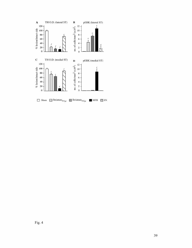
Fig.. 4
39

40

Fig.
. 5
41

Fig.
. 6
42

Fig.. 7
43

Fig.
Fig.
. 8
. 9
44

45
Table 1. Experimental groups and number of animals
Experiment Lesion groups Behavioral tests Treatment
IHC/HPLC L-DOPA Saline
1
Sham (n=8) SN (n=14)
MFB (n=12) ST2x1µl (n=11) ST2x2µl (n=12)
(n=8) (n=14) (n=12) (n=11) (n=12)
(n=6) (n=8) (n=4) (n=5) (n=7)
(n=2) (n=5) (n=5) (n=4) (n=2)
(n=8) (n=13) (n=9) (n=9) (n=9)
2
Sham (n=9) MFB (n=23)
ST2x2µl (n=21)
(n=9) (n=23) (n=21)
(n=5)
(n=17) (n=15)
(n=4) (n=6) (n=6)
(n=9) (n=23) (n=21)
3
Sham (n=11) MFB (n=13)
ST2x2µl (n=17)
(n=11) (n=13) (n=17)
(n=6) (n=7)
(n=11)
(n=5) (n=6) (n=6)
(n=11) (n=13) (n=17)

46
Table 2. Stereological counts of TH-positive neurons Sham ST-LDnd ST-LDdys MFB-LDdys
SNpc les side 4283±226.3 131±11.8c 102±10.1c 44±2.6b int side 4155±160.3 3620±227.4 3575±207.7 3325±215.2 VTA les side 5786±736.4 3592±235.4c 3266±277.9c 2267±157.3b int side 5919±509.7 5776±416.1 5374±379.5 5692±394.1

47
Table 3. Computer-assisted counts of cells immunoreactive for FosB/ΔFosB and TH in the striatum
Sham
ST-Sal
MFB-Sal
ST-LDnd
ST-LDdys
MFB-LDdys
FosB/∆FosB+ cells Medial striatum 1±2.0 8±2.6g 19±2.9g 21±3.8g 48±12.7g 260±41.6e Lateral striatum
1±0.9 5.4±1.3f 14±1.5f 35.4±5.1f 106.7±37e 394±22.1e
TH+ cells n.c. 5±0.4f 7±1.3 f 13±1.9f 37±2.4e 85±5.2e

48
Table 4. Striatal tissue levels of DA, 5-HT and NA Sham
ST-Sal
MFB-Sal
ST-LDnd
ST-LDdys
MFB-LDdys
DA les side 19.04±0.5 1.03±0.3d 0.11±0.06d 1.84±0.46h 0.47±0.08d 0.08±0.02d int side 19.19±0.8 17.66±1.05 20.48±1.24 19.03±0.83 16.68±0.61 21.39±1.23 5-HT les side 0.40±0.03 0.52±0.03 0.51±0.02 0.52±0.04 0.48±0.05 0.54±0.06 int side 0.40±0.04 0.43±0.03 0.46±0.02 0.35±0.05 0.40±0.02 0.57±0.06 NA les side 0.05±0.01 0.02±0.005 0.013±0.004a 0.015±0.003 0.02±0.007 0.02±0.01a int side 0.07±0.01 0.43±0.03 0.46±0.02 0.35±0.05 0.40±0.02 0.52±0.04
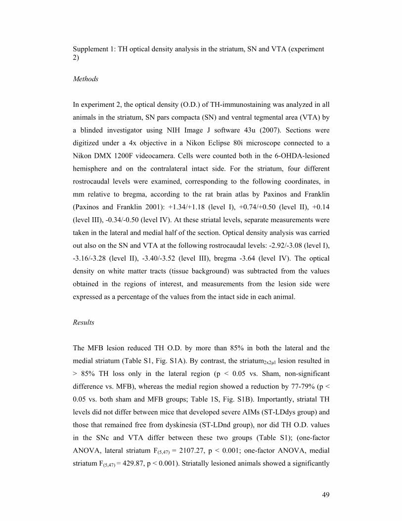
49
Supplement 1: TH optical density analysis in the striatum, SN and VTA (experiment 2)
Methods
In experiment 2, the optical density (O.D.) of TH-immunostaining was analyzed in all
animals in the striatum, SN pars compacta (SN) and ventral tegmental area (VTA) by
a blinded investigator using NIH Image J software 43u (2007). Sections were
digitized under a 4x objective in a Nikon Eclipse 80i microscope connected to a
Nikon DMX 1200F videocamera. Cells were counted both in the 6-OHDA-lesioned
hemisphere and on the contralateral intact side. For the striatum, four different
rostrocaudal levels were examined, corresponding to the following coordinates, in
mm relative to bregma, according to the rat brain atlas by Paxinos and Franklin
(Paxinos and Franklin 2001): +1.34/+1.18 (level I), +0.74/+0.50 (level II), +0.14
(level III), -0.34/-0.50 (level IV). At these striatal levels, separate measurements were
taken in the lateral and medial half of the section. Optical density analysis was carried
out also on the SN and VTA at the following rostrocaudal levels: -2.92/-3.08 (level I),
-3.16/-3.28 (level II), -3.40/-3.52 (level III), bregma -3.64 (level IV). The optical
density on white matter tracts (tissue background) was subtracted from the values
obtained in the regions of interest, and measurements from the lesion side were
expressed as a percentage of the values from the intact side in each animal.
Results
The MFB lesion reduced TH O.D. by more than 85% in both the lateral and the
medial striatum (Table S1, Fig. S1A). By contrast, the striatum2x2µl lesion resulted in
> 85% TH loss only in the lateral region (p < 0.05 vs. Sham, non-significant
difference vs. MFB), whereas the medial region showed a reduction by 77-79% (p <
0.05 vs. both sham and MFB groups; Table 1S, Fig. S1B). Importantly, striatal TH
levels did not differ between mice that developed severe AIMs (ST-LDdys group) and
those that remained free from dyskinesia (ST-LDnd group), nor did TH O.D. values
in the SNc and VTA differ between these two groups (Table S1); (one-factor
ANOVA, lateral striatum F(5,47) = 2107.27, p < 0.001; one-factor ANOVA, medial
striatum F(5,47) = 429.87, p < 0.001). Striatally lesioned animals showed a significantly

50
higher residual TH innervation both in the VTA (about 54%) and in the SNc (about
23%) in comparison with the MFB lesioned mice (about 24% in the VTA and 10% in
the SNc) (Table S1, Fig. S1 D, E, F; one-factor ANOVA, VTA, F(6,46) = 36.77, p <
0.001; SN, F(6,46) = 117.99, p < 0.001). In none of the regions examined was there any
difference in the extent of DA denervation between the three groups with striatal 6-
OHDA lesions (ST-sal, ST-LDnd, and ST-LDdys). Thus, the extent of TH residual
innervation was dependent only on the type of lesion, and was not conditional on L-
DOPA treatment. Among the animals with striatal lesions, the intensity of TH
immunostaining did not predict which animals developed dyskinesia and which did
not.

51
Figure legend
Fig. S1. TH immunostaining (experiment 2). Representative photomicrographs of TH
immunoreactivity in the striatum, VTA and SN were taken from MFB (A and D),
striatal (B and E) and sham-lesion animals (C and F). The side ipsilateral to the lesion
is shown to the right. Scale bar, 1 mm.
Table legend
Table S1. Densitometric analysis of TH immunoreactivity (experiment 2). Values
represent the mean ± SEM per 4 sections throughout striatum, VTA and SN
(experiment 2). They are expressed as percentage of the lesioned side/intact. One-
factor ANOVA and post-hoc Student-Newman-Keuls, p < 0.05; (a) vs. Sham; (b) vs.
Sham and all ST groups; (c) vs. Sham and all MFB groups; (d) vs. Sham and STnd.
SN, substantia nigra pars compacta; VTA, ventral tegmental area; MFB-LDdys, MFB
lesion treated with L-DOPA and dyskinetic; MFB-sal, MFB lesion treated with saline;
ST-LDdys, striatal lesion treated with L-DOPA and dyskinetic; ST-LDnd, striatal
lesion treated with L-DOPA and non-dyskinetic; ST-sal, striatal lesion treated with
saline; Sham, sham lesion.
Fig. S1
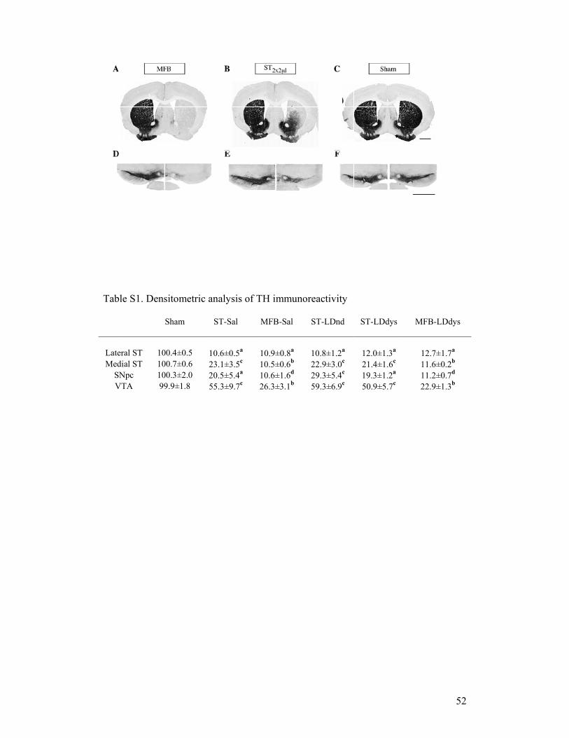
Tab
LateMed
SV
ble S1. Dens
S
eral ST 10dial ST 10
SNpc 10VTA 99
sitometric a
Sham
00.4±0.5 100.7±0.6 200.3±2.0 29.9±1.8 5
analysis of T
ST-Sal
0.6±0.5a 23.1±3.5c 20.5±5.4a 55.3±9.7c
TH immuno
MFB-Sal
10.9±0.8a 10.5±0.6b 10.6±1.6d 26.3±3.1b
oreactivity
ST-LDnd
10.8±1.2a 22.9±3.0c 29.3±5.4c 59.3±6.9c
ST-LDdys
12.0±1.3a 21.4±1.6c 19.3±1.2a 50.9±5.7c
MFB-LD
12.7±1.711.6±0.211.2±0.722.9±1.3
52
dys
7a 2b 7d 3b