fraunhofer iis - spectronet.despectronet.de/portals/visqua/story_docs/intern_spectronet/... ·...
TRANSCRIPT

© Fraunhofer IIS
Fraunhofer IIS
Computer-assisted Analysis of Blood Cells with Smart Microscopes
Dr. Christian Münzenmayer
Group manager Medical image processing
Fraunhofer-Institute for Integrated Circuits IIS
Am Wolfsmantel 33, 91058 Erlangen
E-Mail: [email protected]
Phone: +49-(0)9131-776-7310
© Fraunhofer IIS
Fraunhofer IIS
Fraunhofer Institute for Integrated Circuits IIS
Founded in 1985
More than 730 staff
Budget aprox. € 90 million
Revenue sources
> 75 % income from projects
< 25 % public funding

© Fraunhofer IIS
1% Thrombocytes (Platelets)
Hemostasis / coagulation
96% Erythrocytes (red blood cells)
Oxygen transport
3% Leukocytes (white blood cells)
Immune defense
55%
liquid
(Plasma
)
45%
cellular
Blood
Hematology
Components of blood

© Fraunhofer IIS
Hematology
Basics of hematopoesis
Hematopoesis:
formation of blood
cellular components
Blood cells develop from
haematopoietic stem
cells in the bone marrow
1011 – 1012 cells/day
From: http://commons.wikimedia.org/wiki/File:Hematopoiesis_simple.png

© Fraunhofer IIS
Hematology Laboratory Doctor‘s office
Blood
Withdrawal
Flow
Cytometry Smear Staining
Hematologist / Assistant
Slid
e
Inter-
pretation
Fin
din
g
Microscopy
Sample
Hematology
Laboratory workflow

© Fraunhofer IIS
Staining for microscopic examination
May-Grünwald-Giemsa (MGG) or
Pappenheim-Staining
(Artur Pappenheim, 1870-1916)
Component Staining
Erythrocytes Pink
Nuclei Red violet
Eosinophile Granula Brick-red to red brown
Basophile Granula Dark violet to black
Neutrophile Granula Lilght violet
Zytoplasma of Lymphocytes Light blue
Zytoplasma of Monocytes Gray blue
Hematology
Staining
© Fraunhofer IIS
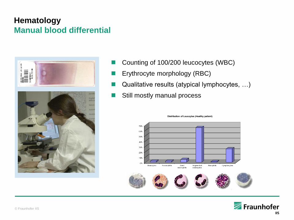
Hematology
Manual blood differential
Counting of 100/200 leucocytes (WBC)
Erythrocyte morphology (RBC)
Qualitative results (atypical lymphocytes, …)
Still mostly manual process

© Fraunhofer IIS
Fast scanning robotic microscope
8x slide handler
Automatic calculation of white blood differential (7 classes)
< 3 Minutes per 100 leukocytes
Red blood cell morphology
Seemless LIS-Integration
Intuitive user interface
Certified Medical Product (IVD) since 10/2010 (98/79/EC Annex I)
Microscopy
HemaCAM™: Computer assisted microscopy in hematology

© Fraunhofer IIS
1. Insert slides, apply immersion oil
2. Identification of slide (barcode, OCR)
3. Automatic distribution of oil
4. Detection and Classification of leucocytes
5. Display of results
6. Optional user interaction
7. Storage, transfer to LIS
Microscopy
HemaCAM™: Process overview

© Fraunhofer IIS
Microscopy
HemaCAM™: Detection of valid area
Detection of valid area and overview image with 10x objective
© Fraunhofer IIS
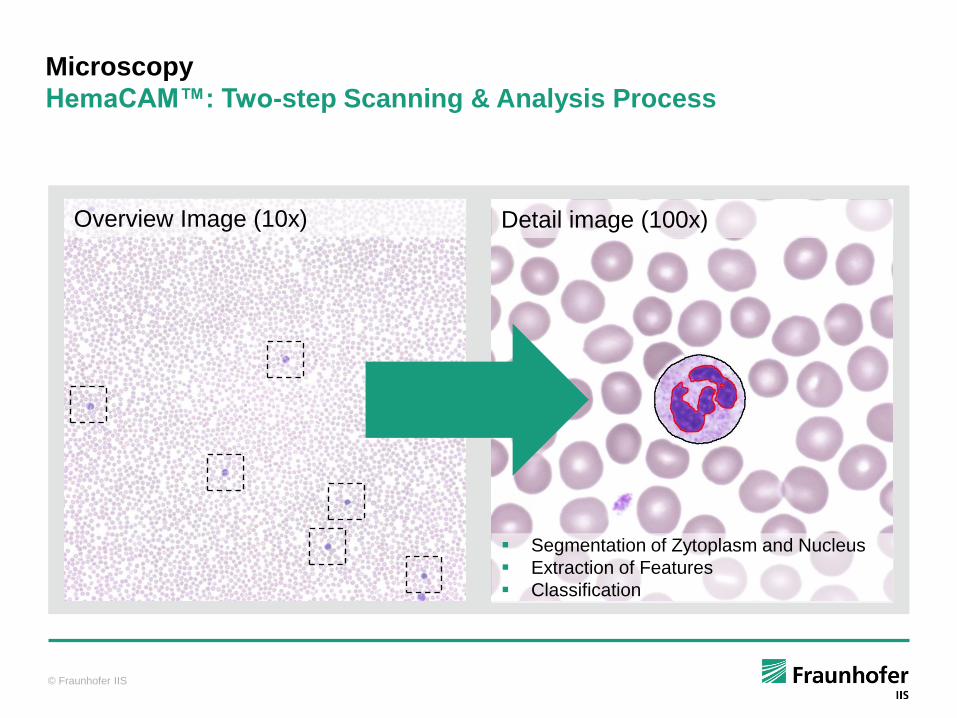
Microscopy
HemaCAM™: Two-step Scanning & Analysis Process
Overview Image (10x) Detail image (100x)
Segmentation of Zytoplasm and Nucleus
Extraction of Features
Classification
© Fraunhofer IIS
Lymphocyte
Neutrophile
14
0
c
c
1
10
100
1000
10000
0 100 200 300 400 500C
ou
nt
Sum i
CorpusCardia
Blau
(x1,y
1)
(x2,y
2)
d
qGrün
(x1,y
1)
(x2,y
2)
d
qRot
(x1,y
1)
(x2,y
2)
d
q
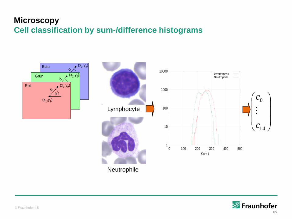
Microscopy
Cell classification by sum-/difference histograms
Lymphocyte
Neutrophile
© Fraunhofer IIS
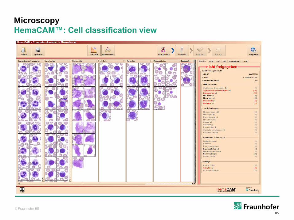
Microscopy
HemaCAM™: Cell classification view
© Fraunhofer IIS
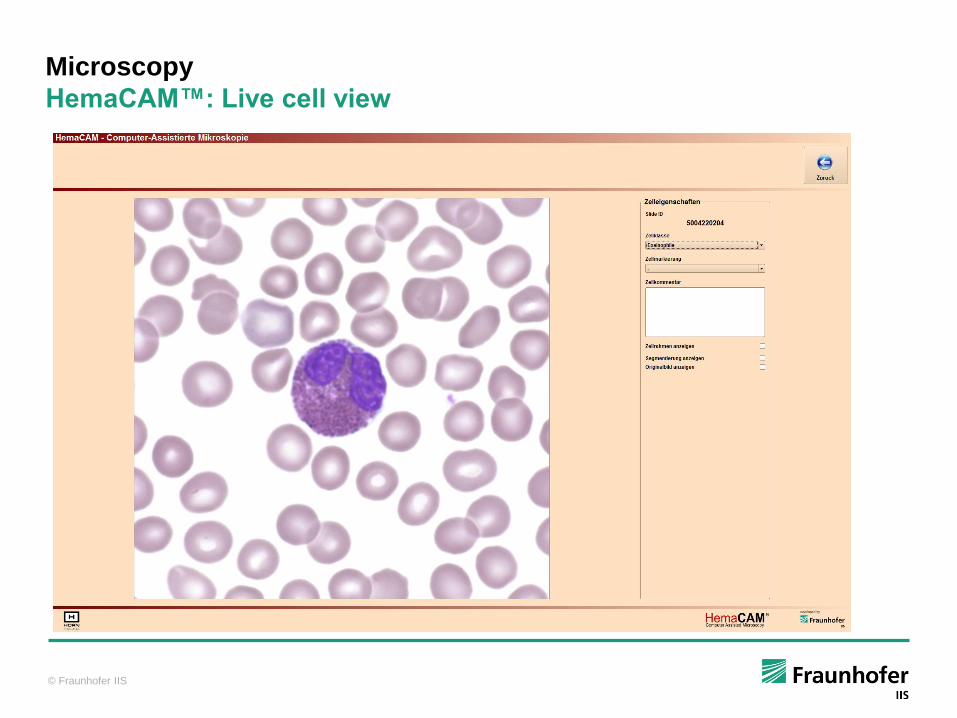
Microscopy
HemaCAM™: Live cell view

© Fraunhofer IIS
Hematology Laboratory Doctor‘s office
Blood
Withdrawal
Flow
Cytometry Smear Staining
Hematologist / Assistant
Slid
e
Inter-
pretation
Fin
din
g
Sample
Hematology
Automated Laboratory workflow
Automated Process
Microscopy Image
Processing Visualization
© Fraunhofer IIS
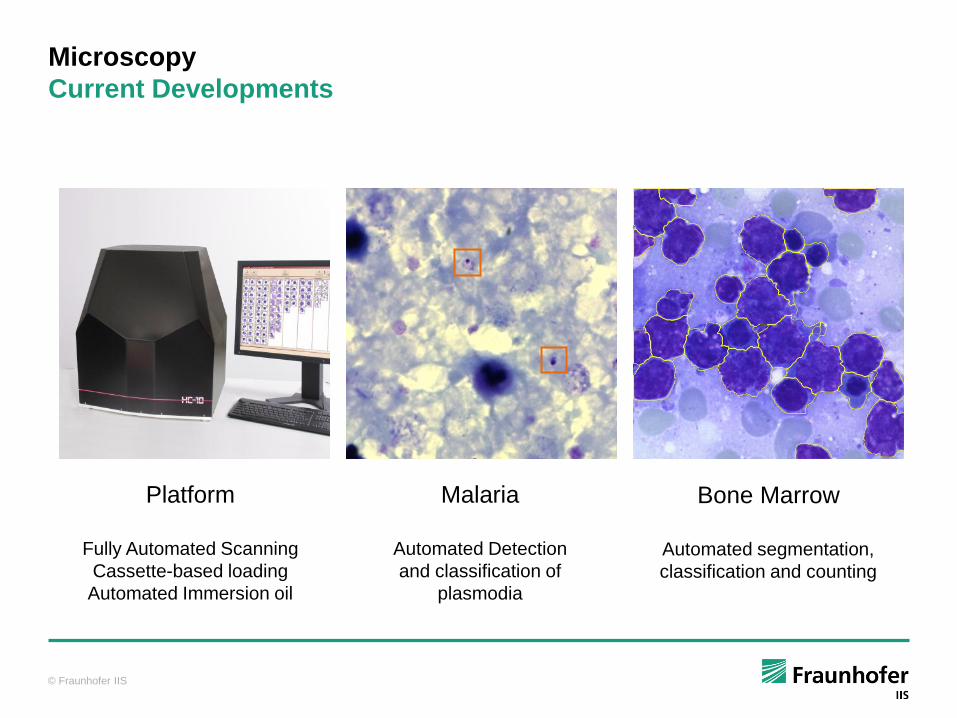
Microscopy
Current Developments
Platform
Fully Automated Scanning
Cassette-based loading
Automated Immersion oil
Malaria
Automated Detection
and classification of
plasmodia
Bone Marrow
Automated segmentation,
classification and counting

© Fraunhofer IIS
Fraunhofer IIS – Medical Image Processing
Research and Development Services
Feasibility Studies
Technical Feasibility, laboratory prototypes
Concepts, risk and cost analysis
Technology & Market Research
Contract R&D Services
Individual R&D for systems / software
Complete Design/Implementation/Test cycle
Documentation, (pre-)clinical studies,
risk management (medical products, IVD)
Licensing of Methods, Systems
Dr. Christian Münzenmayer
Fraunhofer-Institute for Integrated Circuits IIS
Am Wolfsmantel 33, 91058 Erlangen
E-Mail: [email protected]
Phone: +49-(0)9131-776-7310