free energy difference in indolicidin attraction to … energy difference in indolicidin attraction...
TRANSCRIPT

Free Energy Difference in Indolicidin Attraction to Eukaryotic andProkaryotic Model Cell MembranesIn-Chul Yeh,* Daniel R. Ripoll, and Anders Wallqvist
Biotechnology High Performance Computing Software Applications Institute, Telemedicine and Advanced Technology ResearchCenter, U.S. Army Medical Research and Materiel Command, Fort Detrick, Maryland
ABSTRACT: We analyzed the thermodynamic and structuraldeterminants of indolicidin interactions with eukaryotic andprokaryotic cell membranes using a series of atomisticallydetailed molecular dynamics simulations. We used quartz-supported bilayers with two different compositions ofzwitterionic and anionic phospholipids as model eukaryoticand prokaryotic cell membranes. Indolicidin was preferentiallyattracted to the model prokaryotic cell membrane in contrastto the weak adsorption on the eukaryotic membrane. Thenature of the indolicidin surface adsorption depended on anelectrostatic guiding component, an attractive enthalpiccomponent derived from van der Waals interactions, and abalance between entropic factors related to peptide confine-ment at the interface and counterion release from the bilayersurface. Thus, whereas we attributed the specificity of theindolicidin/membrane interaction to electrostatics, theseinteractions were not the sole contributors to the free energy of adsorption. Instead, a balance between an attractive van derWaals enthalpic component and a repulsive entropic component determined the overall strength of indolicidin adsorption.
■ INTRODUCTIONAntimicrobial peptides are small cationic peptides that playimportant roles in the host-defense mechanism of many livingorganisms against bacterial infections.1−3 Antimicrobial pep-tides act rapidly against a broad spectrum of microbes and haveattracted attention as potential novel antibiotics to combatmultidrug-resistant bacterial infections, either as standaloneagents or in combination therapies with other conventionalantibiotics.4−6 Indolicidin is an antimicrobial peptide isolatedfrom bovine neutrophils.7 It consists of 13 amino acid residues(NH3
+-ILPWKWPWWPWRR-NH2) with an uncapped N-terminus and an amidated C-terminus resulting in a netpositive charge of 4 e. The structure of indolicidin is known tobe disordered in aqueous solution but has been shown to forman extended conformation when bound to detergent micelles.8
The mechanism of indolicidin action against both bacterial andhuman cells has been investigated both experimentally9−17 andcomputationally.18−20 Even though several different mecha-nisms of action by indolicidin have been proposed, the exactmechanism remains unclear.14,20 The delineation of themechanism is not straightforward because the biological actionthat ultimately results in lysis of bacteria is multifaceted.Initially, indolicidin must recognize and attach to bacterial cellmembranes. After this attachment, several lytic mechanisms arepossible, for example, membrane thinning resulting in astructural weakening of the membrane and loss of cellhomeostasis, interactions with proteins in the membrane, ortranslocation to the cytosol where the peptide can further
interact with diverse cellular targets, such as RNA and DNA, orother highly charged species. Existing results from bothexperimental8,14,16 and computer simulation18−20 work confirmthat the indolicidin’s mechanism of action includes animportant localization component associated with peptideadsorption to the solvent side of membrane bilayer interface.In this work, we quantify and address the detailedthermodynamic and structural elements of this initial attractionof indolicidin to model prokaryotic and eukaryotic bilayersusing computational approaches.Solid-supported lipid bilayers are widely used as experimental
model systems to investigate detailed interactions of biologicalmolecules with membranes. Such model systems have beenused to investigate the interactions of antimicrobial peptideswith lipid membranes using quartz crystal microbalance(QCM) measurements,12,21 sum frequency generation (SFG)vibrational spectroscopy,22,23 and in situ atomic forcemicroscopy (AFM).14−16 Similarly, one can use computationalmodels to create realistic solid-supported lipid bilayers at theatomic level24−27 to map out the interactions of antimicrobialpeptides with lipid membranes in great detail. Recently, wesuccessfully modeled solid-supported bilayers with a hydrateddimyristoylphosphatidylcholine (DMPC) lipid bilayer depos-ited on a quartz crystal surface.28 Importantly, we showed a
Received: December 9, 2011Revised: February 10, 2012Published: February 16, 2012
Article
pubs.acs.org/JPCB
© 2012 American Chemical Society 3387 dx.doi.org/10.1021/jp211883u | J. Phys. Chem. B 2012, 116, 3387−3396
Report Documentation Page Form ApprovedOMB No. 0704-0188
Public reporting burden for the collection of information is estimated to average 1 hour per response, including the time for reviewing instructions, searching existing data sources, gathering andmaintaining the data needed, and completing and reviewing the collection of information. Send comments regarding this burden estimate or any other aspect of this collection of information,including suggestions for reducing this burden, to Washington Headquarters Services, Directorate for Information Operations and Reports, 1215 Jefferson Davis Highway, Suite 1204, ArlingtonVA 22202-4302. Respondents should be aware that notwithstanding any other provision of law, no person shall be subject to a penalty for failing to comply with a collection of information if itdoes not display a currently valid OMB control number.
1. REPORT DATE 16 FEB 2012 2. REPORT TYPE
3. DATES COVERED 00-00-2012 to 00-00-2012
4. TITLE AND SUBTITLE Free Energy Difference in Indolicidin Attraction to Eukaryotic andProkaryotic Model Cell Membranes
5a. CONTRACT NUMBER
5b. GRANT NUMBER
5c. PROGRAM ELEMENT NUMBER
6. AUTHOR(S) 5d. PROJECT NUMBER
5e. TASK NUMBER
5f. WORK UNIT NUMBER
7. PERFORMING ORGANIZATION NAME(S) AND ADDRESS(ES) U.S. Army Medical Research and Materiel Command,BiotechnologyHigh Performance Computing Software ApplicationsInstitute,Telemedicine and Advanced Technology Research Center,Fort Detrick,MD,21702
8. PERFORMING ORGANIZATIONREPORT NUMBER
9. SPONSORING/MONITORING AGENCY NAME(S) AND ADDRESS(ES) 10. SPONSOR/MONITOR’S ACRONYM(S)
11. SPONSOR/MONITOR’S REPORT NUMBER(S)
12. DISTRIBUTION/AVAILABILITY STATEMENT Approved for public release; distribution unlimited
13. SUPPLEMENTARY NOTES
14. ABSTRACT We analyzed the thermodynamic and structural determinants of indolicidin interactions with eukaryoticand prokaryotic cell membranes using a series of atomistically detailed molecular dynamics simulations.We used quartzsupported bilayers with two different compositions of zwitterionic and anionicphospholipids as model eukaryotic and prokaryotic cell membranes. Indolicidin was preferentiallyattracted to the model prokaryotic cell membrane in contrast to the weak adsorption on the eukaryoticmembrane. The nature of the indolicidin surface adsorption depended on an electrostatic guidingcomponent, an attractive enthalpic component derived from van der Waals interactions, and a balancebetween entropic factors related to peptide confinement at the interface and counterion release from thebilayer surface. Thus, whereas we attributed the specificity of the indolicidin/membrane interaction toelectrostatics, these interactions were not the sole contributors to the free energy of adsorption. Instead, abalance between an attractive van der Waals enthalpic component and a repulsive entropic componentdetermined the overall strength of indolicidin adsorption.
15. SUBJECT TERMS
16. SECURITY CLASSIFICATION OF: 17. LIMITATION OF ABSTRACT Same as
Report (SAR)
18. NUMBEROF PAGES
10
19a. NAME OFRESPONSIBLE PERSON
a. REPORT unclassified
b. ABSTRACT unclassified
c. THIS PAGE unclassified
Standard Form 298 (Rev. 8-98) Prescribed by ANSI Std Z39-18

critical need to implement the appropriate electrostaticboundary conditions for simulations and analyses to capturethe structural and electrostatic properties of DMPC lipidbilayers that correspond to the physical systems being modeled,for example, solvated bilayers representing lamellar systems,nonlamellar bilayers mimicking membranes, and quartz-supported bilayers. These methodological considerations arecritical because the electrostatic interaction between cationicantimicrobial peptides and the anionic lipids in the bacterial cellmembrane is a key feature that governs the selective attractionof antimicrobial peptides to the bacterial cell membrane.3,29 Adetailed understanding of this electrostatic interaction needs totake into consideration all the complex interactions amongexplicit solvent molecules, counterions, and salt ions presentunder physiological conditions. In particular, counterions andsalts strongly diminish the electrostatic interactions betweenantimicrobial peptides and anionic lipids, leading to a morenuanced interpretation of the electrostatic effect associated withcationic antimicrobial peptides.In this work, we investigated the differences in the
interaction of the antimicrobial peptide indolicidin withmodel bacterial and eukaryotic cell membranes by performingatomistically detailed molecular dynamics (MD) simulationsthat included solvent molecules, salt ions, and lipids. Weconstructed model prokaryotic and eukaryotic cell membranesusing mixed (zwitterionic and anionic) and neutral (zwitter-ionic) lipid bilayers respectively deposited on a quartz crystalsurface. To characterize the interaction of indolicidin with thelipid membranes, we calculated the potential of mean force(PMF)30 or free energy profile with respect to the distance ofindolicidin from the lipid membrane using a series of MDsimulations. We confirmed the existence of the selectiveattractive interaction of indolicidin with the model bacterialcell membrane, which results in indolicidin being preferentiallylocated at the membrane interface. We further characterized thenature of the underlying interaction in terms of structural andthermodynamic properties. In particular, we found thatplacement of indolicidin at the membrane surface wasassociated with a minimum PMF of −3.5 ± 0.2 kcal/mol atthe prokaryotic membrane, compared to −0.6 ± 0.6 kcal/molat the eukaryotic membrane. We further used the distance-dependent PMF to estimate the free energy of indolicidinadsorption (ΔG°) to be −1.94 ± 0.56 kcal/mol for the modelprokaryotic cell membrane and −0.05 ± 0.46 kcal/mol for theeukaryotic membrane. The detailed thermodynamic andstructural analysis of these systems provided support for theidea that the specificity of the indolicidin/membraneinteraction can be attributed to electrostatics, but theseinteractions were not the sole contributors to the free energyof adsorption. Instead, a balance between an attractive van derWaals enthalpic component and a repulsive entropiccomponent determined the overall strength of indolicidinadsorption.
■ METHODSSimulations. MD simulations were performed with the
NAMD MD simulation program31 using the CHARMM 27 allatom force field.32 We used DMPC and dimyristoylphosphatidylglycerol (DMPG) as model zwitterionic andanionic phospholipids, respectively. The TIP3P model33 wasused to describe water molecules. The force field for the quartz(011) crystal was taken from one developed by Lopes et al.34 Aprimitive unit cell of quartz (011) of ∼15 Å thickness was
replicated in two dimensions to construct a quartz (011) crystalwith a surface area of 36.5 × 37.0 Å2 in the unit cell, as in theprevious study by Lopes et al.34 and in our recent study.28 Oneside of the constructed crystal was covered with hydrophilicsilanols (Si−OH), and silicon atoms on the other side weresaturated with hydrogens (Si−H). Crystal atoms, except for theO−H groups of silanols, were held fixed to maintain the (011)crystal geometry. Short-range interactions outside a 10 Å cutoffwere truncated. Long-range electrostatic interactions werecalculated with the particle mesh Ewald method35 with acorrection term for the planar vacuum boundary condition,28,36
referred to as EW3DC, which we implemented in NAMD. TheEW3DC correction term, combined with a sufficiently large boxlength (Lz) in the z direction normal to the interface, effectivelyimplements a 2D periodic boundary condition, which isappropriate for solid-supported lipid bilayers. Bonds involvinghydrogens were constrained with the SHAKE algorithm.37 Weused a time step of 2 fs for the time integration, and thetemperature was maintained at 310 K using Langevin dynamicswith a damping coefficient of 10 ps−1.38
Preparation of Quartz-Supported Lipid Bilayers.Quartz-supported lipid bilayers were prepared as illustrated inFigure 1. Hydrated lipid bilayers are in contact with the
hydrophilic side of the quartz crystal. Both a pure DMPCbilayer and a mixed DMPC/DMPG (DMPC/G) bilayer wereprepared as model eukaryotic and bacterial cell membranes,respectively. For a starting point of a pure DMPC bilayer, weused the last configuration from the 200 ns simulation of theDMPC bilayer composed of 44 DMPC molecules (22 on eachleaflet) supported on the quartz surface described in ourprevious work.28 Similarly, a mixed lipid bilayer with 34 DMPCand 10 DMPG molecules (17 DMPC and 5 DMPG on eachleaflet) supported on the quartz surface was prepared after a 45ns equilibration in aqueous solution and a 50 ns equilibrationon the quartz surface. An initial configuration of the mixedDMPC/G lipid bilayer was prepared with the CHARMM-GUI.39 In these configurations, the upper water layer betweenthe lipid bilayer and the water/vacuum interface containedabout twice as many water molecules as the lower water layerbetween the bilayer and the quartz. To increase the bulklikeregion of water to accommodate indolicidin, we doubled the
Figure 1. Snapshot of the hydrated DMPC/G lipid bilayer supportedon a quartz crystal illustrates the placement of the unit cell in thesupported bilayer system. The blue box represents the unit simulationcell with an Lz of 250 Å. The simulation cell is repeated periodically inall three directions. Gray and red lines represent DMPC and DMPGlipids, respectively. Gray and red balls near the membrane interfacesdenote phosphorus atoms of headgroups in DMPC and DMPG lipids,respectively. Smaller pink spheres represent oxygen atoms of watermolecules. Blue and green spheres represent sodium and chloride ions,respectively.
The Journal of Physical Chemistry B Article
dx.doi.org/10.1021/jp211883u | J. Phys. Chem. B 2012, 116, 3387−33963388

number of water molecules in the upper water layer. As a result,the upper water layer contains about four times as many watermolecules as the lower water layer. To observe the effects ofsalt ions on the properties of lipid bilayers, we performedsimulations with and without added NaCl salt for a pureDMPC bilayer. For a simulation with salt, we added 2 and 8pairs of NaCl to the lower and upper water layers respectivelyresulting in a salt concentration of about 0.16 M,commensurate with physiological salt concentrations. For asimulation with the mixed DMPC/G bilayer, 10 extra sodiumcounterions (5 to each water layer) were added to maintain thenet charge neutrality. The area per lipid headgroup is about 60Å2, which is close to the experimental value for DMPC.40 Thebox length (Lz) in the z direction normal to the interface wasfixed at 250 Å, which was large enough to create a water/vacuum interface as shown in Figure 1. This allowed the systemto adjust to an optimum density without the need of constant-pressure simulations. Simulation for each prepared quart-supported lipid bilayer system lasted 100 ns. The initial 20 nswere discarded as equilibration.Preparation of Indolicidin. Five different initial config-
urations of indolicidin were prepared. The first configurationwas taken from the structure of indolicidin bound to adodecylphosphocholine (DPC) micelle as determined by NMR(PDB id: 1g89).8 After a brief energy minimization, weperformed an MD simulation of indolicidin in aqueous solutionfor 100 ns. The other four configurations of indolicidin wereselected from conformations generated by the ECEPPAKprogram.41,42 We selected two conformations where NOEdistances deviated less than 1 Å from experimental values andanother two conformations where the Cα rmsd of the coreresidues 3−11 was less than 1 Å away from the NMR modelstructure. We performed an MD simulation lasting 30 ns inaqueous solution for each of the four selected conformations.We kept the last conformation of indolicidin from each solutionsimulation and placed it in the middle of the upper water layerafter removing water molecules within 2.8 Å of the indolicidinpeptide. The number of counterions in the upper water layerwas adjusted to maintain charge neutrality in the presence ofthe positively (+4) charged indolicidin.Calculation of the Free Energy Profile. For a detailed
understanding of how indolicidin interacts with lipid bilayers,we performed MD simulations with the peptide z positionrestrained at various points in the aqueous phase with NAMD’scolvar module. We used a harmonic biasing potential with aforce constant of 2.0 kcal/(mol Å2) applied to the center ofmass z position of indolicidin. The z position bias wasimplemented at every 1 Å interval from 52 to 96 Å with respectto the quartz surface, and each biased simulation lasted 25 ns.We calculated the free energy or PMF profile by applying theweighted histogram analysis method43,44 to the last 15 ns of thesimulation data. A total of 3.375 μs of the simulation dataderived from five different and independent starting config-urations were used to calculate the free energy profile for eachbilayer system. However, the z position bias for simulationsstarting with the NMR model structure was extended up to 125Å to observe the structural properties of the peptide near thewater/vacuum interface.Lipid Order Parameter. The acyl chain lipid order
parameter profile45 is one of the important quantitiesdescribing the properties of lipid bilayers. To probe possibledisruptions of the membranes caused by indolicidin, wecalculated the carbon-deuterium lipid order parameter SCD for
the aliphatic C−H bond vectors of each carbon atom of the acylchain sn−1 with the following formula:
= θ −S
3cos 12CD
2
(1)
where θ is the angle between the C−H vector and the z axis.The order parameter SCD is zero for a completely unordered(isotropic) system. A perfectly ordered acyl chain in an all-transconformation results in an SCD value of −0.5.45
Electrostatic Property Profiles Across the Interface inPeriodic Boundary Conditions. Distribution of the electricfield along the z direction E(z) was calculated from the chargedensity distribution ρq(z) obtained from the simulations usingthe following relationship:28,36,46
∫=
ρ ′ ′
ε−
E zz z
( )( )dL
z/2 q
0
z
(2)
where ε0 is the vacuum permittivity and Lz is the length of thesimulation cell in the z direction.
■ RESULTS AND DISCUSSIONSStructural and Electrostatic Properties of Quartz-
Supported Neat Lipid Bilayers. To delineate the underlyingdifferences between model eukaryotic and prokaryotic cellmembranes, we analyzed structural and electrostatic propertiesof hydrated DMPC and mixed DMPC/G bilayers supported ona quartz crystal in the absence of the added peptide. Figure 2compares mass density distributions of lipid, water, and sodium
Figure 2. Mass density distributions of components of the quartzcrystal supported lipid bilayers: (a) DMPC bilayer without salt ions,(b) DMPC bilayer with added salt ions, and (c) DMPC/G bilayerwith added salt ions. Black, red, blue, and green lines represent lipid,water, sodium, and chloride, respectively. The z position in this andsubsequent figures is with respect to the membrane center.
The Journal of Physical Chemistry B Article
dx.doi.org/10.1021/jp211883u | J. Phys. Chem. B 2012, 116, 3387−33963389
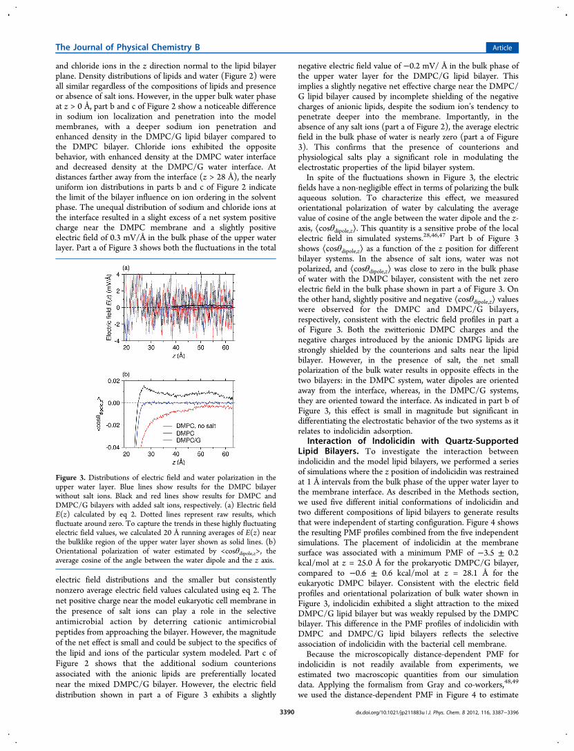
and chloride ions in the z direction normal to the lipid bilayerplane. Density distributions of lipids and water (Figure 2) wereall similar regardless of the compositions of lipids and presenceor absence of salt ions. However, in the upper bulk water phaseat z > 0 Å, part b and c of Figure 2 show a noticeable differencein sodium ion localization and penetration into the modelmembranes, with a deeper sodium ion penetration andenhanced density in the DMPC/G lipid bilayer compared tothe DMPC bilayer. Chloride ions exhibited the oppositebehavior, with enhanced density at the DMPC water interfaceand decreased density at the DMPC/G water interface. Atdistances farther away from the interface (z > 28 Å), the nearlyuniform ion distributions in parts b and c of Figure 2 indicatethe limit of the bilayer influence on ion ordering in the solventphase. The unequal distribution of sodium and chloride ions atthe interface resulted in a slight excess of a net system positivecharge near the DMPC membrane and a slightly positiveelectric field of 0.3 mV/Å in the bulk phase of the upper waterlayer. Part a of Figure 3 shows both the fluctuations in the total
electric field distributions and the smaller but consistentlynonzero average electric field values calculated using eq 2. Thenet positive charge near the model eukaryotic cell membrane inthe presence of salt ions can play a role in the selectiveantimicrobial action by deterring cationic antimicrobialpeptides from approaching the bilayer. However, the magnitudeof the net effect is small and could be subject to the specifics ofthe lipid and ions of the particular system modeled. Part c ofFigure 2 shows that the additional sodium counterionsassociated with the anionic lipids are preferentially locatednear the mixed DMPC/G bilayer. However, the electric fielddistribution shown in part a of Figure 3 exhibits a slightly
negative electric field value of −0.2 mV/ Å in the bulk phase ofthe upper water layer for the DMPC/G lipid bilayer. Thisimplies a slightly negative net effective charge near the DMPC/G lipid bilayer caused by incomplete shielding of the negativecharges of anionic lipids, despite the sodium ion’s tendency topenetrate deeper into the membrane. Importantly, in theabsence of any salt ions (part a of Figure 2), the average electricfield in the bulk phase of water is nearly zero (part a of Figure3). This confirms that the presence of counterions andphysiological salts play a significant role in modulating theelectrostatic properties of the lipid bilayer system.In spite of the fluctuations shown in Figure 3, the electric
fields have a non-negligible effect in terms of polarizing the bulkaqueous solution. To characterize this effect, we measuredorientational polarization of water by calculating the averagevalue of cosine of the angle between the water dipole and the z-axis, ⟨cosθdipole,z⟩. This quantity is a sensitive probe of the localelectric field in simulated systems.28,46,47 Part b of Figure 3shows ⟨cosθdipole,z⟩ as a function of the z position for differentbilayer systems. In the absence of salt ions, water was notpolarized, and ⟨cosθdipole,z⟩ was close to zero in the bulk phaseof water with the DMPC bilayer, consistent with the net zeroelectric field in the bulk phase shown in part a of Figure 3. Onthe other hand, slightly positive and negative ⟨cosθdipole,z⟩ valueswere observed for the DMPC and DMPC/G bilayers,respectively, consistent with the electric field profiles in part aof Figure 3. Both the zwitterionic DMPC charges and thenegative charges introduced by the anionic DMPG lipids arestrongly shielded by the counterions and salts near the lipidbilayer. However, in the presence of salt, the net smallpolarization of the bulk water results in opposite effects in thetwo bilayers: in the DMPC system, water dipoles are orientedaway from the interface, whereas, in the DMPC/G systems,they are oriented toward the interface. As indicated in part b ofFigure 3, this effect is small in magnitude but significant indifferentiating the electrostatic behavior of the two systems as itrelates to indolicidin adsorption.
Interaction of Indolicidin with Quartz-SupportedLipid Bilayers. To investigate the interaction betweenindolicidin and the model lipid bilayers, we performed a seriesof simulations where the z position of indolicidin was restrainedat 1 Å intervals from the bulk phase of the upper water layer tothe membrane interface. As described in the Methods section,we used five different initial conformations of indolicidin andtwo different compositions of lipid bilayers to generate resultsthat were independent of starting configuration. Figure 4 showsthe resulting PMF profiles combined from the five independentsimulations. The placement of indolicidin at the membranesurface was associated with a minimum PMF of −3.5 ± 0.2kcal/mol at z = 25.0 Å for the prokaryotic DMPC/G bilayer,compared to −0.6 ± 0.6 kcal/mol at z = 28.1 Å for theeukaryotic DMPC bilayer. Consistent with the electric fieldprofiles and orientational polarization of bulk water shown inFigure 3, indolicidin exhibited a slight attraction to the mixedDMPC/G lipid bilayer but was weakly repulsed by the DMPCbilayer. This difference in the PMF profiles of indolicidin withDMPC and DMPC/G lipid bilayers reflects the selectiveassociation of indolicidin with the bacterial cell membrane.Because the microscopically distance-dependent PMF for
indolicidin is not readily available from experiments, weestimated two macroscopic quantities from our simulationdata. Applying the formalism from Gray and co-workers,48,49
we used the distance-dependent PMF in Figure 4 to estimate
Figure 3. Distributions of electric field and water polarization in theupper water layer. Blue lines show results for the DMPC bilayerwithout salt ions. Black and red lines show results for DMPC andDMPC/G bilayers with added salt ions, respectively. (a) Electric fieldE(z) calculated by eq 2. Dotted lines represent raw results, whichfluctuate around zero. To capture the trends in these highly fluctuatingelectric field values, we calculated 20 Å running averages of E(z) nearthe bulklike region of the upper water layer shown as solid lines. (b)Orientational polarization of water estimated by <cosθdipole,z>, theaverage cosine of the angle between the water dipole and the z axis.
The Journal of Physical Chemistry B Article
dx.doi.org/10.1021/jp211883u | J. Phys. Chem. B 2012, 116, 3387−33963390
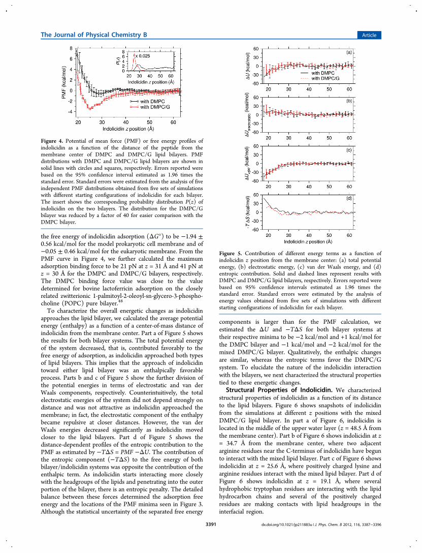
the free energy of indolicidin adsorption (ΔG°) to be −1.94 ±0.56 kcal/mol for the model prokaryotic cell membrane and of−0.05 ± 0.46 kcal/mol for the eukaryotic membrane. From thePMF curve in Figure 4, we further calculated the maximumadsorption binding force to be 21 pN at z = 31 Å and 41 pN atz = 30 Å for the DMPC and DMPC/G bilayers, respectively.The DMPC binding force value was close to the valuedetermined for bovine lactoferricin adsorption on the closelyrelated zwitterionic 1-palmitoyl-2-oleoyl-sn-glycero-3-phospho-choline (POPC) pure bilayer.48
To characterize the overall energetic changes as indolicidinapproaches the lipid bilayer, we calculated the average potentialenergy (enthalpy) as a function of a center-of-mass distance ofindolicidin from the membrane center. Part a of Figure 5 showsthe results for both bilayer systems. The total potential energyof the system decreased, that is, contributed favorably to thefree energy of adsorption, as indolicidin approached both typesof lipid bilayers. This implies that the approach of indolicidintoward either lipid bilayer was an enthalpically favorableprocess. Parts b and c of Figure 5 show the further division ofthe potential energies in terms of electrostatic and van derWaals components, respectively. Counterintuitively, the totalelectrostatic energies of the system did not depend strongly ondistance and was not attractive as indolicidin approached themembrane; in fact, the electrostatic component of the enthalpybecame repulsive at closer distances. However, the van derWaals energies decreased significantly as indolicidin movedcloser to the lipid bilayers. Part d of Figure 5 shows thedistance-dependent profiles of the entropic contribution to thePMF as estimated by −TΔS = PMF −ΔU. The contribution ofthe entropic component (−TΔS) to the free energy of bothbilayer/indolicidin systems was opposite the contribution of theenthalpic term. As indolicidin starts interacting more closelywith the headgroups of the lipids and penetrating into the outerportion of the bilayer, there is an entropic penalty. The detailedbalance between these forces determined the adsorption freeenergy and the locations of the PMF minima seen in Figure 3.Although the statistical uncertainty of the separated free energy
components is larger than for the PMF calculation, weestimated the ΔU and −TΔS for both bilayer systems attheir respective minima to be −2 kcal/mol and +1 kcal/mol forthe DMPC bilayer and −1 kcal/mol and −2 kcal/mol for themixed DMPC/G bilayer. Qualitatively, the enthalpic changesare similar, whereas the entropic terms favor the DMPC/Gsystem. To elucidate the nature of the indolicidin interactionwith the bilayers, we next characterized the structural propertiestied to these energetic changes.
Structural Properties of Indolicidin. We characterizedstructural properties of indolicidin as a function of its distanceto the lipid bilayers. Figure 6 shows snapshots of indolicidinfrom the simulations at different z positions with the mixedDMPC/G lipid bilayer. In part a of Figure 6, indolicidin islocated in the middle of the upper water layer (z = 48.5 Å fromthe membrane center). Part b of Figure 6 shows indolicidin at z= 34.7 Å from the membrane center, where two adjacentarginine residues near the C-terminus of indolicidin have begunto interact with the mixed lipid bilayer. Part c of Figure 6 showsindolicidin at z = 25.6 Å, where positively charged lysine andarginine residues interact with the mixed lipid bilayer. Part d ofFigure 6 shows indolicidin at z = 19.1 Å, where severalhydrophobic tryptophan residues are interacting with the lipidhydrocarbon chains and several of the positively chargedresidues are making contacts with lipid headgroups in theinterfacial region.
Figure 4. Potential of mean force (PMF) or free energy profiles ofindolicidin as a function of the distance of the peptide from themembrane center of DMPC and DMPC/G lipid bilayers. PMFdistributions with DMPC and DMPC/G lipid bilayers are shown insolid lines with circles and squares, respectively. Errors reported werebased on the 95% confidence interval estimated as 1.96 times thestandard error. Standard errors were estimated from the analysis of fiveindependent PMF distributions obtained from five sets of simulationswith different starting configurations of indolicidin for each bilayer.The insert shows the corresponding probability distribution P(z) ofindolicidin on the two bilayers. The distribution for the DMPC/Gbilayer was reduced by a factor of 40 for easier comparison with theDMPC bilayer.
Figure 5. Contribution of different energy terms as a function ofindolicidin z position from the membrane center: (a) total potentialenergy, (b) electrostatic energy, (c) van der Waals energy, and (d)entropic contribution. Solid and dashed lines represent results withDMPC and DMPC/G lipid bilayers, respectively. Errors reported werebased on 95% confidence intervals estimated as 1.96 times thestandard error. Standard errors were estimated by the analysis ofenergy values obtained from five sets of simulations with differentstarting configurations of indolicidin for each bilayer.
The Journal of Physical Chemistry B Article
dx.doi.org/10.1021/jp211883u | J. Phys. Chem. B 2012, 116, 3387−33963391

Part a of Figure 7 shows the distributions of backbone rmsdof indolicidin with respect to the experimental NMR structureaveraged over all configurations in the production runs. Forboth lipid bilayers, the structure of indolicidin deviatedsignificantly from the experimental NMR structure. Part b ofFigure 7 shows backbone rmsd of indolicidin as a function ofindolicidin z position from the membrane center. The averagebackbone rmsd values of indolicidin in bulk water phase andnear the bilayer were similar, whereas indolicidin exposed to thevacuum (z > 60 Å) displayed larger average backbone rmsdvalues. Part c of Figure 7 shows orientational distributions ofindolicidin measured by the average cosine of the anglebetween the z axis and the long axis of indolicidin determinedfrom the inertia tensor (<cosθpeptide,z>). The angle θpeptide,zranged from 0 to 90 degrees with respect to the z axis andcosθpeptide,z ranged from 0 (parallel to the membrane interface)to 1 (perpendicular to the membrane interface). Whenindolicidin was located in the bulk water phase, <cosθpeptide,z>was about 0.5, consistent with an isotropic distribution. Nearthe water/vacuum interface (60 Å < z < 70 Å), indolicidinadopted a perpendicular orientation with respect to theinterface. More importantly, however, when indolicidin waslocated closer to the lipid bilayer, <cosθpeptide,z> was slightlyincreased (30 Å < z < 40 Å) but decreased in region where theminima of the PMF are located (Figure 3). Thus, in theenergetically most favorable configurations, indolicidin adopts aroughly parallel orientation with respect to the membraneinterface. Part c of Figure 6 shows an example of such a
conformation. The structural variations and orientationalpreferences of indolicidin were qualitatively similar for boththe DMPC and DMPC/G bilayer systems.Khandelia and Kaznessis18 observed a persistent cation−π
interaction between TRP11 and ARG13 of indolicidin in thezwitterionic DPC micelle. To determine whether we observe asimilar stabilizing interaction, we calculated the distributions ofthe distance between TRP11 and ARG13 of indolicidin nearDMPC and DMPC/G lipid bilayers as shown in part d ofFigure 7. A larger population of indolicidin with the TRP11-ARG13 distance less than 5 Å was observed near thezwitterionic DMPC bilayer than the mixed DMPC/G bilayer,which is consistent with the simulation results by Khandelia andKaznessis.18 However, no persistent cation−π interaction wasobserved for indolicidin near either lipid bilayer in oursimulations.
Distributions of Positively Charged Residues andTryptophan Residues. In Figure 8, we compared distribu-tions of positively charged residues and hydrophobictryptophan residues from indolicidin adsorbed on DMPC andDMPC/G lipid bilayers. To select the indolicidin interface-contact conformations, we used configurations where thecenter-of-mass of indolicidin was located between 20 and 27 Åaway from the membrane center. Part a of Figure 8 shows thatpositively charged residues were more preferentially distributednear the headgroups of the DMPC/G lipid bilayers than in theDMPC system. However, part b of Figure 8 shows that therewas no significant difference between distributions of
Figure 6. Snapshots of indolicidin at (a) 48.5, (b) 34.7, (c) 25.6, and (d) 19.1 Å away from the center of the DMPC/G lipid bilayer. DMPC andDMPG lipids are represented by gray and red lines, respectively. Phosphorus atoms in the headgroups of DMPC and DMPG lipids are shown as grayand red spheres, respectively. Indolicidin is represented by a ribbon in cyan. Non-hydrogen atoms of positively charged lysine and arginine residuesand N-terminus are explicitly represented as balls and sticks. Carbon, nitrogen, and oxygen atoms are shown in cyan, blue, and red, respectively.Indole rings of hydrophobic tryptophan residues are represented by thick gray bonds. Oxygen atoms of water molecules are shown as small pinkballs.
The Journal of Physical Chemistry B Article
dx.doi.org/10.1021/jp211883u | J. Phys. Chem. B 2012, 116, 3387−33963392

tryptophan residues near the DMPC and DMPC/G lipidbilayers. The primarily hydrophobic interactions stemmingfrom tryptophan/lipid contacts were roughly similar in nature,commensurate with the similar enthalpic profiles shown inFigure 5 for both types of bilayers. Thus, the distinctionbetween the two bilayers in terms of interactions lies primarilyin how the positively charged residues are distributed in theinterface region of the bilayer and how the system of solventand ion molecules accommodate these configurations. Giventhat the enthalpic component derived from electrostaticinteractions between the two systems is not that different dueto charge conservation and counterion shielding, theconformations underlying both distributions in part a of Figure8 are enthalpically equivalent. The difference in density ofpositively charged residues located in the bilayer headgroupregion around z = 20 Å in part a of Figure 8 instead points to amechanism whereby positively charged ions normally asso-ciated with the bilayer are replaced by positive charges fromindolicidin. This type of an effect would essentially be
enthalpically neutral but entropically favorable. This interpre-tation is consistent with the observed difference in entropychanges seen for the two different bilayers.These results indicate that the electrostatic interactions
between positively charged residues of indolicidin and theheadgroups of anionic lipids in the mixed lipid bilayer underliethe specificity, but not the strength of attraction, of indolicidintoward the bacterial cell membrane. Instead, the increasedstrength of adsorption free energy in the DMPC/G modelprokaryotic membrane versus the DMPC eukaryotic membraneis postulated to be derived from a gain in entropy due tocounterion release.49,50 Figure 9 shows the positive chargedistribution in both bilayer systems, both in the presence andabsence of indolicidin at the bilayer surface. Part a of Figure 9shows that the total charge distribution seen in the neat systemhas been replaced by a combination of indolicidin charges andNa+ atoms for the DMPC/G bilayer. Consequently, sodiumatoms strongly localized to the surface in the absence ofindolicidin were released into the bulk solution upon surfaceadsorption of indolicidin, providing a relative gain in entropy.For the DMPC bilayer however, parts b and c of Figure 2 showthat the sodium atoms in the neat solution are not stronglylocalized to the bilayer surface compared to the bulk solution.Consequently, although adsorption of indolicidin at the surfacealso released sodium atoms, it did not provide the same relativegain in entropy as for the DMPC/G bilayer. The observedentropy change is an intrinsic property of the entire system andcannot strictly be decomposed into atomic components.However, in support of our interpretation, Vivcharuk andKaznessis50 found the peptide−membrane attraction to bedominated by the entropy increase due to the release ofcounterions in a POPG/POPE lipid bilayer with MDsimulations where orientations and conformations of thepeptide were constrained.
Figure 7. Structural properties of indolicidin in the lipid bilayersystems. Solid and dashed lines represent results from simulations withDMPC and DMPC/G lipid bilayers, respectively. Backbone rmsd ofindolicidin was calculated with respect to the structure of indolicidinbound to a dodecylphosphocholine micelle as determined by NMR.8
(a) Distributions of rmsd from all production runs. (b) Average rmsdas a function of indolicidin z position from the membrane center. (c)Orientation of indolicidin peptide measured by <cosθ peptide,z>, theaverage cosine of the angle between the peptide long axis and themembrane normal, as a function of indolicidin z position. The data forz > 60 Å corresponds to indolicidin near the water/vacuum interfacecentered around z = 80 Å. (d) Distributions of the distance betweenTRP11 and ARG13 of indolicidin near DMPC and DMPC/G lipidbilayers. The distance between TRP11 and ARG13 (dTRP11‑ARG13) wasdefined as the distance between the CE2 atom of the tryptophanindole ring and the CZ atom of arginine as defined in the PDB file.The distributions were calculated with the conformations ofindolicidin whose z position is between 20 Å and 27 Å away fromthe membrane center.
Figure 8. Distributions of positively charged residues and hydrophobictryptophan residues. Distributions were calculated from configurationswhere indolicidin was located between 20 and 27 Å away from themembrane center. (a) Distributions of positively charged residues wereestimated as the sum of density distributions of the N atom at the N-terminus, the NZ atom of lysine, and the CZ atom of arginine asdefined in the PDB file. (b) Distributions of tryptophan residuescalculated by the density distribution of the CE2 atom of the indolering in the PDB file.
The Journal of Physical Chemistry B Article
dx.doi.org/10.1021/jp211883u | J. Phys. Chem. B 2012, 116, 3387−33963393

Bilayer Perturbations and Lipid Order Parameters.Lastly, we examined the lipid bilayer itself for structural featuresthat could differentiate the response of the bilayers toindolicidin adsorption. We created profiles of the lipid orderparameter −SCD along the lipid acyl chain for DMPC andDMPC/G bilayers using eq 1. We constructed −SCD profilesfor phospholipids that were in contact with indolicidin andcompared them with −SCD profiles obtained when the peptidewas sufficiently distant (z > 50 Å from the membrane center)such that no peptide-phospholipid contact existed. A lipidmolecule in the upper leaflet of the bilayer was considered incontact with the peptide if the center-of-mass z position ofindolicidin was between 20 and 27 Å away from the membranecenter and the radial distance of the phosphorus atom of thelipid from the cylindrical axis along the z direction that passesthrough the center-of-mass position of peptide was within 8 Å.Parts a and b of Figure 10 show the calculated −SCD profiles forthe DMPC and DMPC/G bilayers, respectively. For −SCDprofiles without the peptide−lipid contact, the profilescombined from both upper and lower leaflets of lipid bilayersare shown. We did not observe any significant differencebetween −SCD profiles calculated separately for upper andlower leaflets of bilayers (data not shown). This indicates thatthe effect of the quartz solid support on the structuralproperties of the lipid bilayer was negligible when there was athick water layer between the quartz surface and the lipidbilayer as we observed in our previous study.28 As expected,−SCD values from lipids in contact with the peptide weresmaller than lipids not in contact, indicating a more disorderedacyl chain for lipids in contact with indolicidin. The average zdisplacements of the phosphorus atoms of lipids in contact withthe peptide from those of upper-leaflet lipids not in contact
with the peptide were −1.69 and −2.0 Å for DMPC andDMPC/G bilayers, respectively. Even though −SCD values forcarbon numbers 3 through 9 near the lipid headgroup in theDMPC/G bilayer decreased slightly more than those for theDMPC bilayer in contact with the peptide, we found nosignificant difference in changes in −SCD profiles betweenDMPC and DMPC/G lipid bilayers.
■ SUMMARY AND CONCLUSIONS
In this study, we characterized the interaction of theantimicrobial peptide indolicidin with solid-supported modelbacterial and eukaryotic cell membranes. We based our studyon extensive all-atom molecular dynamics simulations com-bined with rigorous treatment of the electrostatic boundarycondition to derive structural and energetic insights. Thecalculated free energy profiles of indolicidin in proximity tomodel bacterial and eukaryotic cell membranes revealed thatthere was a preferential attraction to the bacterial cellmembrane. Placement of indolicidin at the membrane surfacewas associated with a minimum PMF of −3.5 ± 0.2 kcal/mol atthe prokaryotic membrane, compared to −0.6 ± 0.6 kcal/molat the eukaryotic membrane. We calculated the free energy ofadsorption (ΔG°) from the PMF distance profile to be −1.94± 0.56 kcal/mol for the model prokaryotic cell membrane and−0.05 ± 0.46 kcal/mol for the model eukaryotic membrane.We also observed that indolicidin was preferentially locatednear the membrane interface lying parallel to the interface inagreement with previous experimental and computationalstudies.We analyzed the energetic components and the structural
changes for both types of bilayers to gain insights into thenature of indolicidin adsorption. Although we cannot fullyaddress all possible lytic mechanisms indolicidin is engaged in,the initial association of the peptide with prokaryoticmembranes is a required step for biological activity. Wefound that the effect of counterions and salts at physiologicalconcentrations minimized the direct electrostatic component of
Figure 9. Distributions of Na+ and positively charged residues in thepresence and absence of indolicidin. Distributions for the systems withindolicidin adsorbed onto the bilayer were calculated fromconfigurations where the peptide was located between 20 and 27 Åaway from the membrane center. The contribution from the positivecharged residues of indolicidin was calculated as the sum of densitydistributions of the N atom at the N-terminus, the NZ atom of lysine,and the CZ atom of arginine as defined in the PDB file. (a) The totalpositive charge distribution for the DMPC/G bilayer including Na+
and indolicidin charges (Ile, Lys, Arg, Na+ DMPC/G/IL), the chargedistribution due to Na+ only in the presence of indolicidin (Na+
DMPC/G/IL), and the charge distribution due to Na+ in the neatsystems taken from Figure 2 (Na+ neat DMPC/G). (b) The samedistributions for the DMPC bilayer.
Figure 10. Order parameter −SCD along the lipid acyl chain of (a)DMPC and (b) DMPC/G bilayers calculated with eq 1. The carbonatoms are numbered sequentially from head to tail regions of a lipid.Order parameters for lipids with and without the peptide−lipidcontact are represented by lines with and without circles, respectively.
The Journal of Physical Chemistry B Article
dx.doi.org/10.1021/jp211883u | J. Phys. Chem. B 2012, 116, 3387−33963394

the enthalpic contributions to the PMF. Instead, indolicidinderived the bulk attractive force from van der Waalsinteractions. This attractive force was counteracted by entropicpenalties as the indolicidin molecule approached the bilayerinterface from the solution. These effects were roughly similarfor both model membranes systems, but with one importantdifference. The preferential placement of the positively chargedresidues close to lipid headgroups of the DMPC/G bilayer asopposed to the DMPC bilayer allowed more counterions to bereleased into the bulk aqueous solution, resulting in a reducedentropic penalty.In the current study, we investigated adsorption of a single
indolicidin molecule on to the bilayer. We fixed the simulationbox sizes in directions parallel to the membrane/water interfaceto maintain the geometry of the quartz (011) surface. For abroader understanding of membrane perturbations at deeperpenetrations and higher concentrations51 of indolicidin, as wellas other antimicrobial peptide classes, we would require aflexible simulation cell that can accommodate larger rearrange-ment of the bilayer than seen here. We are currently developinga constant pressure simulation methodology to accommodatelarger lipid bilayer perturbations due to antimicrobial peptides.In summary, the nature of the indolicidin surface adsorption
depended on an electrostatic guiding component, an attractiveenthalpic component derived from van der Waals interactions,and an entropic factor dependent on the amount of counterionrelease from the bilayer surface. Commensurate with this view,introducing additional charged residues into indolicidin will notgreatly alter the strength of adsorption but could alter the typeof bilayer the peptide would adsorb on.17 It also follows thatmodulating the overall van der Waals interactions bysubstituting the bulkier tryptophan residues with smallerphenylalanine residues will, however, noticeably decrease theadsorption free energy.17 Engineering indolicidin-based orother cationic peptides based on maintaining or improvingthe differential surface adsorption profile between eukaryoticand prokaryotic membranes should utilize these principles,whereas the specifics of improving bacterial lysis and reducinghemolytic activity could be guided by different principles.
■ AUTHOR INFORMATION
Corresponding Author*Phone: (301) 619-0702, Fax: (301) 619-1983, E-mail: [email protected].
NotesThe authors declare no competing financial interest.
■ ACKNOWLEDGMENTS
This project was funded in part by a competitive In-houseLaboratory Independent Research (ILIR) grant by the U.S.Army Assistant Secretary of the Army for Acquisition, Logistics,and Technology (ASAALT). Funding support for this workalso came from the Department of Defense (DoD) HighPerformance Computing (HPC) Modernization ProgramOffice, under the HPC Software Applications Instituteinitiative, the U.S. Army Medical Research and MaterielCommand. Computational time was provided by the U.S.Army Research Laboratory, U.S. Army Engineer Research andDevelopment Center, and Navy DoD SupercomputingResource Centers. The opinions or assertions contained hereinare the private views of the authors and are not to be construed
as official or as reflecting the views of the U.S. Army or of theU.S. Department of Defense.
■ REFERENCES(1) Zasloff, M. Nature 2002, 415, 389−395.(2) Yeaman, M. R.; Yount, N. Y. Pharmacol. Rev. 2003, 55, 27−55.(3) Brogden, K. A. Nat. Rev. Microbiol. 2005, 3, 238−250.(4) Hancock, R. E. W. Lancet 1997, 349, 418−422.(5) Hancock, R. E. W.; Lehrer, R. Trends Biotechnol. 1998, 16, 82−88.(6) Hancock, R. E. W.; Sahl, H. G. Nat. Biotechnol. 2006, 24, 1551−1557.(7) Selsted, M. E.; Novotny, M. J.; Morris, W. L.; Tang, Y. Q.; Smith,W.; Cullor, J. S. J. Biol. Chem. 1992, 267, 4292−4295.(8) Rozek, A.; Friedrich, C. L.; Hancock, R. E. W. Biochemistry 2000,39, 15765−15774.(9) Falla, T. J.; Karunaratne, D. N.; Hancock, R. E. W. J. Biol. Chem.1996, 271, 19298−19303.(10) Ladokhin, A. S.; Selsted, M. E.; White, S. H. Biophys. J. 1997, 72,794−805.(11) Bahng, M. K.; Cho, N. J.; Park, J. S.; Kim, K. Langmuir 1998, 14,463−470.(12) Ha, T. H.; Kim, C. H.; Park, J. S.; Kim, K. Langmuir 2000, 16,871−875.(13) Zhang, L. J.; Rozek, A.; Hancock, R. E. W. J. Biol. Chem. 2001,276, 35714−35722.(14) Shaw, J. E.; Alattia, J. R.; Verity, J. E.; Prive, G. G.; Yip, C. M. J.Struct. Biol. 2006, 154, 42−58.(15) Shaw, J. E.; Epand, R. F.; Hsu, J. C. Y.; Mo, G. C. H.; Epand, R.M.; Yip, C. M. J. Struct. Biol. 2008, 162, 121−138.(16) Askou, H. J.; Jakobsen, R. N.; Fojan, P. J. Nanosci. Nanotechnol.2008, 8, 4360−4369.(17) Andrushchenko, V. V.; Aarabi, M. H.; Nguyen, L. T.; Prenner, E.J.; Vogel, H. J. Biochim. Biophys. Acta-Biomembr. 2008, 1778, 1004−1014.(18) Khandelia, H.; Kaznessis, Y. N. J. Phys. Chem. B 2007, 111, 242−250.(19) Hsu, J. C. Y.; Yip, C. M. Biophys. J. 2007, 92, L100−L102.(20) Tsai, C. W.; Hsu, N. Y.; Wang, C. H.; Lu, C. Y.; Chang, Y.; Tsai,H. H. G.; Ruaan, R. C. J. Mol. Biol. 2009, 392, 837−854.(21) Mechler, A.; Praporski, S.; Atmuri, K.; Boland, M.; Separovic, F.;Martin, L. L. Biophys. J. 2007, 93, 3907−3916.(22) Chen, X. Y.; Chen, Z. Biochim. Biophys. Acta-Biomembr. 2006,1758, 1257−1273.(23) Avery, C. W.; Som, A.; Xu, Y. J.; Tew, G. N.; Chen, Z. Anal.Chem. 2009, 81, 8365−8372.(24) Heine, D. R.; Rammohan, A. R.; Balakrishnan, J. Mol. Simul.2007, 33, 391−397.(25) Roark, M.; Feller, S. E. Langmuir 2008, 24, 12469−12473.(26) Xing, C. Y.; Faller, R. J. Phys. Chem. B 2008, 112, 7086−7094.(27) Hoopes, M. I.; Xing, C.; Faller, R. In Biomembrane Frontiers;Faller, R., Longo, M. L., Risbud, S. H., Jue, T., Eds.; Humana Press:New York, 2009; p 101−120.(28) Yeh, I. C.; Wallqvist, A. J. Chem. Phys. 2011, 134, 055109.(29) Skarnes, R. C.; Watson, D. W. Bacteriol. Rev. 1957, 21, 273−294.(30) Roux, B. Comput. Phys. Commun. 1995, 91, 275−282.(31) Kale, L.; Skeel, R.; Bhandarkar, M.; Brunner, R.; Gursoy, A.;Krawetz, N.; Phillips, J.; Shinozaki, A.; Varadarajan, K.; Schulten, K. J.Comput. Phys. 1999, 151, 283−312.(32) MacKerell, A. D.; Bashford, D.; Bellott, M.; Dunbrack, R. L.;Evanseck, J. D.; Field, M. J.; Fischer, S.; Gao, J.; Guo, H.; Ha, S.;Joseph-McCarthy, D.; Kuchnir, L.; Kuczera, K.; Lau, F. T. K.; Mattos,C.; Michnick, S.; Ngo, T.; Nguyen, D. T.; Prodhom, B.; Reiher, W. E.;Roux, B.; Schlenkrich, M.; Smith, J. C.; Stote, R.; Straub, J.; Watanabe,M.; Wiorkiewicz-Kuczera, J.; Yin, D.; Karplus, M. J. Phys. Chem. B1998, 102, 3586−3616.(33) Jorgensen, W. L.; Chandrasekhar, J.; Madura, J. D.; Impey, R.W.; Klein, M. L. J. Chem. Phys. 1983, 79, 926−935.(34) Lopes, P. E. M.; Murashov, V.; Tazi, M.; Demchuk, E.;MacKerell, A. D. J. Phys. Chem. B 2006, 110, 2782−2792.
The Journal of Physical Chemistry B Article
dx.doi.org/10.1021/jp211883u | J. Phys. Chem. B 2012, 116, 3387−33963395

(35) Essmann, U.; Perera, L.; Berkowitz, M. L.; Darden, T.; Lee, H.;Pedersen, L. G. J. Chem. Phys. 1995, 103, 8577−8593.(36) Yeh, I.-C.; Berkowitz, M. L. J. Chem. Phys. 1999, 111, 3155−3162.(37) Ryckaert, J. P.; Ciccotti, G.; Berendsen, H. J. C. J. Comput. Phys.1977, 23, 327−341.(38) Yeh, I. C.; Wallqvist, A. J. Phys. Chem. B 2009, 113, 12382−12390.(39) Jo, S.; Kim, T.; Iyer, V. G.; Im, W. J. Comput. Chem. 2008, 29,1859−1865.(40) Petrache, H. I.; Dodd, S. W.; Brown, M. F. Biophys. J. 2000, 79,3172−3192.(41) Ripoll, D. R.; Liwo, A.; Czaplewski, C.; Scheraga, H. A. T.A.S.K.Quarterly 1999, 3, 313−331.(42) Nemethy, G.; Gibson, K. D.; Palmer, K. A.; Yoon, C. N.;Paterlini, G.; Zagari, A.; Rumsey, S.; Scheraga, H. A. J. Phys. Chem.1992, 96, 6472−6484.(43) Kumar, S.; Bouzida, D.; Swendsen, R. H.; Kollman, P. A.;Rosenberg, J. M. J. Comput. Chem. 1992, 13, 1011−1021.(44) Gallicchio, E.; Andrec, M.; Felts, A. K.; Levy, R. M. J. Phys.Chem. B 2005, 109, 6722−6731.(45) Vermeer, L. S.; de Groot, B. L.; Reat, V.; Milon, A.; Czaplicki, J.Eur. Biophys. J. 2007, 36, 919−931.(46) Yeh, I.-C.; Berkowitz, M. L. J. Chem. Phys. 1999, 110, 7935−7942.(47) Yeh, I.-C.; Hummer, G. Biophys. J. 2004, 86, 681−689.(48) Vivcharuk, V.; Tomberli, B.; Tolokh, I. S.; Gray, C. G. Phys. Rev.E 2008, 77, 11.(49) Tolokh, I. S.; Vivcharuk, V.; Tomberli, B.; Gray, C. G. Phys. Rev.E 2009, 80, 12.(50) Vivcharuk, V.; Kaznessis, Y. J. Phys. Chem. B 2010, 114, 2790−2797.(51) Woo, H. J.; Wallqvist, A. J. Phys. Chem. B 2011, 115, 8122−8129.
The Journal of Physical Chemistry B Article
dx.doi.org/10.1021/jp211883u | J. Phys. Chem. B 2012, 116, 3387−33963396