freedom to see freedom to be freedom to do… anything you want
TRANSCRIPT
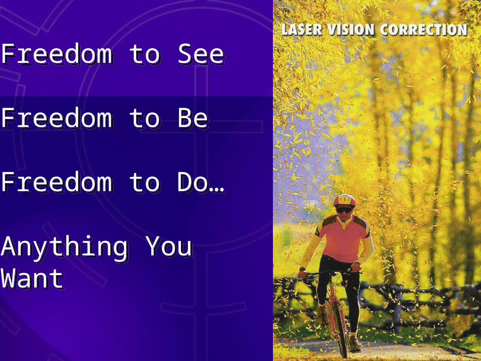
Freedom to See
Freedom to Be
Freedom to Do…
Anything You Want
Freedom to See
Freedom to Be
Freedom to Do…
Anything You Want

• VISX® Star S4 Excimer Laser System Features
• Bausch & Lomb Orbscan
• Lasik Procedure
• Procedure Risks and Benefits
• Surgeon Experience
• Questions
• VISX® Star S4 Excimer Laser System Features
• Bausch & Lomb Orbscan
• Lasik Procedure
• Procedure Risks and Benefits
• Surgeon Experience
• Questions
Discussion TopicsDiscussion Topics

VISX Leadership VISX Leadership • 1983 Stephen Trokel, MD invents laser
vision correction
• 1987 VISX system used to treat 1st human eye
• 1995 VISX StarTM launched
• 1998 VISX Star S2TM launched
• 1999 VISX CAPTM Method launched &–Millionth VISX procedure performed in the United States
• 2001 –VISX WaveScanTM WaveFront System–VISX STAR S3TM
• 1983 Stephen Trokel, MD invents laser vision correction
• 1987 VISX system used to treat 1st human eye
• 1995 VISX StarTM launched
• 1998 VISX Star S2TM launched
• 1999 VISX CAPTM Method launched &–Millionth VISX procedure performed in the United States
• 2001 –VISX WaveScanTM WaveFront System–VISX STAR S3TM
VISX® New TechnologyVISX® New Technology
• Variable Sized Beams (VSS™)
• STAR S4 ActiveTrak™ System
• Larger ablation zones
• Variable Sized Beams (VSS™)
• STAR S4 ActiveTrak™ System
• Larger ablation zones

• Variable Sized Beams (VSS™)– Variable offset scanning beam sizes and
shapes
• Active 3D eye tracker– Tracks entire natural pupil– No dilation required– Tracks eye in X,Y, and Z planes– No pre-imaging required
• Larger Ablation Zones– Reduced Incidence of night vision problems
• Variable Sized Beams (VSS™)– Variable offset scanning beam sizes and
shapes
• Active 3D eye tracker– Tracks entire natural pupil– No dilation required– Tracks eye in X,Y, and Z planes– No pre-imaging required
• Larger Ablation Zones– Reduced Incidence of night vision problems
VISX STAR S4 ActiveTrakTM
key featuresVISX STAR S4 ActiveTrakTM
key features

Beam diameter increases as treatment progresses
Variable Sized BeamsVariable Sized Beams
• Myopic treatments use variable size beam– Treatment starts at 0.65 mm– Treatment finishes at up to 6.5 mm
• Myopic treatments use variable size beam– Treatment starts at 0.65 mm– Treatment finishes at up to 6.5 mm
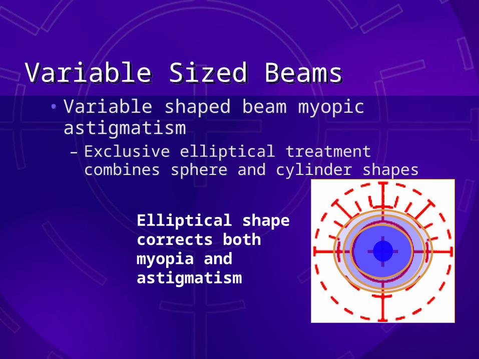
• Variable shaped beam myopic astigmatism– Exclusive elliptical treatment combines
sphere and cylinder shapes
• Variable shaped beam myopic astigmatism– Exclusive elliptical treatment combines
sphere and cylinder shapes
Elliptical shape corrects both myopia and astigmatism
Variable Sized BeamsVariable Sized Beams

Beam scans in periphery to steepen central cornea
Variable Sized BeamsVariable Sized Beams• Offset beam with scanning capabilities
• Hyperopic treatments use offset scanning– Offset rectangular beam scans across the
cornea
• Offset beam with scanning capabilities
• Hyperopic treatments use offset scanning– Offset rectangular beam scans across the
cornea
Variable Sized BeamsVariable Sized Beams• More treatment options than any other
laser
• Variable beam size– Beam changes shape and size to address all
refractive errors
• Scanning capabilities– SmartBeam™ scans across the cornea with
hyperopia and larger ablation zones
• Personalized treatments– Capable of performing individualized
treatments
• More treatment options than any other laser
• Variable beam size– Beam changes shape and size to address all
refractive errors
• Scanning capabilities– SmartBeam™ scans across the cornea with
hyperopia and larger ablation zones
• Personalized treatments– Capable of performing individualized
treatments

VISX STAR S4 ActiveTrak™ Eye Tracker features and benefits
VISX STAR S4 ActiveTrak™ Eye Tracker features and benefits
• Active eye tracker – Moves beam to compensate for all eye
movements– 3D infrared eye tracker– Dual side-mounted infrared cameras monitor
x, y, and z
• No dilation required– Patient comfort and convenience– Maintains LASIK “wow factor”
• Active eye tracker – Moves beam to compensate for all eye
movements– 3D infrared eye tracker– Dual side-mounted infrared cameras monitor
x, y, and z
• No dilation required– Patient comfort and convenience– Maintains LASIK “wow factor”

VISX STAR S4 ActiveTrak™ Eye Tracker features and benefits
VISX STAR S4 ActiveTrak™ Eye Tracker features and benefits
• Flexible, responsive eye tracking– Assures eye remains in optimal 3D window
• No pre-imaging required– Direct measurement of pupil position– Fast patient throughput
• Doctor controls eye tracker function– Doctor engages tracker, controlling centration
and alignment
• Flexible, responsive eye tracking– Assures eye remains in optimal 3D window
• No pre-imaging required– Direct measurement of pupil position– Fast patient throughput
• Doctor controls eye tracker function– Doctor engages tracker, controlling centration
and alignment

VISX STAR S4 ActiveTrak™ Eye TrackerVISX STAR S4 ActiveTrak™ Eye Tracker
• Dual side-mounted, infrared cameras monitor x, y, and z movements
• Oblique IR lighting does not interfere with procedure
• Dual side-mounted, infrared cameras monitor x, y, and z movements
• Oblique IR lighting does not interfere with procedure
VISX STAR S4 ActiveTrak™ Eye TrackerVISX STAR S4 ActiveTrak™ Eye Tracker
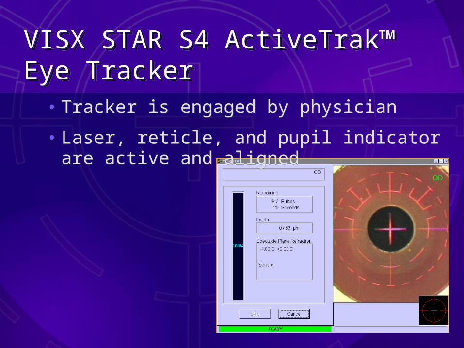
• Tracker is engaged by physician
• Laser, reticle, and pupil indicator are active and aligned
• Tracker is engaged by physician
• Laser, reticle, and pupil indicator are active and aligned
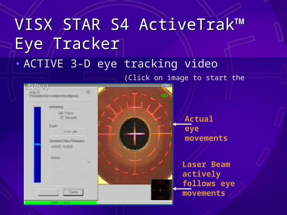
Laser Beam actively follows eye movements
Actual eye movements
VISX STAR S4 ActiveTrak™ Eye TrackerVISX STAR S4 ActiveTrak™ Eye Tracker• ACTIVE 3-D eye tracking video
(Click on image to start the video)
• ACTIVE 3-D eye tracking video (Click on image to start the video)
† Data on file, VISX, Inc.
VISX STAR S4 ActiveTrak™ Eye TrackerVISX STAR S4 ActiveTrak™ Eye Tracker
• Eye movements during LASIK– ActiveTrak™ tracks at 60 Hz– 99.4% of all eye movements can be captured
with a 20 Hz tracker†– Eye position is verified and safety checked
before every laser pulse– Continues to monitor between pulses– Checks eye position 6 times for every pulse
delivered to the cornea
• Eye movements during LASIK– ActiveTrak™ tracks at 60 Hz– 99.4% of all eye movements can be captured
with a 20 Hz tracker†– Eye position is verified and safety checked
before every laser pulse– Continues to monitor between pulses– Checks eye position 6 times for every pulse
delivered to the cornea
VISX STAR S4 ActiveTrak™ Technical ComparisonVISX STAR S4 ActiveTrak™ Technical Comparison
• VISX ® ActiveTrak™ 3D eye tracker – Developed for laser vision correction – No dilation– Tracks X, Y, and Z axis movements
• LadarVision®
– Developed for non-ophthalmic applications– Requires dilation to minimum 7mm– Limited range of motion (no z-axis)
• VISX ® ActiveTrak™ 3D eye tracker – Developed for laser vision correction – No dilation– Tracks X, Y, and Z axis movements
• LadarVision®
– Developed for non-ophthalmic applications– Requires dilation to minimum 7mm– Limited range of motion (no z-axis)

VISX STAR S4 ActiveTrak™ Technical ComparisonVISX STAR S4 ActiveTrak™ Technical Comparison
• VISX ® ActiveTrak™: DIRECT measurement of eye position in real time– Tracks pupil in its natural position– Direct measurement increases system
accuracy
• LadarVision® tracker: Requires pre-imaging for indirect measurement of eye position– Pre-imaging is a source of error– Extrapolates eye position from only 4 points,
based on where the pupil center WAS when pre-imaging, not where it is during the actual surgery

Radar Tracker-only 4 data points used
3-D Infrared Video Tracker- thousands of data points used
Eye Tracker Centration Technical ComparisonEye Tracker Centration Technical Comparison
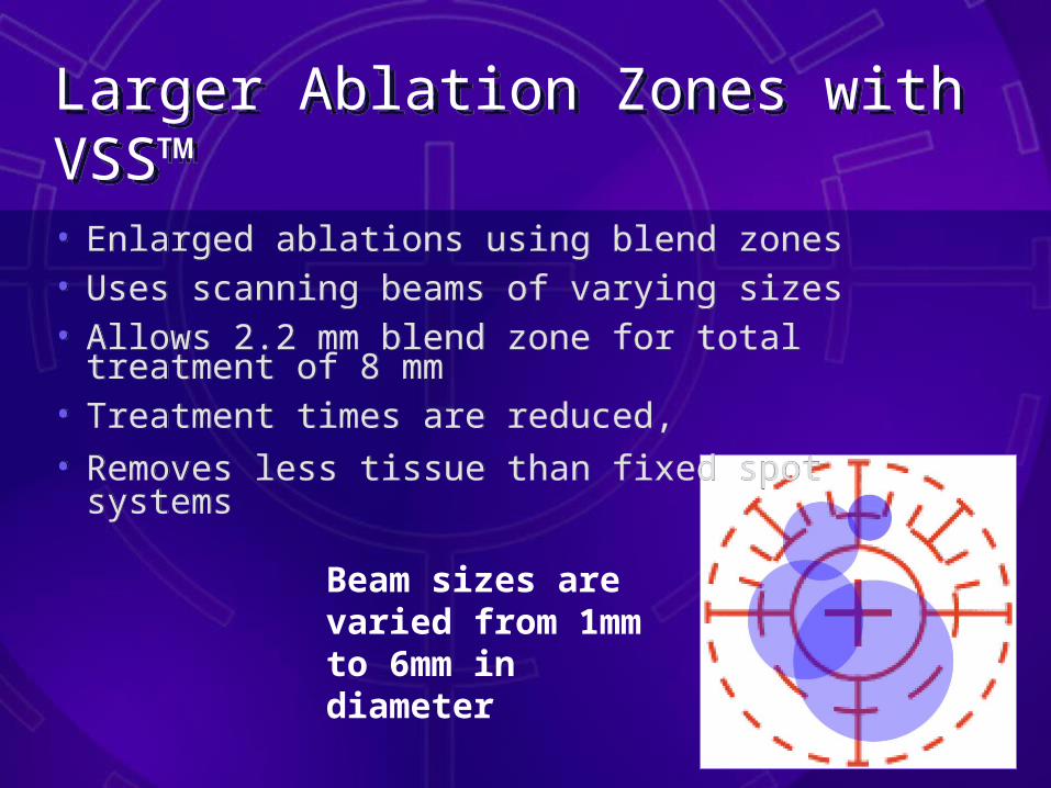
Beam sizes are varied from 1mm to 6mm in diameter
Larger Ablation Zones with VSS™ Larger Ablation Zones with VSS™ • Enlarged ablations using blend zones • Uses scanning beams of varying sizes • Allows 2.2 mm blend zone for total treatment of
8 mm• Treatment times are reduced, • Removes less tissue than fixed spot systems
• Enlarged ablations using blend zones • Uses scanning beams of varying sizes • Allows 2.2 mm blend zone for total treatment of
8 mm• Treatment times are reduced, • Removes less tissue than fixed spot systems
Larger Ablation Zones With VSSLarger Ablation Zones With VSS
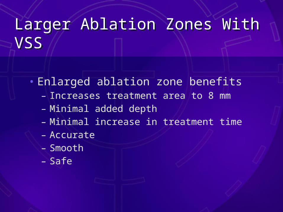
• Enlarged ablation zone benefits– Increases treatment area to 8 mm– Minimal added depth– Minimal increase in treatment time– Accurate– Smooth– Safe
• Enlarged ablation zone benefits– Increases treatment area to 8 mm– Minimal added depth– Minimal increase in treatment time– Accurate– Smooth– Safe

Larger Ablation Zones With VSSLarger Ablation Zones With VSS
• Advantages of Blend Zone– Decreases the angle of ablation edge– May reduce incidence of night vision
problems– May help LASIK flap to lay smoother
and fit better in the bed
• Advantages of Blend Zone– Decreases the angle of ablation edge– May reduce incidence of night vision
problems– May help LASIK flap to lay smoother
and fit better in the bed
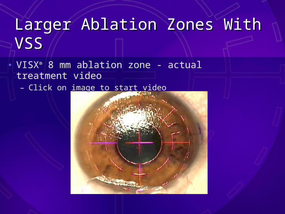
Larger Ablation Zones With VSSLarger Ablation Zones With VSS• VISX® 8 mm ablation zone - actual treatment
video– Click on image to start video
• VISX® 8 mm ablation zone - actual treatment video– Click on image to start video

Larger Ablation Zones With VSSLarger Ablation Zones With VSS
Conclusion
• Larger ablation zones using VSS™ offers a new and unique approach to laser vision correction – Advantages of quicker procedure times and
less tissue removal– Ability to extend ablation zone to 8 mm– Is an accurate and safe treatment
Conclusion
• Larger ablation zones using VSS™ offers a new and unique approach to laser vision correction – Advantages of quicker procedure times and
less tissue removal– Ability to extend ablation zone to 8 mm– Is an accurate and safe treatment

Bausch & Lomb OrbScan™ SystemBausch & Lomb OrbScan™ System
• OrbScanTM
– Patient’s AcuityMap™, a unique “fingerprint” of the eye
» Anterior Float map (left) » Posterior Float map (right)
• OrbScanTM
– Patient’s AcuityMap™, a unique “fingerprint” of the eye
» Anterior Float map (left) » Posterior Float map (right)
OrbScan™ SystemOrbScan™ System
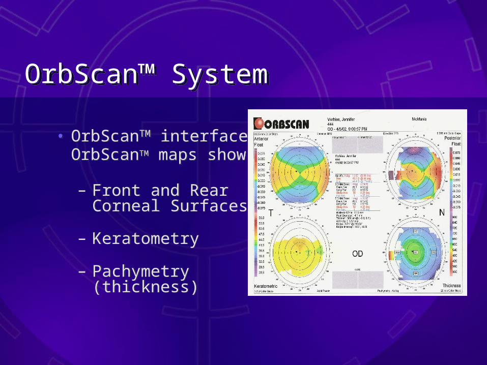
• OrbScanTM interface: OrbScanTM maps show:
– Front and Rear Corneal Surfaces
– Keratometry
– Pachymetry (thickness)
• OrbScanTM interface: OrbScanTM maps show:
– Front and Rear Corneal Surfaces
– Keratometry
– Pachymetry (thickness)

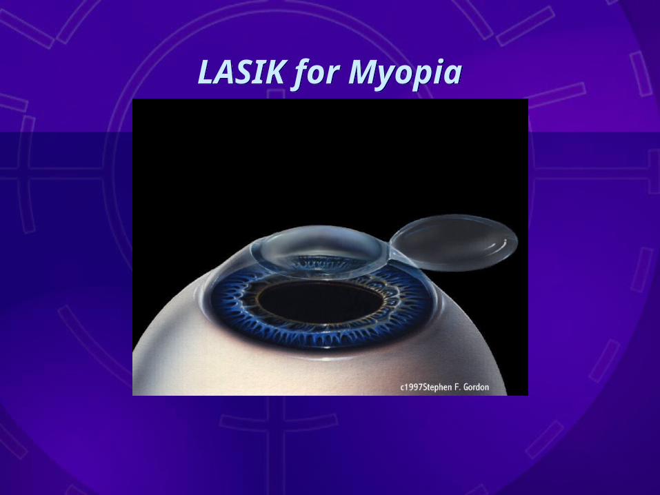
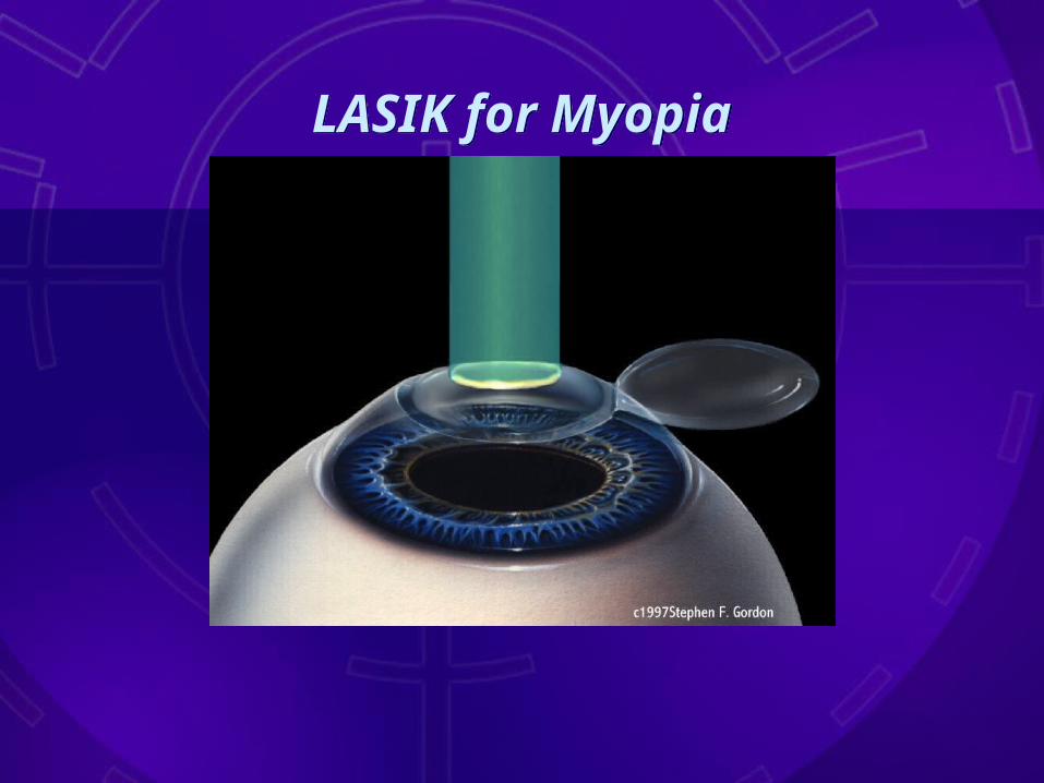
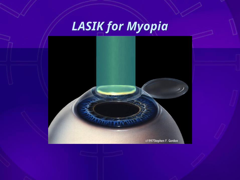
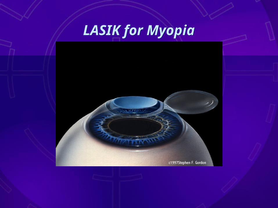
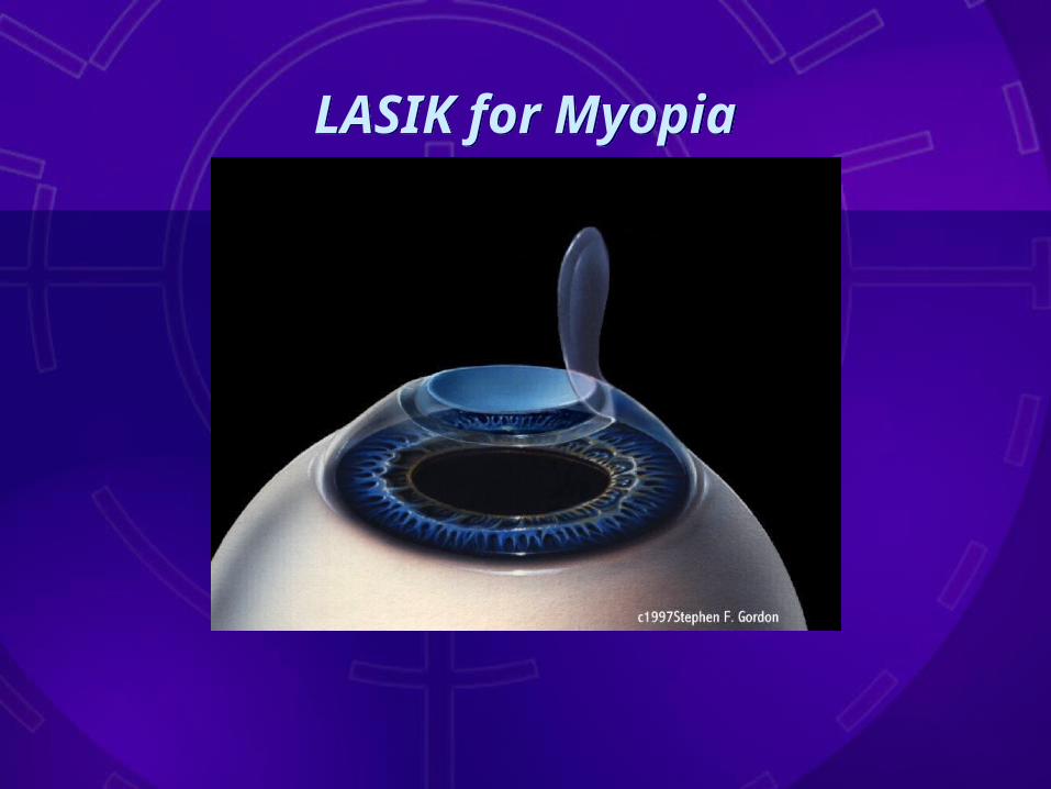
LASIK for MyopiaLASIK for Myopia

LASIK for MyopiaLASIK for Myopia
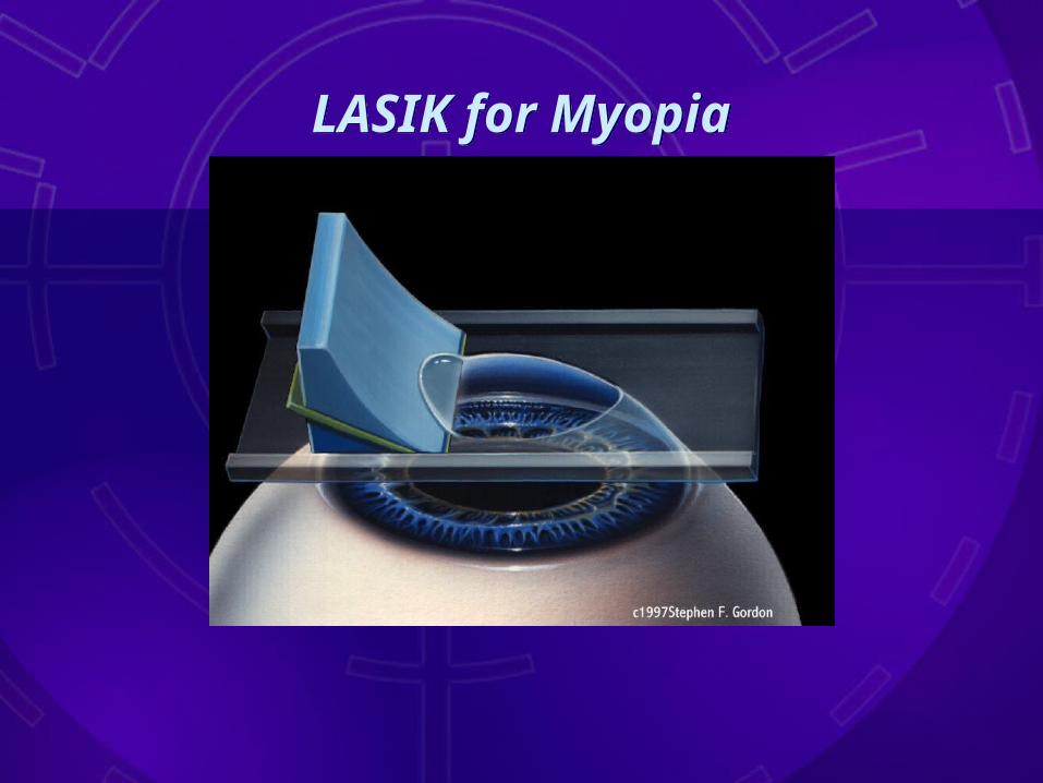
LASIK for MyopiaLASIK for Myopia
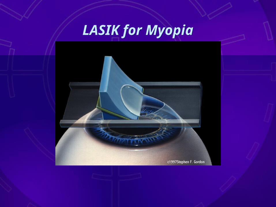
LASIK for MyopiaLASIK for Myopia

LASIK for MyopiaLASIK for Myopia
LASIK for MyopiaLASIK for Myopia

LASIK for MyopiaLASIK for Myopia
LASIK for MyopiaLASIK for Myopia
LASIK for MyopiaLASIK for Myopia
LASIK for MyopiaLASIK for Myopia

LASIK for MyopiaLASIK for Myopia
LASIK for MyopiaLASIK for Myopia

LASIK for MyopiaLASIK for Myopia

LASIK for MyopiaLASIK for Myopia

LASIK for MyopiaLASIK for Myopia
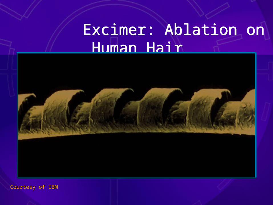
Excimer: Ablation on Human Hair
Excimer: Ablation on Human Hair
Courtesy of IBMCourtesy of IBM

Treatment ParametersTreatment Parameters
• -0.25 to -14.00 D with up to -4.00 D cylinder
• +0.00 to +6.00 with less than -4.00 D cylinder
• Generally 18 years of age or older
• -0.25 to -14.00 D with up to -4.00 D cylinder
• +0.00 to +6.00 with less than -4.00 D cylinder
• Generally 18 years of age or older
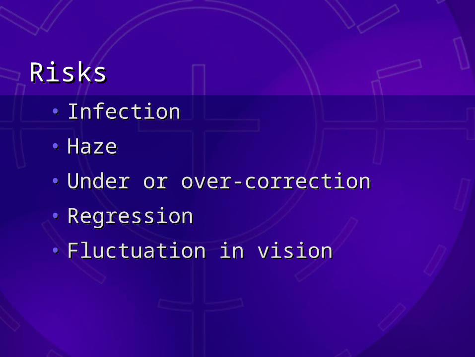
RisksRisks• Infection
• Haze
• Under or over-correction
• Regression
• Fluctuation in vision
• Infection
• Haze
• Under or over-correction
• Regression
• Fluctuation in vision

BenefitsBenefits
• Lifestyle enhanced
• Career opportunities
• Reduced dependence on contacts and glasses
• Lifestyle enhanced
• Career opportunities
• Reduced dependence on contacts and glasses

Consultation AppointmentConsultation Appointment
• No charge- 30-45 minutes appointment
• Connie will counsel patients about lvc and treatment options
• Basic eye measurements taken- auto refraction & lensometry
• Educate about Freedom Laser Center and benefits of lvc
• No charge- 30-45 minutes appointment
• Connie will counsel patients about lvc and treatment options
• Basic eye measurements taken- auto refraction & lensometry
• Educate about Freedom Laser Center and benefits of lvc

Laurence Miller, D.O.Laurence Miller, D.O.
• Associate Professor Department of Surgery, Kirksville College of Osteopathic Medicine
• Member of the American Academy of Ophthalmology
• Member American Osteopathic Association
• Fellowship in the Osteopathic College of Ophthalmology and Otochirolarynogology
• Performed over 10,000 refractive surgery procedures

Questions?Questions?

Thank YouThank You
for your for your AttentionAttention