frog atlas
TRANSCRIPT
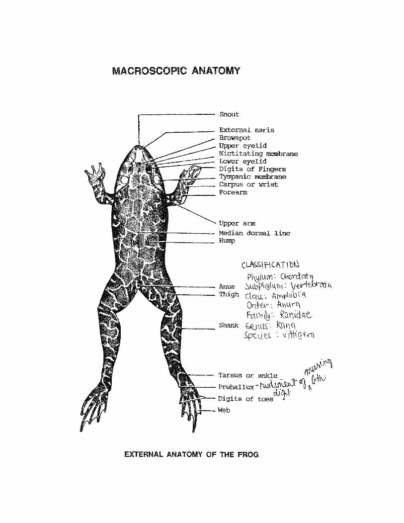
MACROSCOPIC ANATOMY
r-------- Snout
----- External naris Brows pot Upper eyelid Nictitating membrane
~)f~~~~=:::::::::::::=;E;::: Lower eyelid • Digits of Fingers
..J.---"'1~-:S:r-- Tympanic membrane . ....... ,~........... Carpus or wrist
~'l.ldOr--- Forearm
Upper arm r------ Median dorsal line
...------ Hump
(~\ fl Cf\ TID~~ t>h~·\L\W\ ·• (M()n:\Wq .
~N.ir-------- Anus ~l-~b~l,~lu,\\'1: 'v~Arftb~t.n4 WJirfi1il miT---- Thigh C\ q ~ ·, -A IV\"\ \ID ,-G\
Olrcl-tv- ·. A\\ l-\ y-r;\ Fctf,,,b·· Ru\'\idc\t
r---- Shank G(Jil'S. ·. 1\91 t1 C\ ~pt. d \: ~ '. v lll,. g ~.rt\
.. .~.~~ ,W\).tJf!.
- · Tarsus or ankle _ . ,.. :" M·)v oF.~::--- Prehallux- !"V,A(lu:\il:l ~ f\
c\Ji:h\-~.!:,;:::>-- Digits of toes J
Web
EXTERNAL ANATOMY OF THE FROG

~--------Darkly pigmented
A MALE FROG Ventral view
A FEMALE FROG Ventral view
are~ of the skin
SwoJ._J .. en thumb
Diffused skin pigmentation
•-~~--Thumb not swollen

Ischium
SKELETAL SYSTEM Dorsal View
Met atarsals
Astragalus]Tarsals Calcaneum
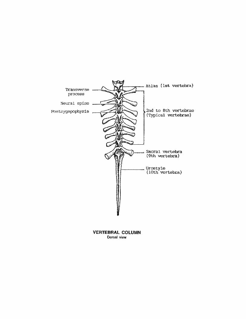
Transverse process
Neural spine
Postzygapophysis
VERTEBRAL COLUMN Dorsal view
2nd to 8th vertebrae (Typical vertebrae)
Sacral vertebra (9th vertebra)

Concavities for articulation lvi th sacral vertebra
ISOLATED VERTEBRAE
ATLAS Antero-dorsal view
Neural spine Neural arch Postzygapophysis
Neural canal Concavity for articulation with occipital condyle Centrun
.--------Neural spine
~-----Postzygapophysis
Transverse process ~~~~-------Prezygapophysis
....._ ___ Neural canal ~~-------Centrum
TYPICAL VETEBRA Antero-dorsal view
SACRAL VERTEBRA Postern-dorsal view
Neural canal
UROSTYLE Antero-latera! view
centrum
of the lOth
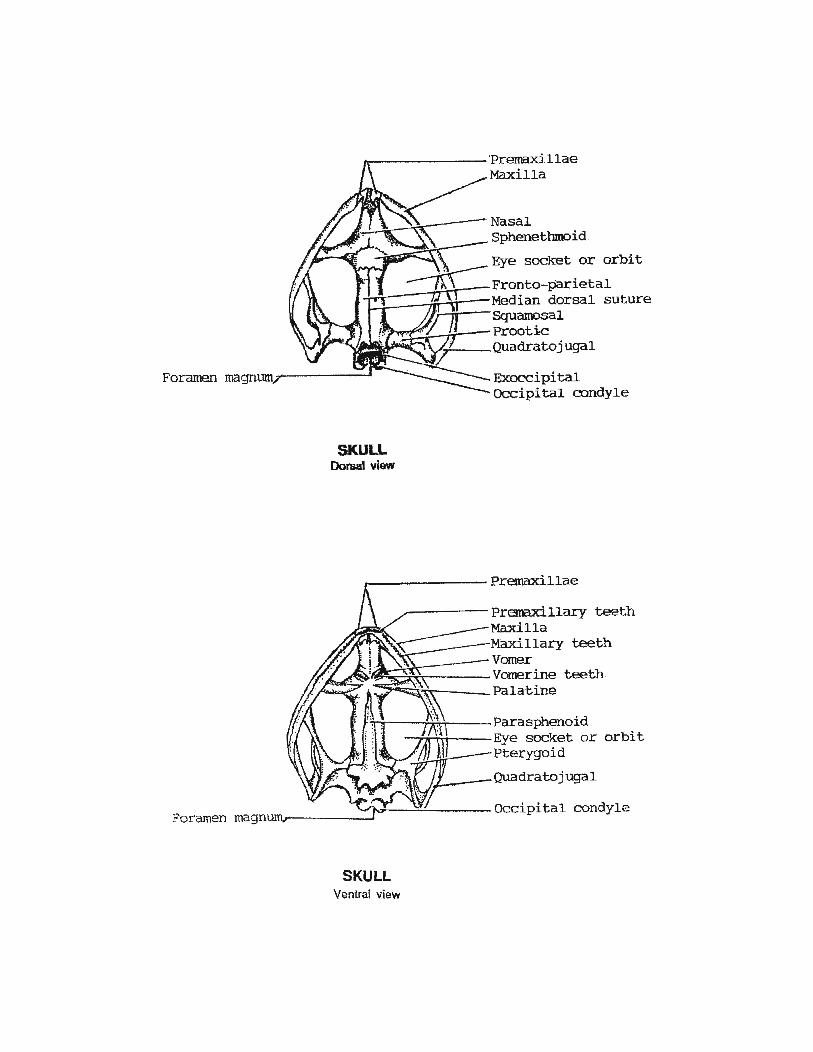
SKULL Dorsal view
Exoccipital Occipital condyle
~------------Premaxillae
orbit
SKULL Ventral view
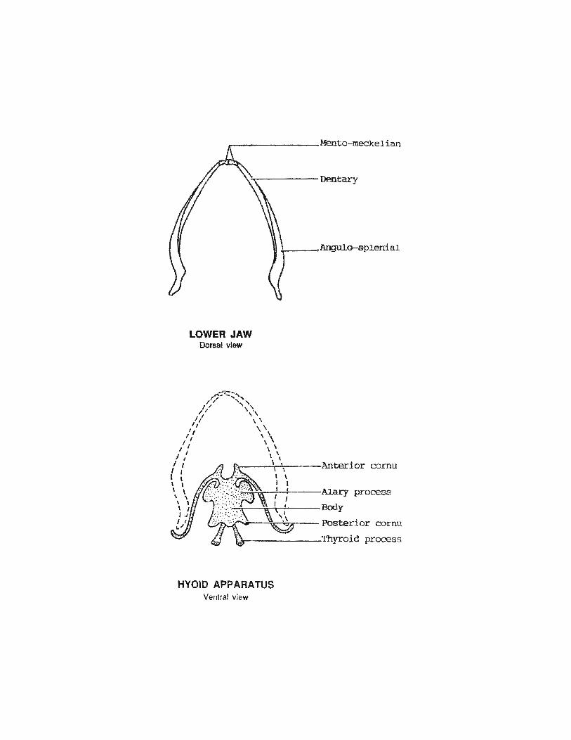
LOWER JAW Dorsal view
HYOID APPARATUS Ventral view
Anterior cornu
Alary process
Body
Posterior cor-nu
Thyroid process
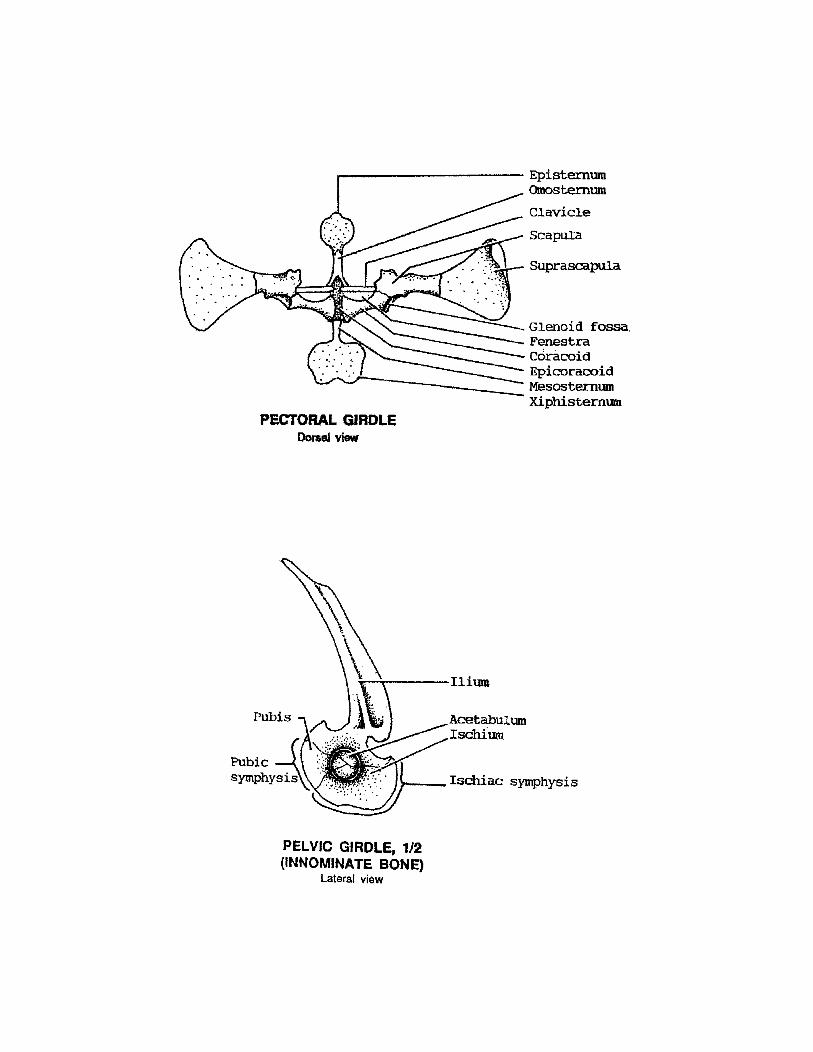
---------------------- Episternum Omosternum
Clavicle
Scapula
Suprascapula
Glenoid fossa. Fenestra coracoid Epicoracoid r-------.::..::: Mesosternum
PECTORAL GIRDLE Dorsal view
PELVIC GIRDLE, 1/2 (INNOMINATE BONE)
Lateral view
Xiphistermn
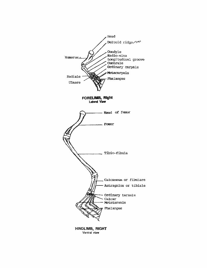
Head
Deltoid ridge/'~·
Condyle Radio-ulna Longitudinal groove Centrale Ordinary carpals
Metacarpals Radiale -"""'o'!;I'J
!l:i(tl.==- Phalanges Ulnare
FOREUMB, R1ght Lataral Vtt1111
Tibio-fibula
~-Calcaneum or fib111are
Astragalus or tibiale
~--~-- Ordinary tarsals Calcar
AA~..,.,'Hf-- Metatarsals
'~~~~~~Phalanges
HINDLIMB, RIGHT Ventral view

~~~------~-rr-----Temporalis
~~:.------nm-;.;y--- Depressor mandibula
~~~~~~~~~~~~==Dorsal1s scapula ~ ~rj;;,r--Cucullaris
~\~~?// Latissimus dorsi IH/IJ/II~r-...;::~=------- External oblique
m.mmr------------Longiss imus dorsi llfi~~-----------Ilio-lumbaris
~~~~~----------Coccygeo-sacralis
~~~~i---==========Coccygeo-iliacus ~ femoris anticus m~~~~wBwm~~-----------Gluteus
:f'(U~~~ ~::m.::rn.------------ Vast us externus \~---------Triceps femoris
Semimembranosus n~~~~---
Gracilis minor
fi--------Tibialis anticus
J:..!!.-.!.----Tendon of Achilles
MUSCULAR SYSTEM Dorsal view

cutaneous~~~~tfl\1 pectoralis
Suanentalis
is 0 tC r.:.(·s Scapulo-humeralis or deltoid
s epicoracofaaa-- -(Anterior pectoralis)
--'"----Pectoralis sternalis AU~--------PeCtoralis ~is
(Posterior pectoralis) Linea alba----~~~~\1:~~~------~Rectus abdominis
Inscriptiones tendinae
Sartori
mag nus
Gracil major
Gracili minor Tibia-. ---;;.;
fibula
vn:;b£~ .·~~i" n;(~~ tt,_,ie;r ~.(.i d<i" ;~ {(I~
~~~~-Semitendinosus Gracilis major (~t) (Rect"US int,ernus major")
L-ww~~-- ·Gracilis minor ) (Rectus internus minor
MUSCULAR qysTEM, ·Ventral view
Extensor cruris anticus t~~'(J\ t~·.
Flexor tarsi anterior
lib;ttli~ lU~\1 t.tl .. '.. l.JC ~;~ -- 1.:~\lCkr T-11 \l' ''~Ji·~.
(The left side has been dissected to show the deeper muscles)

~-------------------- Median subrostral fossa pulvinar rostrale
Lateral subrostral fossa Internal naris or c"hoana1
'---------~per lip fold ~~~~~---------- Vomerine teeth
~~~--------- Sulcus marginalis ~~~--------- Maxillary teeth
-~;,;:;iii;;---- Orbital prominence. Opening into the Esophagus Eustachian tube "fl{·.(.tf·~ -
Laryngeal ·prominence
~---------------------Tuberculum prelinguale
BUCCAL CAVnY OF A MALE FROG
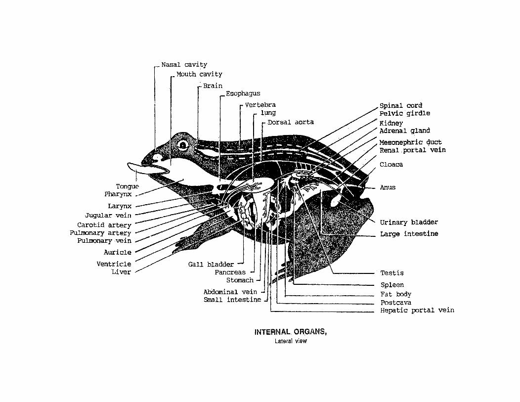
Jugular .vein Carotid artery
Pulmonary artery Pulmonary vein
Nasal cavity Mouth cavity
· Brain Esophagus
Abdominal vein Small intestine
INTERNAL. ORGANS, Lateral view
Spinal cord Pelvic .girdle Kidney Adrenal gland
Mesonephric quct Renal .portal vein
Cloaca
Anus
Urinary bladder
Large intestine
"-"---- · Testis
Spleen Fat body Postcava Hepatic portal vein

...-----419.~- $kin ---~~;--Dorsal subcutaneous
lymph space Epaxial muscles orizontal skeletogenous
septum ·~~~~--Trunk vertebra
Kidney isterna magna
or subvertebral lymph sinus
Gonadl=Jt~~::::::::==~~ Dorsal-mesentery Stomach -~~:r.--rrt
Omentum ____ ~~~~~-J Duodenum _ _:~~;.::?o:;:c--
Linea alba------~~;:~~~~~;;~ Ventro-lateral subcutaneous connective tissue
Ventral subcutaneous lymph space Ventral abdominal ve:i.n
BODY CAVITY
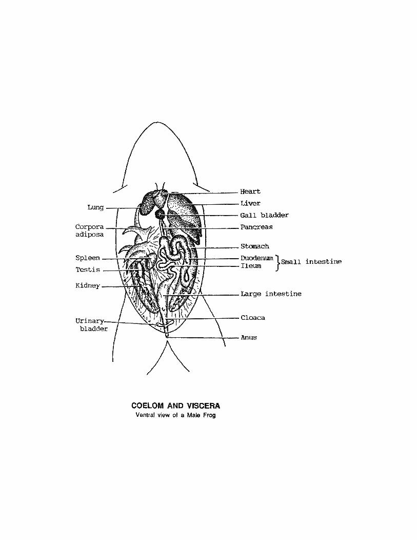
Lung
Corpora adiposa
Spleen
Testis
Kidney
COELOM AND VISCERA Ventral view of a Male Frog
Heart
Liver
Gall bladder
Pancreas
Stanach
Duodenum }Small Ileum
intestine
Large intestine
Cloaca
Anus

Duodenum~--~~~~~
Ileum __ ~------~·~
Mesentery proper or Mesenterium
DIGESTIVE SYSTEM Ventral View
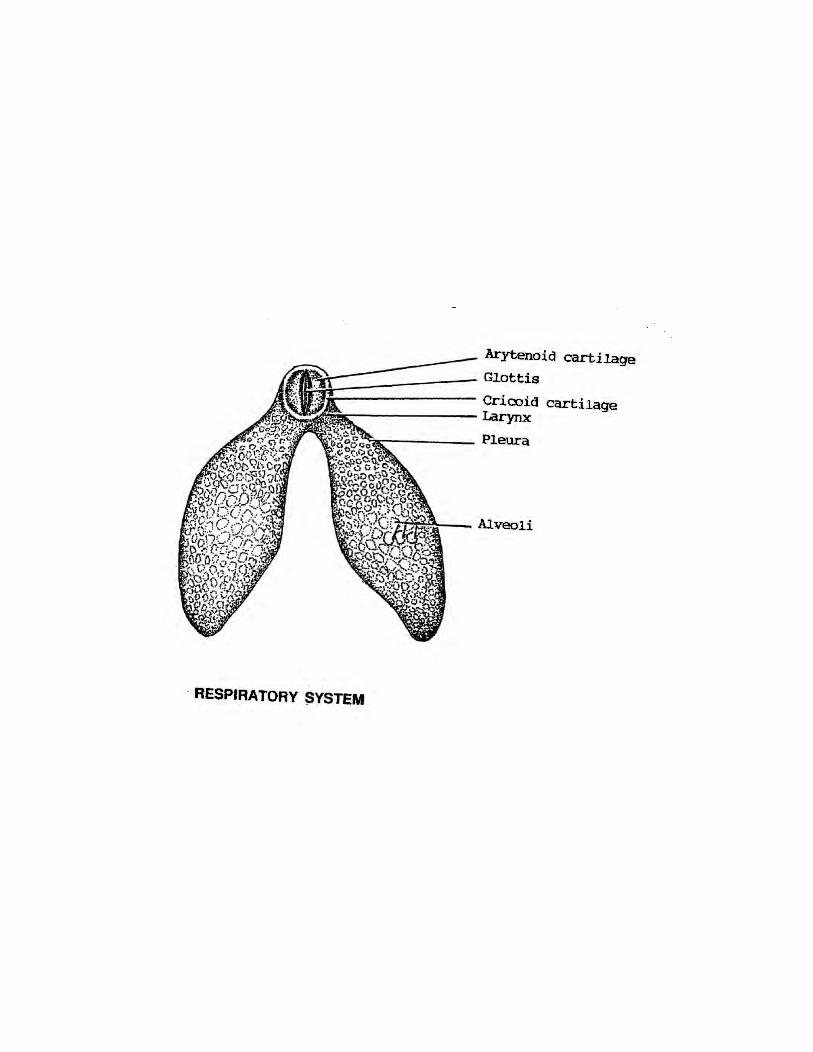
Dr~~------------- Cricoid cartilage ;,_~~~~~----- Laryruc
· RESPIRATORY SYSTEM

Entrance of -~--~----~~ Wolffian duct
MALE UROGENITAL SYSTEM Ventral view
Fat body or Corpora adiposa
Postcava
Testis
Vas efferens
Adrenal gland Kidney
Renal vein Mesonephric duct or Wolffian duct Vestigial oviduct
Entrance of oviduct
Urinary bladder Cloaca
Anus

.~~~r----- Fat body or corpora adiposa
+--1-~~U...---\--- Postcava
~~--~----Kidney
~-~---OViduct
ovary_.,____, with eggs
fri~;.---...._ __ Mesonephric duct or Wolffian duct
of oviduct
~-,~----~---urinary bladder
4--T~----- Cloaca ..__....,&.j~
~--------------Anus
FEMALE UROGENITAL SYSTEM Ventral view
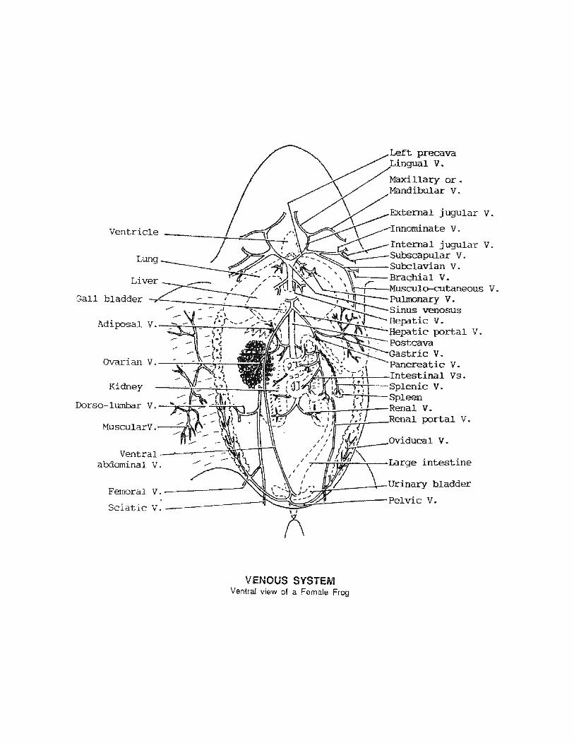
Ventricle
Lung
Liver
Gall bladder
Kidney
Ventra1--~~~~~~r-lr~ abdominal V.
Pulmonary V. Sinus venosus Hepatic v. Bepatic portal V. Postcava
v. v.
v.
v.
intestine
Femoral v.-------------Sciatic V.-----------------,
~~~----~-Urinary bladder
~~~~~---------Pelvic V.
VENOUS SYSTEM Ventral view of a Female Frog
v.
v.

Truncus arteriosus-------
Common carotid A---------------~
carotid A.
Systemic arch __ _
Pulmo-cutaneous A.
Pulmonary A. ____ _ ---=..:...._
Right -lung ----~ Conus arteriosus -----~----~~~~
Syst~~ic A.-------~-~~~·
Coeliaco-mesenteric A.
Adi{:X)sal A.
Anterior mesenteric A.-+~~
ovari an A. ------------~~~ (spermatic ·A. in
A.
A.
A.
A.
Left gastric A.
Hepatic A.
~~~~~~Right gastric A. Pancreatic A.
Jr;'~~:!r=~L Duodenal A. the male) . Intestinal As. K1dney Renal As.-------------t~~~~~~;!.~~2=~JJ~-- Splenic A. Oviducal A. Spleen
Posterior mesenteric A.~~~~--~~•
Right common iliac A---~~~~~ ,...._r:til Epigastric A.~-----~--~~~~~
A.
---~o-=-------- Sciatic A. Epigastrico-vesical
~ecto-vesical A. --------------~
ARTERIAL SYSTEM Ventral view of a Female Frog
A.
A.

Right
arteriosus
HEART Dorsal view
HEART Ventral view
Truncus arteriosus
Right Precava
Pulloonary veins Right auricle Sinus venosus Coronary sulcus
Postcava
Ventricle
Truncus arteriosus
Left precava
Left auricle
Coronary sulcus
Postcava
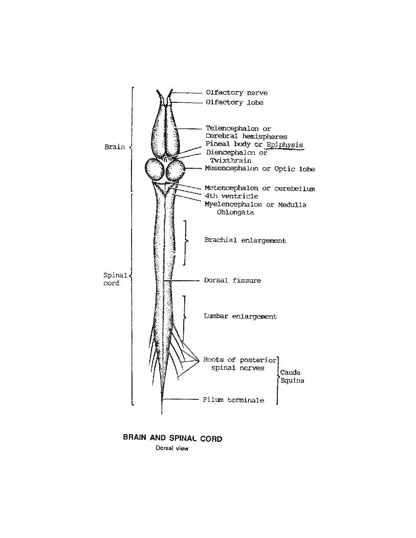
Brain
Spinal cord
.,
.:·: ....
~~. 0
·' . ,.
0
·.· ·.
Olfactory nerve r---- Olfactory lobe
or Optic lobe
or cerebellum
-.~ . or Medulla . ;.';
"·' ·;· •: .. '.: .. Brachial enlargement .. , ·.::
•. Dor sal fissure :: :-:
Lumbar enlargement
~--~ Roots of posterior spi nal nerves
Fi lum tenninale
Cauda Equina
BRAIN AND SPINAL CORD Dorsal view
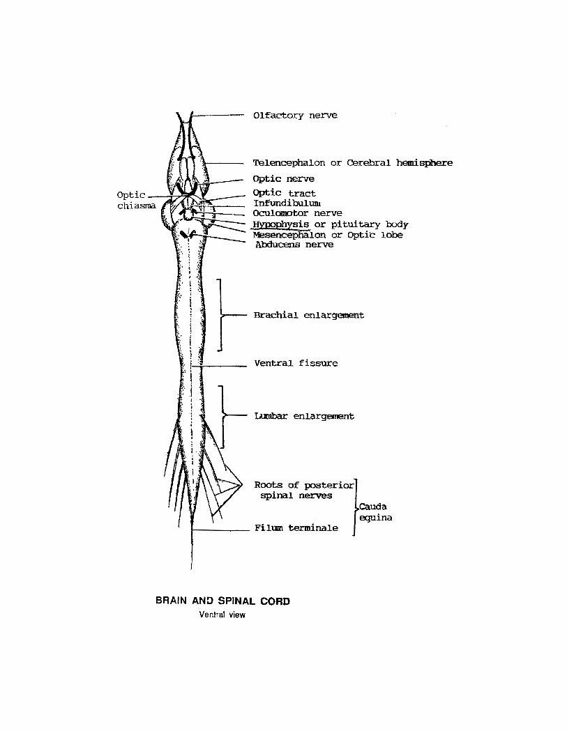
Optic --::ir7'"'"'t.~chiasma
. . . ~ · . . . ; , .
.. ' \
;-
I • t •\' \ ' \ .
I }
·~
.• r-i -;.....----.·. i .. ;· .i
Olfactory nerve
Telencephalon or Cerebral hemisphere
Optic nerve
Optic tract Infundibulum Oculomotor nerve ,Hypophysis or pituitary body Mesencephalon or Optk lobe Abducens nerve
BraChial enlargement
Ventral fissure
L\.Dbar enlargement
Roots of posterior spinal nerves
cauda equina
BRAIN AND SPINAL CORD Ventral view

Metenceph_alon
Olfactory nerve ( 1)
Olfactory lobe
Telencephalon
Pineal body or Epiph_ysis
Diencephalon
Mesencephalon
Trochlear nerve (4) Trigeminal nerve (5) Facial nerve {7) Auditory nerve (8) Glossopharyngeal nerve ( 9) Vagus nerve (1 0)
BRAIN AND CRANIAL NERVES Dorsal view
Spinal
Olfactory nerve (1)
Telencephalon
Optic nerve (2) Optic tract Infundibulum Oculomotor nerve ( 3)
Trigeminal nerve (5) Facial nerve ( 7) Auditory nerve (8) Glossopharyngeal nerve {9) Vagus nerve (10)
BRAIN AND CRANIAL NERVES Ventral view

'~---------- Olfactory nerve J---------- Olfactory lobe
lst and 2nd ventricles or Lateral ventricles
~------~ Telencephalon
Foramen of Monro
3rd ventricle
or
CAVITIES OF THE BRAIN AND SPINAL CORD
Iter