from 'junk' to just unexplored noncoding knowledge: the case of
TRANSCRIPT

From ‘JUNK’ to Just UnexploredNoncoding Knowledge: the caseof transcribed AlusRajesh Pandey and Mitali Mukerji
AbstractNon-coding RNAs (ncRNAs) are increasingly being implicated in diverse functional roles. Majority of these ncRNAshave their origin in the repetitive elements of genome. Significantly, increase in genomic complexity has beencorrelated with increase in repetitive content of the genome. Primate-specific Alu repeats, belonging to SINE classof repeats, is the most abundant repeat class inhabiting the human genome. Of the many possible functional rolesof Alu repeats, they have been shown to modulate human transcriptome by virtue of harboring diverse array offunctional RNA pol II TFBS, cryptic splice-site-mediated Alu exonization and as probable miRNA targets.Retro-transposition of Alu harboring TFBS has shaped up gene-specific regulatory networks. Alu exonized tran-scripts are raw material for dsRNA-mediated A^I editing leading to nuclear retention of transcripts and changein miRNA target. miRNA targets within Alu may titrate the effective miRNA or transcript concentration,thus acting as ‘miRNA sponge’. Differential levels of Alu RNA during different conditions of stress also await clearfunctional understanding. These have contributed toward evolution of complex regulatory repertoire leading tothe evolution of primate-specific functions. Recent reports of co-localization of pol II and pol III binding sites nearthe gene and elsewhere in the genome, increase the possibility of dynamic co-ordination between both pol II andpol III determining the ultimate transcriptional outcome. Dynamic and functional Alu repeats seem to be centrallyplaced to modulate the transcriptional landscape of human genome.
Keywords: non-coding RNA; Alu repeat; TFBS; NAT; miRNA; compartmentalization
Non-coding DNA and GenomeComplexityThe proponents of molecular biology have always
gone to great lengths to emphasize on what we
know today as the Central Dogma. It precisely
holds that for any biological expression to occur at
the genotypic or phenotypic level, a gene must tran-
scribe into an mRNA and this must then go on to
make a protein. This has usually been interpreted
to mean that genetic information flows from DNA
to proteins via RNA, where RNA is a mere inter-
mediate in the information flow cascade. However,
only 2% of our genes functionally code for proteins;
�50–75% of human genome is transcribed, and 98%
of the transcripts do not get translated [1, 2]. Further,
genomic complexity neither correlates with chromo-
some number (popularly known as K value paradox),
genome size (C value paradox) or the number of
genes (N value paradox) [3]. This was confirmed
after human genome sequencing which revealed
that humans have only approximately 21 500
protein-coding genes which are comparable to
lesser complex genomes of Drosophila and
Arabidopsis [4]. In contrast, non-coding component
RajeshPandey During my PhD from CSIR-IGIB, I explored the functional significance of regulatory elements within Alu in human
transcriptome. My research interest is to understand the genome regulatory mechanism being modulated by noncoding RNAs in
general and Alu in particular.
MitaliMukerji My research is in the broad area of genome variation and its effect on human phenotypes and susceptibility to diseases.
My group is studying the role of Alu elements in evolution of novel regulatory networks through functional genomics approaches.
Corresponding authors. Mitali Mukerji, Rajesh Pandey, Genomics and Molecular medicine, Institute of Genomics and Integrative
Biology (CSIR-IGIB), Council of Scientific and Industrial Research (CSIR). Mall Road, Near Jubilee Hall, Delhi 110007, India.
Tel: þ91 11 27662738; Fax: þ91 11 27667471; E-mail: [email protected], [email protected]
BRIEFINGS IN FUNCTIONAL GENOMICS. VOL 10. NO 5. 294^311 doi:10.1093/bfgp/elr029
� The Author 2011. Published by Oxford University Press. All rights reserved. For permissions, please email: [email protected]
Downloaded from https://academic.oup.com/bfg/article-abstract/10/5/294/206990by gueston 31 March 2018
of the genome has increased substantially with
genome complexity. These regions have assumed
importance in recent times as they seem to hold a
large number of cues that might explain the observed
complexity in humans [5, 6]. This makes us sit back
and question some of our good old notions of
biology.
ncRNAs have no apparent ORF for translation,
and it include ‘housekeeping’ ncRNAs [such as trans-
fer RNAs (tRNAs), ribosomal RNAs (rRNAs) and
small nuclear RNAs (snRNAs)], small/short ncRNAs
(miRNA and piRNA), repetitive element-associated
ncRNAs (Alu RNA) and long ncRNAs (lncRNAs).
ncRNAs are transcribed by different RNA polymer-
ase complexes, e.g. Alu is transcribed by pol III
whereas miRNAs, snoRNAs, some snRNAs and
some scRNAs are mostly transcribed by pol II
[7–9]. A large class of ncRNAs like 7SL [10], 7SK
[11] and miRNAs [12] are highly conserved across
species and are being implicated in many genome
functions [13]. Although conserved cis-regulatory
elements in human genome like proximal promoter
elements, enhancers, silencers and insulators comprise
a larger fraction of the regulatory repertoire [14].
ncRNA can assume dynamic secondary structures
and affect various cellular functions like splicing, edit-
ing, silencing through antisense, methylation and nu-
clear retention of transcripts [15–17]. They can also be
a source of miRNA biogenesis and miRNA target.
These RNAs govern diverse aspects of development,
cellular differentiation and external/internal stimuli
driven spatio-temporal expression [18–22]. They are
also known to regulate transcription factors (TFs) as
co-activators or co-repressors [23–26]. Different tiers
of regulation across phyla from bacteria to humans
can be regulated through variety of ncRNAs like
cis/trans antisense RNA and transcribed pseudogene
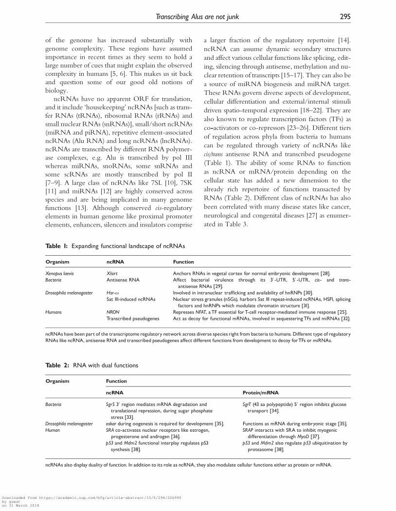
(Table 1). The ability of some RNAs to function
as ncRNA or mRNA/protein depending on the
cellular state has added a new dimension to the
already rich repertoire of functions transacted by
RNAs (Table 2). Different class of ncRNAs has also
been correlated with many disease states like cancer,
neurological and congenital diseases [27] as enumer-
ated in Table 3.
Table 1: Expanding functional landscape of ncRNAs
Organism ncRNA Function
Xenopus laevis Xlsirt Anchors RNAs in vegetal cortex for normal embryonic development [28].Bacteria Antisense RNA Affect bacterial virulence through its 30 -UTR, 50 -UTR, cis- and trans-
antisense RNAs [29].Drosophila melanogaster Hsr-! Involved in intranuclear trafficking and availability of hnRNPs [30].
Sat III-induced ncRNAs Nuclear stress granules (nSGs), harbors Sat III repeat-induced ncRNAs, HSF1, splicingfactors and hnRNPs which modulate chromatin structure [31].
Humans NRON Represses NFAT, aTF essential forT-cell receptor-mediated immune response [25].Transcribed pseudogenes Act as decoy for functional mRNAs, involved in sequesteringTFs and miRNAs [32].
ncRNAs havebeen part of the transcriptomeregulatory network across diverse species right frombacteria to humans.Different type of regulatoryRNAs like ncRNA, antisense RNA and transcribed pseudogenes affect different functions from development to decoy forTFs ormiRNAs.
Table 2: RNAwith dual functions
Organism Function
ncRNA Protein/mRNA
Bacteria SgrS 30 region mediates mRNA degradation andtranslational repression, during sugar phosphatestress [33].
SgrT (43 aa polypeptide) 50 region inhibits glucosetransport [34].
Drosophila melanogaster oskar during oogenesis is required for development [35]. Functions as mRNA during embryonic stage [35].Human SRA co-activates nuclear receptors like estrogen,
progesterone and androgen [36].SRAP interacts with SRA to inhibit myogenicdifferentiation through MyoD [37].
p53 and Mdm2 functional interplay regulates p53synthesis [38].
p53 and Mdm2 also regulate p53 ubiquitination byproteasome [38].
ncRNAs also display duality of function. In addition to its role as ncRNA, they also modulate cellular functions either as protein ormRNA.
Transcribing Alus are not junk 295
Downloaded from https://academic.oup.com/bfg/article-abstract/10/5/294/206990by gueston 31 March 2018

RNARegulation: Economic andEfficient choiceCells can benefit by using RNA molecules for
regulation in diverse ways.
(i) Through its increased regulatory repertoire: The reper-
toire of RNA is enriched through its multi-
dimensional roles based on its location in
50UTR, 30UTR, intronic and intergenic re-
gions. RNAs also have unique ability to fold
in three-dimensional space and hybridize in a
sequence-specific manner to other nucleic
acids. dsRNAs act as substrates for A^I editing
and can form sense–antisense pairs.
(ii) Through transient expression: Regulation by RNA
is more transient as they can be sequestered
based on requirement or titrated through mech-
anisms such as ‘miRNA sponges’ and sense–
antisense pairs.
(iii) At post-transcriptional level: Regulatory RNA can
also act at post-transcriptional level, through
miRNAs and nonsense-mediated decay
(NMD).
(iv) In different cellular compartments: These RNAs
can mediate differential transport of molecules
thereby modulating downstream functions.
Some of these functions like increased regulatory
repertoire by RNA mediated regulation are exclu-
sive to RNA whereas other functions are also shared
by the proteins. Thus it is remarkable that RNA in a
cell has more than one way to achieve a single ob-
jective and provides choice for more than one way
of differential regulation [29]. RNAs are proposed
to modulate networks that process discrete informa-
tion packets to yield continuously varying responses,
thus functioning like digital-to-analog converters,
allowing the expansion of complexity in biological
systems, well beyond the scope of purely protein-
based regulatory networks [55]. One class of
ncRNA that almost reflects all the characteristic fea-
tures displayed by ncRNA community as a whole,
are primate-specific Alu repeats. As highlighted
in Figure 1, transposons constitute �45% of the
human sequences and are comprised of SINE elem-
ents, LINEs, long terminal repeats (LTRs), DNA
transposons, MIR repeats, etc. Among the SINEs,
Alus are the predominant fraction and occupy
nearly 11% of the human genome. Moreover, com-
pared to other repeats, Alu transcripts are also the
most abundant. Alu density is enriched in intragenic
regions (12.5%) compared to intergenic density of
9.6% and nearly 75% of the genes having at least
one or more Alu insertion. Within genes, Alus
occupy 12.8% intronic and 1.6% exonic regions.
Alu repeats were also observed to be significantly
enriched in genes belonging to specific functional
categories like metabolism, transport and signaling
whereas they are sparse in genes coding structural
proteins and information pathways [56].
Genomic organization of AlusThe consensus sequence of Alu is comprised of
�280 bp, formed by head to tail dimerisation of
two similar but non-identical monomers joined by
variable length of functional poly-A and a poly-
morphic poly-A tail at the 30 end [57]. The two
monomers are called as FRAM and FLAM, indicate
the fossil right arm monomer and fossil left arm
monomer, respectively. These monomers, in turn,
have arisen from a 7SL RNA progenitor through a
deletion of 141 nt [57]. The FLAM contains func-
tional bipartite RNA pol III promoter comprising of
Box A and B elements [58]. It also contains binding
sites for many RNA pol II TFs. A schematic repre-
sentation of consensus Alu sequence in Figure 2
summarizes the structure of Alu. Alu harbors a
total of 23 cryptic splice sites in both 50 and 30 orien-
tation. Unlike other pol III promoters, B-box pro-
moter element is capable of initiating weak
Table 3: Dark side of ncRNA
Disease ncRNAs
In cancerBreast cancer SRA [39], H19 [40], BC200 [41],
miR-155 [42] and miR-21 [43].Prostate cancer DD3 [44] and PCGEM1 [45].Colon cancer OCC1 [46].
In neurological disordersAlzheimer’s BC200 [47].Schizophrenia and bipolardisorder
DISC2 [48].
Spinocerebellar ataxia type 8 SCA8 (KLHL1 antisense) [49].In congenital syndromes
Autistic disorder RAY1/ ST7 [50].Prader^Willi syndrome ZNF127AS [51] and IPW [52].DiGeorge syndrome DGCR5 [53].Psoriasis PRINS [54].
Differential levels of ncRNAhave alsobeen functionally correlatedwithdisease states like cancer, neurological disorders, congenital syndromesand developmental disorders.
296 Pandey and Mukerji
Downloaded from https://academic.oup.com/bfg/article-abstract/10/5/294/206990by gueston 31 March 2018

transcription on its own and also acts as a transcrip-
tional enhancer [59].
The fusion of two 7SL RNAs happened �65
million years ago through retro-transposition via a
single-stranded RNA intermediate transcribed by
internal RNA pol III promoter [60]. Importantly,
Alus are the only active SINE in the human
genome capable of retro-transposition via younger
Alu subfamilies. According to recent estimates,
Alus retropose with a rate of approximately 1
Figure 1: Preferential transcription of Alu contrary to its genomic coverage. Broadly, genome is made up of �45%TEs and �55% non-repetitive elements. Transposons are constituted mainly of Alus, LINEs, LTR, DNA transposonsand MIR repeats. Approximate percentage contribution of each class has been mentioned in the schematic diagram.Interestingly, of the total repeat content, Alus occupy �22% of genomic space but contribute �35% of thetranscriptomic pool of repetitive elements.
Figure 2: Basic structure of an Alu repeat showing different sequence features. Alu repeat is characterized by theFLAM and FRAM being joined by poly-A and having a poly-A tail at the 30 end. In addition to Alu being transcribedby nearby pol III promoter of a LINE element, it also harbors RNA pol III binding site in its B-box and a terminatorsequence at variable distance from FRAM. The functional repertoire is enhanced by the presence of RNA pol IIbinding sites within Alu like that for RARE, Vit. D, p53, etc. Asterisks show Transcription Factor Binding Sites(TFBS).
Transcribing Alus are not junk 297
Downloaded from https://academic.oup.com/bfg/article-abstract/10/5/294/206990by gueston 31 March 2018

integration every 20 births [61]. During expansion of
Alu, it is speculated that the master gene acquired
mutations which gave rise to Alu subfamilies, Alu J
(oldest), Alu S (intermediate) and Alu Y (young),
which share �80–85% similarity among them but
can be identified independently by nucleotides at
diagnostic positions [62]. Maximum amplification
rate was observed about 30 million years ago, the
time when Alu Sx subfamily appeared, leading to
their highest representation in the genome [63].
A phylogenetic tree of Alu subfamilies is depicted
in Figure 3 based on repeats catalogued in Repbase
(15.07 release). Of the 1.18� 106 copies of Alu
(hg18) in the human genome, the ancestral form,
i.e. fossil arm monomer (FAM) has 52 097 copies,
Alu J has 310 562 copies, Alu S has 682 137 copies
and the youngest retro-transpositionally active Alu Y
has 142 089 copies (to calculate the exact number
of copies, alternate genome assemblies and unplaced
contigs have been excluded from the analysis).
The percentage contribution of Alu in human
genome is higher among closely related primates
(Table 4). Depending on the location of Alu in the
context of gene-like intergenic, exon, intron, pro-
moter enhancer region or untranslated regions, it can
affect functions at various hierarchies. When present
in the:
(i) Intergenic regions, Alus can mediate non-
homologous recombination leading to genome
shuffling, affect nucleosome positioning/exclu-
sion and chromatin remodeling [64–66], and
alter methylation status/imprinting [67–69].
Alu nucleosomes are proposed to serve as
‘anchors’ in organizing the chromatin in
human cells. Alus are generally hypermethylated
whereas during disease states like cancer and
Alzheimer, they are hypomethylated. Methyla-
tion status of Alu has the potential to be used
as prognostic marker to evaluate progression of
cancer.
(ii) Promoter region, may serve as TFBS or enhan-
cers/repressor and thus regulating gene expres-
sion [70–73]. Alus have been the hub of host
Figure 3: The phylogenetic tree of various subfamilies of Alu. Briefly, the 34 Alu subfamilies consensus sequencewere obtained from Repbase (15.07 release) and were aligned in multiple-sequence alignment mode using MUltipleSequence Comparison by Log-Expectation (MUSCLE). It was analyzed using RepeatMasker (version 3.2.7, January2009). As per count of Alu subfamilies from hg18, FAM is �4.4%, Alu J is 26.2%, Alu S is 57.5% and Alu Y is �12%of the total number of Alus. The intensity of the outer color band is indicative of the Alu subfamily age, newer thesubfamily, darker is the color.
298 Pandey and Mukerji
Downloaded from https://academic.oup.com/bfg/article-abstract/10/5/294/206990by gueston 31 March 2018

of TFs (discussed in detail in ‘Alus and TFBS’
section). Depending upon the position of Alu in
the promoter region, they either act as enhancer
or repressor.
(iii) Intronic region, affect transcriptional activity,
produce alternate splice variants through its
exonization and A–I edited transcripts [74–79].
Alu exons derived from intronic splicing of Alu
are proposed to be one of the basic ingredients
for evolution as they are known to regulate
the human genes and vary in expression across
different tissues.
(iv) Exons, can disrupt reading frame leading to
truncated protein or loss of gene activity (pseudo-
gene formation), genetic disorders or evolution
of new functions [80–82]. Exons derived
from Alu elements have also been associated
with disease states in 40 cases. Alu insertion
leads to human-specific transcript variants as
well as disease like ‘Menkes’ due to insertion
induced mutation.
(v) UTR can affect alternate transcript isoforms in a
tissue-specific manner and provide binding site
for miRNAs [56, 78, 83, 84]. Alternative tran-
scripts mediated by Alu exonization also show
lineage-specific evolutionary events during pri-
mate evolution.
Additionally, intra- and inter-chromosomal
recombination events mediated by Alu retro-
transposition or Alu–Alu non-homologous recom-
bination leads to chromosomal alteration, deletions
and duplications (Table 5) and in some cases has re-
sulted in genetic diseases [95, 96]. A summary of the
regulatory role of Alu by virtue of its presence at
different levels of genomic regulation namely,
DNA, RNA and protein level has been summarized
in Figure 4. The dynamic hub of pol II TFBS and
pol III promoter within Alus make it an interesting
player which modulates both pol II and pol III
mediated transcriptional responses. Through
recombination events, Alus are dispersed in a non-
random manner in the human genome. By virtue of
these characteristics, Alus harboring regulatory sites
could have shaped novel regulatory networks
[56, 72]. The possible non-coding regulatory net-
works in which Alus could be involved extensively
are discussed below.
Alus andTFBSOlder Alu subfamilies, with passage of time, have
accumulated mutations that have led to creation of
TFBS for many nuclear receptors. This has resulted
in widespread distribution of RNA pol II regulatory
sites from these RNA pol III dependant elements
[72]. The plasticity or specificity has also been con-
tributed by different Alu subfamilies, with an im-
portant role of variable midA-stretch joining the
two monomers. Evidences have demonstrated that
Alu elements have evolved to harbor hormone re-
sponse elements (HRE) that overlap with the
Table 5: Alu shapes genomic landscape
Alu Function
Alu insertionpolymorphism
Human population diversity marker [85^87].Population identification [88].DNA fingerprinting [89].
Alu recombination DNA methylation status [69].Canonical polyadenylation signal [90].Positioning of nucleosomes in chromatin
[64, 91].Segmental duplication and CNV [92].Sequence-specific interaction with nuclear
proteins [93].Alu methylation Tissue-specific pattern of methylation.
Especially, it seems to have shapedmethylome of human cerebellum [94].
Alu elements drive genomic rearrangements by virtue of insertion andrecombination events. For example recombination-mediated DNAmethylation imparts tissue-specific pattern of methylation which mayshape the transcriptom in particular tissues.
Table 4: Genomic coverage of Alu in other primates
Primates Genome size (bps) Number of Alu (copies) Genomic coverage of Alu (bps) Percentage of genome
Human (hg18) 3.08�109 1193 407 3.10�108 10.08Chimpanzee (panTro2) 3.35�109 1196 587 3.05�108 9.09Rhesus (rheMac2) 3.01�109 1138 432 2.89�108 9.6
A brief summary of the percentage contribution of Alu repeats in closely related primate genomes.The percentage of Alu is higher in humans andimportantly younger Alu subfamilies are still undergoing retro-transposition.The version used for calculations arementioned in the bracket.
Transcribing Alus are not junk 299
Downloaded from https://academic.oup.com/bfg/article-abstract/10/5/294/206990by gueston 31 March 2018

internal promoter of the Alu. These several hexame-
ric sequences are related to a consensus binding site,
AGGTCA, recognizable by members of the nuclear
receptor super-family of ligand-activated TFs,
including receptors for retinoic acid (RAR) [97],
thyroid hormone (T3) (THR) [98], vitamin D
(VDR) [99], progesterone [71], estrogen [100],
gluco-corticoid [101], growth hormone [102], ster-
oids and a number of orphan receptors. It has been
recently shown that �90% of retinoic acid response
element (RARE) sites reside within Alu and 95.5%
of these are present within Alu S subfamily [103].
Many of the putative binding sites within Alu cor-
respond to TFs associated with early markers of de-
velopment processes [104]. Alus also harbor many
orphan receptors like liver X receptors (LXRs) or
PPAR�, which are members of nuclear receptor
super-family that play important role in functions
like lipid homeostasis and regulation of pro-
inflammatory genes, respectively [105, 106].
In recent times, there have been interesting re-
ports highlighting the role of TFBS within Alu in
modulating immune response. Human cathelicidin
antimicrobial peptide (CAMP) gene is the direct
target of Vitamin D receptor and is strongly
up-regulated in response to 1, 25-dihydroxyvitamin
D3. The binding site (BS) for vitamin D receptor is
present within the Alu repeat present upstream of the
gene [99]. This BS shows primate-specific purifying
selection of the exapted Alu exclusively in human
and primate lineages. Evolutionary selection to place
the CAMP gene under regulation of vitamin D
pathway in primates potentiates the innate immune
response and may counter the anti-inflammatory
properties of vitamin D [107]. It also helps to em-
phasize the point that presence of TFBS within
Alu is a non-random phenomenon as it provides
functional advantage to the host and is under posi-
tive selection. Exaptation of Alu as an additional
promoter for human neuronal apoptosis inhibitory
protein (NAIP) gene has led to a novel protein
isoform of NAIP [108]. Recent studies have also
highlighted that p53 binding site within Alu are
generated by methylation and de-amination of
CpGs on the genomic scale [70, 109]. It has been
recently shown that an ankyrin-repeat containing
protein p200 from an endoparasite Ehrlichiachaffeensis translocates to host nucleus and binds to
Alu Sx elements in promoter and intronic regions
of genes. These are enriched in biological processes
such as transcription, apoptosis, ATPase activity and
structural proteins associated with the nucleus and
membrane-bound organelles [110]. Similarly, origin
of NF-kB BS from Alu families in the proximal pro-
moter region of the IFN-1 regulatory region also
emphasize the role of Alu in innate immune re-
sponse, in response to bacterial lipo poly-saccharide
(LPS) membrane [111].
Figure 4: Regulatory repertoire of Alu shaped by its preferential and non-random genomic distribution. Allthe four-tiers of regulation, namely, genome rearrangement/shuffling by Alu mediated non-homologous recombin-ation, expression by chromatin modulation through Alu nucleosome, transcriptome through Alu-mediatedtranscript diversity and finally at the proteome level by Alu exons. All of this contributes and co-ordinate witheach other to modulate human genomic diversity.
300 Pandey and Mukerji
Downloaded from https://academic.oup.com/bfg/article-abstract/10/5/294/206990by gueston 31 March 2018

Transcribed Alu: Alu RNADuring normal cellular state, Alu RNAs accumulate
at very low levels 103–104 molecules/cell but there is
transient increase of nearly 20-fold in the levels of
Alu RNA during stress, viral infection and cancer
[112, 113]. Such pol III transcribed Alu RNA are
referred to as ‘free Alu RNAs’. Alu is also transcribed
as part of pol II transcription units of protein- and
non-protein coding RNAs, termed as ‘embedded
Alu RNAs’. This is a by-product of integration of
Alu into various locations in the human genome
[114]. As mentioned earlier, transcribed Alus con-
tribute nearly 35% (Figure 1) of transcribed repetitive
elements but an estimation of exact proportion of
Alus has not been determined yet. It is compounded
by the fact that due to the repetitive nature of Alu, it
is not possible to map the transcribed free Alu RNA
although embedded Alu RNAs can be mapped back
to their genomic location. Free Alu RNA and
embedded Alu RNA are regulated by different set
of transcription rules governing pol III and pol II
promoters. Pol III promoter element in older sub-
families have accumulated mutations and hence have
reduced capability of driving transcription but in the
process have gained pol II regulatory signals.
Whereas, though newer subfamilies have less of pol
II regulatory sites, their pol III promoters are in func-
tional state [72]. Thus, older subfamilies would be
transcribed more as embedded Alu RNA and newer
subfamily as free Alu RNA. Most of the embedded
Alus are preferentially present in the 30UTR region
of transcripts. Both free and embedded Alus play an
important role in transcription and translational
modulation. Alu RNA modulates functions like pro-
tein translation, alternative splicing and A–I editing.
During heat shock stress, elevated Alu RNA acts as
transcriptional co-repressor though direct interaction
with RNA pol II in the promoter regions of heat
shock responsive genes [115]. Conversely, the level
of Alu RNA is down-regulated in human macro-
phages infected with Leishmania, a parasitic proto-
zoan, during its initial stage of infection. The
down-regulation is achieved through degradation
of TFIIIC110, which is as essential component of
RNA pol III mediated transcription of Alu via
B-box promoter element [116]. It is possible that
one of the purposes of knocking down of ncRNA
gene expression like Alu RNA and 7SL RNA in
macrophages would be to alleviate ncRNA mediated
transcriptional arrest in these cells so that the parasites
can establish infection as well as proliferate. Alu
RNAs assemble with cellular proteins into ribonu-
cleoprotein complexes and can be processed into the
smaller scAlu RNAs [117]. The potential of Alu
RNA as translational modulator through PKR (pro-
tein Kinase activated by RNA)-dependent mechan-
ism has also been shown [118].
It is also equally plausible that ncRNA transcrip-
tion in general and Alu transcription in particular
could be non-functional. The skepticism about the
functionality of pervasive transcription, mainly con-
tributed by ncRNA has led to studies trying to
understand the veracity of this phenomenon. These
studies conclude that most ‘dark matter’ transcripts
have low abundance than the known exons and that
the genome is not as pervasively transcribed as widely
perceived [119]. There has also been vigorous debate
about what constitutes evidence for functional role
of thousands of ncRNA loci which are located out-
side the protein-coding genes [120]. Possibly, this
can only be answered through cellular phenotypes
arising out of disruption of non-coding loci.
Alu Exonization and A^I EditingIn addition to the transcriptional landscape shaped by
TFBS within Alu, cellular milieu is also replete with
other important genomic variables shaped by Alu
which could contribute to transcriptome diversity.
Alu consensus sequence harbor 9 potential 50 and
14 potential 30 splice site, with most of them being
present in the minus strand [56, 114, 121, 122]. This
could potentiate Alu exonization leading to creation
of novel splice isoforms. This phenomenon termed
as ‘Aluternative splicing’, exhibits tissue-specific ex-
pression patterns [78, 123]. An estimate says that
�5.2% of all identified alternative exons is derived
from Alu elements. Alu exonization also generates
new exons with altered mRNA translational effi-
ciency. This could have global effects in primates
through altered regulation of protein production of
molecules, such as master transcriptional regulators in
specific lineages [124]. Two Alu RNAs when pre-
sent in a head to tail orientation in exonized tran-
scripts are also substrate for A^I editing mediated
by adenosine deaminase that specifically target
double-stranded RNAs (ADAR) [125]. This pro-
cess of RNA editing involves co-transcriptional or
post-transcriptional modification of RNA, most
prevalent being the hydrolytic deamination of ad-
enosine (A) to inosine (I). >90% of all A–I editing
occurs within Alus with ‘A’ at positions 27, 28, 136
and 162 being more susceptible to such editing,
Transcribing Alus are not junk 301
Downloaded from https://academic.oup.com/bfg/article-abstract/10/5/294/206990by gueston 31 March 2018

compared to others [126]. Primates have high level
of such editing occurring in their genomes. This
opens up the avenue for speculating that appearance
of Alu in the primate lineage led to such conspicuous
editing, paving the path for primate evolution [127].
Inosine pairs with cytosine, not uracil, thereby creat-
ing novel secondary structure of RNA. Enhanced
editing levels of Alus have been observed in
human brain. This may add to the known significant
enrichment of edited transcripts in the brain and
their involvement in regulation of brain-specific
transcripts [128]. Association of edited Alus with
neuronal functions hints at the possible contribution
of A^I edited transcripts in the development of
higher brain function. Alu editing may serve as
an alternate information mechanism based on the
binary A/I code [129]. Knockdown experiments of
ADAR1 have shown high editing levels in non-
coding sites of Alu in hESCs. This reverses to
global decrease in editing levels during differenti-
ation, particularly into the neural lineage. This high-
lights the role of A^I editing in development [130].
Alus and NATsNATs are endogenous RNA molecules containing
sequences that are complementary to other tran-
scripts. NATs primarily belong to two subcategories,
cis-NATs, which are transcribed from opposing
DNA strands at the same genomic locus, and trans-NATs, which are transcribed from separate loci.
NATs reflect pervasive nature of transcription from
bacteria to human, which suggests that they are a fun-
damental component of gene regulation [131, 132].
In human, �22% of transcripts form cis sense^anti-
sense pairs [133] and trans-NATs comprise �4% of
the transcriptional units [134]. The size of NATs is
variable ranging from a few bases to several kilobases
[135]. Sense–antisense transcripts tend to be co-ex-
pressed or inversely expressed more frequently
than would be expected by chance [136–138]. The
humongous levels of NAT are also facilitated by
(i) short introns in evolutionarily conserved antisense
genes [139] and (ii) transcription of pseudogenes
[140].
Genome-wide computation study shows that
transcriptional start sites (TSSs) of NATs are overtly
present within transposable elements (TEs) with as
many as 48 718 human gene antisense TSS within
TEs [141]. Interestingly, such TSSs are mostly pre-
sent within 30 ends of the genes which are also most
favored sites for Alu integration. Although it lacks
experimental evidence yet it is tempting to speculate
that relative excess of antisense transcripts initiated
from TEs and their enrichment closer to the 30
ends of TUs might be contributed, in part by Alus.
This may yield cis-NATs with biologically significant
regulatory activities as Alus harbor TFBS which may
initiate transcription. Such circumstantial evidence
awaits experimental validation at the genomic scale
but evidence of human-specific antisense transcripts
like RNF144A, SYNE2 and CAMCK4 induced by
insertion of TEs is an indication in the right direc-
tion. Such NATs may have played a role in acquisi-
tion of human-specific traits [142]. Abundance of
Alu in the human transcriptome especially in the
30UTR of genes and Alus ability to promote tran-
scription potentiates its involvement in NAT
mediated regulation [141]. Besides, Alus as part of
antisense transcriptome has already been reported.
Antisense Alus belong to older subfamily and show
significantly greater sequence divergence from their
consensus sequence than Alus that do not co-locate
with TSS of antisense transcripts [141].
NAT may mediate regulation through tran-
scriptional interference, RNA masking, dsRNA-
dependent mechanisms and RNA interference, and
antisense-mediated methylation and mono-allelic
expression. One way by which cis-NAT can mediate
regulation could be through transcriptional interfer-
ence due to collision between two bulky RNA pol
II complexes transcribing from opposite strands
[143]. The other mechanisms could involve RNA
masking which may hinder processes that require
protein^RNA interactions such as splicing, mRNA
transport, polyadenylation, translation and degrad-
ation. For example, antisense transcript-mediated
masking of splice-site in thyroid hormone receptor
gene erbA� results in shift of balance between the
two splice variants [144]; Wrap53, a NAT to p53,mediate p53 induction in response to DNA damage
by targeting the 50UTR of p53 mRNA [145]; regu-
lation of XIST by an antisense lncRNA Tsix leading
to X chromosome activation [146]. Alu element har-
boring TFBS can activate transcription from pol II
promoter as well as downstream regions. Besides,
Alus can also potentiate transcription of Alu RNA
when present in antisense orientation as well as pro-
vide TFBS for antisense transcription. This could not
only result in transcriptional interference but also
sense–antisense transcript pairing in cis. Due to
their ability to be exonized in antisense orientation
in a gene, Alu exonized transcripts could be
302 Pandey and Mukerji
Downloaded from https://academic.oup.com/bfg/article-abstract/10/5/294/206990by gueston 31 March 2018

substrates for a transcribing sense Alu from opposite
strand or in trans. Alu-mediated genomic rearrange-
ments leading to formation of pseudogenes may also
help to enhance its NAT-mediated regulation.
Alus and miRNAsAlus seem to share many features with miRNAs, the
most characterized of the ncRNAs. These �22 nt
short ncRNAs that now seem to be essential for
rapid and transient response to external or internal
changes are involved in wide variety of functions like
stress response, cell fate determination, morphogen-
esis regulation, synaptic plasticity, apoptosis, mRNA
splicing, DNA methylation, circadian rhythms,
angiogenesis control, cell cycle control, endo-
crinological regulation, immune-modulation and
neuro-protection [147–161]. The regulatory poten-
tial and specificity are also enhanced by editing of
miRNA sequence as well as in the target site of the
miRNA [162–165]. miRNA majorly regulate at
post-transcriptional level by affecting mRNA stabil-
ity after binding to 30UTR region of the transcripts
or at the translational level [166–168]. Each miRNA
on average targets nearly 200 mRNA sequences and
overall �40% of human genes [169].
In recent times, there have been bioinformatics
predictions for origin of mammalian miRNAs from
genomic repeats as well as miRNAs targeting the
repeat elements. miRNAs have been mostly charac-
terized to have target sites in the 30UTR region of
the transcripts. Alu exonization and especially its en-
richment in the 30UTR regions (84%) and the simi-
larity of secondary structure of Alu RNA with the
miRNA precursor potentiate Alu RNA in biogen-
esis of miRNA. Besides, presence of potential
miRNA target sites within Alu also increase the pos-
sibility of Alu involvement in miRNA-based regu-
latory networks [170]. A^I editing of exonized
transcripts adds another dimension as editing within
miRNA or the target region of miRNA have been
shown to increase the functional repertoire of
miRNA. An in silico analysis revealed that around
30 human miRNAs which include paralogous
miRNAs like miR-17-5p, miR-20a, miR-20b,
miR-93, miR-106a and miR-106b, are implicated
in cancer and show 50 seed complementarity against a
specific site in the Alu sequences, which is highly
conserved [83, 171]. Interestingly, several reports in-
dicate that mRNAs containing Alu in their 30UTRs
are as a class associated with growth and differenti-
ation and are subject to translational regulation. It has
also been reported that there has been co-evolution
of Alu and miRNAs in the humans with probable
feedback mechanism helping to maintain homeo-
static state [172]. This dual relationship is postulated
on the premise that duplication events involving Alu
facilitated growth of miRNA cluster and the expres-
sion of miRNAs. On the other hand, the expressed
miRNAs target free Alu RNAs to sequester the level
of Alu RNA. The probability of cross-talk was
increased further with the discovery that in addition
to pol II, miRNAs can also be transcribed by pol III
as in case of C19MC where miRNAs are transcribed
as part of the poly-cistronic transcript along with Alu
by RNA pol III [173, 174]. In the absence of rigor-
ous experimental evidence, it is amply clear that most
of the Alu–miRNA links are circumstantial and it
may be by chance. Thus, one has to have more
than usual stringent controls to understand the func-
tionality of the probable cross-talk.
Compartmentalization of transcripts:Alu involvement?Compartmentalization of transcripts, active or pas-
sive, allows cellular functions to be separated in spe-
cialized organelles and provides an additional level of
gene regulation. The compartmentalization is closely
related to the export kinetics of mRNA from the
nucleus to the cytosol and the degradation rate of
mRNA in the cytosol [175]. Defects in mRNA
export can result in human diseases [176]. Recent
study by Barthelson et al., wherein nuclear and cyto-
plasmic transcripts were studied separately observed
that both the compartments contain different, yet
overlapping populations of transcripts with nuclear
RNA especially enriched in non-coding sequences
comprising of 41% intergenic and 25% intronic se-
quences [177, 178]. Differential compartmentaliza-
tion of alternate transcript isoforms across cell types
has been observed with cytosolic fraction having
more and longer introns in their pre-mRNAs,
more functional RNA folds and unique exons in
the 30-regions whereas nucleus enriched isoforms
are more significantly associated with NMD [179].
Sub-cellular distribution of small RNAs has revealed
that majority of miRNAs are imported to the
nucleus with almost equal distribution of miRNAs
between nuclear and cytosolic fraction [180].
The segregation/targeting are facilitated by par-
ticular signature sequences associated with individual
transcripts. These signatures could be residing in
Alus. Although, it has not been studied but it
Transcribing Alus are not junk 303
Downloaded from https://academic.oup.com/bfg/article-abstract/10/5/294/206990by gueston 31 March 2018

would be worthwhile to explore the importance of
30-UTR Alu integration, its potential to form sec-
ondary structures as embedded Alu RNA, subse-
quent A–I editing and retention of A–I edited
transcripts. It has been reported that �75% of A^I
editing in transcriptome involves Alu repeats [181].
A^I hyper-edited transcripts are preferentially re-
tained in the nucleus in spatio-temporal manner in
membrane-less, transient but discrete ribonucleopro-
tein structure called ‘paraspeckles’. NEAT1 RNA
is essential, along with PSP1, p54 and other factors
regulating A^I editing to initiate the de-novo assem-
bly of paraspeckles. Interestingly, NEAT1 RNA
is not A^I edited which is consistent with its
function of sequestering A^I edited transcripts [182,
183]. Retention of A^I was thought to be a
norm rather than exception, with variable retention
pattern in different tissues reflecting the editing
levels [184] until altered retention pattern in hESCs
were observed in comparison to the differentiated
cells [185].
Future perspectives: Alu elements as‘ornaments’of human genomeWith the paucity of experimental platforms to query
for the functionality of repetitive elements in the
human genome, systematic genome-wide studies
for actively looking into the role of Alu at the
global scale has not been carried out. Most of the
observations have been serendipitous. Genome-wide
study platforms like microarray and tiling array are
devoid of probes to quantify ncRNA transcripts
because of the technical limitations related to speci-
ficity and conservation. But with the availability of
next-generation sequencing (NGS) platforms, it has
been possible to undertake genome-wide studies
involving RNA-seq and ChIP-seq to understand
the role of non-coding part of the genome at the
RNA and DNA level, respectively. NGS would also
be of great help to explore RNA–protein inter-
actions through sequencing of RNA-immuno-
precipitated (RIP) samples.
From its initial days, when it was summarily re-
jected as ‘genomic scrap’, polluting and congesting
the human genome; it has now been accepted and
explored as ‘genomic gems’. It is evident that Alus as
non-coding components of the human genome
could modulate transcriptome in diverse ways. This
would range from providing TFBS, their specific
regulation, exonization, editing and involvement
in antisense to miRNA regulatory networks.
Additionally, through recombination they could fur-
ther distribute these regulatory sites. Do these net-
works cross-talk and where could they assume
importance? Parallel and peripheral information
point toward the fact that ncRNA may be part of
functional redundancy repertoire as a built-in safe-
guard for maintaining accurate regulation of the
genome. Most of the characterized functional role
of Alu repeats has been reported under different con-
dition of stress, thus it is likely that Alus offer an
alternative ‘safe exit’ route for survival during stress.
It has also been reported that heat shock response in
mammalian cells is RNA mediated [186]. Alus may
be the unifying thread during different stress condi-
tions as exemplified by the instances enumerated
below. As Alus seems to be a partner in miRNA
mediated regulatory networks, it would be worth-
while to explore Alu–miRNA cross-talk. During
stress a concomitant transient increase in the level
of Alu RNA along with heat shock proteins
(HSPs) takes place till 12 h and then declines.
Identification of diverse array of functional TFBS
within Alu raises the possibility of Alu also harboring
heat shock factor (HSF) binding site which could
induce expression of Alu RNA and also heat shock
responsive genes directly. Alu RNA may mediate
complementary binding and sequester the expression
levels of sense transcripts. Though Alu antisense tran-
scripts have been reported to be abundant in the
human genome, there are very few reports probing
functional relevance of these antisense transcripts. It
is possible that these transcripts are transcribed in re-
sponse to rapidly changing environmental stimuli.
The other possibility could be through a direct
cross talk between pol II and pol III machinery in
stress as Alu RNA levels are elevated and they are
also known to act as transcriptional co-repressor.
It would be important to mention that Alu is not
itself recognized by pol III machinery instead
‘B-Box’ promoter is recognized. The transcriptional
machinery thus transcribes both ‘free’ and
‘embedded’ Alu RNAs. Recent observations show
close association of pol III and many TFs, like c-Fos,
c-Jun and c-Myc that are otherwise tightly associated
with pol II genes [187]. The preferential association
of human pol III complexes near functional pol II
promoters indicates that TFIIIC mediated recruit-
ment of TFIIIB is regulated in a locus-specific
manner. As Alu ‘B-Box’ is recognized by the pol
III machinery, specifically the TFIIIC complex to
initiate its transcription, these elements might be
304 Pandey and Mukerji
Downloaded from https://academic.oup.com/bfg/article-abstract/10/5/294/206990by gueston 31 March 2018

involved in such cross-talk. On the other hand, pol II
modulating pol III activity is exemplified by the
presence of pol II epigenetic marks, like histone
acetylation with pol III transcribed ncRNA
genes. It has been observed that pol III bound and
expressed ncRNA are marked by H3K4me1/2/3
modifications. The widespread co-occurrence of
pol II and pol III perhaps reflect much larger
role of this phenomenon in organizing the human
genome into discrete functional domains and
TE like Alu could play such ‘insulator roles’ [188,
189]. Alu has already been shown to confer such
position-independent expression in transgenic mice.
Since Alus are predicted to be targets for
miRNAs, they can affect the function of miRNA
through titration of miRNA by providing alternate
target sites. miRNA mediated post-transcriptional
degradation of non-canonical transcript isoforms
made in stress, may be one of the possible mechan-
isms by which such transcripts can be cleared from
the cellular milieu when the gene product is not of
immediate use [190]. Such transcripts can be ear-
marked by the presence of exonized Alu,
which may act like ‘miRNA sponges’ to protect
the genome against the increased levels of miRNA
in any particular condition/stimuli. With evidence of
pseudogene, like PTENP1, regulating the corres-
ponding protein-coding mRNA by acting as a
decoy for miRNA that bind to the common site in
the 30UTR region, such a function mediated by Alu
is also possible [32, 191]. Localization of miRNA
associated Argonaute to the stress granules (SG),
which are temporarily formed foci for undecided
transcripts during condition of stress, also highlights
the close association between miRNA and stress
[147, 192].
Stress response is also mediated through the
activation of signaling pathways which lead to phos-
phorylation of target splicing factors and change their
sub-cellular distribution, activity and/or association
with multi-protein complexes. In response to stress,
hnRNPA1, a nucleo-cytoplasmic shuttling protein
that antagonizes serine/arginine-rich (SR) proteins
during alternative splicing, accumulates in the cyto-
plasm localizing to the SGs. This accumulation
causes an altered ratio of the antagonistic alternative
splicing factors SF2/ASF and hnRNPA1 in the nu-
cleus and consequently affects alternative splicing
regulation. Similarly, cell stress induces phosphoryl-
ation of the splicing factor RBM4 (RNA-binding
motif protein 4) and drives its cytoplasmic
accumulation and targeting to SGs, via the MKK3/
6–p38 signaling pathway, where it inhibits transla-
tion. Overall it seems that hnRNAp A1 and RBM4are not the only exception but stress stimuli also in-
fluence the sub-cellular localization of several other
RNA binding proteins, including the second step
splicing factor hSlu7, required for correct 30 splice-site choice [193, 194] and HuR (human antigen R)
[195]. These splicing factors may act on cryptic splice
sites within Alu and add it as part of the coding exon.
This may determine the fate of the transcripts, e.g. its
retention in nucleus and compartmentalization or
differential sensitivity to stress response. A summary
of the validated and proposed regulatory functions
mediated by Alu is present in Figure 5.
Finally, the increased levels of Alu RNA leading
to toxicity of the cell in case of age-related macular
degeneration has opened a totally new domain of
research because till date it was only known that
Alu RNA levels were either elevated or down-regu-
lated. It has been shown that staufen-1 (STAU1)-mediated mRNA decay requires binding of STAU1to the 30UTR of translationally active mRNA.
This binding is facilitated by the presence of Alu
in 30UTR of mRNA and another Alu element
in a cytoplasmic, polyadenylated lncRNA [196].
Although this is not a universal phenomena but it
reflects the genome plasticity contributed by Alu
which may vary in time and space, different tissues,
stimuli and interacting partners. The functional role
of transcribed Alus is just being realized whereby
new quanta of information are being strung together
to understand the Alu RNA mediated regulation.
The variable levels of Alu RNA during infection
further substantiate its functional involvement in
physiological homeostasis. The role of Alu in gov-
erning the dynamics of host–pathogen interaction
would be an interesting avenue to explore. Human
complexity and plasticity is characterized by its ability
to adjust to different conditions. To achieve this,
there must be stand-by scenario available which pro-
vides flexibility. The abundance of TE like Alu may
capacitate the human genome with such flexibility.
Key points
� ncRNA may be part of functional redundancy repertoire as abuilt-in safeguard for maintaining accurate regulation of thegenome.
� Differential levels of Alu RNA during stress conditions like heatshock, cancer and infection potentiates it for functional role inhost^pathogen interaction.
Transcribing Alus are not junk 305
Downloaded from https://academic.oup.com/bfg/article-abstract/10/5/294/206990by gueston 31 March 2018

� Possible direct cross-talk between pol II and pol III machineryduring condition of stress which may be mediated by the pres-ence of binding sites for bothwithin Alu repeats.
� Probable miRNA targets within Alu potentiate the role of Aluas ‘miRNA sponge’ in response to external/internal stimuli.
� Alu exonised transcripts may determine the compartmentaliza-tion of transcripts across nucleo-cytosolic fractions.
AcknowledgementFinancial support to M.M. and R.P. from Council of Scientific
and Industrial Research (CSIR) grant (NWP-0036) and
Council of Scientific and Industrial Research-Senior Research
Fellowship (CSIR-SRF to R.G.) is acknowledged. The authors
would like to acknowledge Amit K. Mandal for valuable
discussions.
FUNDINGFinancial support to M.M. and R.P. from Council of Scientific
and Industrial Research (CSIR) grant (NWP-0036) and Council
of Scientific and Industrial Research-Senior Research Fellowship
(CSIR-SRF to R.P.) is duly acknowledged.
References1. Huttenhofer A, Schattner P, Polacek N. Non-coding
RNAs: hope or hype? Trends Genet 2005;21:289–97.
2. Prasanth KV, Spector DL. Eukaryotic regulatory RNAs: ananswer to the ’genome complexity’ conundrum. GenesDev2007;21:11–42.
3. Eddy SR. Non-coding RNA genes and the modern RNAworld. Nat Rev Genet 2001;2:919–29.
4. Lander ES, Linton LM, Birren B, et al. Initial sequencingand analysis of the human genome. Nature 2001;409:860–921.
5. Taft RJ, Pheasant M, Mattick JS. The relationship betweennon-protein-coding DNA and eukaryotic complexity.Bioessays 2007;29:288–99.
6. Mattick JS. The functional genomics of noncoding RNA.Science 2005;309:1527–8.
7. Hannon GJ. RNA interference. Nature 2002;418:244–51.
8. He L, Hannon GJ. MicroRNAs: small RNAs with a bigrole in gene regulation. Nat Rev Genet 2004;5:522–31.
9. Lipovich L, Johnson R, Lin CY. MacroRNA underdogsin a microRNA world: evolutionary, regulatory, andbiomedical significance of mammalian long non-protein-coding RNA. Biochim Biophys Acta 2010;1799:597–615.
10. Blencowe BJ. Transcription: surprising role for an elusivesmall nuclear RNA. Curr Biol 2002;12:R147–9.
11. Yang Z, Zhu Q, Luo K, et al. The 7SK small nuclear RNAinhibits the CDK9/cyclin T1 kinase to control transcrip-tion. Nature 2001;414:317–22.
12. Friedman RC, Farh KK, Burge CB, et al. Most mammalianmRNAs are conserved targets of microRNAs. Genome Res2009;19:92–105.
Figure 5: Transcriptomic diversity leading to genome plasticity and complexity being modulated and mediated bytranscribed Alus. Arrows with continuous line represent known experimental/computational facts whereas dottedlines are plausible functions based on peripheral and parallel evidences. Asterisk on dsRNA represent shared func-tions. Alu repeats seem to be an integral component of genome regulatory repertoire particularly for maintainingcellular homeostasis during stress and also for variability/specificity in cellular functions.
306 Pandey and Mukerji
Downloaded from https://academic.oup.com/bfg/article-abstract/10/5/294/206990by gueston 31 March 2018

13. Chodroff RA, Goodstadt L, Sirey TM, et al. Long noncod-ing RNA genes: conservation of sequence and brain expres-sion among diverse amniotes. Genome Biol 2010;11:R72.
14. Riethoven JJ. Regulatory regions in DNA: promoters, en-hancers, silencers, and insulators. MethodsMol Biol 2010;674:33–42.
15. Sleutels F, Zwart R, Barlow DP. The non-coding Air RNAis required for silencing autosomal imprinted genes. Nature2002;415:810–3.
16. Leeb M, Steffen PA, Wutz A. X chromosome inactivationsparked by non-coding RNAs. RNABiol 2009;6:94–9.
17. Goodrich JA, Kugel JF. From bacteria to humans, chroma-tin to elongation, and activation to repression: the expand-ing roles of noncoding RNAs in regulating transcription.Crit Rev BiochemMol Biol 2009;44:3–15.
18. Okamoto I, Arnaud D, Le BP, et al. Evidence for de novoimprinted X-chromosome inactivation independent of mei-otic inactivation in mice. Nature 2005;438:369–73.
19. Rinn JL, Kertesz M, Wang JK, etal. Functional demarcationof active and silent chromatin domains in human HOX lociby noncoding RNAs. Cell 2007;129:1311–23.
20. Duret L, Chureau C, Samain S, et al. The Xist RNAgene evolved in eutherians by pseudogenization of aprotein-coding gene. Science 2006;312:1653–5.
21. Johnson R, Teh CH, Kunarso G, et al. REST regulatesdistinct transcriptional networks in embryonic and neuralstem cells. PLoS Biol 2008;6:e256.
22. Sheik MJ, Gaughwin PM, Lim B, et al. Conserved longnoncoding RNAs transcriptionally regulated by Oct4 andNanog modulate pluripotency in mouse embryonic stemcells. RNA 2010;16:324–37.
23. Zhou Y, Zhong Y, Wang Y, et al. Activation of p53 byMEG3 non-coding RNA. J Biol Chem 2007;282:24731–42.
24. Martianov I, Ramadass A, Serra BA, et al. Repression of thehuman dihydrofolate reductase gene by a non-coding inter-fering transcript. Nature 2007;445:666–70.
25. Willingham AT, Orth AP, Batalov S, et al. A strategy forprobing the function of noncoding RNAs finds a repressorof NFAT. Science 2005;309:1570–3.
26. Goodrich JA, Kugel JF. Dampening DNA binding: acommon mechanism of transcriptional repression forboth ncRNAs and protein domains. RNA Biol 2010;7:305–9.
27. Tufarelli C, Stanley JA, Garrick D, et al. Transcription ofantisense RNA leading to gene silencing and methylation asa novel cause of human genetic disease. NatGenet 2003;34:157–65.
28. Allen L, Kloc M, Etkin LD. Identification and characteriza-tion of the Xlsirt cis-acting RNA localization element.Differentiation 2003;71:311–21.
29. Gripenland J, Netterling S, Loh E, etal. RNAs: regulators ofbacterial virulence. Nat RevMicrobiol 2010;8:857–66.
30. Prasanth KV, Rajendra TK, Lal AK, et al. Omega speckles -a novel class of nuclear speckles containing hnRNPs asso-ciated with noncoding hsr-omega RNA in Drosophila.J Cell Sci 2000;113(Pt 19):3485–97.
31. Sandqvist A, Sistonen L. Nuclear stress granules: the awa-kening of a sleeping beauty? J Cell Biol 2004;164:15–7.
32. Poliseno L, Salmena L, Zhang J, etal. A coding-independentfunction of gene and pseudogene mRNAs regulates tumourbiology. Nature 2010;465:1033–38.
33. Maki K, Morita T, Otaka H, et al. A minimal base-pairingregion of a bacterial small RNA SgrS required for transla-tional repression of ptsG mRNA. Mol Microbiol 2010;76:782–92.
34. Wadler CS, Vanderpool CK. A dual function for a bacterialsmall RNA: SgrS performs base pairing-dependent regula-tion and encodes a functional polypeptide. ProcNatlAcadSciUSA 2007;104:20454–9.
35. Jenny A, Hachet O, Zavorszky P, et al. A translation-independent role of oskar RNA in early Drosophila oogen-esis. Development 2006;133:2827–33.
36. Colley SM, Leedman PJ. SRA and its binding partners: anexpanding role for RNA-binding coregulators in nuclearreceptor-mediated gene regulation. Crit Rev Biochem MolBiol 2009;44:25–33.
37. Hube F, Velasco G, Rollin J, et al. Steroid receptorRNA activator protein binds to and counteracts SRARNA-mediated activation of MyoD and muscle differenti-ation. Nucleic Acids Res 2011;39:513–25.
38. Candeias MM, Malbert-Colas L, Powell DJ, et al. P53mRNA controls p53 activity by managing Mdm2 functions.Nat Cell Biol 2008;10:1098–105.
39. Hube F, Guo J, Chooniedass-Kothari S, et al. Alternativesplicing of the first intron of the steroid receptor RNA ac-tivator (SRA) participates in the generation of coding andnoncoding RNA isoforms in breast cancer cell lines. DNACell Biol 2006;25:418–28.
40. Lottin S, Adriaenssens E, Dupressoir T, etal. Overexpressionof an ectopic H19 gene enhances the tumorigenicproperties of breast cancer cells. Carcinogenesis 2002;23:1885–95.
41. Iacoangeli A, Lin Y, Morley EJ, et al. BC200 RNA in in-vasive and preinvasive breast cancer. Carcinogenesis 2004;25:2125–33.
42. Tam W, Dahlberg JE. miR-155/BIC as an oncogenicmicroRNA. Genes Chromosomes Cancer 2006;45:211–2.
43. Iorio MV, Ferracin M, Liu CG, et al. MicroRNA gene ex-pression deregulation in human breast cancer. Cancer Res2005;65:7065–70.
44. Bussemakers MJ, van BA, Verhaegh GW, et al. DD3: a newprostate-specific gene, highly overexpressed in prostatecancer. Cancer Res 1999;59:5975–9.
45. Srikantan V, Zou Z, Petrovics G, et al. PCGEM1, aprostate-specific gene, is overexpressed in prostate cancer.Proc Natl Acad Sci USA 2000;97:12216–21.
46. Pibouin L, Villaudy J, Ferbus D, et al. Cloning of themRNA of overexpression in colon carcinoma-1: a sequenceoverexpressed in a subset of colon carcinomas. CancerGenetCytogenet 2002;133:55–60.
47. Lukiw WJ, Handley P, Wong L, et al. BC200 RNA innormal human neocortex, non-Alzheimer dementia(NAD), and senile dementia of the Alzheimer type (AD).Neurochem Res 1992;17:591–7.
48. Millar JK, James R, Brandon NJ, et al. DISC1 and DISC2:discovering and dissecting molecular mechanisms underly-ing psychiatric illness. AnnMed 2004;36:367–78.
49. Mutsuddi M, Marshall CM, Benzow KA, et al. The spino-cerebellar ataxia 8 noncoding RNA causes neurodegenera-tion and associates with staufen in Drosophila. Curr Biol2004;14:302–8.
50. Vincent JB, Petek E, Thevarkunnel S, et al. The RAY1/ST7 tumor-suppressor locus on chromosome 7q31
Transcribing Alus are not junk 307
Downloaded from https://academic.oup.com/bfg/article-abstract/10/5/294/206990by gueston 31 March 2018

represents a complex multi-transcript system. Genomics2002;80:283–94.
51. Jong MT, Gray TA, Ji Y, et al. A novel imprinted gene,encoding a RING zinc-finger protein, and overlappingantisense transcript in the Prader-Willi syndrome criticalregion. HumMol Genet 1999;8:783–93.
52. Wevrick R, Kerns JA, Francke U. Identification of a novelpaternally expressed gene in the Prader-Willi syndromeregion. HumMol Genet 1994;3:1877–1882.
53. Sutherland HF, Wadey R, McKie JM, etal. Identification ofa novel transcript disrupted by a balanced translocation asso-ciated with DiGeorge syndrome. AmJHumGenet 1996;59:23–31.
54. Sonkoly E, Bata-Csorgo Z, Pivarcsi A, et al. Identificationand characterization of a novel, psoriasis susceptibility-related noncoding RNA gene, PRINS. J Biol Chem 2005;280:24159–67.
55. Mattick JS, Gagen MJ. Mathematics/computation. Acceler-ating networks. Science 2005;307:856–8.
56. Grover D, Kannan K, Brahmachari SK, etal. ALU-ring elem-ents in the primate genomes. Genetica 2005;124:273–89.
57. Ullu E, Tschudi C. Alu sequences are processed 7SL RNAgenes. Nature 1984;312:171–2.
58. Fuhrman SA, Deininger PL, LaPorte P, et al. Analysis oftranscription of the human Alu family ubiquitous repeatingelement by eukaryotic RNA polymerase III. Nucleic AcidsRes 1981;9:6439–56.
59. Perez-Stable C, Shen CK. Competitive and cooperativefunctioning of the anterior and posterior promoter elementsof an Alu family repeat. Mol Cell Biol 1986;6:2041–52.
60. Weiner AM, Deininger PL, Efstratiadis A. Nonviralretroposons: genes, pseudogenes, and transposable elementsgenerated by the reverse flow of genetic information. AnnuRev Biochem 1986;55:631–61.
61. Cordaux R, Hedges DJ, Herke SW, et al. Estimating theretrotransposition rate of human Alu elements. Gene 2006;373:134–7.
62. Shen MR, Batzer MA, Deininger PL. Evolution of themaster Alu gene(s). JMol Evol 1991;33:311–20.
63. Britten RJ. Evidence that most human Alu sequences wereinserted in a process that ceased about 30 million years ago.Proc Natl Acad Sci USA 1994;91:6148–50.
64. Englander EW, Howard BH. Nucleosome positioning byhuman Alu elements in chromatin. J Biol Chem 1995;270:10091–96.
65. Tanaka Y, Yamashita R, Suzuki Y, et al. Effects of Aluelements on global nucleosome positioning in the humangenome. BMCGenomics 2010;11:309.
66. Bettecken T, Frenkel ZM, Trifonov EN. Humannucleosomes: special role of CG dinucleotides and Alu-nucleosomes. BMCGenomics 2011;12:273.
67. Bollati V, Galimberti D, Pergoli L, et al. DNA methylationin repetitive elements and Alzheimer disease. Brain BehavImmun 2011;25:1078–83.
68. Fabris S, Bollati V, Agnelli L, et al. Biological and clinicalrelevance of quantitative global methylation of repetitiveDNA sequences in chronic lymphocytic leukemia.Epigenetics 2011;6:188–94.
69. Witherspoon DJ, Watkins WS, Zhang Y, et al. Alu repeatsincrease local recombination rates. BMCGenomics 2009;10:530.
70. Cui F, Sirotin MV, Zhurkin VB. Impact of Alu repeats on theevolution of human p53 binding sites. BiolDirect 2011;6:2.
71. Jacobsen BM, Jambal P, Schittone SA, et al. ALU repeats inpromoters are position-dependent co-response elements(coRE) that enhance or repress transcription by dimericand monomeric progesterone receptors. Mol Endocrinol2009;23:989–1000.
72. Shankar R, Grover D, Brahmachari SK, etal. Evolution anddistribution of RNA polymerase II regulatory sites fromRNA polymerase III dependant mobile Alu elements.BMCEvol Biol 2004;4:37.
73. Oei SL, Babich VS, Kazakov VI, et al. Clusters of regulatorysignals for RNA polymerase II transcription associated withAlu family repeats and CpG islands in human promoters.Genomics 2004;83:873–82.
74. Pastor T, Talotti G, Lewandowska MA, et al. An Alu-derived intronic splicing enhancer facilitates intronic pro-cessing and modulates aberrant splicing in ATM. NucleicAcids Res 2009;37:7258–67.
75. Schwartz S, Gal-Mark N, Kfir N, et al. Alu exonizationevents reveal features required for precise recognition ofexons by the splicing machinery. PLoS Comput Biol 2009;5:e1000300.
76. Schmitz J, Brosius J. Exonization of transposed elements: achallenge and opportunity for evolution. Biochimie 2011.
77. Sakurai M, Yano T, Kawabata H, et al. Inosine cyanoethy-lation identifies A-to-I RNA editing sites in the humantranscriptome. Nat Chem Biol 2010;6:733–40.
78. Lin L, Shen S, Tye A, et al. Diverse splicing patterns ofexonized Alu elements in human tissues. PLoS Genet2008;4:e1000225.
79. Athanasiadis A, Rich A, Maas S. Widespread A-to-I RNAediting of Alu-containing mRNAs in the human transcrip-tome. PLoS Biol 2004;2:e391.
80. Vorechovsky I. Transposable elements in disease-associatedcryptic exons. HumGenet 2010;127:135–54.
81. Gu Y, Kodama H, Watanabe S, et al. The first reported caseof Menkes disease caused by an Alu insertion mutation.Brain Dev 2007;29:105–8.
82. Kim DS, Hahn Y. Identification of human-specific tran-script variants induced by DNA insertions in the humangenome. Bioinformatics 2011;27:14–21.
83. Smalheiser NR, Torvik VI. Alu elements within humanmRNAs are probable microRNA targets. Trends Genet2006;22:532–6.
84. Lee JR, Huh JW, Kim DS, et al. Lineage specific evolution-ary events on SFTPB gene: Alu recombination-mediateddeletion (ARMD), exonization, and alternative splicingevents. Gene 2009;435:29–35.
85. Roy-Engel AM, Carroll ML, Vogel E, et al. Alu insertionpolymorphisms for the study of human genomic diversity.Genetics 2001;159:279–90.
86. Arcot SS, Wang Z, Weber JL, etal. Alu repeats: a source forthe genesis of primate microsatellites. Genomics 1995;29:136–44.
87. Batzer MA, egria-Hartman M, Deininger PL. A consensusAlu repeat probe for physical mapping. GenetAnalTechAppl1994;11:34–8.
88. Novick GE, Novick CC, Yunis J, etal. Polymorphic humanspecific Alu insertions as markers for human identification.Electrophoresis 1995;16:1596–601.
308 Pandey and Mukerji
Downloaded from https://academic.oup.com/bfg/article-abstract/10/5/294/206990by gueston 31 March 2018

89. Novick GE, Gonzalez T, Garrison J, et al. The use of poly-morphic Alu insertions in human DNA fingerprinting. EXS1993;67:283–91.
90. Roy-Engel AM, El-Sawy M, Farooq L, et al. Human retro-elements may introduce intragenic polyadenylation signals.Cytogenet Genome Res 2005;110:365–71.
91. Tanaka Y, Yamashita R, Suzuki Y, et al. Effects of Aluelements on global nucleosome positioning in the humangenome. BMCGenomics 2010;11:309.
92. Kim PM, Lam HY, Urban AE, etal. Analysis of copy numbervariants and segmental duplications in the human genome:Evidence for a change in the process of formation in recentevolutionary history. GenomeRes 2008;18:1865–74.
93. Tomilin NV, Bozhkov VM, Bradbury EM, et al.Differential binding of human nuclear proteins to Alu sub-families. Nucleic Acids Res 1992;20:2941–5.
94. Xie H, Wang M, Bonaldo MF, et al. High-throughputsequence-based epigenomic analysis of Alu repeats inhuman cerebellum. Nucleic Acids Res 2009;37:4331–40.
95. Deininger PL, Batzer MA. Alu repeats and human disease.MolGenetMetab 1999;67:183–93.
96. Callinan PA, Batzer MA. Retrotransposable elements andhuman disease. GenomeDyn 2006;1:104–15.
97. Vansant G, Reynolds WF. The consensus sequence of amajor Alu subfamily contains a functional retinoic acidresponse element. Proc Natl Acad Sci USA 1995;92:8229–33.
98. Piedrafita FJ, Molander RB, Vansant G, et al. An Alu elem-ent in the myeloperoxidase promoter contains a compositeSP1-thyroid hormone-retinoic acid response element. JBiolChem 1996;271:14412–20.
99. Gombart AF, Borregaard N, Koeffler HP. Human catheli-cidin antimicrobial peptide (CAMP) gene is a direct targetof the vitamin D receptor and is strongly up-regulated inmyeloid cells by 1,25-dihydroxyvitamin D3. FASEBJ 2005;19:1067–77.
100.Norris J, Fan D, Aleman C, et al. Identification of a newsubclass of Alu DNA repeats which can function as estrogenreceptor-dependent transcriptional enhancers. J Biol Chem1995;270:22777–82.
101.Babich V, Aksenov N, Alexeenko V, et al. Association ofsome potential hormone response elements in human geneswith the Alu family repeats. Gene 1999;239:341–49.
102.Goodyer CG, Zheng H, Hendy GN. Alu elements inhuman growth hormone receptor gene 5’ untranslatedregion exons. JMol Endocrinol 2001;27:357–66.
103.Laperriere D, Wang TT, White JH, et al. Widespread Alurepeat-driven expansion of consensus DR2 retinoic acidresponse elements during primate evolution. BMCGenomics 2007;8:23.
104.Polak P, Domany E. Alu elements contain many bindingsites for transcription factors and may play a role in regula-tion of developmental processes. BMC Genomics 2006;7:133.
105.Reynolds WF, Kumar AP, Piedrafita FJ. The human mye-loperoxidase gene is regulated by LXR and PPARalphaligands. Biochem Biophys Res Commun 2006;349:846–54.
106.Li Y, Bolten C, Bhat BG, et al. Induction of human liver Xreceptor alpha gene expression via an autoregulatory loopmechanism. Mol Endocrinol 2002;16:506–14.
107.Gombart AF, Saito T, Koeffler HP. Exaptation of anancient Alu short interspersed element provides a highly
conserved vitamin D-mediated innate immune response inhumans and primates. BMCGenomics 2009;10:321.
108.Romanish MT, Nakamura H, Lai CB, etal. A novel proteinisoform of the multicopy human NAIP gene derivesfrom intragenic Alu SINE promoters. PLoS One 2009;4:e5761.
109.Zemojtel T, Kielbasa SM, Arndt PF, et al. Methylation anddeamination of CpGs generate p53-binding sites on a gen-omic scale. Trends Genet 2009;25:63–6.
110.Zhu B, Nethery KA, Kuriakose JA, et al. Nuclear translo-cated Ehrlichia chaffeensis ankyrin protein interacts with aspecific adenine-rich motif of host promoter and intronicAlu elements. Infect Immun 2009;77:4243–55.
111.Thomson SJ, Goh FG, Banks H, et al. The role of transpos-able elements in the regulation of IFN-lambda1 gene ex-pression. Proc Natl Acad Sci USA 2009;106:11564–9.
112.Panning B, Smiley JR. Activation of RNA polymerase IIItranscription of human Alu elements by herpes simplexvirus. Virology 1994;202:408–17.
113.Tang RB, Wang HY, Lu HY, et al. Increased level of poly-merase III transcribed Alu RNA in hepatocellular carcin-oma tissue. Mol Carcinog 2005;42:93–6.
114.Hasler J, Samuelsson T, Strub K. Useful ’junk’: Alu RNAsin the human transcriptome. Cell Mol Life Sci 2007;64:1793–800.
115.Mariner PD, Walters RD, Espinoza CA, et al. HumanAlu RNA is a modular transacting repressor ofmRNA transcription during heat shock. Mol Cell 2008;29:499–509.
116.Rana T, Misra S, Mittal MK, et al. Mechanism of down-regulation of RNA polymerase III-transcribed NcRNAgenes in macrophages by Leishmania. J Biol Chem 2011;286:6614–26.
117.Berger A, Strub K. Multiple roles of Alu-related noncodingRNAs. ProgMol Subcell Biol 2011;51:119–46.
118.Rubin CM, Kimura RH, Schmid CW. Selective stimula-tion of translational expression by Alu RNA. Nucleic AcidsRes 2002;30:3253–61.
119.van BH, Nislow C, Blencowe BJ, et al. Most ‘‘dark matter’’transcripts are associated with known genes. PLoSBiol 2010;8:e1000371.
120.Ponting CP, Belgard TG. Transcribed dark matter: meaningor myth? HumMol Genet 2010;19:R162–8.
121.Makalowski W, Mitchell GA, Labuda D. Alu sequences inthe coding regions of mRNA: a source of protein variabil-ity. Trends Genet 1994;10:188–93.
122.Sorek R, Ast G, Graur D. Alu-containing exons are alter-natively spliced. GenomeRes 2002;12:1060–7.
123.Kreahling J, Graveley BR. The origins and implications ofAluternative splicing. Trends Genet 2004;20:1–4.
124.Shen S, Lin L, Cai JJ, et al. Widespread establishment andregulatory impact of Alu exons in human genes. Proc NatlAcad Sci USA 2011;108:2837–42.
125.Kim DD, Kim TT, Walsh T, etal. Widespread RNA editingof embedded alu elements in the human transcriptome.Genome Res 2004;14:1719–25.
126.Levanon EY, Eisenberg E, Yelin R, et al. Systematic iden-tification of abundant A-to-I editing sites in the humantranscriptome. Nat Biotechnol 2004;22:1001–5.
127.Eisenberg E, Nemzer S, Kinar Y, et al. Is abundant A-to-IRNA editing primate-specific? TrendsGenet 2005;21:77–81.
Transcribing Alus are not junk 309
Downloaded from https://academic.oup.com/bfg/article-abstract/10/5/294/206990by gueston 31 March 2018

128. Jepson JE, Reenan RA. RNA editing in regulatinggene expression in the brain. Biochim Biophys Acta 2008;1779:459–70.
129.Paz-Yaacov N, Levanon EY, Nevo E, et al. Adenosine-to-inosine RNA editing shapes transcriptome diversity inprimates. Proc Natl Acad Sci USA 2010;107:12174–9.
130.Osenberg S, Paz YN, Safran M, et al. Alu sequences inundifferentiated human embryonic stem cells displayhigh levels of A-to-I RNA editing. PLoS One 2010;5:e11173.
131.Sharma CM, Hoffmann S, Darfeuille F, et al. The primarytranscriptome of the major human pathogen Helicobacterpylori. Nature 2010;464:250–5.
132.Yelin R, Dahary D, Sorek R, et al. Widespread occurrenceof antisense transcription in the human genome. NatBiotechnol 2003;21:379–86.
133.Chen J, Sun M, Kent WJ, et al. Over 20% of human tran-scripts might form sense-antisense pairs. Nucleic Acids Res2004;32:4812–20.
134.Li JT, Zhang Y, Kong L, et al. Trans-natural antisensetranscripts including noncoding RNAs in 10 species: impli-cations for expression regulation. Nucleic Acids Res 2008;36:4833–44.
135.He Y, Vogelstein B, Velculescu VE, et al. The antisensetranscriptomes of human cells. Science 2008;322:1855–7.
136. Impey S, McCorkle SR, Cha-Molstad H, etal. Defining theCREB regulon: a genome-wide analysis of transcriptionfactor regulatory regions. Cell 2004;119:1041–54.
137.Lercher MJ, Blumenthal T, Hurst LD. Coexpressionof neighboring genes in Caenorhabditis elegans is mostlydue to operons and duplicate genes. Genome Res 2003;13:238–43.
138.Wang P, Yin S, Zhang Z, et al. Evidence for common shortnatural trans sense-antisense pairing between transcriptsfrom protein coding genes. Genome Biol 2008;9:R169.
139.Lapidot M, Pilpel Y. Genome-wide natural antisense tran-scription: coupling its regulation to its different regulatorymechanisms. EMBORep 2006;7:1216–22.
140.Muro EM, ndrade-Navarro MA. Pseudogenes as an alter-native source of natural antisense transcripts. BMCEvol Biol2010;10:338.
141.Conley AB, Miller WJ, Jordan IK. Human cis natural anti-sense transcripts initiated by transposable elements. TrendsGenet 2008;24:53–56.
142.Kim DS, Hahn Y. Human-specific antisense transcriptsinduced by the insertion of transposable element. Int J MolMed 2010;26:151–7.
143.Prescott EM, Proudfoot NJ. Transcriptional collision be-tween convergent genes in budding yeast. Proc Natl AcadSci USA 2002;99:8796–801.
144.Hastings ML, Milcarek C, Martincic K, et al. Expressionof the thyroid hormone receptor gene, erbAalpha, in Blymphocytes: alternative mRNA processing is independentof differentiation but correlates with antisense RNA levels.Nucleic Acids Res 1997;25:4296–300.
145.Mahmoudi S, Henriksson S, Corcoran M, et al. Wrap53, anatural p53 antisense transcript required for p53 inductionupon DNA damage. Mol Cell 2009;33:462–71.
146.Kiyosawa H, Abe K. Speculations on the role of naturalantisense transcripts in mammalian X chromosome evolu-tion. Cytogenet Genome Res 2002;99:151–6.
147.Marsit CJ, Eddy K, Kelsey KT. MicroRNA responses tocellular stress. Cancer Res 2006;66:10843–8.
148.Bilen J, Liu N, Bonini NM. A new role for microRNApathways: modulation of degeneration induced bypathogenic human disease proteins. Cell Cycle 2006;5:2835–8.
149.Nelson PT, Wang WX, Rajeev BW. MicroRNAs(miRNAs) in neurodegenerative diseases. Brain Pathol2008;18:130–8.
150.Reinhart BJ, Slack FJ, Basson M, et al. The 21-nucleotidelet-7 RNA regulates developmental timing in Caenorhab-ditis elegans. Nature 2000;403:901–6.
151.Harfe BD, McManus MT, Mansfield JH, et al. TheRNaseIII enzyme Dicer is required for morphogenesis butnot patterning of the vertebrate limb. ProcNatlAcadSciUSA2005;102:10898–903.
152.Nelson PT, Keller JN. RNA in brain disease: no longer just"the messenger in the middle". J Neuropathol Exp Neurol2007;66:461–8.
153.Bothwell M, Giniger E. Alzheimer’s disease: neurodevelop-ment converges with neurodegeneration. Cell 2000;102:271–3.
154.Makeyev EV, Zhang J, Carrasco MA, etal. The MicroRNAmiR-124 promotes neuronal differentiation by triggeringbrain-specific alternative pre-mRNA splicing. Mol Cell2007;27:435–48.
155.Garzon R, Liu S, Fabbri M, et al. MicroRNA-29b inducesglobal DNA hypomethylation and tumor suppressor genereexpression in acute myeloid leukemia by targeting directlyDNMT3A and 3B and indirectly DNMT1. Blood 2009;113:6411–8.
156.Cheng HY, Papp JW, Varlamova O, et al. microRNAmodulation of circadian-clock period and entrainment.Neuron 2007;54:813–29.
157.Yang WJ, Yang DD, Na S, et al. Dicer is required for em-bryonic angiogenesis during mouse development. J BiolChem 2005;280:9330–5.
158.Vasudevan S, Tong Y, Steitz JA. Cell-cycle control ofmicroRNA-mediated translation regulation. Cell Cycle2008;7:1545–9.
159.Linsley PS, Schelter J, Burchard J, et al. Transcripts targetedby the microRNA-16 family cooperatively regulate cellcycle progression. Mol Cell Biol 2007;27:2240–52.
160.Cuellar TL, McManus MT. MicroRNAs and endocrinebiology. J Endocrinol 2005;187:327–32.
161.Rodriguez A, Vigorito E, Clare S, et al. Requirement ofbic/microRNA-155 for normal immune function. Science2007;316:608–11.
162.Kawahara Y, Megraw M, Kreider E, et al. Frequency andfate of microRNA editing in human brain. NucleicAcidsRes2008;36:5270–80.
163.Blow MJ, Grocock RJ, van DS, et al. RNA editing ofhuman microRNAs. Genome Biol 2006;7:R27.
164.Kawahara Y, Zinshteyn B, Sethupathy P, et al. Redirectionof silencing targets by adenosine-to-inosine editing ofmiRNAs. Science 2007;315:1137–40.
165.Liang H, Landweber LF, Hypothesis. RNA editing ofmicroRNA target sites in humans? RNA 2007;13:463–7.
166.Guo H, Ingolia NT, Weissman JS, et al. MammalianmicroRNAs predominantly act to decrease target mRNAlevels. Nature 2010;466:835–40.
310 Pandey and Mukerji
Downloaded from https://academic.oup.com/bfg/article-abstract/10/5/294/206990by gueston 31 March 2018

167.Selbach M, Schwanhausser B, Thierfelder N, et al.Widespread changes in protein synthesis induced bymicroRNAs. Nature 2008;455:58–63.
168.Baek D, Villen J, Shin C, et al. The impact of microRNAson protein output. Nature 2008;455:64–71.
169.Krek A, Grun D, Poy MN, etal. Combinatorial microRNAtarget predictions. Nat Genet 2005;37:495–500.
170.Daskalova E, Baev V, Rusinov V, et al. 3’UTR-locatedALU elements: donors of potential miRNA target sitesand mediators of network miRNA-based regulatory inter-actions. Evol BioinformOnline 2006;2:103–20.
171.Smalheiser NR, Torvik VI. Mammalian microRNAsderived from genomic repeats. Trends Genet 2005;21:322–6.
172.Lehnert S, Van LP, Thilakarathne PJ, et al. Evidence forco-evolution between human microRNAs and Alu-repeats.PLoSOne 2009;4:e4456.
173.Borchert GM, Lanier W, Davidson BL. RNA polymeraseIII transcribes human microRNAs. Nat StructMol Biol 2006;13:1097–101.
174.Bortolin-Cavaille ML, Dance M, Weber M, et al. C19MCmicroRNAs are processed from introns of large Pol-II,non-protein-coding transcripts. Nucleic Acids Res 2009;37:3464–73.
175.Martin KC, Ephrussi A. mRNA localization: gene expres-sion in the spatial dimension. Cell 2009;136:719–30.
176.Hurt JA, Silver PA. mRNA nuclear export and humandisease. DisModelMech 2008;1:103–8.
177.Barthelson RA, Lambert GM, Vanier C, et al. Comparisonof the contributions of the nuclear and cytoplasmic com-partments to global gene expression in human cells. BMCGenomics 2007;8:340.
178.Trask HW, Cowper-Sal-lari R, Sartor MA, etal. Microarrayanalysis of cytoplasmic versus whole cell RNA reveals aconsiderable number of missed and false positive mRNAs.RNA 2009;15:1917–28.
179.Chen L. A global comparison between nuclear and cytosolictranscriptomes reveals differential compartmentalization ofalternative transcript isoforms. Nucleic Acids Res 2010;38:1086–97.
180.Liao JY, Ma LM, Guo YH, et al. Deep sequencing ofhuman nuclear and cytoplasmic small RNAs reveals an un-expectedly complex subcellular distribution of miRNAs andtRNA 3’ trailers. PLoSOne 2010;5:e10563.
181.Barak M, Levanon EY, Eisenberg E, etal. Evidence for largediversity in the human transcriptome created by Alu RNAediting. Nucleic Acids Res 2009;37:6905–15.
182.Clemson CM, Hutchinson JN, Sara SA, et al. An architec-tural role for a nuclear noncoding RNA: NEAT1 RNA is
essential for the structure of paraspeckles. MolCell 2009;33:717–26.
183.Mao YS, Sunwoo H, Zhang B, et al. Direct visualization ofthe co-transcriptional assembly of a nuclear body by non-coding RNAs. Nat Cell Biol 2011;13:95–101.
184.Levanon K, Eisenberg E, Rechavi G, et al. Letter from theeditor: Adenosine-to-inosine RNA editing in Alu repeats inthe human genome. EMBORep 2005;6:831–5.
185.Chen LL, Carmichael GG. Altered nuclear retention ofmRNAs containing inverted repeats in human embryonicstem cells: functional role of a nuclear noncoding RNA.Mol Cell 2009;35:467–78.
186.Shamovsky I, Ivannikov M, Kandel ES, et al. RNA-mediated response to heat shock in mammalian cells.Nature 2006;440:556–60.
187.Barski A, Chepelev I, Liko D, et al. Pol II and its associatedepigenetic marks are present at Pol III-transcribed noncod-ing RNA genes. Nat StructMol Biol 2010;17:629–34.
188.Raha D, Wang Z, Moqtaderi Z, et al. Close association ofRNA polymerase II and many transcription factors with PolIII genes. Proc Natl Acad Sci USA 2010;107:3639–44.
189.Moqtaderi Z, Wang J, Raha D, et al. Genomic bindingprofiles of functionally distinct RNA polymerase III tran-scription complexes in human cells. NatStructMolBiol 2010;17:635–40.
190.Yoon OK, Brem RB. Noncanonical transcript forms inyeast and their regulation during environmental stress.RNA 2010;16:1256–67.
191.Ebert MS, Neilson JR, Sharp PA. MicroRNA sponges:competitive inhibitors of small RNAs in mammalian cells.NatMethods 2007;4:721–6.
192.Pothof J, Verkaik NS, Hoeijmakers JH, et al. MicroRNAresponses and stress granule formation modulate the DNAdamage response. Cell Cycle 2009;8:3462–8.
193.Shomron N, Alberstein M, Reznik M, et al. Stress alters thesubcellular distribution of hSlu7 and thus modulates alter-native splicing. J Cell Sci 2005;118:1151–9.
194.Chua K, Reed R. The RNA splicing factor hSlu7 isrequired for correct 3’ splice-site choice. Nature 1999;402:207–10.
195.Gallouzi IE, Brennan CM, Stenberg MG, et al. HuR bind-ing to cytoplasmic mRNA is perturbed by heat shock. ProcNatl Acad Sci USA 2000;97:3073–8.
196.Gong C, Maquat LE. lncRNAs transactivate STAU1-mediated mRNA decay by duplexing with 3’ UTRs viaAlu elements. Nature 2011;470:284–8.
Transcribing Alus are not junk 311
Downloaded from https://academic.oup.com/bfg/article-abstract/10/5/294/206990by gueston 31 March 2018