from the institute of neurophysiology, university of oslo, oslo
TRANSCRIPT
J. Phyaiol. (1978), 275, pp. 391-402 391With 4 text-figure8Printed in Great Britain
CONTROL OF ACETYLCHOLINE SENSITIVITY AND SYNAPSEFORMATION BY MUSCLE ACTIVITY
BY T. L0MO AND C. R. SLATER*From the Institute of Neurophysiology, University of Oslo, Oslo, Norway
(Received 25 August 1977)
SUMMARY
1. The formation of ectopic junctions between the 'foreign' superficial fibularnerve and the soleus muscle of adult rats, and its relation to changes in extra-junctional sensitivity to acetylcholine (ACh), has been studied by denervating themuscle 3-6 weeks after implanting the foreign nerve.
2. The earliest signs of nerve-muscle transmission were seen 25-3 days afterdenervation, in those fibres where the extrajunctional ACh sensitivity first reachedits full post-denervation level. The number of innervated fibres continued to increasethroughout the first week after denervation until 70-100% of fibres underlying theforeign nerve growth were innervated.
3. Direct stimulation of muscles with chronically implanted electrodes from thetime of denervation prevents the formation of functional neuromuscular junctions(n.m.j.s). If stimulation begins 2 or 4 days after denervation, some functionaln.m.j.s are formed which can be detected 7-9 days after denervation, though notas many as in the absence of stimulation.
4. Direct stimulation of muscles from the time of denervation prevents the develop-ment of detectable extrajunctional ACh sensitivity. If stimulation begins 2 days afterdenervation nearly maximal sensitivity develops during the third day and thenrapidly declines to undetectable levels by the beginning of the eighle day afterdenervation.
INTRODUCTION
Following denervation, many changes in the surface of vertebrate skeletal muscleoccur. One of these is the development of sensitivity to acetylcholine (ACh) outsidethe vicinity of the nerve-muscle junction (n.m.j.) (Axelsson & Thesleff, 1959; Miledi,1960a). This sensitivity is known to depend upon the presence of a complex glyco-protein receptor for ACh which appears in the extrajunctional membrane afterdenervation (see Brockes, Berg & Hall, 1976, for review). Another change in theextrajunctional region is the appearance of the ability to make new n.m.j.s. withexperimentally introduced nerves (Elsberg, 1917; Aitken, 1950; Fex & Thesleff,1967). In contrast to ACh sensitivity, nothing is known about the molecular basis ofthis property.The possibility that the ACh binding site on the receptor might itself play an
essential role in initiating the events in n.m.j. formation, though attractive (cf. Katz* Present address: Muscular Dystrophy Group Laboratories, Newcastle General Hospital,
Westgate Road, Newcastle upon Tyne.

T. L0MO AND C. R. SLATER
& Miledi, 1964), appears to be ruled out by experiments in which pharmacologicalblock of that site in a non-innervated muscle cells fails to prevent differentiation ofthe terminal of an ingrowing motor axon (Crain & Peterson, 1971; Cohen, 1972;Jansen & Van Essen, 1975). On the other hand, a number of experiments show thatsensitivity to ACh and the ability to make new n.m.j.s often appear together. Forexample, block of action potentials in the motor nerve (Jansen, L0mo, Nicolaysen &Westgaard, 1973) or local treatment with botulinum toxin (Fex et al. 1966) both leadto increased extrajunctional ACh sensitivity and to the ability to form new n.m.j.s.Since direct stimulation of denervated muscles prevents both these events (L0mo &Rosenthal, 1972; Jansen et al. 1973) a key feature of these treatments may well bethe resulting paralysis of the muscle. While apparently having different molecularbases, these properties of the muscle fibre surface may thus be subject to similarcontrolling influences.In this study, we have examined in greater detail how the formation ofnew ectopic
n.m.j.s. in denervated adult muscle is related to the development of extrajunctionalACh sensitivity, and how both processes are influenced by imposed electricalstimulation.
METHODS
All experiments were made on male white rats which weighed approximately 200 g at the timeof the initial operation.
Surgical procedures. Under Nembutal anaesthesia, the superficial fibular nerve was cut awayfrom the fibular muscles and the central end placed in the space between the soleus and gastro-cnemius muscles, near the proximal end of the soleus. Subsequent denervation of the soleus wasaccomplished by removing several millimetres of the tibial bundle of the sciatic nerve in thethigh, under ether anaesthesia.
Chronic stimulation. The procedure for chronic implantation of electrodes and subsequentstimulation has been described in detail elsewhere (L0mo & Westgaard, 1975). In all theexperiments reported here, the temporal pattern of stimulation was as follows; once every 100 seca train of 100 stimuli, each 1 msec long, at a frequency of 100 Hz was given (over-all meanfrequency 1 Hz). To ensure adequate stimulation of the entire soleus muscle, careful positioningof the electrode in the leg was important, and currents of 20-25 mA were used. Inadequatestimulation of denervated muscles was indicated by the presence of fibres with high AChsensitivity, and such muscles were not included in the results.
Acute experiment. Both soleus muscles, together with the soleus and/or fibular nerve, wereremoved from each rat, and mounted in a chamber (ca. 10 ml. volume) perfused with gassed(95% 02-5% C02) mammalian saline solution at a rate of 1-2 ml/min. The composition of thebathing solution was (mM): Na+, 149; K+, 5; Ca2+, 2; Mg2+, 1; H2P04-, 1; HC03-, 12; Cl-, 147;D-glucose, 11.The preparations were transilluminated so that the extent of the fibular nerve could be seen
(see Results). In determining the extent of new innervation by the fibular nerve, only thosesurface muscle fibres which were clearly within the visible zone of nerve growth were considered.Intracellular membrane potentials were recorded with conventional micropipettes filled with4 M-K acetate, which had resistances of 20-60 MO.
Sensitivity to ACh, applied iontophoretically, was determined as previously described (L0mo& Westgaard, 1975) and expressed as mV depolarization per nC of charge passed through thedrug pipette (Miledi, 1960a).The pulse which ejected ACh was generally kept at 10-8 A and the duration adjusted to give
a response of less than 5 mV.
392

SYNAPSE FORMATION
RESULTS
During the first 2 weeks after implanting the proximal end of the cut fibular nerveonto the soleus muscle, the nerve grows over the surface of the muscle and formsa highly vascularized growth zone which contains numerous axonal sprouts (Frank,Jansen, L0mo & Westgaard, 1975). The extent and character of the growth is rathervariable, depending, it seems, on exactly how the nerve comes to lie over the muscle.In about 25% of the operations we performed (31/132) the axonal sprouts eithergrew off into the intramuscular connective tissue or into the connective tissue
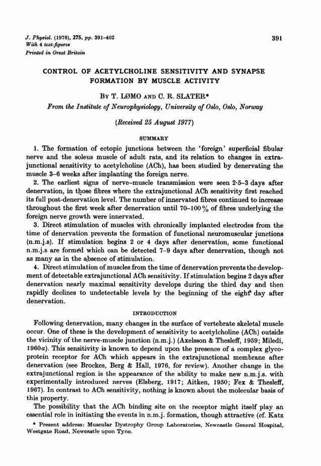
(A) Innervated fibres (B) Innervated fibres withaction potentials
100 (2T_111 -l)j10T0 (2
50/I(8)~~~~~~~~~~~~ %50) 1250 - %~~~~50 6)T (8)
0 2 4 6 8 9-26 0 2 4 6 8 9-26
Time after cutting tibial nerve (days)Fig. 1. Development of new innervation of denervated soleus muscles by previouslytransplanted fibular nerves. A, fraction of soleus muscle fibres underlying visible sproutsof the fibular nerve which showed some signs of innervation by the fibular nerve (eitherevoked or spontaneous). B, fraction of innervated fibres which responded to fibularnerve stimulation with an action potential. Values were calculated for each muscle, andthe average and range of these are shown (number of muscles in brackets). Usuallyfifteen to thirty fibres were studied in each muscle.
immediately overlying the muscle, in which case the entire growth could be dissectedaway. In most cases, however, the neural growth adheres tightly to the muscle andthe axons seem to establish intimate contact with the muscle fibres. Within severalweeks, many of these sprouts become myelinated, sometimes to within 100 Jim orless of their terminations (as visualized after staining with methylene blue, Waerhaug& Korneliussen, 1974). The spatial limits of the adherent growth zone can usuallybe determined fairly unambiguously by inspection of the freshly isolated prep-aration with the dissecting microscope (see Methods). It rarely comes closer to theoriginal end-plate band than 1-2 mm and usually extends about half-way across themuscle.So long as the soleus nerve was intact, very few of the fibular nerve axons inner-
vated soleus muscle fibres. In ten out of thirty-one muscles with an intact soleusnerve, studied 15-60 days after nerve transplantation, a few isolated muscle fibres(usually fewer than five in any one muscle) could be seen to contract on stimulationof the fibular nerve. It was only after some searching that these fibres could be
393

T. LOMO AND C. R. SLATERidentified and impaled with a micro-electrode, and they might well have been missedduring a routine study of the surface fibres.
Development offoreign innervation following denervationTo initiate extensive formation of new n.m.j.s, the tibial nerve was cut, after
allowing a period of 3-6 weeks for the fibular nerve to grow into the muscle (Fex &Thesleff, 1967). Neuromuscular transmission had failed at most of the isolated soleus
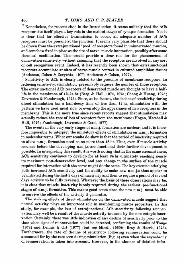
A Innervated Denervated100
(8/133)Ch
CN50
0
100
Ce
50
0
Uom
100
50
(3/24)
0
(8/106)(A
-o
o 'I I r1IIIm II I
0.1 1 10100 1000 0.1 1 10 1001000ACh sensitivity (mV/nC)
Figs. 2A and B. Distribution of extrajunctional ACh sensitivity of soleus muscle fibresat different stages during the development of ectopic innervation by the fibular nerve.Fibres innervated by the fibular nerve are shown in the left column, while thoseremaining fully denervated are shown on the right. The time (days) after cutting thetibial nerve is shown for each pair of histograms. Numbers in brackets are the numberof muscles and fibres in each sample.
394

SYNAPSE FORMATION 395
nerve endings 18 hr later. The first signs of a response to stimulation of the fibularnerve were seen 2-5-3 days after denervation. During the next few days, the numberof innervated fibres steadily increased and by the end of the first week after dener-vation, about 75 % of the fibres underlying the visible growth zone showed somesigns of innervation (Fig. 1 A).
B Innervated Denervated
100
W
(0
50
0
100
(10/134)
Co
50
0
100
Cn
c
-0
CD0E
U,)
m
(n
(4/57)
100 o(9/27) (3/8)
50-
oI~ ll1-LII001 1 10100 1000 01 1 10 100 1000ACh sensitivity (mV/nC)
Fig. 2B. For legend see facing page.
When tested in isolated nerve-muscle preparations at room temperature, theefficiency of transmission at newly formed synapses was low. Only about 1O% of theinnervated fibres examined 25-3 days after cutting the tibial nerve responded tofibular nerve stimulation with an action potential. In the rest, only subthresholdend-plate potentials, similar in amplitude to the spontaneously occurring miniature

T. LOMO AND C. R. SLATER
end-plate potentials, were seen. During the following few days, the efficiency oftransmission increased markedly, so that 8 days after cutting the tibial nerve, anaverage of more than 85% of innervated fibres (or about 65 %/0 of all fibres underlyingthe growth zone) gave action potentials (Fig. 1 B).
While not studied in detail here, it is evident that the morphology of newly formedectopic n.m.j.s is still primitive even 8-10 days after denervation of the soleus(Korneliussen & Sommerschild, 1976). Our preliminary observations show thathistochemically detectable cholinesterase activity (demonstrated with the methodof Buckley & Heaton, 1968) develops gradually at the new n.m.j.s. Signs of enzymeactivity were generally first seen 6-7 days after denervating the soleus, but a patternof staining qualitatively similar to that at mature n.m.j.s was not seen for a furtherweek or so.
Changes in extrajunctional ACh sensitivity associated with new innervationTo study how the development of new innervation is related to changes in extra-
junctional ACh sensitivity, the response of muscle fibres to locally applied ACh wasdetermined at different times after cutting the tibial nerve. To reduce the influence ofany local effects which the new n.m.j.s might have had on ACh sensitivity, fibres weretested at the edge of the zone of fibular nerve growth. Those fibres which respondedto fibular nerve stimulation with action potentials or a detectable end-plate potentialwere considered to be innervated. Fibres which gave no detectable response wereconsidered still denervated, though this group would also have included any fibreswith a subthreshold end-plate potential too small to be detected at the recordingsite.The results of these studies are shown in Fig. 2, in which the distribution of
sensitivities in samples of fibres deemed innervated or denervated by the abovecriteria is shown as a function of time after denervation. For ease of comparison, themedian sensitivity of each distribution is plotted against time after denervation inFig. 3.Development of sensitivity. In confirmation of earlier studies (e.g. Axelsson &
Thesleff, 1959; L0mo & Rosenthal, 1972) the ACh sensitivity of denervated soleusfibres increased approximately 1000-fold during the second and third days afterdenervation (Figs. 3, 4).When the first responses to stimulation of the fibular nerve were seen, 65-66 hr
after denervation, many fibres were not yet fully sensitive (Fig. 2). However, thosefibres which were innervated at this time were as sensitive as expected for fibresinnervated 3 days or more, and as a group were therefore slightly more sensitive thanthe non-innervated fibres in the same muscles (Fig. 3). Further, at this time, theinnervated fibres represented nearly half (25/58) of all the fibres tested whosesensitivity was greater than 100 mV/nC. In contrast, virtually no fibres (2/102)with sensitivities less than 100 mV/nC were innervated. Thus, signs of transmissionat the new nerve-muscle junctions were not detectable until after the extrajunctionalsensitivity had reached its final high level.
Decline of sensitivity. Fibres which became innervated by the fibular nerve losttheir extrajunctional ACh sensitivity during the first 2 weeks after cutting the soleusnerve. The first newly innervated fibres with sensitivities lower than 30 mV/nC
396

SYNAPSE FORMATION(a value encountered in only 2/406 denervated fibres studied 3 days or more afterdenervation) were seen 5-6 days after cutting the soleus nerve (Fig. 2). By the8th day after denervation, the sensitivity of some fibres was already too low to bedetected, and by the 15th day, nearly all innervated fibres were insensitive.
1000
100
ChE~10
C
01 Li0 2 4 6 8 10 12 14 16
Time after denervation (days)
Fig. 3. Extrajunctional ACh sensitivity of soleus fibres at different stages during thedevelopment of ectopic innervation by the fibular nerve. Values are the medians of thesamples shown in Fig. 2, as well as those from several additional times. 0O denervatedfibres; 0, fibres innervated by the fibular nerve. (Point with downward arrow indicatesno detectable sensitivity.)
Effects of stimulation on n.m.j. formationIn their study of the effect of direct stimulation on the ectopic reinnervation of rat
soleus muscle, Jansen, L0mo, Nicolaysen & Westgaard (1973) assessed the extent ofinnervation by measuring the strength of contraction elicited by fibular nervestimulation. We have examined the state of innervation of individual fibres withintracellular recordings and have fully confirmed their basic findings. When directstimulation was started at the time of denervation and was deemed fully effective(see Methods) no innervated fibres were found 4-6 days later (Table 1). In contrast,unstimulated control muscles were well innervated at this time (Fig. 1, Table 1).
It was of interest to learn how long a period of muscle inactivity was required toallow the formation of stable innervation. Experiments were therefore made in whichdirect stimulation was started either 2 or 4 days after denervating the soleus muscle.Once again, the extent of innervation was tested 4-6 days after stimulation began,when an average of more than 70 % of muscle fibres underlying the foreign nervegrowth were innervated in control muscles.Whether stimulation was started 2 or 4 days after denervation, an appreciable
397

398 T. L0MO AND C. R. SLATERnumber of innervated fibres was found (an average of 20 or 35% respectively,Table 1). Thus, a period of inactivity lasting only 2 days is adequate to allowformation of some functional n.m.j.s. At the same time, though the sample sizes aresmall, the data in Table 1 suggest that fewer muscle fibres were innervated in thestimulated muscles than in the unstimulated controls.
TABLE 1. Effect of chronic stimulation of denervated soleus muscles on the development ofinnervation by a previously transplanted fibular nerve
Stimulation (for parameters see Methods) began 0, 2, or 4 days after cutting theoriginal soleus innervation. In the acute experiments the response of a number (n) ofmuscle fibres in the stimulated muscle and in the unstimulated contralateral muscleto stimulation of the fibular nerve was tested. Any evoked response was taken asa sign of innervation. In some cases, the foreign nerve had not grown over the controlmuscle and these are indicated by blanks in the Table
Acuteexperi-
Onset of ment Fibres with foreign innervationstimulation (days A
(days after after Stimulated muscle Unstimulated muscledener- dener- ,vation) vation) % n % n
0 4 0 (No 32 25contraction)
0 6 0 10 - -0 4 0 20 17 180 4 0 28 50 282 8 28 32 82 332 8 23 47 94 182 8 22 40 96 292 8 17 12 -2 7 20 50 -4 8 24 17 -4 9 33 484 8 50 32 100 37
Effects of stimulation on extrajunctional ACh sensitivityIt is known that direct stimulation of denervated soleus muscles from the time of
denervation prevents the appearance of extrajunctional ACh sensitivity, ifthe patternof stimulation we have used is employed (L0mo & Westgaard, 1976). If stimulationdoes not begin until 5 days after denervation, when the muscle is fully sensitive, thenthat sensitivity is abolished within about 10 days (L0mo & Rosenthal, 1972; L0mo& Westgaard, 1975). The experiments in the previous section show that even whendirect stimulation begins 2 days after denervation - that is, before full ACh sensitivityhas developed - synapses can form. To see how ACh sensitivity changes in thesecircumstances we stimulated denervated soleus muscles, which had not receiveda fibular nerve transplant, starting 2 days after denervation (Fig. 4).In spite of the vigorous activity of the muscles during the third day after dener-
vation, the median ACh sensitivity (tested in the region of the muscle far from end-plates) rose to very nearly the full post-denervation level, only to begin to decline

SYNAPSE FORMATIONalmost immediately. Within 5 days of the onset of stimulation, the median sensitivitywas once again in the range expected for a normally innervated muscle.
In contrast, when stimulation was started only one day after denervation, onlya very small increase in sensitivity was seen at the end of the third day afterdenervation. If the onset of stimulation was delayed until 3 days after denervation,when full sensitivity had already developed, than the decline of sensitivity occurredat a rate similar to that seen after 2 days or 5 days (L0mo & Westgaard, 1975). Ineach of these three cases, the maximum rate of decline of sensitivity begins about2 days after the onset of stimulation and corresponds roughly to an exponentialdecline with a half-time of 6-9 hr.
1000Onset of stimulation* * A4 4 4 °
0 A~~0100 L0 A A
0C
E0
*5 10
o 0
0 1 _ -| |X0 1 2 3 4 5 6 7 8
Time after denervation (days)Fig. 4. Effect on extrajunctional ACh sensitivity of denervated soleus fibres of directelectrical stimulation with chronically implanted electrodes (see Methods) starting atdifferent times after denervation. Stimulation started 1 (U), 2 (@), or 3 (A) days afterdenervation. Open symbols show results from unstimulated contralateral musclesdenervated at the same time. Values are medians of samples of 20-120 fibres in one tosix muscles (average, sixty fibres in three muscles). Points with downward arrowsindicate no detectable sensitivity.
DISCUSSION
These experiments confirm the close parallels between extrajunctional sensitivityto ACh and the ability to form new functional n.m.j.s. Thus, in the development ofectopic innervation, the first fibres to become innervated were also those which firstbecame fully sensitive to ACh. In the stimulation experiments, 2 days of inactivitywas long enough to allow some new n.m.j.s to form, though not as many as inunstimulated muscles, and was also just long enough to allow the development of fullACh sensitivity in most fibres. This suggests that the kinetics of the response of boththese properties of the muscle to imposed activity is similar.
399
T. LOMO AND C. R. SLATERy Nonetheless, for reasons cited in the Introduction, it seems unlikely that the AChreceptor site itself plays a key role in the earliest stages of synapse formation. Yet itis clear that for effective transmission to occur, an adequate number of AChreceptors must be present at the junction. It seems very plausible that these shouldbe drawn from the extrajunctional 'pool' of receptors found in uninnervated muscles,and somehow fixed in place at the site of nerve-muscle interaction, possibly after somechemical modification. This would provide a clear role for the phenomenon ofdenervation sensitivity without assuming that the receptors are involved in any sortof cell recognition event. Indeed, it has recently been shown that extrajunctionalreceptors accumulate at skes of nerve-muscle contact in cultured amphibian tissues(Anderson, Cohen & Zorychta, 1977; Anderson & Cohen, 1977).
Sensitivity to ACh is clearly related to the presence of membrane receptors. Inreducing sensitivity, stimulation presumably reduces the number of those receptors.The extrajunctional ACh receptors of denervated muscle are thought to have a half-life in the membrane of 12-24 hr (Berg & Hall, 1974, 1975; Chang & Huang, 1975;Devreotes & Fambrough, 1975). Since, at its fastest, the decline of sensitivity duringdirect stimulation has a half-decay time of less than 12 hr, stimulation with thepattern we have used must slow or even stop the appearance of new receptors in themembrane. This is the more true since recent reports suggest that stimulation mayactually reduce the rate of loss of receptors from the membrane (Hogan, Marshall &Hall, 1976; Fambrough, Devreotes & Card, 1977).The events in the very early stages of n.m.j. formation are unclear, and it is there-
fore impossible to interpret the inhibitory effects of stimulation on n.m.j. formationin molecular terms. What our results do show is that the period of inactivity requiredto allow n.m.j. formation need be no more than 48 hr. Thus, even if muscle activityresumes before the developing n.m.j.s are functional their further development ispossible. In interpreting this result, it is worth noting that in the same circumstances,ACh sensitivity continues to develop for at least 24 hr ultimately reaching nearlyits maximum post-denervation level, and any change in the surface of the musclerequired for interaction with the nerve might do the same. The key events underlyingboth increased ACh sensitivity and the ability to make new n.m.j.s thus appear tobe initiated during the first 2 days of inactivity and then to require a period of severaldays activity to be fully reversed. Whatever the basis of these observations may be,it is clear that muscle inactivity is only required during the earliest, pre-functionalstages of n.m.j. formation. This makes good sense since the new n.m.j. must be ableto survive the effects of the activity it generates.The striking effects of direct stimulation on the denervated muscle suggest that
normal activity plays an important role in maintaining muscle properties. In thisstudy, for example, the loss of extrajunctional ACh sensitivity following reinner-vation may well be a result of the muscle activity induced by the new ectopic inner-vation. Certainly, there was little indication of any decline of sensitivity prior to thetime when signs of reinnervation could be detected, confirming the results of Tonge(1974) and Dennis & Ort (1977) (but see Miledi, 1960b; Bray & Harris, 1975).Furthermore, the rate of decline of sensitivity following reinnervation could beaccounted for by the rate seen during stimulation (Fig. 4) even when the asynchronyof reinnervation is taken into account. However, in the absence of detailed infor-
400

SYNAPSE FORMATIONmation about the activity of the muscles in vivo, it is not possible critically to testthe idea that the activity of the muscles is the sole factor contributing to the declinein sensitivity.
In conclusion, we suggest that activity is able to maintain the extrajunctionalregion of the muscle fibre surface in a normal state with respect both to ACh sensi-tivity and to the ability to accept new innervation. However, even a short period(2 days) of inactivity allows the expression of the full post-denervation effect, if onlytransiently. The nature of the process of n.m.j. formation ensures that during sucha brief period of altered conditions, stable nerve-muscle interactions can form which,though primitive in both structure and function, can develop into transmittingjunctions even if intense muscle activity resumes.
Many of the surgical operations required for this study were performed by Elizabeth Djupvikand Sigrid Schaller, whose excellent technical assistance we gratefully acknowledge. C. R. S. wassupported by a fellowship from the Norwegian Research Council for Science and the Humanities.
REFERENCES
AITKEN, J. T. (1950). Growth of nerve implants in voluntary muscle. J. Anat. 84, 38-48.ANDERSON, M. J. & COHEN, M. W. (1977). Nerve induced and spontaneous redistribution of
acetylcholine receptors on cultured muscle cells. J. Physiol. 268, 757-773.ANDERSON, M. J., COHEN, M. W. & ZORYCHTA, E. (1977). Effects ofinnervation on the distribution
of acetylcholine receptors on cultured muscle cells. J. Phy8iol. 268, 731-756.AXELSSON, J. & THESLEFF, S. (1959). A study of supersensitivity in denervated mammalian
skeletal muscle. J. Phy8iol. 147, 178-193.BERG, D. K. & HALL, Z. W. (1974). Fate of a-bungarotoxin bound to acetylcholine receptors ofnormal and denervated muscle. Science, N.Y. 184, 473-475.
BERG, D. & HALL, Z. W. (1975). Loss of a-bungarotoxin from junctional and extrajunctionalacetylcholine receptors in rat diaphragm in vivo and in organ culture. J. Phy8iol. 252, 771-789.
BRAY, J. J. & HARRIS, A. J. (1975). Dissociation between nerve-muscle transmission and nervetrophic effects on rat diaphragm using type D botulinum toxin. J. Physiol. 253, 53-77.
BROCKES, J. B., BERG, D. K. & HALL, Z. W. (1976). The biochemical properties and regulationof acetylcholine receptors in normal and denervated muscle. Cold Spring Harbor Symp. quaint.Biol. 40, 253-262.
BUCKLEY, G. A. & HEATON, J. (1968). A quantitative study of cholinesterase in myoneuraljunctions from rat and guinea pig extraocular muscles. J. Physiol. 199, 743-749.
CHANG, C. C. & HUANG, M. C. (1975). Turnover of junctional and extrajunctional acetylcholinereceptors of the rat diaphragm. Nature, Lond. 253, 643-644.
COHEN, M. W. (1972). The development of neuromuscular connexions in the presence ofD-tubocurarine. Brain Res. 41, 457-463.
CRAIN, S. M. & PETERSON, E. M. (1971). Development of paired explants of foetal spinal cordand adult skeletal muscle during chronic exposure to curare and hemicholinium. In vitro 6,373.
DENNIS, M. J. & ORT, C. A. (1977). The distribution of acetylcholine receptors on muscle fibresof regenerating Salamander limbs. J. Physiol. 266, 765-776.
DEVREOTES, P. & FAMBROUGH, D. M. (1975). Acetylcholine receptor turnover in membranes ofdeveloping muscle fibres. J. cell Biol. 65, 335-358.
ELSBERG, C. A. (1917). Experiments on motor nerve regeneration and direct neurotization ofparalysed muscles by their own and foreign nerves. Science, N.Y. 45, 318-320.
FAMBROUGH, D. M., DEVREOTES, P. N. & CARD, D. J. (1977). The synthesis and degradation ofacetylcholine receptors. In Synapses, ed COTTRELL, G. A. & USHERWOOD, P. N. R., pp. 202-263. Glasgow: Blackie & Son.
FEX, S., SONESSON, S., THESLEFF, S. & ZELENX, J. (1966). Nerve implants in botulinum poisonedmammalian muscle. J. Physiol. 184, 872-882.
401

T. LOMO AND C. R. SLATERFEX, S. & THESLEFF, S. (1967). The time required for innervation of denervated muscles by
nerve implants. Life Sci., Oxford 6, 635-639.FRANK, E., JANSEN, J. K. S., L0Mo, T. & WESTGAARD, R. (1975). The interaction between
foreign and original motor nerves innervating the soleus muscle of rats. J. PhyIiol. 247,725-743.
HOGAN, P. G., MARSHALL, J. M. & HALL, Z. W. (1976). Muscle activity decreases rate ofdegradation of a-bungarotoxin bound to extrajunctional acetylcholine receptors. Nature,Lond. 261, 328-330.
JANSEN, J. K. S., LoMo, T., NICOLAYSEN & WESTGAARD, R. H. (1973). Hyperinnervation ofskeletal muscle fibres: dependence on muscle activity. Science, N.Y. 181, 559-561.
JANSEN, J. K. S. & VAN ESSEN, D. C. (1975). Re-innervation of rat skeletal muscle in the presenceof a-bungarotoxin. J. Physiol. 250, 651-667.
KATZ, B. & MILEDI, R. (1964). The development of acetylcholine sensitivity in nerve-freesegments of skeletal muscle. J. Phyaiol. 170, 389-396.
KORNELIUSSEN, H. & SOMMERSCHILD, H. (1976). Ultrastructure of the new neuromuscularjunctions formed during reinnervation of rat soleus muscle muscle by a 'foreign' nerve. CellTie8. Re8. 167, 439-452.
L0Mo, T. & ROSENTHAL, J. (1972). Control of ACh sensitivity by muscle activity in the rat.J. Phyaiol. 221, 493-513.
L0Mo, T. & WESTGAARD, R. H. (1975). Further studies on the control of ACh sensitivity bymuscle activity in the rat. J. Physiol. 252, 603-626.
LoMo, T. & WESTGAARD, R. H. (1976). Control of ACh sensitivity in rat muscle fibres. ColdSpring Harbor Symp. quant. Biol. 40, 263-274.
MILEDI, R. (1960a). The acetylcholine sensitivity of frog muscle fibres after complete or partialdenervation. J. Physiol. 151, 1-23.
MILEDI, R. (1960b). Properties of regenerating neuromuscular synapses in the frog. J. Physiol.154, 190-205.
TONGE, D. A. (1974). Physiological characteristics of reinnervation of skeletal muscle in themouse. J. Physiol. 241, 141-153.
WAERHAUG, 0. W. and KORNELIUSSEN, H. (1974). Morphological types of motor nerve terminalsin rat hind limb muscles, possibly innervating different muscle fiber types. Z. Anat. EntwGesch.144, 237-247.
402