functional analysis of the bacillus subtilis bacteriophage spp1 pac
TRANSCRIPT

Nucleic Acids Research, Vol. 18, No. 10 2881
Functional analysis of the Bacillus subtilis bacteriophageSPP1 pac site
Alicia Bravo, Juan C.Alonso* and Thomas A.TrautnerMax-Planck-Institut fur Molekulare Genetik, Ihnestrasse 73, D-1000 Berlin 33, FRG
Received February 26, 1990; Revised and Accepted April 18, 1990 EMBL accession no. X52481
ABSTRACT
Encapsidation of the DNA of the virulent Bacillussubtilis phage SPP1 follows a processive unidirectionalheadful-mechanism and initiates at a unique genomiclocation (pac). We have cloned a fragment of SPP1 DNAcontaining the pac site flanked by reporter genes intothe chromosome of B. subtilis. Infection of such cellswith SPP1 led to highly efficient packaging, initiatedat the inserted pac site, of chromosomal DNA. Thedirectionality in the packaging of this DNA was thesame as observed with vegetative phage DNA.Mutagenizing the chromosomal pac insert defined an83 base pair segment containing the pac cleavage sitewhich is sufficient to direct phage specific DNAencapsidation. The packaging recognition signal asdefined can also be utilized by the SPP1 related phages41c, SF6 and Q15.
INTRODUCTION
One of the most remarkable events in the life cycle of a viruswithin an infected cell is the packaging of its nucleic acid intoprogeny virus particles. Among the many facets of this process,the selective recognition of virus DNA within a universe of hostDNA has attracted interest as an intriguing case of highly specificprotein-DNA interaction. Studying bacteriophage packaging, itwas realized that in many instances specificity of packaging isprovided by the recognition by the phage coded packagingmachinery of a 'signal sequence' within replicating DNA (1).Such signal sequences occur only once per genome.
In the case of phage X, packaging is initiated by the bindingof a phage-encoded nuclease (terminase) to cosB. At least in vitro,the binding of the terminase at cosB generates a protein-DNAcomplex and introduces staggered nicks at the nearby cosN site(a region of dyad symmetry) (2, 3). Cleavage of such sequenceswithin the concatemer generates unit-length molecules carryingidentical genetic information which are encapsidated into thephage proheads. In this case and those of other 'unit-length' DNAphages (T3, T7), the size of the DNA to be packaged isdetermined essentially by the distance between neighboringpackaging signals within concatemeric DNA (4).
In other phages (TI, P1, P22, SPP1), whose DNA also goesthrough a concatemeric replication intermediate, discrete
packaging signals were also identified (5-1 1). Here, however,the packaging signal only serves to initiate packaging by theformation of a double strand cleavage at the pac site. Startingfrom this initial cleavage site, packaging proceeds processivelyand unidirectionally, leading to the encapsidation through furthercleavages at non defined sites, of amounts of DNA which areslightly bigger than one genome. The length of the packagedDNA in these phages is not determined by the unit genome lengthspacing of packaging signals in the DNA but by parameters ofthe packaging machinery, possibly including the packagingcapacity of the phage head. This processive headful mechanismleads to the generation of terminally redundant and partiallycircularly permuted DNA molecules (1,12). A highly degenerateversion of this mode of packaging is the case of bacteriophageT4, where also a processive mode of DNA packaging occurs,which produces a population of terminally redundant andcircularly permuted DNA molecules, but where a specificinitiation site for encapsidation could not be identified (13).
In this communication we are concerned with the packagingsignal of the virulent B. subtilis phage SPPl. We have insertedfragments of SPP1 DNA containing the pac cleavage sitesequence (9) into the chromosome of B. subtilis. DNA flankingthe pac insert is packaged into phage particles following infectionwith SPP1, showing that the phage encoded packaging machineis interacting in trans with the packaging signal placed in thechromosome. By following the packaging of chromosomal DNAflanking the pac region, after infection of cells carrying the insert,we were able to establish the minimal sequence required fordirectional packaging of SPP1 DNA.
MATERIALS AND METHODSBacterial strains and phagesB. subtilis strain YB886 (14) and its derivatives bearing the ble-pac-cat cassette were used. A DNA segment from the SPPIEcoRl fragment I containing pac [coordinates 1 to 891 ofDeichelbohrer et al. (9)] was flanked by the ble [coordinates 1548to 2081 of McKenzie et al. (15)] and cat [coordinates 610 to 2013of Horinouchi and Weisblum (16)] genes from pUBI10 andpC194 respectiveley (ble-pac-cat cassette). A chromosomal DNAregion with a low density of EcoRI restriction sites (add region)(J. Kooistra, pers. comm.) was selected for integration of the
* To whom correspondence should be addressed
.=) 1990 Oxford University Press

X HI I
pac cleavage site
TX B AI I I I
Clone40
l l ll--CZMii2/o 35
pacactivity
+
36I I I
I I I - --
+
3738
3934
+
cat
Fig. 1. Physical map of the ble-pac-cat cassette and its deletion derivatives. Thin lines represent phage DNA. SPP1 deleted fragments were generated in vitro byrestriction enzyme digestion of clone 40. The size of the SPP1 DNA in the cassettes is 891 bp (clone 40), 455 bp (35), 197 bp (36), 172 bp (37), 484 bp (38),865 bp (39) and 840 (clone 34). Stippled bars represent pUBl 10 DNA containing the ble gene. Striped bars denote pC194 DNA carrying the cat gene. The arrows
indicate direction of transcription. Abbreviations: A, AhaII; B, BsmI; Ba, BamHI; H, HindII; T, 7haI; X, XmaIII.
ble-pac-cat cassette. For this, the cassette was joined to an 1.4kb segment of the add chromosomal DNA region and introducedinto YB886 competent cells by transformation. Chloramphenicol(Cm) resistant transformants were selected and the cassettesubjected to DNA amplification as described by Young (17) usingphleomycine (Pm) as selective marker (strain BG40). In vitrodeletions into the SPP1 region on a plasmid-based ble-pac-catcassette (clone 40) were performed. As revealed in Figure 1,clones 35, 36 and 37 lack the 436 bp BamHI-Hindll, 694 bpBamHI-TaI or 720 bp BamHI-XmnaH fragments respectively.In clone 35 both sites were destroyed by construction, whereasin clones 36 and 37 the BamHI and XmalII sites, respectively,are present. For the construction of clones 39 and 34, the 26bp ThaI-XnalI and 49 bp BsmI-AhaII DNA segments,respectively, were deleted. In both constructs the restriction sitesinvolved were destroyed. Clone 38 has an internal XMafI deletion(407 bp). The deletions were verified for the presence or absenceof landmarking restriction sites. The ble-pac-cat cassettes werethen integrated and amplified as described above. The clonenumber gives the name of the strain (i.e. clone 35 gives raiseto strain BG35).
Bacteriophages SPP1 (18), 41c, Q 15 and SF6 (19) were used.
Transformation and mediaThe method of Rottlander and Trautner (20) was used fortransforming B. subtilis competent cells.TY broth (21) was used throughout as liquid and solid medium.
It was supplemented with 5 yg/ml of Cm or 0.2 lsg/ml of Pm.
DNA techniquesDNA packaged into phage particles was purified as follows:cultures of B. subtilis cells carrying the ble-pac-cat cassette weregrown in TY supplemented with Pm, at 370C, to an OD560=0.8and infected at a multiplicity of 5 phage/cell. The lysates were
then treated with DNAse (1 14g/ml) and RNAse (1 ,tg/ml). Phage
particles were precipitated with PEG 20%, 2M NaCl (v/v). TheDNA packaged was purified by extraction with phenol anddialyzed against 10 mM Tris-HCl, 1 mM EDTA pH 8.0.DNA preparations digested with the appropiate restriction
enzymes were electrophoresed overnight in 0.8% agarose.Southern transfer to nylon membranes (GeneScreen, NENResearch products) and hybridization to 32P-labeled DNA probeswere carried out as described by Maniatis et al. (22). The bleprobe is a 0.55 Kb EcoRI-XbaI fragment containing the ThaI-HaeIII sequence from pUB 110 [coordinates 1548 to 2081 ofMckenzie et al. (15)]. The cat probe is a 0.73 Kb HindIJI-NcoIfragment carrying the HpaII-NcoI sequence from pC194[coordinates 969 to 1688 of Horinouchi and Weisblum (16)]. Theprobes were end-labeled with polynucleotide kinase accordingto the procedure of Maniatis et al. (22). DNA was visualizedby autoradiography with Kodak XAR film. The relative amountof DNA present in any particular band identified either by stainingwith EtBr or Southern hybridization was determined by densityscanning of films using a laser densitometer (LKB Ultro ScanXL). To determine the degree of amplification of the ble-pac-cat cassette on the chromosome, DNA was digested with an
enzyme that cleaves once within the amplified unit and the ratiobetween the amount of the amplified DNA segment with respectto the non-amplified one, identified by Southern blot using thecat probe, was determined as described above.
Restriction endonucleases and DNA modifying enzymes were
purchased from Boehringer, Mannheim (FRG) or from BRL,Gaithersburg, Md. (U.S.A.) and were used as specified by themanufacturers.
RESULTSThe SPP1 pac region is recognized as a signal for packagingwhen present in the B. subtilis chromosomeTo analyze the requirements of a pac signal to serve as theinitiator of packaging, it is desirable to place this signal in trans
2882 Nucleic Acids Research, Vol. 18, No. 10
Ba
I I II---
L--777,-l =-,7 -
I I.
ble
0 m a a qzzzzzZZ777,14- 1 1
.............. bmmj.
I
I
I

Nucleic Acids Research, Vol. 18, No. 10 2883
no 34 35 36 371 2 3 4 5 6 7 8 9 '0 11 '2
_. 1
no 34 35 36 37
2 3 4 5 6 7 8 9 10 11 12
B 38 39 40 no
1 2 3 4 5 6 7I B 9 10
0 38 39 40 no
2 3 4 5 6 7 8 0 10
a-.do__a
Fig. 2. Analysis of DNA packaged into SPP1 particles by agarose gel electrophoresis (A and B) and Southern blot (C and D) using the cat probe. DNA from SPP1particles grown on bacterial strains, carrying in the chromosome the SPP1 cassettes indicated or no insert, was either digested with EcoRI (odd numbered lanes)or non-digested (even numbered lanes). The arrows indicate the position of reference fragments of 3.8 Kb (lane 1) and 2.1 Kb (A: lane 12 and B: lane 10) containingthe cat gene.
on a replicon whose packaging following phage infection can
easily be monitored. Stemnberg and Coulby (6) achieved thissituation in their studies on P1 packaging by lysogenizing X phage,into which the P1 packaging signal had been inserted. In our case,
an 891 bp fragment from the SPP1 EcoRI fragment 1 containingthe pac cleavage site was flanked by specific markers, the bleand cat genes (Fig. 1). The resulting cassette (ble-pac-cat) was
linked to defined B. subtilis DNA and integrated, by chromosomaltransformation, into the B. subtilis chromosome in the vicinityof the addA gene. In the presence of 0.2 ,Ag/ml of Pm in thegrowth media this cassette was amplified about 6 fold (strainBG40, see Materials and Methods). No chromosomal EcoRIDNA fragments, containing the amplified cassette, smaller than35 kb should be expected.
Strain BG40 was infected with SPPl, and after lysis the DNApackaged into SPP1 particles was purified. Part of this DNA was
digested with EcoRI, which neither cuts the cassette ble-pac-catnor the chromosomal DNA surrounding it. Such DNA was
analyzed by agarose gel electrophoresis and Southernhybridization using as probes end-labeled restriction fragmentsfrom both the ble (ble probe) and cat (cat probe) genes. Withinthe standard EcoRI restriction pattern of SPPI DNA an additionalband, which comigrates with undigested phage DNA, was
detected (Fig. 2B, lane 7). This material represents about 1%of the total DNA packaged, as estimated by densitometry of theEtBr stained DNA. Only this EcoRI resistant DNA specificallyhybridizes with the cat probe (Fig. 2D, lane 7). Identical resultswere obtained when the ble probe was used (data not shown).Therefore, such DNA packaged into the SPPI particles mustderive from the amplified cassette integrated into thechromosome. When the encapsidated DNA from phages grownon strain BG40, was digested with AflJ (this enzyme cuts bacterialDNA but not phage DNA) a band which hybridizes with the catand ble probes was detected (see below). These results indicatethat chromosomal DNA adjacent to the pac region is encapsidatedand that the presence of the 891 bp SPPI fragment in the B.
A
C
2884 Nucleic Acids Research, Vol. 18, No. 10
4 1F":""pl5.../41 p15^
1 2 3. L4
-IF.-
12 j/
0
3.1 kb- .k4b
2 .4 5 k b
e o ia
i............b.........t -A
i3
Fig. 3. Directionality of chromosomal DNA packaging. Upper panel: DNA fromSPP1 particles grown on strains BG34, BG35 or BG36 was digested with AflIl,separated on 0.8% agarose and blot hybridized with either the ble probe or thecat probe, as shown. The lengths of two restriction fragments used as referencesfor hybridization are indicated at the left of the gel. Lower panel: scheme of thestructure generated in the chromosome of the strains used in this work. Stippledbar denotes the ble gene, striped bar denotes the cat gene, the open box indicatesphage DNA containing the pac site (closed circle), thin line represents thechromosomal DNA fragment (1.4 Kb) used to place the ble-pac-cat cassette inthe chromosome. The AflJ (A) restriction sites are indicated. The size of theAflll DNA fragment containing the pac site is indicated for the different strains.The cat probe does not include DNA from the smaller AlfJl DNA fragment. Thearrows show the defined terminus of chromosomal DNA molecules which areexpected to be present into SPP1 particles if chromosomal DNA packagingproceeds from the SPP1 pac site either to the left or to the right. The lengthof the putative Aflll-pac end fragments in strains BG34, BG35 and BG36 isindicated. For simplicity, in the drawing a threefold amplification of the cassetteis assumed.
subtilis chromosome is sufficient for specific packaging ofchromosomal DNA.
Identical results were obtained when the ble-pac-cat cassettewas not subjected to amplification. However, in this case thesignal of chromosomal DNA packaged into SPP1 particlesdetected by Southern-blot was very weak (data not shown). Thehybridizing band is, as expected, smaller (ranging from 15 Kbto 20 Kb depending on the strain under assay) due to the lackof amplification. This suggests that the pac site, as in the phageconcatemer, is used only once. Hence, to avoid the detectionproblem we used in all cases strains carrying amplification ofthe cassette.
A functional pac site is located within an 83 bp regionTo narrow down the size of a functional pac recognition site,deletions in the 891 bp region of SPP1 DNA were generated invitro (Fig. 1). This involved deletions both at the right (clone34) or at the left of the pac cleavage site sequence (clones 35,36, 37, 38, and 39). The resulting cassettes were placed in the
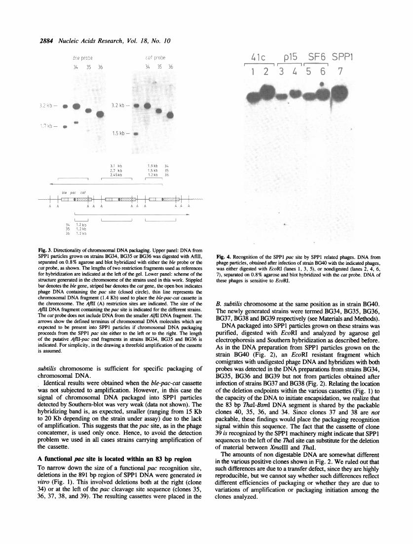
Fig. 4. Recognition of the SPP1 pac site by SPP1 related phages. DNA fromphage particles, obtained after infection of strain BG40 with the indicated phages,was either digested with EcoRI (lanes 1, 3, 5), or nondigested (lanes 2, 4, 6,7), separated on 0.8% agarose and blot hybridized with the cat probe. DNA ofthese phages is sensitive to EcoRI.
B. subtilis chromosome at the same position as in strain BG40.The newly generated strains were termed BG34, BG35, BG36,BG37, BG38 and BG39 respectively (see Materials and Methods).DNA packaged into SPP1 particles grown on these strains was
purified, digested with EcoRI and analyzed by agarose gelelectrophoresis and Southern hybridization as described before.As in the DNA preparation from SPP1 particles grown on thestrain BG40 (Fig. 2), an EcoRI resistant fragment whichcomigrates with undigested phage DNA and hybridizes with bothprobes was detected in the DNA preparations from strains BG34,BG35, BG36 and BG39 but not from particles obtained afterinfection of strains BG37 and BG38 (Fig. 2). Relating the locationof the deletion endpoints within the various cassettes (Fig. 1) tothe capacity of the DNA to initiate encapsidation, we realize thatthe 83 bp ThaI-BsmI DNA segment is shared by the packableclones 40, 35, 36, and 34. Since clones 37 and 38 are notpackable, these findings would place the packaging recognitionsignal within this sequence. The fact that the cassette of clone39 is recognized by the SPPI machinery might indicate that SPP1sequences to the left of the ThaI site can substitute for the deletionof material between XmaIIl and ThaI.The amounts of non digestable DNA are somewhat different
in the various positive clones shown in Fig. 2. We ruled out thatsuch differences are due to a transfer defect, since they are highlyreproducible, but we cannot say whether such differences reflectdifferent efficiencies of packaging or whether they are due tovariations of amplification or packaging initiation among theclones analyzed.
S- F 6r--
r-, b
SPP17
'.5"..k- 35L.29kb 36' .5 K( E 43e' k, , Al 6
r
1:4 I (.
3 '21 k .- o 460
Is
.i; .11 -,6.4 j i,
... & 000

Nucleic Acids Research, Vol. 18, No. 10 2885
GENOME
~~~~~~~~~~:.... ...
:
pac-DDIRECTION OF MATURATION
HEADFUL 104%
I\
II It It ill
12i I 1 4 1410 8 5 13 2 15 7 11
: -_~~~~~~~~~~~~~~~r
695 T5 sIl+i5'CGGACGTGCGG6 CATGTTCTCCGCCGCL TAGTCGAA
T5----I---n777GCAGAC CTATAAC CCGIT CCGC;TT GT
I__, _ J
6 3 16 912
Fig. 5. Proposed processive headful mechanism for SPPl DNA maturation. Thesequential and unidirectional DNA packaging from thepac end starts by recognitionand cleavage of the pac site (closed circle) on a concatemer. The EcoRI restrictionmap of the first encapsidated DNA molecule is shown. Numbers indicate theEcoRI fragment positions on agarose gel. The right end (pac end) is generatedby specific cleavage at the pac site, while the left end is generated by a non-specific cleavage (headful cut). The nucleotide sequence of the 83 bp 7haI-BsmIregion including the pac site is shown. The thickness and length of the verticalarrows indicate the frequency of cutting at the pac cleavage site.
Chromosomal DNA is packaged unidirectionally from theSPP1 pac siteTo determine whether the 83 bp SPPI region definesdirectionality of packaging and is used only once, as in the phageconcatemer, DNA from SPP1 particles grown on bacteriacarrying the pac site in the chromosome, was digested with Afllland analyzed by Southern hybridization using both the cat andble probes (Fig. 3). No restriction sites for the Afm enzyme arepresent in the SPP1 DNA. If packaging of bacterial DNA startsat any SPP1 pac site within the amplified region and proceeds,from such a point, unidirectionally in the same direction reportedfor the phage DNA (from right to left in Fig. 3), we expect,in addition to an Aflll fragment which would hybridize with bothprobes, a minor fragment present in non-stoichiometric amountswhich would hybridize only with the ble probe. If the bacterialDNA is packaged in the opposite direction, then a minor bandwhich would hybridize only with the cat probe should be detected.If cutting would occur at all pac sites within the amplified region,we expect only a DNA fragment which would hybridize eitherwith the ble or cat probe. We analyzed DNA from SPP1 particlesgrown on the strains BG34, BG35 or BG36. AMlI restrictionfragments of 3.1 Kb, 2.7 Kb or 2.45 Kb respectively, which
hybridize with both probes, were detected (see Fig. 3). In additionto these major bands, minor fragments of 1.9 Kb, 1.5 Kb or 1.2Kb, respectively, were observed only when the ble probe wasused. These results indicate that chromosomal DNA is packagedunidirectionally from the SPP1 pac site and in the same directionreported for the phage DNA (toward the EcoRI fragment 12,see Fig. 5). In addition, we conclude from those data that thepac site present in the chromosomal amplified region is utilizedonly once.
The SPP1 pac site is recognized by SPP1 related phagesBy various criteria the B. subtilis bacteriophages 41c, Ql5 andSF6 are highly related to SPP1 (19, 23, 24). To determinewhether these phages recognize the SPP1 pac site as a signalfor packaging, strain BG40 was infected with the phages 41c,QlS or SF6 and, after lysis, the DNA packaged into phageparticles was purified and part of it was digested with EcoRI.Analysis of such DNAs by Southern hybridization using the end-labeled cat probe shows that also these phages are able tospecifically encapsidate chromosomal DNA (Fig. 4).Quantification of the amount of this chromosomal DNA, bydensitometric scanning of the autorradiogram, revealed thatphages 41c and SF6 recognize the SPP1 pac site with anefficiency similar to SPP1. In contrast, phage Qi5 recognizesthe SPP1 pac site with a 3-fold lower efficiency.
DISCUSSION
To facilitate an analysis of the DNA packaging mechanism bybacteriophage SPP1 we have engineered a fragment of SPP1DNA, containing the previously identified pac cleavage site (9)into the chromosome of B. subtilis. Following infection of suchcells by SPP1, this inserted phage DNA serves as an initiationsite for encapsidation of chromosomal DNA, indicating that theSPPI insert carries the recognition sequence for the SPP1packaging machinery. Systematically reducing the size of theinserted SPP1 DNA, we could allocate the capacity of the DNAto become recognized to an 83 bp subfragment of the originalinsert.The nucleotide sequence of the 83 bp region shows some
peculiar features (Fig. 5). Located towards the middle of thesequence (coordinates 722 to 726) are the major and the minorpac cleavage sites, which had been described before (9). Thecleavage sites fall into a pentanucleotide sequence GCCGC whichis repeated within the 83 bp fragment at coordinates 758 to 762and 767 to 771. This sequence is the 5' end of a decanucleotidesequence GCCGCAATAG which is repeated beginning atcoordinate 758. Furthermore, sequences TTTTT are locatedsymmetrically around the pac cleavage site contained in thedecanucleotide. It is difficult to assess the significance of thenucleotide sequence motifs and their arrangement in recognitionand cleavage at the pac site. Most likely the minimal length forpac recognition and cleavage could be less than 83 bp. The useof fragments smaller than 83 bp in our packaging assay, producedby systematic exonuclease digestion from the ThaI and BsmI ends,could lead to an accurate determination of the minimal size forpac utilization.
Comparing the SPP1 pac site with that of P1, as defined byStemnberg and Coulby (6, 7), we realize that a highly structuredDNA sequence is also involved here. The minimal length of P1DNA required for packaging and its directionality amounts toabout 160 bp. Also here the pac cleavage site is rather
E llle 1.111

2886 Nucleic Acids Research, Vol. 18, No. 10
symmetrically imbedded in a series of seven direct repeatsequences of 6 bp. It is of interest that both in the SPP1 and P1cleavage site the same nucleotide sequence GCCGC is involved.The B. subtilis phages 41c, SF6 and Q 15, are closely related
to SPP1 (24). By electron-microscopic and Southern hybridizationanalysis, heterologous and homologous genome regions betweenthese four phages have been defined (23). The phages 41c andSF6 recognize the SPP1 pac site as signal for packaging initiationwith an efficiency similar to SPP1, while the recognitionefficiency is about 3 fold lower for Q 15 when is compared withSPP1 (Fig. 4). These results suggest a higher divergence betweenQ15 and SPP1 with respect to their packaging systems.Comparisons to the nucleotide level between both systems wouldbe of particular interest for studying molecular evolution.
ACKNOWLEDGEMENTSThis research was partially supported by DeutscheForschungsgemeinschaft (Al 284/1-1). A.B. was supported byan EMBO long term fellowship.
REFERENCES1. Casjens,S.R. (1985) In Virus Structure and Assembly (Casjens,S.R. ed.)
pp. 75-147, Jones and Bartlett Publishers Inc.2. Becker,A. and Gold,M. (1978) Proc. Nat. Acad. Sci. U.S.A. 75,4199-4203.3. Feiss,M., Widner,W., Miller,G., Johnson,G. and Christiansen,S. (1983)
Gene 24,207 -218.4. Yamagishi,M., Fujisawa,H. and Minagawa,T. (1985) Virology
144,502-515.5. Ramsay,N. and Ritchie,D.A. (1983) Nature 301,264-266.6. Sternberg,N. and Coulby,J. (1987) J. Mol. Biol. 194,453-468.7. Sternberg,N. and Coulby,J. (1987) J. Mol. Biol. 194,469-479.8. Casjens,S.R., Huang,W.M., Hayden,M. and Parr,R. (1987) J. Mol. Biol.
194,411-422.9. Deichelbohrer,I., Messer,W. and Trautner,T.A. (1982) J. Virol. 42,83-90.
10. Casjens,S.R. and Huang,W. (1982) J. Mol. Biol. 157, 287-298.11. Backhaus,H. (1985) J. Virol. 55, 458-465.12. Black,L.W. (1988) The Bacteriophages (Calendar,R. ed.) Vol. 2 pp.
321-373. Plenum Press. New York and London.13. Kalinski,A. and Black,L.W. (1986) J. Virol. 58,951-954.14. Yasbin,R.E., Field.P.I. and Anderson,B.J. (1980) Gene 12,155-159.15. Mckenzie,T., Hoshino,T., Tanaka,T., Sueoka,N. (1986) Plasmid 15,93-105.16. Horinouchi,S. and Weisblum,B. (1982) J. Bacteriol. 150,815-825.17. Young,M. (1984) J. Gen. Microbiol. 130,1613-1621.18. Riva,S., Polsinelli,M. and Falaschi,A. (1968) J. Mol. Biol. 35,347-356.19. Santos,M.A., de Lencastre,H. and Archer,L.J. (1984) J. Gen. Virol.
65,2067-2072.20. Rottlander,E. and Trautner,T.A. (1970) Mol. Gen. Genet. 108,47-60.21. Biswal,N., Kleinschmidt,A.K., Spatz,H.C. and Trautner,T.A. (1967) Mol.
Gen. Genet. 100,39-55.22. Maniatis,T., Fritsch,E.F. and Sambrook,J. (1982) Cold Spring Harbor
Laboratory, Cold Spring Harbor, N.Y.23. Santos,M.A., Almeida,J., de Lencastre,H., Morelli,G., Kamke,M. and
Trautner,T.A. (1986) J. Virol. 60,702-707.24. Ratcliff,S.W., Luh,J., Ganesan,A.T., Behrens,B., Thompson, R.,
Montenegro,M.A., Morelli,G. and Trautner,T.A. (1979) Mol. Gen. Genet.168,165-172.