functional and genetic characterization of the tap efflux pump in
TRANSCRIPT

Functional and Genetic Characterization of the Tap Efflux Pump inMycobacterium bovis BCG
Santiago Ramón-García,a,b Virginie Mick,a Elisa Dainese,c Carlos Martín,a Charles J. Thompson,b Edda De Rossi,d Riccardo Manganelli,c
and José A. Aínsaa
Departamento de Microbiología, Medicina Preventiva y Salud Pública, Universidad de Zaragoza, Zaragoza, Spain, and CIBER Enfermedades Respiratoriasa‡; Department ofMicrobiology and Immunology and Centre for Tuberculosis Research, Life Sciences Centre, University of British Columbia, Vancouver, British Columbia, Canadab;Department of Histology, Microbiology, and Medical Biotechnologies, University of Padova, Padova, Italyc; and Dipartimento di Genetica e Microbiologia, Universita degliStudi di Pavia, Pavia, Italyd
Efflux pumps extrude a wide variety of chemically unrelated compounds conferring multidrug resistance and participating innumerous physiological processes. Mycobacterium tuberculosis possesses many efflux pumps, and their roles in drug resistanceand physiology are actively investigated. In this work we found that tap mutant cells showed changes in morphology and a pro-gressive loss of viability upon subcultivation in liquid medium. Transcriptome analysis in Mycobacterium bovis BCG revealedthat disruption of the Rv1258c gene, encoding the Tap efflux pump, led to an extensive change in gene expression patterns dur-ing stationary phase, with no changes during exponential growth. In stationary phase, Tap inactivation triggered a general stressresponse and led to a general repression of genes involved in cell wall biosynthesis, in particular the formation of the peptidogly-can; this suggested the accumulation of an unknown Tap substrate that reaches toxic concentrations during stationary phase.We also found that both disruption and overexpression of tap altered susceptibility to many clinically approved antibiotics in M.bovis BCG. Acriflavine and tetracycline accumulation assays and carbonyl cyanide m-chlorophenylhydrazone (CCCP) potentia-tion experiments demonstrated that this phenotype was due to an active efflux mechanism. These findings emphasize the impor-tant role of the Tap efflux pump in bacterial physiology and intrinsic drug resistance.
Bacterial efflux pumps are energy-dependent membrane pro-teins capable of exporting a wide variety of compounds from
the cytoplasm. Substrates of efflux pumps include antimicrobials,synthetic compounds, lipids, toxic metabolites, host-derived an-timicrobial agents, and virulence factors, among others. Such aheterogeneous substrate profile allows bacterial efflux pumps toplay diverse roles in bacterial cell physiology, drug resistance, de-toxification, and virulence (41). In fact, the ability of many bacte-rial pathogens such as Salmonella enterica serovar Typhimurium,Pseudomonas aeruginosa, Campylobacter jejuni, or Neisseria gon-orrhoeae to cause disease relies on the activities of efflux pumps(42, 47). In addition, efflux-mediated drug resistance has becomeclinically relevant in some bacterial pathogens, such as P. aerugi-nosa, and attempts have been made to develop efflux pump inhib-itors as a strategy to overcome drug resistance (27). Efflux pumpsusually confer low levels of drug resistance but can also play crit-ical roles in the evolution to high levels of resistance. The activitiesof drug transporters can facilitate the progressive acquisition ofchromosomal mutations conferring higher levels of resistance to aparticular antibiotic (46). In addition to their roles in drug resis-tance and virulence, efflux pumps have also been associated withcell division (11, 25) and have physiological roles supporting pHhomeostasis and alkali tolerance (24). Interestingly, the physio-logical functions of these pumps may foster their persistence aspotential resistance determinants (24).
In Mycobacterium, the genus including the important humanpathogen Mycobacterium tuberculosis, clinically relevant drug re-sistance is mainly due to chromosomal mutations in genes encod-ing the drug target or prodrug-activating enzymes (29). As inother bacterial pathogens, drug efflux pumps could contribute tothe acquisition of such mutations in M. tuberculosis and explainwhy mutations in the target genes were not found in many low-
level resistant strains (8). In this regard, rifampin resistance in M.tuberculosis has been traditionally correlated with specific muta-tions in the gene encoding the �-subunit of the RNA polymerase(rpoB). It has recently been proposed that the level of rifampinresistance in these mutant strains is defined by efflux (28); more-over, it was shown that activation of efflux pump genes by rifam-pin led to cross-resistance, i.e., a decrease in susceptibility toofloxacin. The loss of particular efflux pumps also results in M.tuberculosis strains having a decreased virulence phenotype in ananimal model of infection (6), probably due to their inabilities toproperly secrete and locate essential cell envelope components(5). In addition, efflux pumps in mycobacteria also play funda-mental roles in intrinsic drug resistance, oxidative stress re-sponses, cell wall assembly, and growth (8, 15, 36, 53). These find-ings highlight the relevance of efflux pumps for establishinglatency, in which a subpopulation of mycobacteria that are slowlydividing, metabolically active, and drug tolerant is able to persistin tuberculosis (TB) patients. The persistent state of mycobacte-rium has some similarities to cultures in stationary growth phase(23).
We have previously characterized the Tap efflux pumps from
Received 13 October 2011 Returned for modification 31 October 2011Accepted 30 December 2011
Published ahead of print 9 January 2012
Address correspondence to Santiago Ramón-García, [email protected].
‡ For this virtual institution, see http://www.ciberes.org/.
Supplemental material for this article may be found at http://aac.asm.org/.
Copyright © 2012, American Society for Microbiology. All Rights Reserved.
doi:10.1128/AAC.05946-11
2074 aac.asm.org 0066-4804/12/$12.00 Antimicrobial Agents and Chemotherapy p. 2074–2083
Dow
nloa
ded
from
http
s://j
ourn
als.
asm
.org
/jour
nal/a
ac o
n 24
Jan
uary
202
2 by
45.
183.
93.9
4.

Mycobacterium fortuitum and M. tuberculosis, focusing on theircontributions to drug resistance. These genes were expressed inthe fast-growing nonpathogenic species Mycobacterium smegma-tis. Here we extend these studies, further documenting the role ofTap efflux pumps in providing intrinsic drug resistance as well asits essential roles in physiology, growth, and cell morphology inMycobacterium bovis BCG Pasteur, a slow-growing, more closelyrelated model system for M. tuberculosis.
MATERIALS AND METHODSBacterial strains, growth conditions, and chemicals. Strains used in thisstudy are listed in Table 1. Sequences of oligonucleotides and plasmidsused are available upon request. M. bovis BCG was cultivated at 37°C and5% CO2 in Middlebrook 7H9 broth (Difco) supplemented with 10%Middlebrook ADC (Difco) and 0.05% (vol/vol) Tween 80 or on Middle-brook 7H10 agar plates (Difco) supplemented with 10% (vol/vol) oleicacid-albumin-dextrose-catalase (OADC; Difco). Escherichia coli wasgrown at 37°C in LB broth or on LB 1.5% agar plates. For the selection ofvectors in mycobacteria, hygromycin or kanamycin was added to culturesat final concentrations of 10 mg/liter or 20 mg/liter, respectively. Plasmidswere maintained in E. coli with appropriate antibiotics for selection (am-picillin 100 mg/liter, kanamycin 20 mg/liter).
DNA manipulations. DNA manipulations were carried out accordingto standard techniques (54). Mycobacterial genomic DNA isolation wasperformed as previously described (43). Southern blotting was done usingthe ECL direct nucleic acid labeling and detection system (AmershamBiosciences) according to the manufacturer’s instructions. Both E. coliand mycobacteria were transformed by electroporation with a gene pulser(Bio-Rad Laboratories Inc., Richmond, CA) (43).
Strain construction. The nucleotide sequence of the M. tuberculosisRv1258c gene (http://genolist.pasteur.fr/TubercuList/) is identical to thatof BCG1316c from M. bovis BCG Pasteur 1173 P2 (http://genolist.pasteur.fr/BCGList/). In this study, both Rv1258c and BCG1316c will be referredto as tapTB and the tap gene from M. fortuitum as tapFR.
(i) Overexpression. The tapTB and tapFR genes expressed under thecontrol of their respective promoters were cloned previously into thepSUM36 vector, yielding pPAZ11 (9) and pAC48 (2). Plasmids pPAZ11and pAC48 were introduced to M. bovis BCG, resulting in M. bovis BCGPAZ11 and M. bovis BCG AC48, respectively.
(ii) Disruption. A suicide delivery plasmid was constructed contain-ing the tapTB gene interrupted by the insertion of a hygromycin resistancecassette (�hyg) with flanking termination sequences (49). Briefly, a 2.3-kbPCR product from M. tuberculosis H37Rv genomic DNA containing tapTB
was cloned into pUC19. The tapTB gene was then interrupted by the in-sertion of the �hyg cassette. The tap::�hyg fragment was isolated by PstIdigestion and cloned into the PstI-digested p2NIL vector (44), yieldingpVZ16. A cassette containing lacZ and sacB genes from pGOAL17 (44)was then cloned into the single PacI site of pVZ16 to generate the suicidedelivery vector pVZ17. pVZ17 was used to transform M. bovis BCG. Sin-gle- and double-crossover (DXO) transformants were selected as de-
scribed elsewhere (44). Candidates for DXO were analyzed by PCR withprimers for the tapTB gene flanking the �hyg insertion point. The mutantDNA generated a large PCR fragment compared to the wild-type frag-ment, because it included the inserted hygromycin cassette (data notshown). Candidates with the expected PCR patterns were streaked ontoplates with X-Gal (5-bromo-4-chloro-3-indolyl-�-D-galactopyranoside)and either kanamycin or hygromycin for phenotypic analysis. Finally,Southern blotting was done to confirm DXO (Fig. 1). The M. bovis BCGstrain with the disrupted tapTB gene was named M. bovis BCG KOTap(Table 1).
Drug susceptibility assays. MICs were determined using 2-fold serialdilutions of compounds. For determination in liquid medium, the resaz-urin assay was carried out essentially as previously described (51), exceptplates were incubated for 8 days at 37°C and for two additional days afterthe addition of the redox indicator (resazurin). For agar-based determi-nations, exponential-phase cultures were diluted to 105 cells/ml, and 10 �lwas spotted onto 25-ml 7H10 agar plates containing serial 2-fold antibi-otic dilutions. Additionally, 10-fold serial dilutions of a 107-cell/ml expo-
TABLE 1 Strains used in this study
Strain Description Reference or source
M. bovis BCG Pasteur 1173 Wild type Laboratory collectionM. bovis SUM36 Derivative of wild type containing vector pSUM36 This studyM. bovis KOTap Derivative of wild type with the tap gene disrupted; tapTB::�hyg This studyM. bovis PAZ11 Derivative of wild type expressing the tapTB gene under its own
promoter in the multicopy pSUM36 plasmidThis study
M. bovis AC48 Derivative of wild type expressing the tapFR gene under its ownpromoter in the multicopy pSUM36 plasmid
This study
M. bovis KOP55 Derivative of wild type with the p55 gene disrupted; p55::�hyg 53M. bovis KO2333 Derivative of wild type with the stp gene disrupted; stp::�hyg 52
FIG 1 Southern blot analysis for tap gene inactivation in M. bovis BCG.Genomic DNA isolated from M. bovis BCG wild type (lane 1) and the M. bovisBCG double-crossover (DXO) strain (lane 2) was digested with HindIII andhybridized to a probe corresponding to a 0.86-kb tapTB internal region flank-ing the �hyg insertion point. HindIII digestion released the �hyg cassette,yielding two fragments of 5.2 kb and 2.2 kb for the DXO strain, while a single7.4-kb fragment was observed in the wild-type strain. Loading wells and un-digested DNA can be also seen on the upper part of the image. The diagramshowing the expected HindIII digestion fragment is not shown to scale. In M.bovis BCG Pasteur, the RD13 deletion (4) comprises the region containing theRv1255c, Rv1256c, and Rv1257c genes, compared to the M. tuberculosis H37Rvgenome. The chimeric gene after the fusion of the Rv1255c and Rv1257c genesis shown in white and black.
Role of Mycobacterial Tap Efflux Pumps
April 2012 Volume 56 Number 4 aac.asm.org 2075
Dow
nloa
ded
from
http
s://j
ourn
als.
asm
.org
/jour
nal/a
ac o
n 24
Jan
uary
202
2 by
45.
183.
93.9
4.

nential-phase culture were inoculated on a fixed subinhibitoryconcentration of antibiotic using a Steer replicator. A conventional discdiffusion assay was used to test the redox compounds. Briefly, an expo-nential-phase culture dilution containing 106 cells/ml was spread onMiddlebrook 7H10 agar plates, and discs containing the redox com-pounds were placed onto the lawn. Visible growth was scored after 21 daysof incubation. Experiments were carried out in triplicate and repeated atleast three times.
Accumulation experiments. (i) [3H]tetracycline accumulation. Up-take experiments were essentially performed as previously described (10,35). Exponential-phase cultures of M. bovis BCG wild-type and KOTapstrains growing in 7H9 broth were harvested by centrifugation at roomtemperature, washed twice with 0.1 M potassium phosphate (pH 7.0), andresuspended in prewarmed assay phosphate buffer (0.1 M potassiumphosphate [pH 7.0], 1 mM MgCl2). Aliquots of 1 ml were preincubatedfor 15 min at 37°C with vigorous aeration by shaking, and the assay wasstarted by the addition of [3H]tetracycline (0.76 Ci/mmol; New EnglandNuclear) to a final concentration of 5 �M. At various time intervals there-after, 50 �l of the suspension was removed, diluted in 1 ml of ice-cold 0.1M potassium phosphate (pH 7.0) buffer containing 0.1 M LiCl, and im-mediately filtered through a 0.45-�m-pore-size filter (Millipore). The fil-ter was rapidly washed twice with 4 ml of the same buffer and dried. Theradioactivity was then determined in a Beckman LS 7000 liquid scintilla-tion counter by using Ecolume scintillation cocktail (ICN Biomedicals).Uptake experiments were performed at least three times.
Acriflavine accumulation. Exponential-phase cultures of M. bovisBCG SUM36 and PAZ11 cultures grown in 7H9 broth were washed twiceand resuspended in 0.1 M potassium phosphate (pH 7.0). Cells were in-cubated at 37°C for 15 min to allow stabilization of the intrinsic fluores-cence (F0) of the cells. The experiment was initiated by the addition ofacriflavine at a final concentration of 5 �M. At every time point, fluores-cence was measured for 20 s, and the highest [F1(max)] and lowest [F1(min)]values were recorded. The mean fluorescence of the two values was thencalculated (F1). Acriflavine accumulation was expressed as an arbitraryunit (F1/F0). Acriflavine has an excitation wavelength (�ex) of 485 nm andan emission wavelength (�ex) of 501 nm. Fluorescence was determinedusing a Perkin Elmer LS-3 fluorimeter.
RNA extraction, cDNA labeling, microarray hybridization, and dataanalysis. Two independent cultures of M. bovis BCG wild-type andKOTap strains grown in 75-cm2 tissue culture standing flasks were har-vested during exponential growth phase (1 week; optical density at 600nm [OD600] � 0.2 to 0.3) or stationary growth phase (6 weeks; OD600 �0.8 to 1.0). Cultures were pelleted at room temperature. After removingthe supernatant, pellets were frozen on dry ice and stored at �80°C. RNAextraction was performed as previously described (30). Fluorescently la-beled cDNA copies of total RNA were prepared by direct incorporation offluorescent nucleotide analogues during a first-strand reverse transcrip-tion (RT) reaction as previously described (30). M. tuberculosis oligoar-rays were obtained from the Center for Applied Genomics, InternationalCenter for Public Health (Newark, NJ). These microarrays consist of4,295 70-mer oligonucleotides representing 3,924 open reading frames(ORFs) from M. tuberculosis strain H37Rv and 371 unique ORFs fromstrain CDC1551 that are not present in H37Rv. Each microarray wasprocessed for hybridization as previously described (30). Hybridizationswere performed using RNA extracted from two cultures, each startedfrom different colonies. Each sample was hybridized twice through re-verse labeling of their cDNAs. Fluorescence intensity data from each arraywere collected with an Affymetrix 428 scanner. Fluorescence intensities ofCy3 and Cy5 dyes at each spot were quantified using the ImaGene soft-ware 5.0 (BioDiscovery, Inc.), and data obtained from qualified spots oneach chip were normalized using the print-tip Lowess implementationincluded in GEPAS v1.1 (62). The expression ratio for the wild-type andthe mutant genes was determined from the normalized fluorescence in-tensity and was calculated as the average fold change. Significance analysisof microarrays (SAM) identified genes whose expression was affected by
the disruption of tap (60), defined by a q value (percent chance that thegene is a false positive) of �1% and a minimum change in expression of2.0- or 0.5-fold.
RT-qPCR. Reverse transcription was performed with random hex-amer primers using murine leukoblastoma virus retrotranscriptase (Ap-plied Biosystems), as previously described (30). Quantitative PCR (qPCR)was performed with SYBR green master mix (Applied Biosystems). After10 min at 95°C to activate the enzyme, 40 amplification cycles were per-formed using an Applied Biosystems 7700 Prism spectrofluorometricthermal cycler (Perkin-Elmer) under the following conditions: 1 min de-naturation at 95°C, 30 s annealing at 60°C, and 30 s extension at 72°C.Results were normalized to the amount of sigA mRNA, as previously de-scribed (31). RNA samples that had not been reverse transcribed wereincluded in all experiments to exclude samples with significant DNA con-tamination. For each sample, melting curves were performed to confirmthe purity of the products. Sequences of the primers for qRT-PCR areavailable upon request.
Auramine O stain. Samples were taken to an OD600 of 0.5 and fixed ona heating block for 1 h at 70 to 80°C. A fluorescent stain kit for mycobac-teria (Fluka) was used. Glass slides were flooded with phenolic auramineO solution and stained for 30 min. Samples were decolorized for 1 minwith an acid-alcohol solution. Finally, counterstaining was performed for2 min using a potassium permanganate solution. Samples were air driedand visualized by fluorescence microscopy. Images were captured with aHamamatsu Orca camera system coupled to a Nikon Eclipse TE2000-Umicroscope equipped with a 100� objective and a 10� ocular. Micro-graph analyses were performed using ImageJ.
Microarray data accession number. Microarray data have been de-posited in the Gene Expression Omnibus public database (http://www.ncbi.nlm.nih.gov/geo) under the accession number GSE32249.
RESULTS AND DISCUSSIONKOTap cells had a progressive growth defect and altered mor-phology. In the course of this study, we found that KOTap cells(M. bovis BCG with a disrupted homolog of the tapTB gene) grewless rapidly and arrested growth prematurely after cycles of sub-cultivation in liquid cultures (Fig. 2). This progressive growth de-fect could be reversed by cultivation on solid medium; when thesenew single colonies were reinoculated into liquid medium, growthkinetics were similar to those of the original transformants. Inter-estingly, after subcultivation in liquid, the mutant bacilli becamemore elongated (Fig. 3). The progressive growth defect and alteredmorphology phenotypes were specific for the KOTap strain; otherefflux pump mutants (p55 [53] and stp [52]) were included forcomparison and did not display the progressive growth defect(Fig. 2A) or elongated bacillus morphology (data not shown).Surprisingly, KOTap cells resembled the morphology observedafter overexpression of ftsZ (12) or an E. coli AcrEF-TolC effluxpump mutant (25). The ftsZ gene determines molecular eventsinvolved in cell division in mycobacteria. Dziadek et al. (12)showed that overproduction of FtsZ generated filamentous cellsthat lacked visible septa and were not viable. Finally, while the p55colonies were smaller than those of its parental strain (53), thisphenotype was not observed in KOTap or KO2333 (data notshown), suggesting different physiological roles for these effluxpumps.
Inactivation of the Tap efflux pump in M. bovis BCG greatlyalters global gene expression in stationary phase but not duringexponential growth. Efflux pump studies have traditionally fo-cused on their roles in conferring intrinsic drug resistance in bac-teria (26), a function having potential clinical implications (46).However, other documented roles in physiological homeostasisand virulence processes (47) suggest that antibiotic resistance
Ramón-García et al.
2076 aac.asm.org Antimicrobial Agents and Chemotherapy
Dow
nloa
ded
from
http
s://j
ourn
als.
asm
.org
/jour
nal/a
ac o
n 24
Jan
uary
202
2 by
45.
183.
93.9
4.

genes, particularly those encoding multidrug efflux transporters,could have evolved from existing genes with other physiologicalfunctions (24).
Because of the progressive growth defect observed in theKOTap strain, we used transcriptomic approaches to study the
role of the Tap efflux pump during exponential and stationarygrowth and to define both in vitro growth dynamics and potentialroles of Tap in TB latency. M. bovis BCG KOTap transformantswere initially viable, indicating that tapTB is not essential forgrowth in vitro during exponential growth. When gene expression
FIG 2 Growth kinetics of M. bovis BCG wild-type and efflux pump mutant strains. Single colonies were inoculated in 10 ml of broth as preinoculum andsubcultivated at 37°C, 5% CO2 in 100-ml cultures in 75-cm2 tissue culture standing flasks with occasional shaking. After 1 week, strains were subcultivated infresh media. Bacteria were subcultured on a weekly basis, i.e., the second subculture was inoculated from the first, and the third subculture from the second. (A)Wild type (circles), KOP55 (open squares), KO2333 (diamonds) and KOTap (inverted empty triangles). Growth curves are the means from the three subculturesand one representative of two experiments. Error bars represent standard errors of the mean (SEM). (B) Consecutive individual subcultivation growth curves areshown for KOTap (first subculture [squares], second subculture [triangles], and third subculture [open triangles]). The growth curve of the wild type is shownfor comparison.
FIG 3 Morphology of M. bovis BCG wild-type and KOTap cells. Samples correspond to the second subcultivation at day 14. Two fields with aggregates andindividual cells are shown for comparison. Bars, 5 �m.
Role of Mycobacterial Tap Efflux Pumps
April 2012 Volume 56 Number 4 aac.asm.org 2077
Dow
nloa
ded
from
http
s://j
ourn
als.
asm
.org
/jour
nal/a
ac o
n 24
Jan
uary
202
2 by
45.
183.
93.9
4.
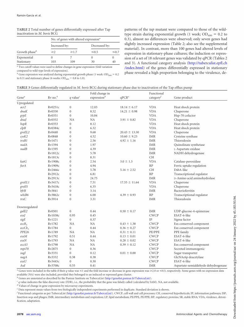
patterns of the tap mutant were compared to those of the wild-type strain during exponential growth (1 week; OD600 � 0.2 to0.3), almost no differences were observed; only seven genes hadslightly increased expression (Table 2; also see the supplementalmaterial). In contrast, more than 100 genes had altered levels ofexpression in stationary-phase cultures; the induction or repres-sion of a set of 18 relevant genes was validated by qPCR (Tables 2and 3). A functional category analysis (http://tuberculist.epfl.ch/index.html) of the genes differentially expressed in stationaryphase revealed a high proportion belonging to the virulence, de-
TABLE 2 Total number of genes differentially expressed after Tapinactivation in M. bovis BCG
No. of genes with altered expressiona
Growth phaseb
Increased by: Decreased by:
�2 �1.7 �0.5 �0.7
Exponential 0 7 0 0Stationary 103 109 30 40a Two cutoff values were used to define changes in gene expression (fold variationcompared to wild-type levels of expression).b Gene expression was analyzed during exponential growth phase (1 week: OD600 � 0.2to 0.3) and stationary phase (6 weeks: OD600 � 0.8 to 1.0).
TABLE 3 Genes differentially regulated in M. bovis BCG during stationary phase due to inactivation of the Tap efflux pump
Genea Rv no.b q valuec
Fold change inexpressiond qPCRe
Functionalcategoryf Gene product
Upregulatedacr2 Rv0251c 0 12.03 18.14 � 6.17 VDA Heat shock proteindnaK Rv0350 0 8.52 14.21 � 0.98 VDA ChaperonegrpE Rv0351 0 10.84 VDA Hsp-70 cofactordnaJ Rv0352 NA NA 3.91 � 0.82 VDA ChaperonehspR Rv0353 0 8.12 VDA Heat shock proteinclpB Rv0384c 0 6.32 VDA Heat shock proteingroEL2 Rv0440 0 9.68 20.43 � 13.50 VDA ChaperonecysK2 Rv0848 0 4.52 10.60 � 9.23 IMR Cysteine synthasetrxB1 Rv1471 0 2.56 4.92 � 1.16 IMR ThioredoxinnadA Rv1594 0 1.97 IMR Quinolinate synthetasenadB Rv1595 0 4.39 IMR L-Aspartate oxidase
Rv1812c 0 5.78 IMR NADH dehydrogenaseRv1813c 0 8.31 CH
katG Rv1908c 0 2.54 3.0 � 1.3 VDA Catalase-peroxidasefurA Rv1909c 0 4.94 RP Ferric uptake regulation
Rv2466c 0 5.78 5.16 � 2.52 CH DsbA-likeRv2912c 0 6.81 RP Transcriptional regulatorRv2913c 0 24.75 IMR D-Amino acid aminohydrolase
groEL1 Rv3417c 0 7.51 17.55 � 11.64 VDA ChaperonegroES Rv3418c 0 6.35 VDA ChaperonebfrB Rv3841 0 3.14 IMR BacterioferritinwhiB6 Rv3862c 0 6.00 4.39 � 0.93 RP Transcriptional regulatortrxC Rv3914 0 3.31 IMR Thioredoxin
DownregulatedgalE2 Rv0501 0 0.44 0.50 � 0.17 IMR UDP-glucose-4-epimeraseesxJ Rv1038c 0.95 0.45 CWCP ESAT-6-likesigE Rv1221 0 0.37 IP Sigma factoreccB5 Rv1782 NA NA 0.43 � 1.38 CWCP Esx conserved componenteccCb5 Rv1784 0 0.44 0.36 � 0.27 CWCP Esx conserved componentPPE26 Rv1789 NA NA 0.31 � 0.11 PE/PPE PPE familyesxM Rv1792 0.51 0.44 0.13 � 0.01 CWCP ESAT-6-likeesxN Rv1793 NA NA 0.28 � 0.02 CWCP ESAT-6-likeeccA5 Rv1798 NA NA 0.39 � 0.12 CWCP Esx conserved componentmpt70 Rv2875 0 0.36 CWCP Secreted immunogenicsugI Rv3331 0 0.12 0.01 � 0.00 CWCP Sugar transporternagA Rv3332 0.38 0.38 CWCP GlcNAc6p deacetylaseesxU Rv3445c 0 0.30 CWCP ESAT-6-likeAsd Rv3708c 0.55 0.41 IMR Aspartate-semialdehyde dehydrogenase
a Genes were included in the table if their q value was �1 and the fold increase or decrease in gene expression was �2.0 or �0.5, respectively. Some genes with no expression dataavailable (NA) were also included, provided they belonged to an induced or repressed gene cluster.b Genes are annotated as described by the Pasteur Institute on TubercuList (http://genolist.pasteur.fr/TubercuList/).c q value indicates the false discovery rate (FDR), i.e., the probability that the gene was falsely called (calculated by SAM). NA, not available.d Values of change in gene expression by microarray experiments.e Data represent mean values from two biologically independent experiments performed in duplicate. Standard deviation is shown.f Functional categories as per TubercuList (http://genolist.pasteur.fr/TubercuList/). CWCP, cell wall and cell processes; CH, conserved hypotheticals; IP, information pathways; ISP,Insertion seqs and phages; IMR, intermediary metabolism and respiration; LP, lipid metabolism; PE/PPE, PE/PPE; RP, regulatory proteins; SR, stable RNA; VDA, virulence, detoxi-fication, adaptation.
Ramón-García et al.
2078 aac.asm.org Antimicrobial Agents and Chemotherapy
Dow
nloa
ded
from
http
s://j
ourn
als.
asm
.org
/jour
nal/a
ac o
n 24
Jan
uary
202
2 by
45.
183.
93.9
4.
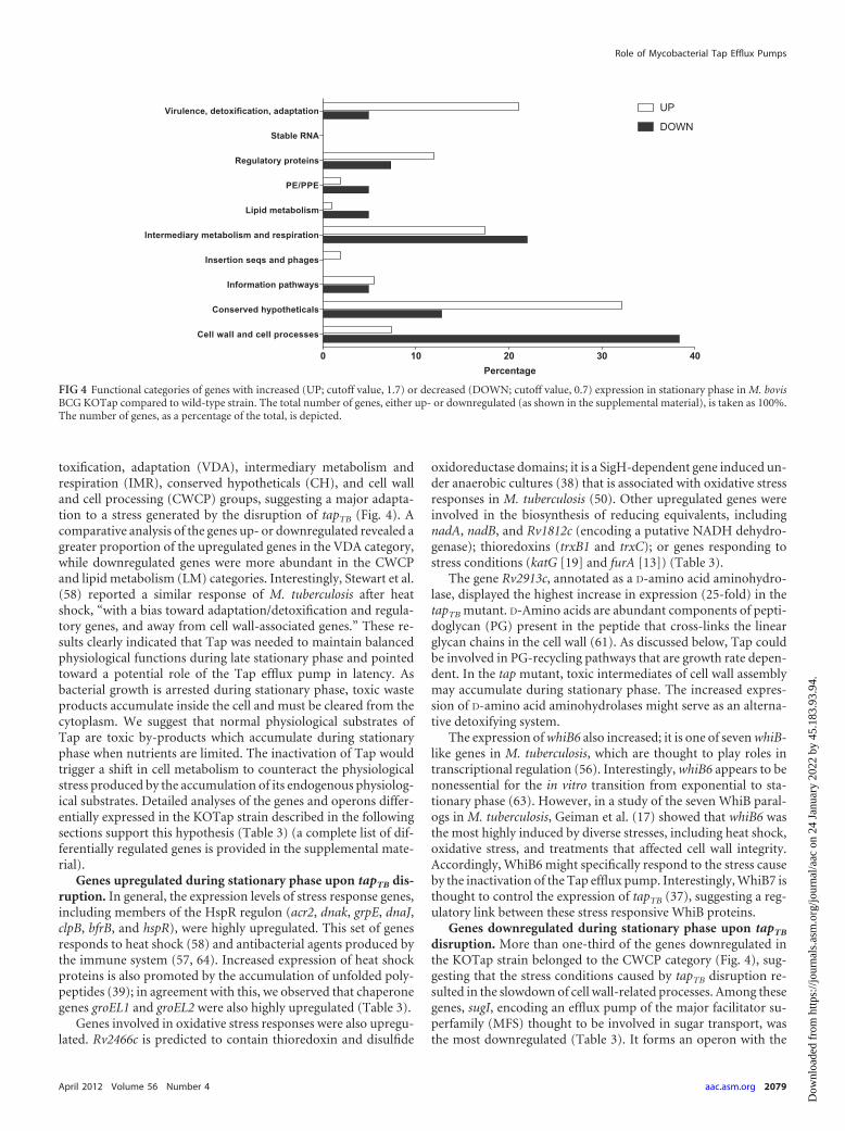
toxification, adaptation (VDA), intermediary metabolism andrespiration (IMR), conserved hypotheticals (CH), and cell walland cell processing (CWCP) groups, suggesting a major adapta-tion to a stress generated by the disruption of tapTB (Fig. 4). Acomparative analysis of the genes up- or downregulated revealed agreater proportion of the upregulated genes in the VDA category,while downregulated genes were more abundant in the CWCPand lipid metabolism (LM) categories. Interestingly, Stewart et al.(58) reported a similar response of M. tuberculosis after heatshock, “with a bias toward adaptation/detoxification and regula-tory genes, and away from cell wall-associated genes.” These re-sults clearly indicated that Tap was needed to maintain balancedphysiological functions during late stationary phase and pointedtoward a potential role of the Tap efflux pump in latency. Asbacterial growth is arrested during stationary phase, toxic wasteproducts accumulate inside the cell and must be cleared from thecytoplasm. We suggest that normal physiological substrates ofTap are toxic by-products which accumulate during stationaryphase when nutrients are limited. The inactivation of Tap wouldtrigger a shift in cell metabolism to counteract the physiologicalstress produced by the accumulation of its endogenous physiolog-ical substrates. Detailed analyses of the genes and operons differ-entially expressed in the KOTap strain described in the followingsections support this hypothesis (Table 3) (a complete list of dif-ferentially regulated genes is provided in the supplemental mate-rial).
Genes upregulated during stationary phase upon tapTB dis-ruption. In general, the expression levels of stress response genes,including members of the HspR regulon (acr2, dnak, grpE, dnaJ,clpB, bfrB, and hspR), were highly upregulated. This set of genesresponds to heat shock (58) and antibacterial agents produced bythe immune system (57, 64). Increased expression of heat shockproteins is also promoted by the accumulation of unfolded poly-peptides (39); in agreement with this, we observed that chaperonegenes groEL1 and groEL2 were also highly upregulated (Table 3).
Genes involved in oxidative stress responses were also upregu-lated. Rv2466c is predicted to contain thioredoxin and disulfide
oxidoreductase domains; it is a SigH-dependent gene induced un-der anaerobic cultures (38) that is associated with oxidative stressresponses in M. tuberculosis (50). Other upregulated genes wereinvolved in the biosynthesis of reducing equivalents, includingnadA, nadB, and Rv1812c (encoding a putative NADH dehydro-genase); thioredoxins (trxB1 and trxC); or genes responding tostress conditions (katG [19] and furA [13]) (Table 3).
The gene Rv2913c, annotated as a D-amino acid aminohydro-lase, displayed the highest increase in expression (25-fold) in thetapTB mutant. D-Amino acids are abundant components of pepti-doglycan (PG) present in the peptide that cross-links the linearglycan chains in the cell wall (61). As discussed below, Tap couldbe involved in PG-recycling pathways that are growth rate depen-dent. In the tap mutant, toxic intermediates of cell wall assemblymay accumulate during stationary phase. The increased expres-sion of D-amino acid aminohydrolases might serve as an alterna-tive detoxifying system.
The expression of whiB6 also increased; it is one of seven whiB-like genes in M. tuberculosis, which are thought to play roles intranscriptional regulation (56). Interestingly, whiB6 appears to benonessential for the in vitro transition from exponential to sta-tionary phase (63). However, in a study of the seven WhiB paral-ogs in M. tuberculosis, Geiman et al. (17) showed that whiB6 wasthe most highly induced by diverse stresses, including heat shock,oxidative stress, and treatments that affected cell wall integrity.Accordingly, WhiB6 might specifically respond to the stress causeby the inactivation of the Tap efflux pump. Interestingly, WhiB7 isthought to control the expression of tapTB (37), suggesting a reg-ulatory link between these stress responsive WhiB proteins.
Genes downregulated during stationary phase upon tapTB
disruption. More than one-third of the genes downregulated inthe KOTap strain belonged to the CWCP category (Fig. 4), sug-gesting that the stress conditions caused by tapTB disruption re-sulted in the slowdown of cell wall-related processes. Among thesegenes, sugI, encoding an efflux pump of the major facilitator su-perfamily (MFS) thought to be involved in sugar transport, wasthe most downregulated (Table 3). It forms an operon with the
FIG 4 Functional categories of genes with increased (UP; cutoff value, 1.7) or decreased (DOWN; cutoff value, 0.7) expression in stationary phase in M. bovisBCG KOTap compared to wild-type strain. The total number of genes, either up- or downregulated (as shown in the supplemental material), is taken as 100%.The number of genes, as a percentage of the total, is depicted.
Role of Mycobacterial Tap Efflux Pumps
April 2012 Volume 56 Number 4 aac.asm.org 2079
Dow
nloa
ded
from
http
s://j
ourn
als.
asm
.org
/jour
nal/a
ac o
n 24
Jan
uary
202
2 by
45.
183.
93.9
4.

N-acetylglucosamine-6-phosphate (GlcNAc6p) deacetylase gene,nagA, also highly downregulated, which is involved in the pathwayfor recycling the amino sugar of the PG. It is estimated that 40% to50% of the PG is degraded and reused each generation. In E. coli,NagA catalyzes the conversion of GlcNAc6p into glucosamine-6-phosphate (GlcN6P), which is then converted into UDP-GlcNAc,the main precursor of the PG biosynthetic pathway, by the GlmMand GlmU enzymes. Newton and coworkers (40) have suggested arole for SugI in the import of myo-inositol, a sugar needed formycothiol biosynthesis and as a precursor to mycobacterial mem-brane and cell wall components. If so, why would inactivation ofthe Tap efflux pump have such a dramatic effect on the expressionof the sugI-nagA operon during stationary phase but not duringexponential growth? In addition to having a structural role, aminosugars are an important energy source for bacteria because theysupply both carbon and nitrogen. This could be of particular im-portance in stationary phase, in which nutrients are limited. Inthis context, Tap could be involved in the maintenance of sugarphosphate homeostasis. The inactivation of Tap would lead toGlcN6P accumulation, causing toxicity (21, 48), which would ex-plain the upregulation of genes involved in general stress re-sponses. A model in which PG recycling is shut down to preventthe accumulation of sugar phosphates is supported by the obser-vation that other genes involved in cell wall processes are alsorepressed (Table 3). These include the galE2 gene encoding a pos-sible UDP-glucose-4-epimerase, involved in the biosynthetic ara-binogalactan-peptidoglycan complex (MAPc) pathway of M. tu-berculosis (7), and asd, encoding an L-aspartic-�-semialdehydedehydrogenase. The asd gene forms an operon with ask, an aspar-tokinase; both genes are essential for the biosynthetic pathway ofdiaminopimelate (DAP), which forms essential interpeptide PGcross-linking (45). These observations suggest a general repres-sion of genes involved in cell wall biosynthesis, in particular theformation of the PG.
Most sigma factors are involved in the regulation of stress re-sponses, nutrient adaptation, and cell differentiation. In our anal-ysis, sigma E was the only sigma factor whose expression was al-tered in the KOTap mutant strain (Table 3). The sigE gene isessential for growth in macrophages and virulence in mice (32,34). In addition, a sigE mutant is more sensitive to membrane-disrupting agents and vancomycin as well as heat shock and oxi-dative compounds such as hydrogen peroxide or plumbagin (33).Interestingly, in addition to vancomycin the KOTap strain wasmore sensitive to the oxidative compounds diamide, hydrogenperoxide, and plumbagin (Tables 4 and 5), suggesting a role (di-
rect or indirect) in cell wall stability and in the maintenance of theoxidative balance within the cell, as previously described for an-other efflux pump in mycobacteria (53).
Finally, several genes (eccB5, eccCb5, eccA5, esxJ, esxM, esxN,and esxU) encoding members of the immunologically active earlysecretory antigenic target-6 (ESAT-6) family of proteins, alongwith the major secreted immunogenic Mtp70 protein, were re-pressed in the KOTap strain (Table 3). The esat-6 gene is found inthe RD1 (region of difference 1; absent from all M. bovis BCGstrains), and it has been correlated with virulence (18). We con-firmed the repression of the genes contained in the ESAT-6 genecluster region 5 of M. tuberculosis (Table 3), involved in antigensecretion (18). These results suggest a link between the Tap effluxpump and virulence.
Mycobacterial Tap efflux pumps contribute to intrinsic anti-biotic resistance in M. bovis BCG. The mycobacterial Tap effluxpumps from M. fortuitum and M. tuberculosis were originally re-ported to confer tetracycline and aminoglycoside resistance in M.smegmatis, a fast-growing Mycobacterium (2). The levels of resis-tance conferred and the substrate specificities differed between thetwo efflux pumps; in general, TapFR provided higher levels of re-sistance to a larger number of drugs than TapTB (2). TapFR usedthe electrochemical gradient to extrude tetracycline from the cell,and this efflux activity could be inhibited by several compounds,such as the protonophore carbonyl cyanide m-chlorophenyl-hydrazone (CCCP) (51). In the present study, the contribution ofthese Tap efflux pumps to antimicrobial resistance was character-
TABLE 4 Sensitivity of M. bovis BCG KOTap to redox compounds
Redox compounda
Disc load(�mol)
Inhibition zoneb (mm) forM. bovis BCG strains
Wild type KOTapc
Hydrogen peroxide 80 15 1840 13 14
Plumbagin 0.01 26 30Diamide 20 0 18a Dimethyl sulfoxide, which was used as the solvent for some of these compounds, hadno effect on the growth of either strain (data not shown).b Zones of inhibition were recorded after 21 days at 37°C.c KOTap is M. bovis BCG with the tapTB gene inactivated.
TABLE 5 Antimicrobial susceptibility of M. bovis BCG Tap-derivativestrains
Compound
MIC for M. bovis BCG strains (mg/liter)a
Wild type KOTapb PAZ11c AC48d
2=-N-Ethylnetilmicin 1 0.5 ND ND6=-N-Ethylnetilmycin 2 1 ND NDAcriflavine 1 1–0.5 4 2Chloramphenicol 4 4 8 8Clarithromycin 0.03 0.03–0.015 0.03 0.06–0.25Ethambutol 4 4 8 4Gentamicin 1 0.5 2–4 8Isoniazid 0.2 0.2 0.4 0.4p-Aminosalicylate 0.125 0.03 1 0.25Rifampin 0.012 0.012 0.012 0.025Streptomycin 0.2 0.2 0.4 1.6Spectinomycin 1 0.25 4 4Tetracycline 1 0.25 4 4Tetracycline � CCCPe ND ND 1 1Triclosan 8 4 32 8–16Vancomycin 10 5 20–40 40a MICs were assayed over a range of 2-fold dilutions of antibiotics. No difference insensitivity between wild-type and Tap-derivative strains was observed for the followingcompounds: amikacin (MICWT � 0.125 mg/liter), carbonyl cyanidem-chlorophenylhydrazone (MICWT � 5 mg/liter), ciprofloxacin (MICWT � 0.125 mg/liter), clofazimine (MICWT � 0.05 to 0.1 mg/liter), chlorpromazine (MICWT � 4 mg/liter), econazole (MICWT � 6.4 mg/liter), ethionamide (MICWT � 4 mg/liter),fluconazole (MICWT, �64 mg/liter), gatifloxacin (MICWT � 0.03 mg/liter),moxifloxacin (MICWT � 0.03 to 0.06 mg/liter), and ofloxacin (MICWT � 0.25mg/liter). ND, not determined.b KOTap, M. bovis BCG with the tapTB gene inactivated.c PAZ11, M. bovis BCG strain with the pPAZ11 plasmid (tapTB cloned into pSUM36).d AC48, M. bovis BCG containing pAC48 plasmid (tapFR cloned in pSUM36).e CCCP was present at a concentration of 2 mg/liter.
Ramón-García et al.
2080 aac.asm.org Antimicrobial Agents and Chemotherapy
Dow
nloa
ded
from
http
s://j
ourn
als.
asm
.org
/jour
nal/a
ac o
n 24
Jan
uary
202
2 by
45.
183.
93.9
4.

ized in M. bovis BCG, a much closer relative to M. tuberculosis thanM. smegmatis.
M. bovis BCG derivatives were constructed overexpressingtapTB or tapFR genes, or with the tapTB gene interrupted by theinsertion of a hygromycin resistance cassette (Table 1; Fig. 1).Antimicrobial susceptibility to a set of compounds with diversecellular targets was determined for these strains (Table 5). In gen-eral, variations in sensitivity due to the interruption of the tapTB
gene were lower (2- to 4-fold) than those due to overexpressingeither tapTB or tapFR (4- to 8-fold). The substrate specificity profilewas also larger when either tap gene (tapTB or tapFR) was overex-pressed; there are two possible explanations for these differences.First, the disruption of tapTB could lead to a compensatory in-crease in the levels of expression of other efflux pump genes withsimilar substrate recognition profiles. Second, it is well recognizedthat efflux pumps nonspecifically extrude a wide variety of chem-ically and structurally unrelated compounds and that constitutiveoverexpression may provide for increased resistance, a phenotypethat would not be detected in studies of efflux pump mutants (42);here, tapTB and tapFR were overexpressed on a multicopy plasmid(5 to 10 copies per cell). A �4-fold change in sensitivity was ob-served for acriflavine, gentamicin, p-aminosalicylic acid (PAS),streptomycin, spectinomycin, tetracycline, triclosan, and vanco-mycin (Table 5). The degree of drug sensitivity probably correlateswith the specificity of Tap for a particular substrate. In this regard,PAS (a second-line oral anti-TB agent), spectinomycin, and tetra-cycline were identified as specific Tap substrates in M. bovis BCGsince a �4-fold change in sensitivity was observed in mutant andoverexpressing strains compared to the wild-type strain.
Two complementary approaches were used to confirm thatthis phenotype was due to an efflux mechanism. First, tetracyclinesusceptibility assays in the presence of CCCP were carried outusing tap-overexpressing strains (Table 5); in M. bovis BCG,CCCP reduced Tap activity by inhibiting the electrochemical gra-dient across the membrane, similar to what was previously ob-served in M. smegmatis (51). Second, tetracycline and acriflavineaccumulation experiments were performed (Fig. 5). [3H]tetracy-cline efflux experiments showed that KOTap cells accumulatedapproximately twice as much tetracycline as wild-type cells,achieving a steady-state level of accumulation within about 3 min(Fig. 5A). Similarly, acriflavine accumulation assays were per-formed with the M. bovis PAZ11 strain (tapTB overexpression).When acriflavine is accumulated inside the cells, its fluorescenceemission is quenched upon binding to the DNA. The tapTB-over-expressing PAZ11 culture had higher levels of fluorescence (Fig.5B) and a 4-fold increase in MIC to acriflavine (Table 5). To-gether, these data showed that TapTB provided tetracycline andacriflavine resistance in M. bovis BCG by active efflux.
The contribution of Tap to intrinsic resistance to first-line an-ti-TB drugs ethambutol, rifampin, and isoniazid was first sug-gested by the fact that small increases in resistance were reproduc-ibly observed in overexpressing strains (Table 5). To validate theseresults, 10-�l portions of 10-fold serial dilutions of the culture(original inoculum, 107 cell/ml) were inoculated on agar plates ata fixed subinhibitory concentration of isoniazid (0.05 mg/liter).Under these conditions, the wild-type strain grew at a 10�2 dilu-tion; the overexpressing strain (tapTB or tapFR) grew at a 10�4
dilution (data not shown), documenting its ability to better adaptto or tolerate isoniazid.
The levels of drug resistance conferred by efflux pumps are
generally low compared to what is seen for mutations in drugtarget genes and, unlike other bacteria (46), no clinically relevantefflux pump mutations have yet been identified in M. tuberculosis.While some authors have reported increases in tapTB expressionafter antibiotic treatment in clinical isolates (20, 55), a direct cor-relation between tapTB levels of expression and its contribution toantibiotic resistance has not been demonstrated in these clinicalisolates. In fact, increased expression of an efflux pump gene uponantibiotic exposure does not necessarily mean that the antibiotic isa substrate of the efflux pump. Antibiotics might have deleteriouscellular effects beyond their molecular targets (22) and thus gen-erate other metabolic signals. It is possible that the induction ofefflux pump genes might be a response to counteract antibioticdownstream effects, including oxidative stress (16). In addition,increased expression of efflux pumps may permit bacteria to sur-vive otherwise lethal concentrations of substrate antibiotics andthus allow selection for target mutations that increase drug resis-tance levels. This phenomenon is known as “drug target resistancemasking” (14). Together, this underlines the role of efflux pumpsin the development of high levels of multidrug resistance.
FIG 5 Tetracycline and acriflavine accumulation by intact cells of M. bovisTap-derivative strains. (A) [3H]tetracycline uptake by wild-type (squares) andKOTap mutant (circles) cells. (B) Acriflavine uptake by SUM36 (squares) andthe tapTB-overexpressing PAZ11 (triangles) cells. [3H]tetracycline and acrifla-vine were added to the cells at time zero. Acriflavine fluorescence emission isquenched upon binding to the DNA; thus, in contrast to [3H]tetracycline, alower value reading indicates intracellular accumulation. The results are theaverage of three experiments, and error bars indicate standard deviations.
Role of Mycobacterial Tap Efflux Pumps
April 2012 Volume 56 Number 4 aac.asm.org 2081
Dow
nloa
ded
from
http
s://j
ourn
als.
asm
.org
/jour
nal/a
ac o
n 24
Jan
uary
202
2 by
45.
183.
93.9
4.

Importantly, in an era in which tuberculosis (TB) therapeuticoptions are limited and multidrug-resistant (MDR) and exten-sively drug-resistant (XDR) TB strains are on the rise (23), a Tapinhibitor could provide new strategies for TB therapy allowingshortened treatment, perhaps by targeting the drug-tolerant sub-population of cells (1). One interesting approach to accelerate thisprocess would be to identify drugs clinically approved for othertherapeutic applications as Tap inhibitors, such as verapamil (1).This approach, known as “repurposing” (3), would allow thetimely introduction of new TB therapies. A recent report validatedthis concept in vivo. Louw et al. (28) infected mice with anMDR-TB strain and showed improved efficacy of rifampin whenadministered in combination with verapamil. However, the use ofverapamil for TB therapy generates concerns about the pharma-cological effects on cardiac conduction and potential extrapyra-midal disorders associated with calcium channel blockers (59).
Perspectives. In this study, we have characterized the impor-tant physiological role of Tap in stationary phase (probablythrough its contribution to the maintenance of the cell wall struc-ture) and to intrinsic drug resistance. Our results build on thestudy by Adams et al. (1), suggesting the importance of Tap indrug tolerance. This subpopulation of M. tuberculosis cells toler-ant to anti-TB drugs is one of the causes of prolonged TB therapy.Therefore, the development of Tap inhibitors could provide avaluable new weapon to combat TB. Such inhibitors would haveimportant therapeutic applications. First, they would allow theintroduction of already available although clinically non-TB-ef-fective antibiotics such as spectinomycin or tetracycline and makesecond-line anti-TB drugs such as PAS and streptomycin moreeffective. Second, assuming that Tap has a primary role in thedevelopment of antibiotic tolerance, such an inhibitor would po-tentially reduce the duration of the current standard treatment.However, further research needs to be performed to validate thesehypotheses, which could provide new avenues for TB therapy.
ACKNOWLEDGMENTS
We are grateful to Roberta Provvedi for helpful advice with the microarrayexperiment. We also thank Dmitry Apel, Jesús Gonzalo, and Ainhoa Ar-bués for microscopy technical support and Erin Gaynor for use of thefluorescence microscope.
This work was supported by Spanish Ministry of Science and Innova-tion grant BIO2009-09405 (J.A.A.), by FAR 2010 of the University ofPavia (E.D.R.), European Community’s Seventh Framework Programme(FP7/2007–2013) under grant agreement N. 260872 (R.M.), and The Ca-nadian Institute of Health Research to C.J.T. (MOP-82745 and MOP-82855). This work was also supported by fellowships from the SpanishMinistry of Science and Education (AP2001-1114), the European Molec-ular Biology Organization (ASTF 74.00-06), and the Programa EuropaXXI de Estancias de Investigación from the Government of Aragon(Spain) and the Caja de Ahorros de la Immaculada (S.R.-G.).
REFERENCES1. Adams KN, et al. 2011. Drug tolerance in replicating mycobacteria me-
diated by a macrophage-induced efflux mechanism. Cell 145:39 –53.2. Ainsa JA, et al. 1998. Molecular cloning and characterization of Tap, a
putative multidrug efflux pump present in Mycobacterium fortuitum andMycobacterium tuberculosis. J. Bacteriol. 180:5836 –5843.
3. Boguski MS, Mandl KD, Sukhatme VP. 2009. Drug discovery. Repur-posing with a difference. Science 324:1394 –1395.
4. Brosch R, et al. 2000. Comparative genomics uncovers large tandemchromosomal duplications in Mycobacterium bovis BCG Pasteur. Yeast17:111–123.
5. Camacho LR, et al. 2001. Analysis of the phthiocerol dimycocerosate
locus of Mycobacterium tuberculosis. Evidence that this lipid is involved inthe cell wall permeability barrier. J. Biol. Chem. 276:19845–19854.
6. Camacho LR, Ensergueix D, Perez E, Gicquel B, Guilhot C. 1999.Identification of a virulence gene cluster of Mycobacterium tuberculosis bysignature-tagged transposon mutagenesis. Mol. Microbiol. 34:257–267.
7. Crick DC, Mahapatra S, Brennan PJ. 2001. Biosynthesis of the arabi-nogalactan-peptidoglycan complex of Mycobacterium tuberculosis. Glyco-biology 11:107R–118R.
8. De Rossi E, Ainsa JA, Riccardi G. 2006. Role of mycobacterial effluxtransporters in drug resistance: an unresolved question. FEMS Microbiol.Rev. 30:36 –52.
9. De Rossi E, et al. 2002. The multidrug transporters belonging to majorfacilitator superfamily in Mycobacterium tuberculosis. Mol. Med. 8:714 –724.
10. De Rossi E, et al. 1998. Molecular cloning and functional analysis of anovel tetracycline resistance determinant, tet(V), from Mycobacteriumsmegmatis. Antimicrob. Agents Chemother. 42:1931–1937.
11. Dhamdhere G, Zgurskaya HI. 2010. Metabolic shutdown in Escherichiacoli cells lacking the outer membrane channel TolC. Mol. Microbiol. 77:743–754.
12. Dziadek J, Madiraju MV, Rutherford SA, Atkinson MA, RajagopalanM. 2002. Physiological consequences associated with overproduction ofMycobacterium tuberculosis FtsZ in mycobacterial hosts. Microbiology148:961–971.
13. Escolar L, Perez-Martin J, de Lorenzo V. 1999. Opening the iron box:transcriptional metalloregulation by the Fur protein. J. Bacteriol. 181:6223– 6229.
14. Fange D, Nilsson K, Tenson T, Ehrenberg M. 2009. Drug efflux pumpdeficiency and drug target resistance masking in growing bacteria. Proc.Natl. Acad. Sci. U. S. A. 106:8215– 8220.
15. Farrow MF, Rubin EJ. 2008. Function of a mycobacterial major facilitatorsuperfamily pump requires a membrane-associated lipoprotein. J. Bacte-riol. 190:1783–1791.
16. Fraud S, Poole K. 2011. Oxidative stress induction of the MexXY multi-drug efflux genes and promotion of aminoglycoside resistance develop-ment in Pseudomonas aeruginosa. Antimicrob. Agents Chemother. 55:1068 –1074.
17. Geiman DE, Raghunand TR, Agarwal N, Bishai WR. 2006. Differentialgene expression in response to exposure to antimycobacterial agents andother stress conditions among seven Mycobacterium tuberculosis whiB-likegenes. Antimicrob. Agents Chemother. 50:2836 –2841.
18. Gey Van Pittius NC, et al. 2001. The ESAT-6 gene cluster of Mycobacte-rium tuberculosis and other high G�C Gram-positive bacteria. GenomeBiol. 2:RESEARCH0044.
19. Heym B, Zhang Y, Poulet S, Young D, Cole ST. 1993. Characterizationof the katG gene encoding a catalase-peroxidase required for the isoniazidsusceptibility of Mycobacterium tuberculosis. J. Bacteriol. 175:4255– 4259.
20. Jiang X, et al. 2008. Assessment of efflux pump gene expression in aclinical isolate Mycobacterium tuberculosis by real-time reverse transcrip-tion PCR. Microb. Drug Resist. 14:7–11.
21. Kadner RJ, Murphy GP, Stephens CM. 1992. Two mechanisms forgrowth inhibition by elevated transport of sugar phosphates in Escherichiacoli. J. Gen. Microbiol. 138:2007–2014.
22. Kohanski MA, Dwyer DJ, Hayete B, Lawrence CA, Collins JJ. 2007. Acommon mechanism of cellular death induced by bactericidal antibiotics.Cell 130:797– 810.
23. Koul A, Arnoult E, Lounis N, Guillemont J, Andries K. 2011. Thechallenge of new drug discovery for tuberculosis. Nature 469:483– 490.
24. Krulwich TA, Lewinson O, Padan E, Bibi E. 2005. Do physiological rolesfoster persistence of drug/multidrug-efflux transporters? A case study.Nat. Rev. Microbiol. 3:566 –572.
25. Lau SY, Zgurskaya HI. 2005. Cell division defects in Escherichia colideficient in the multidrug efflux transporter AcrEF-TolC. J. Bacteriol. 187:7815–7825.
26. Li XZ, Nikaido H. 2009. Efflux-mediated drug resistance in bacteria: anupdate. Drugs 69:1555–1623.
27. Lomovskaya O, et al. 2001. Identification and characterization of inhib-itors of multidrug resistance efflux pumps in Pseudomonas aeruginosa:novel agents for combination therapy. Antimicrob. Agents Chemother.45:105–116.
28. Louw GE, et al. 2011. Rifampicin reduces susceptibility to ofloxacin inrifampicin-resistant Mycobacterium tuberculosis through efflux. Am. J. Re-spir. Crit. Care Med. 184:269 –276.
Ramón-García et al.
2082 aac.asm.org Antimicrobial Agents and Chemotherapy
Dow
nloa
ded
from
http
s://j
ourn
als.
asm
.org
/jour
nal/a
ac o
n 24
Jan
uary
202
2 by
45.
183.
93.9
4.

29. Louw GE, et al. 2009. A balancing act: efflux/influx in mycobacterial drugresistance. Antimicrob. Agents Chemother. 53:3181–3189.
30. Maciag A, et al. 2007. Global analysis of the Mycobacterium tuberculosisZur (FurB) regulon. J. Bacteriol. 189:730 –740.
31. Manganelli R, Dubnau E, Tyagi S, Kramer FR, Smith I. 1999. Differ-ential expression of 10 sigma factor genes in Mycobacterium tuberculosis.Mol. Microbiol. 31:715–724.
32. Manganelli R, et al. 2004. The extra cytoplasmic function sigma factorsigma(E) is essential for Mycobacterium tuberculosis virulence in mice.Infect. Immun. 72:3038 –3041.
33. Manganelli R, Provvedi R. 2010. An integrated regulatory network in-cluding two positive feedback loops to modulate the activity of sigma(E)in mycobacteria. Mol. Microbiol. 75:538 –542.
34. Manganelli R, Voskuil MI, Schoolnik GK, Smith I. 2001. The Mycobac-terium tuberculosis ECF sigma factor sigma(E): role in global gene expres-sion and survival in macrophages. Mol. Microbiol. 41:423– 437.
35. McMurry L, Petrucci RE, Jr, Levy SB. 1980. Active efflux of tetracyclineencoded by four genetically different tetracycline resistance determinantsin Escherichia coli. Proc. Natl. Acad. Sci. U. S. A. 77:3974 –3977.
36. Milano A, et al. 2009. Azole resistance in Mycobacterium tuberculosis ismediated by the MmpS5-MmpL5 efflux system. Tuberculosis (Edinb.)89:84 –90.
37. Morris RP, et al. 2005. Ancestral antibiotic resistance in Mycobacteriumtuberculosis. Proc. Natl. Acad. Sci. U. S. A. 102:12200 –12205.
38. Muttucumaru DG, Roberts G, Hinds J, Stabler RA, Parish T. 2004.Gene expression profile of Mycobacterium tuberculosis in a non-replicatingstate. Tuberculosis (Edinb.) 84:239 –246.
39. Narberhaus F. 1999. Negative regulation of bacterial heat shock genes.Mol. Microbiol. 31:1– 8.
40. Newton GL, Ta P, Bzymek KP, Fahey RC. 2006. Biochemistry of theinitial steps of mycothiol biosynthesis. J. Biol. Chem. 281:33910 –33920.
41. Nikaido H. 2009. Multidrug resistance in bacteria. Annu. Rev. Biochem.78:119 –146.
42. Nishino K, Nikaido E, Yamaguchi A. 2009. Regulation and physiologicalfunction of multidrug efflux pumps in Escherichia coli and Salmonella.Biochim. Biophys. Acta 1794:834 – 843.
43. Parish T, Stoker NG. 1998. Mycobacterial protocols. In Parish T,Stoker NG (ed), Methods in molecular biology, vol 101. HumanaPress, Totowa, NJ.
44. Parish T, Stoker NG. 2000. Use of a flexible cassette method to generatea double unmarked Mycobacterium tuberculosis tlyA plcABC mutant bygene replacement. Microbiology 146:1969 –1975.
45. Pavelka MS, Jr, Jacobs WR, Jr. 1996. Biosynthesis of diaminopimelate,the precursor of lysine and a component of peptidoglycan, is an essentialfunction of Mycobacterium smegmatis. J. Bacteriol. 178:6496 – 6507.
46. Piddock LJ. 2006. Clinically relevant chromosomally encoded multidrugresistance efflux pumps in bacteria. Clin. Microbiol. Rev. 19:382– 402.
47. Piddock LJ. 2006. Multidrug-resistance efflux pumps—not just for resis-tance. Nat. Rev. Microbiol. 4:629 – 636.
48. Plumbridge J. 2009. An alternative route for recycling of N-acetyl-glucosamine from peptidoglycan involves the N-acetylglucosamine phos-photransferase system in Escherichia coli. J. Bacteriol. 191:5641–5647.
49. Prentki P, Krisch HM. 1984. In vitro insertional mutagenesis with aselectable DNA fragment. Gene 29:303–313.
50. Raman S, et al. 2001. The alternative sigma factor SigH regulates majorcomponents of oxidative and heat stress responses in Mycobacterium tu-berculosis. J. Bacteriol. 183:6119 – 6125.
51. Ramon-Garcia S, Martin C, Ainsa JA, De Rossi E. 2006. Characteriza-tion of tetracycline resistance mediated by the efflux pump Tap fromMycobacterium fortuitum. J. Antimicrob. Chemother. 57:252–259.
52. Ramon-Garcia S, Martin C, De Rossi E, Ainsa JA. 2007. Contribution ofthe Rv2333c efflux pump (the Stp protein) from Mycobacterium tubercu-losis to intrinsic antibiotic resistance in Mycobacterium bovis BCG. J. An-timicrob. Chemother. 59:544 –547.
53. Ramon-Garcia S, Martin C, Thompson CJ, Ainsa JA. 2009. Role of theMycobacterium tuberculosis P55 efflux pump in intrinsic drug resistance,oxidative stress responses, and growth. Antimicrob. Agents Chemother.53:3675–3682.
54. Sambrook J, Russell DW. 2001. Molecular cloning: a laboratory manual,3rd ed. Cold Spring Harbor Laboratory Press, Cold Spring Harbor, NY.
55. Siddiqi N, et al. 2004. Mycobacterium tuberculosis isolate with a distinctgenomic identity overexpresses a tap-like efflux pump. Infection 32:109 –111.
56. Soliveri JA, Gomez J, Bishai WR, Chater KF. 2000. Multiple paralogousgenes related to the Streptomyces coelicolor developmental regulatory genewhiB are present in Streptomyces and other actinomycetes. Microbiology146:333–343.
57. Stewart GR, et al. 2005. The stress-responsive chaperone alpha-crystallin2 is required for pathogenesis of Mycobacterium tuberculosis. Mol. Micro-biol. 55:1127–1137.
58. Stewart GR, et al. 2002. Dissection of the heat-shock response in Myco-bacterium tuberculosis using mutants and microarrays. Microbiology 148:3129 –3138.
59. Sweetman SC, ed. 2011. Martindale: the complete drug reference. Phar-maceutical Press, London, United Kingdom.
60. Tusher VG, Tibshirani R, Chu G. 2001. Significance analysis of microar-rays applied to the ionizing radiation response. Proc. Natl. Acad. Sci.U. S. A. 98:5116 –5121.
61. van Heijenoort J. 2001. Formation of the glycan chains in the synthesis ofbacterial peptidoglycan. Glycobiology 11:25R–36R.
62. Vaquerizas JM, et al. 2005. GEPAS, an experiment-oriented pipeline forthe analysis of microarray gene expression data. Nucleic Acids Res. 33:W616 –W620.
63. Voskuil MI, Visconti KC, Schoolnik GK. 2004. Mycobacterium tubercu-losis gene expression during adaptation to stationary phase and low-oxygen dormancy. Tuberculosis (Edinb.) 84:218 –227.
64. Wilkinson KA, et al. 2005. Infection biology of a novel alpha-crystallin ofMycobacterium tuberculosis: Acr2. J. Immunol. 174:4237– 4243.
Role of Mycobacterial Tap Efflux Pumps
April 2012 Volume 56 Number 4 aac.asm.org 2083
Dow
nloa
ded
from
http
s://j
ourn
als.
asm
.org
/jour
nal/a
ac o
n 24
Jan
uary
202
2 by
45.
183.
93.9
4.