functional characterization of 21 rare allelic cyp1a2
TRANSCRIPT

Journal of
Personalized
Medicine
Article
Functional Characterization of 21 Rare Allelic CYP1A2 VariantsIdentified in a Population of 4773 Japanese Individuals byAssessing Phenacetin O-Deethylation
Masaki Kumondai 1,2,† , Evelyn Marie Gutiérrez Rico 1,†, Eiji Hishinuma 3,4, Yuya Nakanishi 1, Shuki Yamazaki 1,Akiko Ueda 3, Sakae Saito 3,4, Shu Tadaka 4, Kengo Kinoshita 4, Daisuke Saigusa 4, Tomoki Nakayoshi 5,Akifumi Oda 5, Noriyasu Hirasawa 1,2,3 and Masahiro Hiratsuka 1,2,3,4,*
�����������������
Citation: Kumondai, M.; Gutiérrez
Rico, E.M.; Hishinuma, E.; Nakanishi,
Y.; Yamazaki, S.; Ueda, A.; Saito, S.;
Tadaka, S.; Kinoshita, K.; Saigusa, D.;
et al. Functional Characterization of
21 Rare Allelic CYP1A2 Variants
Identified in a Population of 4773
Japanese Individuals by Assessing
Phenacetin O-Deethylation. J. Pers.
Med. 2021, 11, 690. https://doi.org/
10.3390/jpm11080690
Academic Editor: Su-Jun Lee
Received: 11 June 2021
Accepted: 20 July 2021
Published: 22 July 2021
Publisher’s Note: MDPI stays neutral
with regard to jurisdictional claims in
published maps and institutional affil-
iations.
Copyright: © 2021 by the authors.
Licensee MDPI, Basel, Switzerland.
This article is an open access article
distributed under the terms and
conditions of the Creative Commons
Attribution (CC BY) license (https://
creativecommons.org/licenses/by/
4.0/).
1 Laboratory of Pharmacotherapy of Life-Style Related Diseases, Graduate School of Pharmaceutical Sciences,Tohoku University, Sendai 980-8578, Japan; [email protected] (M.K.);[email protected] (E.M.G.R.); [email protected] (Y.N.); [email protected] (S.Y.);[email protected] (N.H.)
2 Department of Pharmaceutical Sciences, Tohoku University Hospital, Sendai 980-8574, Japan3 Advanced Research Center for Innovations in Next-Generation Medicine, Tohoku University,
Sendai 980-8575, Japan; [email protected] (E.H.); [email protected] (A.U.);[email protected] (S.S.)
4 Tohoku Medical Megabank Organization, Tohoku University, Sendai 980-8575, Japan;[email protected] (S.T.); [email protected] (K.K.); [email protected] (D.S.)
5 Faculty of Pharmacy, Meijo University, Nagoya 468-8503, Japan; [email protected] (T.N.);[email protected] (A.O.)
* Correspondence: [email protected]; Tel./Fax: +81-22-717-7049† Co-first author, these authors contributed equally to this work.
Abstract: Cytochrome P450 1A2 (CYP1A2), which accounts for approximately 13% of the total hepaticcytochrome content, catalyzes the metabolic reactions of approximately 9% of frequently used drugs,including theophylline and olanzapine. Substantial inter-individual differences in enzymatic activityhave been observed among patients, which could be caused by genetic polymorphisms. Therefore,we functionally characterized 21 novel CYP1A2 variants identified in 4773 Japanese individuals bydetermining the kinetic parameters of phenacetin O-deethylation. Our results showed that mostof the evaluated variants exhibited decreased or no enzymatic activity, which may be attributedto potential structural alterations. Notably, the Leu98Gln, Gly233Arg, Ser380del Gly454Asp, andArg457Trp variants did not exhibit quantifiable enzymatic activity. Additionally, three-dimensional(3D) docking analyses were performed to further understand the underlying mechanisms behindvariant pharmacokinetics. Our data further suggest that despite mutations occurring on the proteinsurface, accumulating interactions could result in the impairment of protein function through thedestabilization of binding regions and changes in protein folding. Therefore, our findings provideadditional information regarding rare CYP1A2 genetic variants and how their underlying effectscould clarify discrepancies noted in previous phenotypical studies. This would allow the improve-ment of personalized therapeutics and highlight the importance of identifying and characterizingrare variants.
Keywords: cytochrome P450 1A2; genetic variation; phenacetin; drug metabolism
1. Introduction
The individual impact and clinical significance of genetic factors on drug metabolismhave been well established in vivo and in vitro [1–5]. Several gene-coding alleles fordrug-metabolizing enzymes and transporters correspond to distinct phenotypes [6,7],making it necessary to account for distinctive environmental factors and inter-individualgenetic differences while establishing rational prescribing decisions to ensure an optimalpharmacological response.
J. Pers. Med. 2021, 11, 690. https://doi.org/10.3390/jpm11080690 https://www.mdpi.com/journal/jpm

J. Pers. Med. 2021, 11, 690 2 of 17
The drug-metabolizing enzyme cytochrome P450 1A2 (CYP1A2) is constitutivelyexpressed in the human liver and accounts for approximately 13% of the total hepaticcytochrome (CYP) content [8,9]. It catalyzes the metabolism of approximately 9% of com-monly used drugs, including the oxidation of drugs such as theophylline, olanzapine, andpropranolol [10,11]. CYP1A2 plays an important role in the metabolism of psychotropicdrugs and other drugs targeting the central nervous system, either involved in the catalysisof the main metabolic route or directly inhibited by these drugs. Previous studies haveidentified a 40- to 130-fold inter-individual difference in CYP1A2 activity and a 40-folddifference in protein expression among individuals; of these, 35–75% of differences areattributed to genetic factors [12]. To date, 40 polymorphic variants of the CYP1A2 genehave been reported (https://www.pharmvar.org/gene/CYP1A2 accessed on 21 July 2021),of which the most frequent and most highly characterized variant is the -163C>A poly-morphism (CYP1A2*1F), located in the promoter region. High frequencies of this varianthave been reported in populations worldwide, including among the Japanese population(60%) [13,14]. To date, several studies have examined the effects of polymorphisms ofthe CYP1A2 gene on enzyme inducibility. Examples include a report of decreased bloodlevels of olanzapine in schizophrenic patients with the -163C>A polymorphism, and onediscussing increased clearance of theophylline in non-smoker asthmatic patients carryingthe -3860C>A polymorphism [15,16].
In a continuous effort to improve medical outcomes, identify potential risks asso-ciated with treatments, and establish population-specific clinical tools (including dos-ing algorithms and adverse effect predictors), the Tohoku Medical Megabank Organiza-tion (ToMMo) reported whole-genome sequences of 4773 Japanese individuals (https://www.megabank.tohoku.ac.jp/english/timeline/20190913_01 accessed on 23 March2021) up to September 2019. Twenty-one single-nucleotide variations were identifiedin the regions coding for CYP1A2. Amino acid substitutions may result in alterations inthe CYP1A2 protein structure and consequently affect enzyme function; this, in turn, maysignificantly affect drug efficacy and increase the risk of adverse drug reactions. Whilethe importance of evaluating the clinical impact of these variants in patients is paramountto predicting treatment outcomes, polymorphic variants of the CYP1A2 gene causingprotein structure alteration are rare within the Japanese population, with a frequencyof approximately only 1% [17–19]. This, along with the increased patient risk and thepotential for adverse effects specific to CYP1A2 substrates, makes it challenging to conductclinical trials. Therefore, in vitro functional analysis of CYP1A2 variants has been widelyperformed [3,5,20–22], including our previous comprehensive analysis of the enzymaticfunction of 19 CYP1A2 variants. Such analyses make it possible to identify the causes ofinter-individual variations in response to endogenous substances, and to devise strategiesto identify patients at risk of therapeutic failure or adverse drug reactions.
This study aimed to comprehensively elucidate the in vitro functional changes ofthe 21 novel structural variants arising from CYP1A2 gene polymorphisms, which wereidentified in 4773 Japanese individuals. We conducted recombinant protein expression in293FT cells, with co-expression of cytochrome P450 oxidoreductase (CPR) and cytochromeb5. Characterization of enzymatic activity was performed using phenacetin as the pre-ferred probe for the in vitro screening of CYP1A2-based metabolism [23–25]. To furtheranalyze the mechanisms underlying these differences, carbon monoxide (CO)-differencespectroscopy and three-dimensional (3D) docking analyses were performed.
2. Materials and Methods2.1. Chemicals
Reagents used in this study were purchased from the following sources: phenacetin(Nacalai Tesque, Kyoto, Japan); 4-acetamidophenol (Sigma-Aldrich, St. Louis, MO, USA);4-acetamidophenol-d4 (Cayman Chemical, Ann Arbor, MI, USA); oxidized β-nicotinamide-adenine dinucleotide phosphate (NADP+), glucose-6-phosphate (G-6-P), glucose-6-phosphatedehydrogenase (G-6-PDH), reduced β-nicotinamide-adenine dinucleotide (NADH), ox-

J. Pers. Med. 2021, 11, 690 3 of 17
idized β-nicotinamide-adenine dinucleotide, and reduced β-nicotinamide-adenine din-ucleotide phosphate (NADPH) (Oriental Yeast, Tokyo, Japan); polyclonal anti-humanCYP1A2 antibody (cat. no. ab170204; Abcam, Cambridge, UK); horseradish peroxidase(HRP)-conjugated goat anti-rabbit IgG (ProteinSimple, Tokyo, Japan); and sodium cyanideand cytochrome c from horse hearts (Nacalai Tesque, Kyoto, Japan). All other chemicalsand reagents were of the highest quality and commercially available.
2.2. Sanger Sequencing Analysis for the Detection of CYP1A2 Sequence Alterations
Peripheral blood leukocytes were isolated from the whole blood of participatingJapanese subjects of the community-based cohort study conducted by ToMMo. Writteninformed consent was obtained from all subjects prior to sample collection [26]. Polymerasechain reaction (PCR) amplification of the genomic DNA extracted from the cells wasconducted using a Gentra Puregene Blood Kit (Qiagen, Hilden, Germany). More than10 ng genomic DNA, 2 × AmpliTaq Gold 360 Master Mix (Applied Biosystems, FosterCity, CA, USA), and 0.5 µM of each primer (Supplementary Table S1), in a total volume of20 µL were used for PCR amplification. The thermal cycling conditions included an initialdenaturation at 95 ◦C for 10 min, followed by 30 cycles of denaturation at 95 ◦C for 30 s,annealing at 60 ◦C for 30 s, extension at 72 ◦C for 1 min (exons 2, 4 and 5) or 30 s (exons3, 6, and 7), and a final extension at 72 ◦C for 7 min. The PCR products were purified ona column and analyzed by Sanger sequencing using the same primers for each exon asemployed for PCR.
2.3. Expression of CYP1A2 Variants in 293FT Cells
Wild-type and variant CYP1A2 cDNAs subcloned into the pcDNA3.4 vector werepurchased from GenScript (Piscataway, NJ, USA). Plasmids carrying CPR or cytochrome b5cDNAs subcloned into the pcDNA3.4 vector were prepared as previously described [27].293FT cells (ThermoFisher Scientific, Waltham, MA, USA) were cultured in Dulbecco’s mod-ified Eagle’s medium (Nacalai Tesque) containing 10% fetal bovine serum at 37 ◦C under5% CO2. Microsomal fractions containing each CYP1A2 variant, CPR, and cytochrome b5were prepared according to the previously described protocol [27]. Protein concentrationswere determined using a BCA Protein Assay Kit (ThermoFisher Scientifics).
2.4. Western Blotting
Immunoassays were performed using the Wes (ProteinSimple, San Jose, CA, USA)and Compass for SW ver. 4.1.0 (ProteinSimple) software to evaluate CYP1A2 proteinexpression levels. Briefly, 100 ng of microsomes containing CYP1A2 wild-type and variantproteins were loaded into each well. CYP1A2 levels were detected using a polyclonalanti-CYP1A2 antibody (1:100 dilution) and HRP-conjugated goat anti-rabbit IgG. A totalprotein assay was performed to normalize each signal using 100 ng of microsomes, as perthe manufacturer’s instructions [28].
2.5. Determination of Cytochrome (CYP), Cytochrome P450 Oxidoreductase (CPR), andCytochrome b5 Content
CYP1A2 holoprotein, CPR, and cytochrome b5 contents were spectrophotometricallymeasured by ultraviolet-visible spectrophotometry (Cary 300 UV-Vis spectrophotometer,Agilent Technologies, Santa Clara, CA, USA), as previously reported [27,29]. Data anal-ysis was conducted using Jasco Spectra Manager (JASCO Corporation, Sendai, Japan).Cuvettes (Sub-Micro Cells; 16.50-Q-10/Z20) were purchased from Starna Scientific, Ltd.(London, UK).
2.6. Phenacetin O-Deethylation
The extent of O-deethylation of phenacetin by CYP1A2 was measured using a modifi-cation of the method previously reported by Ito et al. [21,27]. The reaction mixture (150 µL)was composed of the microsomal fraction (25 µg), phenacetin (2.5, 5, 10, 25, 50, 100, 250,
J. Pers. Med. 2021, 11, 690 4 of 17
or 500 µM), 3.3 mM MgCl2, and 50 mM potassium phosphate buffer (pH 7.4). After pre-incubating at 37 ◦C for 3 min, reactions were initiated by adding the NADPH-generationsystem, consisting of 1.3 mM NADP+, 3.3 mM G-6-P, and 0.4 U/mL G-6-PDH. The mixturewas incubated at 37 ◦C for 40 min, before terminating the reactions by the addition of150 µL acetonitrile, containing 5 µM acetamidophenol-d4 as an internal standard. Afterprotein removal by centrifugation at 15,400× g for 10 min, 5 µL of the supernatant wasinjected into a liquid chromatography-tandem mass spectrometry system, as previouslydescribed [27].
2.7. 3D Structural Modeling of CYP1A2
The 3D structural modeling of CYP1A2 was conducted based on the X-ray structureof CYP1A2 reported by Sansen et al. (Protein Data Bank code: 2HI4) [30]. Phenacetin wascoordinated with the CYP1A2 wild-type model structure using the CDOCKER protocol ofDiscovery Studio 2.5 (BIOVIA, San Diego, CA, USA). Docking iterations were conductedconsidering the binding orientations and binding energies under the conditions previouslydescribed by Oda et al. [31]. After each substitution, structural optimization was performedas previously reported [32]. Alongside 3D docking analysis, Polymorphism PhenotypingV-2 (PolyPhen-2) [33] and Sorting Intolerant From Tolerant (SIFT) [34] predictive softwarewere used to evaluate the structural impact of the amino acid substitutions in CYP1A2variants on the functionality of the enzyme [35].
2.8. Data Analysis
Kinetic parameters (Km; Michaelis constant, Vmax; maximum velocity, and CLint; in-trinsic clearance) were determined using the Enzyme Kinetics Module of SigmaPlot 12.5(Systat Software, Inc., Chicago, IL, USA), a curve-fitting program based on non-linearregression analysis. All values are expressed as the mean ± standard deviation (SD) ofexperiments performed in triplicate. Statistical analyses for multiple comparisons wereperformed through variance analysis by Dunnett’s T3 test or the Kruskal–Wallis method,using IBM SPSS Statistics Ver. 22 (International Business Machines, Armonk, NY, USA).The normality of our datasets was initially assessed using the Shapiro-Wilk test. Differenceswere considered statistically significant when P values were less than 0.05.
3. Results
Twenty-one novel structural variants of CYP1A2 were identified in a cohort of4773 Japanese individuals by whole-genome sequencing (allele frequencies ranging from0.01–0.10%) and confirmed by Sanger sequencing, using the primer pairs listed in Sup-plementary Table S1. The structure-based enzymatic functionalities of all the variantswere predicted using the PolyPhen-2 and SIFT software. PolyPhen-2 analysis revealed atotal of ten potentially damaging substitutions, while SIFT analysis classified 12 variantsas potentially damaging based on the effects of the detected amino acid substitutions onprotein structure (Table 1).
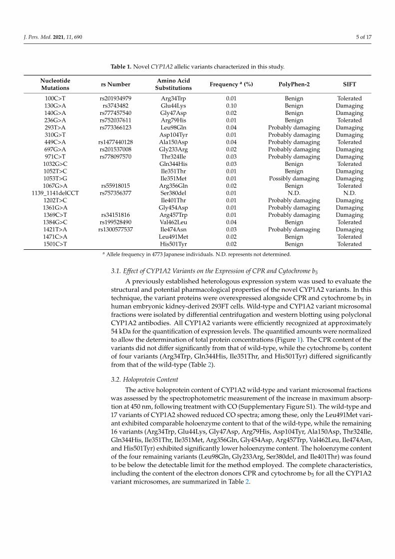
J. Pers. Med. 2021, 11, 690 5 of 17
Table 1. Novel CYP1A2 allelic variants characterized in this study.
NucleotideMutations rs Number Amino Acid
Substitutions Frequency a (%) PolyPhen-2 SIFT
100C>T rs201934979 Arg34Trp 0.01 Benign Tolerated130G>A rs3743482 Glu44Lys 0.10 Benign Damaging140G>A rs777457540 Gly47Asp 0.02 Benign Damaging236G>A rs752037611 Arg79His 0.01 Benign Tolerated293T>A rs773366123 Leu98Gln 0.04 Probably damaging Damaging310G>T Asp104Tyr 0.01 Probably damaging Damaging449C>A rs1477440128 Ala150Asp 0.04 Probably damaging Tolerated697G>A rs201537008 Gly233Arg 0.02 Probably damaging Damaging971C>T rs778097570 Thr324Ile 0.03 Probably damaging Damaging
1032G>C Gln344His 0.03 Benign Tolerated1052T>C Ile351Thr 0.01 Benign Damaging1053T>G Ile351Met 0.01 Possibly damaging Damaging1067G>A rs55918015 Arg356Gln 0.02 Benign Tolerated
1139_1141delCCT rs757356377 Ser380del 0.01 N.D. N.D.1202T>C Ile401Thr 0.01 Probably damaging Damaging1361G>A Gly454Asp 0.01 Probably damaging Damaging1369C>T rs34151816 Arg457Trp 0.01 Probably damaging Damaging1384G>C rs199528490 Val462Leu 0.04 Benign Tolerated1421T>A rs1300577537 Ile474Asn 0.03 Probably damaging Damaging1471C>A Leu491Met 0.02 Benign Tolerated1501C>T His501Tyr 0.02 Benign Tolerated
a Allele frequency in 4773 Japanese individuals. N.D. represents not determined.
3.1. Effect of CYP1A2 Variants on the Expression of CPR and Cytochrome b5
A previously established heterologous expression system was used to evaluate thestructural and potential pharmacological properties of the novel CYP1A2 variants. In thistechnique, the variant proteins were overexpressed alongside CPR and cytochrome b5 inhuman embryonic kidney-derived 293FT cells. Wild-type and CYP1A2 variant microsomalfractions were isolated by differential centrifugation and western blotting using polyclonalCYP1A2 antibodies. All CYP1A2 variants were efficiently recognized at approximately54 kDa for the quantification of expression levels. The quantified amounts were normalizedto allow the determination of total protein concentrations (Figure 1). The CPR content of thevariants did not differ significantly from that of wild-type, while the cytochrome b5 contentof four variants (Arg34Trp, Gln344His, Ile351Thr, and His501Tyr) differed significantlyfrom that of the wild-type (Table 2).
3.2. Holoprotein Content
The active holoprotein content of CYP1A2 wild-type and variant microsomal fractionswas assessed by the spectrophotometric measurement of the increase in maximum absorp-tion at 450 nm, following treatment with CO (Supplementary Figure S1). The wild-type and17 variants of CYP1A2 showed reduced CO spectra; among these, only the Leu491Met vari-ant exhibited comparable holoenzyme content to that of the wild-type, while the remaining16 variants (Arg34Trp, Glu44Lys, Gly47Asp, Arg79His, Asp104Tyr, Ala150Asp, Thr324Ile,Gln344His, Ile351Thr, Ile351Met, Arg356Gln, Gly454Asp, Arg457Trp, Val462Leu, Ile474Asn,and His501Tyr) exhibited significantly lower holoenzyme content. The holoenzyme contentof the four remaining variants (Leu98Gln, Gly233Arg, Ser380del, and Ile401Thr) was foundto be below the detectable limit for the method employed. The complete characteristics,including the content of the electron donors CPR and cytochrome b5 for all the CYP1A2variant microsomes, are summarized in Table 2.

J. Pers. Med. 2021, 11, 690 6 of 17J. Pers. Med. 2021, 11, x FOR PEER REVIEW 6 of 17
Figure 1. Representative Western blots showing immunoreactive cytochrome P450 1A2 (CYP1A2)
proteins (A) and total proteins (B). Average CYP1A2 intensity was normalized according to total
protein content (C). All assays and measurements were performed in triplicate using a single mi-
crosomal preparation. Mock 1 represents transfection with 10 μg mock plasmid. Mock 2 represents
transfection with 9.6 μg mock plasmid, 0.2 μg cytochrome P450 oxidoreductase (CPR) plasmid, and
0.2 μg cytochrome b5 plasmid. N.D. represents not determined. * p < 0.05 compared with wild-type
CYP1A2 by Kruskal–Wallis method.
Table 2. The characterization of microsomes prepared from 293FT cells co-expressed with CYP1A2 variants, CPR, and
cytochrome b5.
Variants
CYP Holoenzyme
Content
(pmol/mg Protein)
CPR Content
(pmol/mg Protein)
Cytochrome b5
Content (pmol/mg
Protein)
CYP:CPR Ratio CYP:Cytochrome b5
Ratio
Figure 1. Representative Western blots showing immunoreactive cytochrome P450 1A2 (CYP1A2)proteins (A) and total proteins (B). Average CYP1A2 intensity was normalized according to totalprotein content (C). All assays and measurements were performed in triplicate using a single mi-crosomal preparation. Mock 1 represents transfection with 10 µg mock plasmid. Mock 2 representstransfection with 9.6 µg mock plasmid, 0.2 µg cytochrome P450 oxidoreductase (CPR) plasmid, and0.2 µg cytochrome b5 plasmid. N.D. represents not determined. * p < 0.05 compared with wild-typeCYP1A2 by Kruskal–Wallis method.

J. Pers. Med. 2021, 11, 690 7 of 17
Table 2. The characterization of microsomes prepared from 293FT cells co-expressed with CYP1A2 variants, CPR, andcytochrome b5.
VariantsCYP Holoenzyme
Content(pmol/mg Protein)
CPR Content(pmol/mg Protein)
Cytochrome b5 Content(pmol/mg Protein) CYP:CPR Ratio CYP:Cytochrome
b5 Ratio
Wild-type 145.91 ± 4.11 80.52 ± 16.79 21.11 ± 2.44 1.81 6.91Arg34Trp 34.50 ± 11.84 * 135.50 ± 45.33 28.34 ± 2.11 # 0.25 1.22Glu44Lys 41.69 ± 4.62 *** 89.69 ± 20.20 27.14 ± 3.24 0.46 1.54Gly47Asp 23.21 ± 6.09 *** 98.12 ± 31.17 27.54 ± 2.01 0.24 0.84Arg79His 21.31 ± 3.02 *** 158.64 ± 24.52 27.09 ± 3.18 0.13 0.79Leu98Gln N.D. 146.98 ± 9.95 25.43 ± 1.49 N.D. N.D.Asp104Tyr 15.21 ± 1.52 *** 150.87 ± 57.19 26.46 ± 1.69 0.10 0.57Ala150Asp 20.75 ± 1.41 *** 127.57 ± 47.56 19.28 ± 1.74 0.16 1.08Gly233Arg N.D. 109.67 ± 52.40 24.82 ± 2.06 N.D. N.D.Thr324Ile 21.89 ± 6.31 *** 103.76 ± 15.09 17.46 ± 3.16 0.21 1.25Gln344His 18.83 ± 2.93 *** 156.04 ± 4.92 30.98 ± 2.52 ### 0.12 0.61Ile351Thr 36.09 ± 2.85 *** 133.09 ± 12.25 27.72 ± 2.79 # 0.27 1.30Ile351Met 43.77 ± 1.61 *** 129.07 ± 6.34 17.71 ± 1.17 0.34 2.47Arg356Gln 88.92 ± 0.88 * 129.54 ± 1.51 25.77 ± 3.52 0.69 3.45Ser380del N.D. 140.78 ± 25.66 19.72 ± 2.48 N.D. N.D.Ile401Thr N.D. 95.05 ± 33.43 18.27 ± 3.62 N.D. N.D.
Gly454Asp 12.88 ± 4.98 *** 120.75 ± 18.54 16.95 ± 1.45 0.11 0.76Arg457Trp 14.34 ± 2.24 *** 126.46 ± 16.37 17.00 ± 2.12 0.11 0.84Val462Leu 14.59 ± 5.64 *** 113.72 ± 62.19 23.71 ± 3.57 0.13 0.62Ile474Asn 33.80 ± 4.70 *** 136.52 ± 28.20 26.53 ± 3.32 0.25 1.27
Leu491Met 144.17 ± 6.56 109.22 ± 35.05 23.54 ± 1.60 1.32 6.13His501Tyr 43.22 ± 2.31 *** 141.73 ± 9.07 31.52 ± 4.02 ### 0.30 1.37
Data represent the means ± SDs of the three independently performed catalytic assays. * p < 0.05 and *** p < 0.005 compared with wild-typeCYP1A2 by Dunnett’s T3 test. # p < 0.05 and ### p < 0.005 compared with wild-type CYP1A2 by Dunnett’s t-test. N.D. represents notdetermined. All assays and measurements were performed in triplicate using a single microsomal preparation.
3.3. O-Deethylation of Phenacetin
Phenacetin O-deethylation activity was measured using a modification of previouslydescribed methods; kinetic parameters were subsequently determined using Michaelis–Menten curves (Figure 2). Enzymatic activities were normalized to the correspondingCYP1A2 total protein quantified by Western blotting (Table 3). The O-deethylation ofphenacetin was assessed using microsomal protein fractions containing wild-type or vari-ant CYP1A2 (25 µg); this revealed that 4-acetamidophenol was formed linearly, in atime-dependent manner, for incubation times of up to 40 min (data not shown). Amongthese, 13 variants (Glu44Lys, Gly47Asp, Arg79His, Asp104Tyr, Ala150Asp, Gly233Arg,Thr324Ile, Ile351Thr, Ile351Met, Arg356Gln, Ile401Thr, Val462Leu, Ile474Asn, Leu491Met,and His501Tyr) were found to show significantly lower CLint and Vmax values than the wild-type. Conversely, the Km values for all the variants evaluated did not differ significantlyfrom those of the wild-type. Regarding Gln344His, this variant was found to have a CLintvalue slightly lower than that of the wild-type, while its Vmax and Km values did not differsignificantly. The kinetic parameters of the remaining five variants (Leu98Gln, Gly233Arg,Ser380del, Gly454Asp, and Arg457Trp) could not be determined, since 4-acetamidophenolformation was negligible.

J. Pers. Med. 2021, 11, 690 8 of 17
J. Pers. Med. 2021, 11, x FOR PEER REVIEW 8 of 17
wild-type. Conversely, the Km values for all the variants evaluated did not differ signifi-
cantly from those of the wild-type. Regarding Gln344His, this variant was found to have
a CLint value slightly lower than that of the wild-type, while its Vmax and Km values did not
differ significantly. The kinetic parameters of the remaining five variants (Leu98Gln,
Gly233Arg, Ser380del, Gly454Asp, and Arg457Trp) could not be determined, since 4-ac-
etamidophenol formation was negligible.
Figure 2. Michaelis–Menten curves for CYP1A2 variants. Determined kinetic parameters of phenac-
etin O-deethylation. All assays and measurements were performed in triplicate using a single mi-
crosomal preparation.
Table 3. Kinetic parameters of phenacetin O-deethylation by microsomes from 293FT cells express-
ing wild-type and variant CYP1A2 proteins.
Variants Km (μM) Vmax
(pmol/min/mg Protein)
CLint
(µL/min/mg Protein)
(% of Wild-Type)
Wild-type 10.14 ± 0.68 972.99 ± 39.96 96.10 ± 2.47 (100.00)
Arg34Trp 12.97 ± 0.65 1224.92 ± 48.86 94.51 ± 1.30 (98.35)
Glu44Lys 8.86 ± 0.27 462.54 ± 12.05 * 52.23 ± 2.04 *** (54.35)
Gly47Asp 7.42 ± 0.32 142.46 ± 3.76 ** 19.22 ± 0.50 *** (20.00)
Arg79His 11.06 ± 0.74 599.00 ± 17.84 * 54.23 ± 1.99 *** (56.43)
Leu98Gln N.D. N.D. N.D.
Asp104Tyr 7.83 ± 0.43 234.53 ± 5.62 ** 30.04 ± 2.06 *** (31.26)
Ala150Asp 7.71 ± 0.27 178.11 ± 3.53 ** 23.12 ± 1.13 *** (24.06)
Gly233Arg N.D. N.D. N.D.
Thr324Ile 110.07 ± 2.08 153.36 ± 0.59 ** 1.39 ± 0.02 *** (1.45)
Gln344His 13.18 ± 0.30 1019.25 ± 28.36 77.36 ± 2.97 * (80.50)
Ile351Thr 12.92 ± 1.10 473.86 ± 16.59 ** 36.93 ± 4.50 *** (38.44)
Ile351Met 7.37 ± 0.58 285.80 ± 16.34 *** 38.85 ± 0.84 *** (40.43)
Arg356Gln 8.03 ± 0.21 423.55 ± 25.17 *** 52.74 ± 1.75 *** (54.88)
Ser380del N.D. N.D. N.D.
Ile401Thr 2.31 ± 0.26 57.05 ± 1.90 ** 24.88 ± 2.09 *** (25.89)
Gly454Asp N.D. N.D. N.D.
Arg457Trp N.D. N.D. N.D.
Val462Leu 9.97 ± 1.39 334.57 ± 8.54 ** 34.06 ± 5.64 ** (35.44)
Ile474Asn 12.41 ± 1.45 511.48 ± 10.98 * 41.63 ± 5.48 * (43.31)
Figure 2. Michaelis–Menten curves for CYP1A2 variants. Determined kinetic parameters of phenacetin O-deethylation. Allassays and measurements were performed in triplicate using a single microsomal preparation.
Table 3. Kinetic parameters of phenacetin O-deethylation by microsomes from 293FT cells expressing wild-type and variantCYP1A2 proteins.
Variants Km (µM) Vmax(pmol/min/mg Protein)
CLint(µL/min/mg Protein)
(% of Wild-Type)
Wild-type 10.14 ± 0.68 972.99 ± 39.96 96.10 ± 2.47 (100.00)Arg34Trp 12.97 ± 0.65 1224.92 ± 48.86 94.51 ± 1.30 (98.35)Glu44Lys 8.86 ± 0.27 462.54 ± 12.05 * 52.23 ± 2.04 *** (54.35)Gly47Asp 7.42 ± 0.32 142.46 ± 3.76 ** 19.22 ± 0.50 *** (20.00)Arg79His 11.06 ± 0.74 599.00 ± 17.84 * 54.23 ± 1.99 *** (56.43)Leu98Gln N.D. N.D. N.D.Asp104Tyr 7.83 ± 0.43 234.53 ± 5.62 ** 30.04 ± 2.06 *** (31.26)Ala150Asp 7.71 ± 0.27 178.11 ± 3.53 ** 23.12 ± 1.13 *** (24.06)Gly233Arg N.D. N.D. N.D.Thr324Ile 110.07 ± 2.08 153.36 ± 0.59 ** 1.39 ± 0.02 *** (1.45)Gln344His 13.18 ± 0.30 1019.25 ± 28.36 77.36 ± 2.97 * (80.50)Ile351Thr 12.92 ± 1.10 473.86 ± 16.59 ** 36.93 ± 4.50 *** (38.44)Ile351Met 7.37 ± 0.58 285.80 ± 16.34 *** 38.85 ± 0.84 *** (40.43)Arg356Gln 8.03 ± 0.21 423.55 ± 25.17 *** 52.74 ± 1.75 *** (54.88)Ser380del N.D. N.D. N.D.Ile401Thr 2.31 ± 0.26 57.05 ± 1.90 ** 24.88 ± 2.09 *** (25.89)
Gly454Asp N.D. N.D. N.D.Arg457Trp N.D. N.D. N.D.Val462Leu 9.97 ± 1.39 334.57 ± 8.54 ** 34.06 ± 5.64 ** (35.44)Ile474Asn 12.41 ± 1.45 511.48 ± 10.98 * 41.63 ± 5.48 * (43.31)
Leu491Met 6.97 ± 0.41 574.36 ± 9.20 * 82.58 ± 4.18 (85.93)His501Tyr 16.83 ± 1.29 404.49 ± 14.69 ** 24.08 ± 1.13 *** (25.06)
Data represent the means ± SD of the three independently performed catalytic assays. * p < 0.05, ** p < 0.01, and *** p < 0.005 comparedwith wild-type CYP1A2 by Dunnett’s T3 tests. N.D. represents not determined. All assays and measurements were performed in triplicateusing a single microsomal preparation.
3.4. 3D Molecular Modeling
To evaluate the molecular properties of the identified novel variants, 3D structuralmodeling was performed; the structural integrity and molecular interactions withinwild-type or CYP1A2 variant proteins were assessed when coordinated with phenacetin(Figure 3).

J. Pers. Med. 2021, 11, 690 9 of 17
J. Pers. Med. 2021, 11, x FOR PEER REVIEW 9 of 17
Leu491Met 6.97 ± 0.41 574.36 ± 9.20 * 82.58 ± 4.18 (85.93)
His501Tyr 16.83 ± 1.29 404.49 ± 14.69 ** 24.08 ± 1.13 *** (25.06)
Data represent the means ± SD of the three independently performed catalytic assays. * p < 0.05, **
p < 0.01, and *** p < 0.005 compared with wild-type CYP1A2 by Dunnett’s T3 tests. N.D. represents
not determined. All assays and measurements were performed in triplicate using a single microso-
mal preparation.
3.4. 3D Molecular Modeling
To evaluate the molecular properties of the identified novel variants, 3D structural
modeling was performed; the structural integrity and molecular interactions within wild-
type or CYP1A2 variant proteins were assessed when coordinated with phenacetin (Fig-
ure 3).
Figure 3. Diagram of the overall structure of CYP1A2. The heme group is shown in red; phenacetin
is shown in blue. The distance between the central iron and the metabolic site was calculated as 4.32
Å .
The Gly233Arg substitution introduced additional interactions with several residues,
including additional hydrogen bonds with A’ helix members Val54 and Gly58; in turn,
Gly58 formed additional interactions with Leu55, which resulted in the unraveling of a
helical segment from Leu55 to Gly58. The lengthening of the carbon-hydrogen bond be-
tween the substituted Arg233 and Pro235 was also observed, resulting in interactions
causing the G’ helix (Phe239 to Leu242) to unravel. Additional hydrogen bonds were
formed between Asp320, Thr324, phenacetin, and heme in proximity to the substrate
recognition site. This resulted in the loss of several interactions present with the wild-type
enzyme, including interactions between Leu219 and Thr223; Asp320 and Gly316; π-alkyl
interactions between phenacetin and heme; π-alkyl interactions between phenacetin and
Ala317, and heme and Ala317; and π-π stacking interactions between heme, Phe451, and
Phe381 (Figure 4A).
Figure 3. Diagram of the overall structure of CYP1A2. The heme group is shown in red; phenacetin isshown in blue. The distance between the central iron and the metabolic site was calculated as 4.32 Å.
The Gly233Arg substitution introduced additional interactions with several residues,including additional hydrogen bonds with A’ helix members Val54 and Gly58; in turn,Gly58 formed additional interactions with Leu55, which resulted in the unraveling ofa helical segment from Leu55 to Gly58. The lengthening of the carbon-hydrogen bondbetween the substituted Arg233 and Pro235 was also observed, resulting in interactionscausing the G’ helix (Phe239 to Leu242) to unravel. Additional hydrogen bonds wereformed between Asp320, Thr324, phenacetin, and heme in proximity to the substraterecognition site. This resulted in the loss of several interactions present with the wild-typeenzyme, including interactions between Leu219 and Thr223; Asp320 and Gly316; π-alkylinteractions between phenacetin and heme; π-alkyl interactions between phenacetin andAla317, and heme and Ala317; and π-π stacking interactions between heme, Phe451, andPhe381 (Figure 4A).
The substitution of the highly conserved Thr324 residue with Ile324 lead to the lossof the carbon-hydrogen bond between Ile324 and Thr321 and the two hydrogen bondsbetween the Ile324 and Asp320 residues. Meanwhile, additional π-alkyl interactions wereintroduced between Ile324 and Trp328, and Phe381 and Lys500, resulting in the loss ofhydrophobic interactions between phenacetin and heme. New hydrogen bonds to Gly316were also formed, while amide π-stacking interactions involving Gly316, Ala317, andphenacetin were lost (Figure 4B).
In the Ile401Thr variant, Thr401 formed an additional interaction with Thr394, but lostπ-alkyl interactions with Leu86 and Cys405. Leu86 formed additional interactions withVal75 and Pro42. In the binding pocket, phenacetin did not show a hydrophobic interactionwith Ala317, but did form an additional hydrogen bond with Gly316. A new interactionwas introduced between heme and Ala325; however, the hydrophobic interactions betweenheme and Ile314 were lost. Additional interactions of Arg392 (attractive charges withAsp103 and carbon-hydrogen bond with Thr391) were observed, resulting in cascadinginteractions affecting Asp103 and Asp104, ultimately leading to the unwinding of thehelical structure formed by Gly102–Lys106 (Figure 4C).

J. Pers. Med. 2021, 11, 690 10 of 17J. Pers. Med. 2021, 11, x FOR PEER REVIEW 10 of 17
Figure 4. Diagram pairs showing the partial crystal structure of CYP1A2.1 (left image) and
A CYP1A2 (Wild-type)
B CYP1A2 (Wild-type) CYP1A2 variant (Thr324Ile)
C CYP1A2 (Wild-type) CYP1A2 variant (Ile401Thr)
D CYP1A2 (Wild-type) CYP1A2 variant (His501Tyr)
Gly233
Pro235
Gly58
Leu55
Ala54
CYP1A2 variant (Gly233Arg)
Arg233
Pro235
Leu55 Ala54
Gly58
Ala317 Thr324
Phe281 Trp328
Trh321
Gly316 Lys500 Asp320
Ala317
Gly316
Ile324
Phe281 Trp328
Trh321
Lys500 Asp320
Cys405
Asp393 Thr394
Arg392 Asp103
Ile401 Thr391
Leu86
Cys405
Asp393 Thr394
Arg392 Asp103
Thr401 Thr391
Leu86
Gly316 Ala502
Ser327
Trp328
His501
Gly316 Ala502 Ser327
Trp328
Tyr501
Gly316
Figure 4. Diagram pairs showing the partial crystal structure of CYP1A2.1 (left image) and CYP1A2 variants (right image)for Gly233Arg (A), Thr324Ile (B), Ile401Thr (C), and His501Tyr (D). The heme group is shown in red; phenacetin is shownin blue, and amino acid substitutions are shown in green. Unwound helical sections are shown in teal blue. Pink line:hydrophobic interactions. Green line: conventional hydrogen bonds. Gray line: carbon-hydrogen bonds. Orange line:electrostatic interaction. Purple line: Pi-Pi T-shaped interaction.

J. Pers. Med. 2021, 11, 690 11 of 17
In the His501Tyr variant, Tyr501 lost several interactions with important structuraland functional residues, including hydrogen bonds with the I helix residue Ser327 and thevariable residue Ala502. Several π-σ and π-π T-shaped interactions with Trp328 were alsolost, resulting in a change in the orientation of the alkyl interactions and the unraveling ofthe helix formed by residues Leu359 to Asp361. Additionally, phenacetin formed additionalhydrogen bonds with Gly316, but lost two alkyl interactions between phenacetin and heme(Figure 4D).
4. Discussion
CYP1A2 is one of the major CYP enzymes in the human liver, forming 13% of thetotal hepatic CYP content [8,9]. CYP1A2 participates in the metabolic activation of dietaryheterocyclic and aromatic amines, and has been shown to participate in the metabolicactivation of procarcinogens along with CYP1A1 (another member of the CYP1A sub-family) [36,37]. Previous studies have linked the levels of expression of enzymes in thissub-family to the risk of carcinogenesis [38,39]. Considerable inter-individual differencesare observed in the in vitro and in vivo enzymatic activity of CYP1A2. These differencesbecome clinically relevant with respect to an individual’s response to CYP1A2-metabolizeddrugs [2,14,40,41]. Several studies have also addressed inter-individual variability throughassociative studies between the clearance of drugs metabolized by CYP1A2 and geneticpolymorphisms [37,42–44]. However, the functional consequences of most CYP1A2 poly-morphisms have not yet been determined, prompting the need for a detailed study ofCYP1A2 variants, including rare, region-specific ones.
This study functionally characterized 21 CYP1A2 novel variants identified in a cohortof 4773 Japanese individuals. We heterologously expressed variants in an innovativemammalian expression system to measure CO spectra, CPR, and cytochrome b5 levels. Wealso conducted enzyme kinetic studies using phenacetin as a probe substrate, as well as insilico protein-ligand docking analysis using 3D structural models.
Analysis of the active holoprotein content revealed that only the Leu491Met vari-ant exhibited holoenzyme content comparable to that of the wild-type. In contrast, 16variants (Arg34Trp, Glu44Lys, Gly47Asp, Arg79His, Asp104Tyr, Ala150Asp, Thr324Ile,Gln344His, Ile351Thr, Ile351Met, Arg356Gln, Gly454Asp, Arg457Trp, Val462Leu, Ile474Asn,and His501Tyr) exhibited significantly lower holoenzyme contents. The holoenzyme con-tent and enzyme activities of the four remaining variants (Leu98Gln, Gly233Arg, Ser380del,and Ile401Thr) could not be quantified. In silico 3D structural analysis of these four variantsrevealed differences in interactions of amino acid residues adjacent to the heme-bindingsite, which could explain their lack of detectable amounts of holoprotein content. Addi-tional structural modifications observed at the substrate recognition site (SRS) and activesite further supported the hypothesis that structural alterations could be responsible forthe lack of enzymatic activity shown by these variants.
The Gly233Arg substitution, located in a highly conserved region, caused notablestructural changes. The hydrophobic Gly residue often grants greater flexibility to theprotein structure than Arg [45,46]. This substitution appears to cause changes in theproximity between the F helix and heme and Arg, allowing further stabilization of theprotein structure and potentially altering its folding capacity. The uncoiling of the A’and G’ helical segments (Leu55 to Gly58 and Phe239 to Leu424, respectively) may lowerprotein stability and affect the folding capacity and substrate binding ability throughproximity alterations between the residues in the E and J helices; this could result in thetightening of the active site cavity (Figure 4A). Further changes alter the planar bindingplatform characteristic of CYP1A2 within the side chains of the F and I helixes; the Arg233substitution causes the loss of alkyl interactions of both heme and the substrate withresidues in the substrate-binding cavity (Thr223 and Asp320, Gly316, and Ala317), thussevering the Gly316-Ala317 peptide bond [47]. Further structural changes affecting hemeinclude the loss of several stabilizing interactions, including alkyl interactions and π-πstacking interactions with Phe381 and the Cys-pocket residue Phe451. These structural

J. Pers. Med. 2021, 11, 690 12 of 17
changes could result in a highly unstable protein, which is incapable of properly bindingheme and exhibits altered substrate docking or recognition.
Phenacetin O-deethylation activity studies revealed that of the 21 variants assayed, 13(Glu44Lys, Gly47Asp, Arg79His, Asp104Tyr, Ala150Asp, Gly233Arg, Thr324Ile, Ile351Thr,Ile351Met, Arg356Gln, Ile401Thr, Val462Leu, Ile474Asn, Leu491Met, and His501Tyr) showedsignificantly decreased activity, resulting in the overall activity of the variants ranging froma slight decrease seen in Gln344His (80%), to the negligible enzymatic activity of Thr324(1.45%), compared to wild-type. While four variants exhibited differences in cytochrome b5content (Arf34Trp, Gln344His, Ile351Thr, and His501Tyr), this did not significantly impacttheir activities, as it is believed that CPR is predominantly responsible for electron transferfor CYP1A2 via an NADPH-dependent mechanism [48].
The highly conserved Thr324 residue, found within the I helix, is involved in thebinding of oxygen to iron, and in the supply of protons required for enzyme activation. Asa component of SRS-4, it acts as a proton donor for hydrogen bond formation [47–49]. Theholoenzyme content of the Thr324Ile variant was found to be approximately 15% that ofthe wild-type. Consequently, this variant also showed the lowest clearance ratio amongall the variants assayed in this study (1.45%). Ile is a hydrophobic residue, so substitutionwith Ile324 could cause the I helix to bend towards the core of the protein, reducingthe active site cavity and resulting in the loss of interactions with Asp320 and Thr321in SRS-4 (Figure 4B); these structural changes, along with the significantly increased Kmand decreased Vmax values, suggest that this substitution may cause functional alterationsthat could impede proper substrate binding and recognition. Similarly, since CYP1A2substrates bind above the heme group [48,50,51], additional interactions involving Trp328,Phe381, and Lys500 further impede proper substrate binding, as evidenced by the loss ofstabilizing interactions between heme and phenacetin; this further supports our findings.Therefore, substitution with Ile324 may impair substrate binding and recognition, resultingin insufficient enzymatic activity. At the same time, heme stability is affected by furtheraltering binding and stereoselectivity.
Western blotting results revealed that the Asp104Tyr variant showed significantlylower expression levels. The Tyr104 substitution, located in the B, B’ helix region, formedan additional hydrogen bond with Ile401 in the β 2-2 strand. As with the Arg79Hissubstitution, this substitution also caused uncoiling from Gly102 to Lys106. These changescould lead to the misfolding of the resulting protein, leading to a decrease in the quantityof active protein, while possibly shifting substrate access channels; this would result in adecrease in the enzymatic activity without any significant effect on substrate affinity.
In the case of Ile401Thr, located in the 2-2 β-strand, the substitution of the hydrophobicIle with the polar Thr caused substantial structural changes, resulting in a destabilizedprotein structure with altered enzymatic characteristics, as shown by its decreased enzy-matic activity toward phenacetin (25.89% of the wild-type). Thr401 formed additionalinteractions with Thr401 and Thr394 in the 2-1 β sheet (Figure 4C), and differed in thestabilizing interactions with Val75, Leu86, and Cys405 of the 1-1, 1-2, and 1-3 β strands,respectively. It also formed additional alkyl interactions with Pro42 in the proline-richregion. Additionally, this substitution affected interactions with the substrate-bindingresidues, Gly316 and Ala317, and Ile314 (SRS-4), resulting in cascading interactions thatlead to the unwinding of the Gly102–Lys106 helical structure, which flanks SRS-1. Modify-ing the highly conserved β sheets 1 and 2 could directly affect the hydrophobic substrateaccess channels, leading to poor substrate recognition [52,53]. Furthermore, CYP1A1 insilico studies have shown that although it is not directly involved in heme binding, theproline-rich region directly interacts with heme through the β-sheet and SRS segments,influencing holoenzyme formation [54,55]. While the holoenzyme content of the Ile401Thrvariant could not be determined, it did exhibit enzymatic activity, albeit considerablyreduced. The heme-bound fraction of this variant may have been formed by heme insertionearly in the process of apoprotein folding [53]; while the formation may not have been

J. Pers. Med. 2021, 11, 690 13 of 17
substantial enough for holoenzyme to be detected using our methods, it may have beensufficient to exhibit minor enzymatic activity.
Gly454Asp and Arg457Trp, located in the heme-binding region, belong to a highlyconserved ten-residue motif (FXXGXRXCXG) centered around Cys458, with Gly454 as thefourth, and Arg457 as the seventh base from the N-terminal side [56,57]. In the wild-typeenzyme, Gly454 and Arg457 form a hydrogen bond, which was lost with the Asp454substitution, while a carbon-hydrogen bond interaction with Leu98 was also lost, which inturn affected its interactions with Lys455. While the Trp457 substitution caused the lossof the helical turn from Met453 to Arg457. Substitutions in the 454th residue increasedthe distance between the metabolic iron and the phenacetin metabolic site from 4.32 Åin the wild-type, to 5.13 Å and 4.89 Å in the Asp454 and Trp457 variants, respectively;this indicates a significant change in the mobility of the surrounding area. The enzymaticactivity of phenacetin was possibly abolished because of these changes. Moreover, theholoenzyme content for both variants was below 10% than that of the wild-type. A changein the proximity to the heme group and differences in regional flexibility could cause achange in the electrostatic interactions, affecting enzymatic activities and leading to inactiveenzymes [58].
The His501Tyr variant showed reduced activity toward phenacetin (25.06%) comparedto the wild-type. While the Km was higher than that of the wild-type, active holoenzymecontent was significantly reduced, suggesting possible changes to the binding pocket.Ionic interactions between the residues may also be affected by the difference in pKabetween His and Tyr [59]. Tyr501 lacked interactions with residues in the I helix, includingthe hydrogen bond with the highly conserved residue Ser327, and the π-σ and π-π T-shaped interactions with Trp328. Moreover, the alkyl interactions observed in this variantdiffered in their orientation and proximity to heme, leading to a change in substratepositioning, and possibly affecting substrate affinity. Likewise, the Tyr501 substitutionled to cascading changes in the interactions, affecting Tyr189, Val220, Thr498, and Lys500(members of a network of hydrogen-bonded water molecules and side chains) and Phe319.Phe319 appears to be involved in oxygen activation during catalysis, since mutations ofthis residue have been found to affect the coordination of iron and the kinetics of CObinding [30]. Therefore, even though this substitution occurs far from the center of theprotein, accumulating modifications in the interaction could impair enzyme function bydestabilizing key binding regions and affecting protein folding, as expected in the uncoiledregion between Leu359 and Asp361.
In this study, 21 CYP1A2 variants were functionally characterized to gain a morecomprehensive understanding of the structural effects of single amino acid modificationson enzyme activity, as quantified by their capacities for phenacetin O-deethylation. No-tably, all but two variants (Arg34Trp and Leu491Met) showed significantly decreasedenzymatic activity, while Leu98Gln, Gly233Arg, Ser380del, Gly454Asp, and Arg457Trp didnot exhibit any quantifiable activity. Although we determined CLint ratios for phenacetinO-deethylation, thus outlining the possible influence of these genetic variants on drugtherapeutics, further assays are required to discern the substrate specificity of these novelvariants and to predict their impact in clinical settings. Additionally, while in silico eval-uations are instrumental in preliminary studies, discrepancies between the output ofpredictive software and the in vitro results further underline the importance of detailedmultiapproach studies to avoid data misinterpretation. Current diagnostic tools for thegenetic study of the CYP1A2 enzyme are limited to representative allelic variants; ourfindings further support the need for regional guidelines and greater scrutiny of raregenetic variants to improve therapeutic approaches and discern possible risk, includingthe influence of environmental factors.
5. Conclusions
The enzymatic activities of 21 novel CYP1A2 variants expressed in 293FT cells werecharacterized by their ability to catalyze phenacetin O-deethylation. A majority of the

J. Pers. Med. 2021, 11, 690 14 of 17
evaluated variants (90%) showed either decreased or negligible enzymatic activity. There-fore, patients harboring these rare genetic variants could be at higher risk of compromisedtreatment outcomes or experience increased risk from exposure to CYP1A2-inducing envi-ronmental factors. The CYP1A2 enzyme has been highly implicated in several pathologies,including increased carcinogenesis, myocardial infarction, and insulin resistance. Whileour findings provide additional information regarding rare genetic variants found inthe Japanese population, additional correlation studies using different clinically relevantCYP1A2 substrates are required to determine the risks associated with treatment andpredict potential clinical outcomes. While rare allelic variants can be restricted to specificpopulations and thus overlooked in traditional diagnostic techniques, their detailed studycould improve regional therapeutics by building patient-specific treatment approachesand advancing biomedical research by establishing novel therapeutic targets based onmultidisciplinary data regarding CYP1A2 enzyme function.
Supplementary Materials: The following are available online at https://www.mdpi.com/article/10.3390/jpm11080690/s1, Figure S1: Representative CO-difference spectra of CYP1A2 variant proteinsexpressed in 293FT cells. All assays and measurements were performed in triplicate using a singlemicrosomal preparation. Mock 1 represents transfection with 10 µg mock plasmid. Mock 2 representstransfection with 9.6 µg mock plasmid, 0.2 µg CPR plasmid, and 0.2 µg cytochrome b5 plasmid.Table S1: PCR primers used to amplify sequences of the human CYP1A2 gene.
Author Contributions: Conceptualization, M.H.; methodology, M.K., E.M.G.R., E.H., Y.N. and M.H.;software, T.N., A.O., S.T. and K.K.; validation, M.K., E.M.G.R., E.H., Y.N., S.Y., A.U. and S.S.; formalanalysis, M.K. and E.M.G.R.; investigation, M.K., E.M.G.R., E.H., Y.N., S.Y., A.U. and S.S.; resources,A.O., K.K., D.S., N.H. and M.H.; data curation, M.K., E.M.G.R. and S.S.; writing—original draft, M.K.and E.M.G.R.; writing—review and editing, M.H.; visualization, M.K. and E.M.G.R.; supervision,A.O., K.K., N.H. and M.H.; project administration, M.H.; funding acquisition, M.K., S.S., S.T., K.K.,D.S. and M.H. All authors have read and agreed to the published version of the manuscript.
Funding: M.H. was supported by grants from the Japan Agency for Medical Research and Develop-ment (AMED) (grant no. JP19kk0305009), Takahashi Industrial and Economic Research Foundation,and Smoking Research Foundation. M.K. was supported by the Japan Society for the Promotion ofScience (grant no. 19J10744) and the Pharmaceutical Society of Japan (grant no. N-170603). S.S., K.K.,S.T. and D.S. were supported by grants from AMED (grant nos. JP20km0105001 and JP20km0105002).This research was also supported in part by the Tohoku Medical Megabank Project: Promoting PublicUtilization of Advanced Research Infrastructure, and the Sharing and Administrative Network forResearch Equipment funded by the Ministry of Education, Culture, Sports, Science and Technology(MEXT). K.K. and S.T. were supported by grants for the Facilitation of R&D Platform for AMEDGenome Medicine Support from AMED (grant no. JP20km0405001).
Institutional Review Board Statement: This study was conducted according to the guidelines of theDeclaration of Helsinki and the protocols approved by the Tohoku University Graduate School ofPharmaceutical Sciences committee (permission number 14-08) and the Tohoku Medical MegabankOrganization committee (permission number 2017-4-26, 2017-4-58, and 2017-4-090).
Informed Consent Statement: Prior written informed consent was obtained from all participants.
Data Availability Statement: Data available on request due to restrictions, e.g., privacy or ethical.
Acknowledgments: We thank the Biomedical Research Core at Tohoku University Graduate Schoolof Medicine for technical support.
Conflicts of Interest: The authors declare no conflict of interest.
References1. Belle, D.J.; Singh, H. Genetic factors in drug metabolism. Am. Fam. Physician 2008, 77, 1553–1560.2. Zhou, S.F.; Liu, J.P.; Chowbay, B. Polymorphism of human cytochrome P450 enzymes and its clinical impact. Drug Metab. Rev.
2009, 41, 89–295. [CrossRef] [PubMed]3. Hiratsuka, M. In vitro assessment of the allelic variants of cytochrome P450. Drug Metab. Pharm. 2012, 27, 68–84. [CrossRef]
[PubMed]
J. Pers. Med. 2021, 11, 690 15 of 17
4. Song, Y.; Li, C.; Liu, G.; Liu, R.; Chen, Y.; Li, W.; Cao, Z.; Zhao, B.; Lu, C.; Liu, Y. Drug-Metabolizing Cytochrome P450 EnzymesHave Multifarious Influences on Treatment Outcomes. Clin. Pharm. 2021, 60, 585–601. [CrossRef]
5. Solus, J.F.; Arietta, B.J.; Harris, J.R.; Sexton, D.P.; Steward, J.Q.; McMunn, C.; Ihrie, P.; Mehall, J.M.; Edwards, T.L.; Dawson, E.P.Genetic variation in eleven phase I drug metabolism genes in an ethnically diverse population. Pharmacogenomics 2004, 5, 895–931.[CrossRef] [PubMed]
6. Zanger, U.M.; Schwab, M. Cytochrome P450 enzymes in drug metabolism: Regulation of gene expression, enzyme activities, andimpact of genetic variation. Pharm. Ther. 2013, 138, 103–141. [CrossRef]
7. Ahmed, S.; Zhou, Z.; Zhou, J.; Chen, S.Q. Corrigendum to “Pharmacogenomics of Drug Metabolizing Enzymes and Transporters:Relevance to Precision Medicine” [Genomics Proteomics Bioinformatics 14 (5) (2016) 298-313]. Genom. Proteom. Bioinform. 2018,16, 152–153. [CrossRef]
8. Achour, B.; Barber, J.; Rostami-Hodjegan, A. Expression of hepatic drug-metabolizing cytochrome p450 enzymes and theirintercorrelations: A meta-analysis. Drug Metab. Dispos. 2014, 42, 1349–1356. [CrossRef]
9. Shimada, T.; Yamazaki, H.; Mimura, M.; Inui, Y.; Guengerich, F.P. Interindividual variations in human liver cytochrome P-450enzymes involved in the oxidation of drugs, carcinogens and toxic chemicals: Studies with liver microsomes of 30 Japanese and30 Caucasians. J. Pharm. Exp. Ther. 1994, 270, 414–423.
10. Klein, K.; Winter, S.; Turpeinen, M.; Schwab, M.; Zanger, U.M. Pathway-Targeted Pharmacogenomics of CYP1A2 in Human Liver.Front. Pharm. 2010, 1, 129. [CrossRef] [PubMed]
11. Ghodke-Puranik, Y.A.; Lamba, J.K. Chapter 7—Pharmacogenomics. In Innovative Approaches in Drug Discovery; Patwardhan, B.,Chaguturu, R., Eds.; Academic Press: Boston, MA, USA, 2017; pp. 195–234.
12. Zhou, S.F.; Chan, E.; Zhou, Z.W.; Xue, C.C.; Lai, X.; Duan, W. Insights into the structure, function, and regulation of humancytochrome P450 1A2. Curr. Drug Metab. 2009, 10, 713–729. [CrossRef]
13. Zhou, S.F.; Yang, L.P.; Zhou, Z.W.; Liu, Y.H.; Chan, E. Insights into the substrate specificity, inhibitors, regulation, and polymor-phisms and the clinical impact of human cytochrome P450 1A2. AAPS J. 2009, 11, 481–494. [CrossRef] [PubMed]
14. Gunes, A.; Dahl, M.L. Variation in CYP1A2 activity and its clinical implications: Influence of environmental factors and geneticpolymorphisms. Pharmacogenomics 2008, 9, 625–637. [CrossRef]
15. Yim, E.Y.; Kang, H.R.; Jung, J.W.; Sohn, S.W.; Cho, S.H. CYP1A2 polymorphism and theophylline clearance in Korean non-smokingasthmatics. Asia Pac. Allergy 2013, 3, 231–240. [CrossRef] [PubMed]
16. Laika, B.; Leucht, S.; Heres, S.; Schneider, H.; Steimer, W. Pharmacogenetics and olanzapine treatment: CYP1A2*1F andserotonergic polymorphisms influence therapeutic outcome. Pharm. J. 2010, 10, 20–29. [CrossRef] [PubMed]
17. Myrand, S.P.; Sekiguchi, K.; Man, M.Z.; Lin, X.; Tzeng, R.Y.; Teng, C.H.; Hee, B.; Garrett, M.; Kikkawa, H.; Lin, C.Y.; et al.Pharmacokinetics/genotype associations for major cytochrome P450 enzymes in native and first- and third-generation Japanesepopulations: Comparison with Korean, Chinese, and Caucasian populations. Clin. Pharm. Ther. 2008, 84, 347–361. [CrossRef]
18. Soyama, A.; Saito, Y.; Hanioka, N.; Maekawa, K.; Komamura, K.; Kamakura, S.; Kitakaze, M.; Tomoike, H.; Ueno, K.; Goto, Y.;et al. Single nucleotide polymorphisms and haplotypes of CYP1A2 in a Japanese population. Drug Metab. Pharm. 2005, 20, 24–33.[CrossRef]
19. Ota, T.; Kamada, Y.; Hayashida, M.; Iwao-Koizumi, K.; Murata, S.; Kinoshita, K. Combination analysis in genetic polymorphismsof drug-metabolizing enzymes CYP1A2, CYP2C9, CYP2C19, CYP2D6 and CYP3A5 in the Japanese population. Int. J. Med. Sci.2015, 12, 78–82. [CrossRef]
20. Palma, B.B.; Silva, E.S.M.; Urban, P.; Rueff, J.; Kranendonk, M. Functional characterization of eight human CYP1A2 variants: Therole of cytochrome b5. Pharm. Genom. 2013, 23, 41–52. [CrossRef]
21. Ito, M.; Katono, Y.; Oda, A.; Hirasawa, N.; Hiratsuka, M. Functional characterization of 20 allelic variants of CYP1A2. Drug Metab.Pharm. 2015, 30, 247–252. [CrossRef]
22. Saito, Y.; Hanioka, N.; Maekawa, K.; Isobe, T.; Tsuneto, Y.; Nakamura, R.; Soyama, A.; Ozawa, S.; Tanaka-Kagawa, T.; Jinno, H.;et al. Functional analysis of three CYP1A2 variants found in a Japanese population. Drug Metab. Dispos. 2005, 33, 1905–1910.[CrossRef]
23. Venkatakrishnan, K.; von Moltke, L.L.; Greenblatt, D.J. Human cytochromes P450 mediating phenacetin O-deethylation in vitro:Validation of the high affinity component as an index of CYP1A2 activity. J. Pharm. Sci. 1998, 87, 1502–1507. [CrossRef] [PubMed]
24. Tassaneeyakul, W.; Birkett, D.J.; Veronese, M.E.; McManus, M.E.; Tukey, R.H.; Quattrochi, L.C.; Gelboin, H.V.; Miners, J.O.Specificity of substrate and inhibitor probes for human cytochromes P450 1A1 and 1A2. J. Pharm. Exp. Ther. 1993, 265, 401–407.
25. Tucker, G.T.; Houston, J.B.; Huang, S.M. Optimizing drug development: Strategies to assess drug metabolism/transporterinteraction potential–towards a consensus. Br. J. Clin. Pharm. 2001, 52, 107–117. [CrossRef] [PubMed]
26. Nagasaki, M.; Yasuda, J.; Katsuoka, F.; Nariai, N.; Kojima, K.; Kawai, Y.; Yamaguchi-Kabata, Y.; Yokozawa, J.; Danjoh, I.; Saito,S.; et al. Rare variant discovery by deep whole-genome sequencing of 1,070 Japanese individuals. Nat. Commun. 2015, 6, 8018.[CrossRef]
27. Kumondai, M.; Hishinuma, E.; Gutierrez Rico, E.M.; Ito, A.; Nakanishi, Y.; Saigusa, D.; Hirasawa, N.; Hiratsuka, M. Heterologousexpression of high-activity cytochrome P450 in mammalian cells. Sci. Rep. 2020, 10, 14193. [CrossRef]
28. Simple Western Size-Based Total Protein Assays. Available online: https://www.proteinsimple.com/simple_western_size-based_total_protein_assays.html (accessed on 11 July 2021).

J. Pers. Med. 2021, 11, 690 16 of 17
29. Guengerich, F.P.; Martin, M.V.; Sohl, C.D.; Cheng, Q. Measurement of cytochrome P450 and NADPH-cytochrome P450 reductase.Nat. Protoc. 2009, 4, 1245–1251. [CrossRef]
30. Sansen, S.; Yano, J.K.; Reynald, R.L.; Schoch, G.A.; Griffin, K.J.; Stout, C.D.; Johnson, E.F. Adaptations for the oxidation ofpolycyclic aromatic hydrocarbons exhibited by the structure of human P450 1A2. J. Biol. Chem. 2007, 282, 14348–14355. [CrossRef]
31. Kumondai, M.; Ito, A.; Gutierrez Rico, E.M.; Hishinuma, E.; Ueda, A.; Saito, S.; Nakayoshi, T.; Oda, A.; Tadaka, S.; Kinoshita, K.;et al. Functional Assessment of 12 Rare Allelic CYP2C9 Variants Identified in a Population of 4773 Japanese Individuals. J. Pers.Med. 2021, 11, 94. [CrossRef]
32. Oda, A.; Yamaotsu, N.; Hirono, S. New AMBER force field parameters of heme iron for cytochrome P450s determined by quantumchemical calculations of simplified models. J. Comput. Chem. 2005, 26, 818–826. [CrossRef]
33. Adzhubei, I.A.; Schmidt, S.; Peshkin, L.; Ramensky, V.E.; Gerasimova, A.; Bork, P.; Kondrashov, A.S.; Sunyaev, S.R. A method andserver for predicting damaging missense mutations. Nat. Methods 2010, 7, 248–249. [CrossRef] [PubMed]
34. Sim, N.L.; Kumar, P.; Hu, J.; Henikoff, S.; Schneider, G.; Ng, P.C. SIFT web server: Predicting effects of amino acid substitutionson proteins. Nucleic Acids Res. 2012, 40, W452–W457. [CrossRef]
35. Dong, C.; Wei, P.; Jian, X.; Gibbs, R.; Boerwinkle, E.; Wang, K.; Liu, X. Comparison and integration of deleteriousness predictionmethods for nonsynonymous SNVs in whole exome sequencing studies. Hum. Mol. Genet. 2015, 24, 2125–2137. [CrossRef][PubMed]
36. Turesky, R.J.; Le Marchand, L. Metabolism and biomarkers of heterocyclic aromatic amines in molecular epidemiology studies:Lessons learned from aromatic amines. Chem. Res. Toxicol. 2011, 24, 1169–1214. [CrossRef] [PubMed]
37. Koonrungsesomboon, N.; Khatsri, R.; Wongchompoo, P.; Teekachunhatean, S. The impact of genetic polymorphisms on CYP1A2activity in humans: A systematic review and meta-analysis. Pharm. J. 2018, 18, 760–768. [CrossRef] [PubMed]
38. Androutsopoulos, V.P.; Tsatsakis, A.M.; Spandidos, D.A. Cytochrome P450 CYP1A1: Wider roles in cancer progression andprevention. BMC Cancer 2009, 9, 187. [CrossRef]
39. Eaton, D.L.; Gallagher, E.P.; Bammler, T.K.; Kunze, K.L. Role of cytochrome P4501A2 in chemical carcinogenesis: Implications forhuman variability in expression and enzyme activity. Pharmacogenetics 1995, 5, 259–274. [CrossRef]
40. Xie, C.; Pogribna, M.; Word, B.; Lyn-Cook, L., Jr.; Lyn-Cook, B.D.; Hammons, G.J. In vitro analysis of factors influencing CYP1A2expression as potential determinants of interindividual variation. Pharm. Res. Perspect. 2017, 5, e00299. [CrossRef]
41. Lobo, E.D.; Bergstrom, R.F.; Reddy, S.; Quinlan, T.; Chappell, J.; Hong, Q.; Ring, B.; Knadler, M.P. In vitro and in vivo evaluationsof cytochrome P450 1A2 interactions with duloxetine. Clin. Pharm. 2008, 47, 191–202. [CrossRef]
42. Xiong, S.; Li, L. The effect of CYP1A2 gene polymorphism on the metabolism of theophylline. Exp. Ther. Med. 2018, 15, 109–114.[CrossRef] [PubMed]
43. Melkersson, K.I.; Scordo, M.G.; Gunes, A.; Dahl, M.L. Impact of CYP1A2 and CYP2D6 polymorphisms on drug metabolism andon insulin and lipid elevations and insulin resistance in clozapine-treated patients. J. Clin. Psychiatry 2007, 68, 697–704. [CrossRef]
44. Saiz-Rodriguez, M.; Ochoa, D.; Belmonte, C.; Roman, M.; Vieira de Lara, D.; Zubiaur, P.; Koller, D.; Mejia, G.; Abad-Santos, F.Polymorphisms in CYP1A2, CYP2C9 and ABCB1 affect agomelatine pharmacokinetics. J. Psychopharmacol. 2019, 33, 522–531.[CrossRef]
45. Yan, B.X.; Sun, Y.Q. Glycine residues provide flexibility for enzyme active sites. J. Biol. Chem. 1997, 272, 3190–3194. [CrossRef]46. Shalit, Y.; Tuvi-Arad, I. Side chain flexibility and the symmetry of protein homodimers. PLoS ONE 2020, 15, e0235863. [CrossRef]47. Zhou, S.F.; Wang, B.; Yang, L.P.; Liu, J.P. Structure, function, regulation and polymorphism and the clinical significance of human
cytochrome P450 1A2. Drug Metab. Rev. 2010, 42, 268–354. [CrossRef]48. Campelo, D.; Esteves, F.; Brito Palma, B.; Costa Gomes, B.; Rueff, J.; Lautier, T.; Urban, P.; Truan, G.; Kranendonk, M. Probing the
Role of the Hinge Segment of Cytochrome P450 Oxidoreductase in the Interaction with Cytochrome P450. Int. J. Mol. Sci. 2018,19, 3914. [CrossRef]
49. Pochapsky, T.C.; Kazanis, S.; Dang, M. Conformational plasticity and structure/function relationships in cytochromes P450.Antioxid. Redox Signal. 2010, 13, 1273–1296. [CrossRef] [PubMed]
50. Gay, S.C.; Roberts, A.G.; Halpert, J.R. Structural features of cytochromes P450 and ligands that affect drug metabolism as revealedby X-ray crystallography and NMR. Future Med. Chem. 2010, 2, 1451–1468. [CrossRef] [PubMed]
51. Ying, B.; Zhong, Y.; Wang, J. Impact of peripheral mutations on the access channels of human cytochrome P450 1A2. J. Biomol.Struct. Dyn. 2020, 38, 4906–4913. [CrossRef] [PubMed]
52. Jang, H.H.; Jamakhandi, A.P.; Sullivan, S.Z.; Yun, C.H.; Hollenberg, P.F.; Miller, G.P. Beta sheet 2-alpha helix C loop of cytochromeP450 reductase serves as a docking site for redox partners. Biochim. Biophys. Acta 2010, 1804, 1285–1293. [CrossRef]
53. Johnson, E.F.; Connick, J.P.; Reed, J.R.; Backes, W.L.; Desai, M.C.; Xu, L.; Estrada, D.F.; Laurence, J.S.; Scott, E.E. Correlatingstructure and function of drug-metabolizing enzymes: Progress and ongoing challenges. Drug Metab. Dispos. 2014, 42, 9–22.[CrossRef] [PubMed]
54. Lee, S.H.; Kang, S.; Dong, M.S.; Park, J.D.; Park, J.; Rhee, S.; Ryu, D.Y. Characterization of the Ala62Pro polymorphic variant ofhuman cytochrome P450 1A1 using recombinant protein expression. Toxicol. Appl. Pharm. 2015, 285, 159–169. [CrossRef]
55. Lee, S.H.; Yu, H.J.; Lee, S.; Ryu, D.Y. Characterization of the Gly45Asp variant of human cytochrome P450 1A1 using recombinantexpression. Toxicol. Lett. 2015, 239, 81–89. [CrossRef] [PubMed]

J. Pers. Med. 2021, 11, 690 17 of 17
56. Watanabe, Y.; Kato, K.; Fukuyoshi, S.; Hiratsuka, M.; Yamaotsu, N.; Hirono, S.; Gouda, H.; Oda, A. Effect of the Arg456Hismutation on the three-dimensional structure of cytochrome P450 1A2 predicted by molecular dynamics simulations. J. Phys. Conf.Ser. 2018, 1136, 012023. [CrossRef]
57. Kemper, B. Structural basis for the role in protein folding of conserved proline-rich regions in cytochromes P450. Toxicol. Appl.Pharm. 2004, 199, 305–315. [CrossRef] [PubMed]
58. Kim, H.J.; Lee, S.B.; Guengerich, F.P.; Park, Y.I.; Dong, M.S. Effects of N-terminal modification of recombinant human cytochromeP450 1A2 on catalytic activity. Xenobiotica 2007, 37, 356–365. [CrossRef]
59. Khan, M.K.A.; Akhtar, S.; Arif, J.M. Development of In Silico Protocols to Predict Structural Insights into the Metabolic ActivationPathways of Xenobiotics. Interdiscip. Sci. 2018, 10, 329–345. [CrossRef]