functional electrical stimulation (fes) of long-term dener ... · functional electrical stimulation...
TRANSCRIPT

- 291 -
Functional Electrical Stimulation (FES) of Long-Term Dener-vated Muscles in Humans: Clinical Observations and Laboratory Findings Helmut Kern, Christian Hofer, Michaela Mödlin, Claudia Forstner, Winfried Mayr(1) and Wolfgang Richter(2)
Ludwig Boltzmann Institute of Electrostimulation and Physical Rehabilitation, De-partment of Physical Medicine, Wilhelminenspital (1) Department of Biomedical Engineering and Physics, University of Vienna and (2) Department of Radiology, Wilhelminenspital Wien, Vienna, Austria
Abstract High intensity stimulation is a feasible clinical method for preventing effects of early den-ervation, however in long-term denervation exponential currents could not elicit tetanic contractions of denervated-degenerated muscles (DDM) powerful enough to sustain motil-ity functions. In our earlier clinical work, we demonstrated that electrical stimulation with exponential current could slow down atrophy of the DDM in human. Here we show that a modulated long-term training program restore function up to supported standing. To elicit single muscle twitches of the DDM we use biphasic electric impulses with dura-tions of 120-150 ms at the beginning of muscle training. Contractions of whole thigh mus-cles are elicited by using anatomically shaped large-size electrodes. In the progress of train-ing, sustained contractions are achieved with the following parameters: pulse duration of 30-50 ms, frequency of 16-25 Hz, amplitudes of up to 250 mA. This stimulation protocol effectively improves the structural and metabolic characteristics of the denervated-degenerated human muscles. Following these procedures trophism and power of DDM im-prove even if denervation lasts up to 20 years. Effectiveness of electrical stimulation protocols of DDM is demonstrated by measuring contraction force. Measurements by CT-scans reveal high variation in the increase of thigh muscle cross-sectional area during the first years of functional electrical stimulation (FES). At present we are conducting a co-operative European research project with the goal to strengthen the scientific bases of this innovative rehabilitation procedure for people with permanent chronic denervation of skeletal muscles. Our working hypothesis is based on sound knowledge of regeneration capabilities of satellite cells in long-term denervated skeletal muscles and on the expected additional contribution of myoblast replication acti-vated by the above-described FES training of degenerated-denervated muscles. All patients with complete conus cauda lesion are welcome to join the European partners in the effort to develop this new rehabilitation procedure. We are confident that the goal will be scored and the quality of life of this specific group of patients will improve during the four-year period of the EU research project “RISE”. Key words: clinical observations, FES, functional electrical stimulation, human, long-term denervated muscles, paraplegia, rehabilitation, SCI.
Basic Appl Myol 12 (6): 291-299, 2002
Functional electrical stimulation (FES) is an effective clinical rehabilitation practice for upper motoneuron le-sions (peroneal FES of drop foot, Cleveland freehand system for tetraplegic hand, leg pacemaker for walking and cycling, etc.) [7, 9, 17]. On the other hand, in the patients with lower motoneuron lesions, like complete
conus and cauda equina lesion, it is impossible to re-store and maintain trophism and function of the perma-nent denervated muscles after they degenerated due to long term absence of contractile activity (denervated and degenerated muscle, DDM) by the common thera-
Functional Electrical Stimulation (FES) of long-term denervated muscles in humans
- 292 -
peutic procedures that elicit muscle contractions by means of exponential currents [11, 12].
During the last twenty years we developed the re-quired clinical and technical methods for functional electrical stimulation (FES) of long-term denervated de-generated muscles (DDM) in human. In the past we per-formed clinical experiments and some laboratory evaluations to find out if and under what conditions electrical stimulation effectively improve long-term de-nervated muscles. Our aims were to reverse atrophy and degeneration of the leg muscles, then to rebuild muscle force and use it for knee extension, supported stand up and standing. In those patients with complete conus cauda lesion (no peripheral nerves and areflexia) only direct electrical stimulation of muscle fibers elicits muscle contraction [8, 10, 12, 14, 16, 19, 20].
At present we co-ordinate a European project to con-solidate the scientific foundations of our innovative re-habilitation procedure, which stands on sound experi-mental evidence of spontaneous and inducible muscle regeneration in long term denervated muscles [1, 3, 4, 5]. Our working hypothesis is that by tuning the FES protocol the contribution of myoblast-driven regenera-tion of myofibers [6] could shorten the lag phase and raise the steady-state level of trophism and power of permanently denervated muscles [15].
Methods and Patients Characteristics
Patients characteristics We examined 14 subjects (male, age 32 ± 5,2 SD,
range 23-52 years). All subjects experienced traumatic conus cauda lesion. Interval between lesion and onset of electrical stimulation was 1,9 ± 0,62 SD years (range 0.9-3.6). All patients did not present any other neurological disorders.
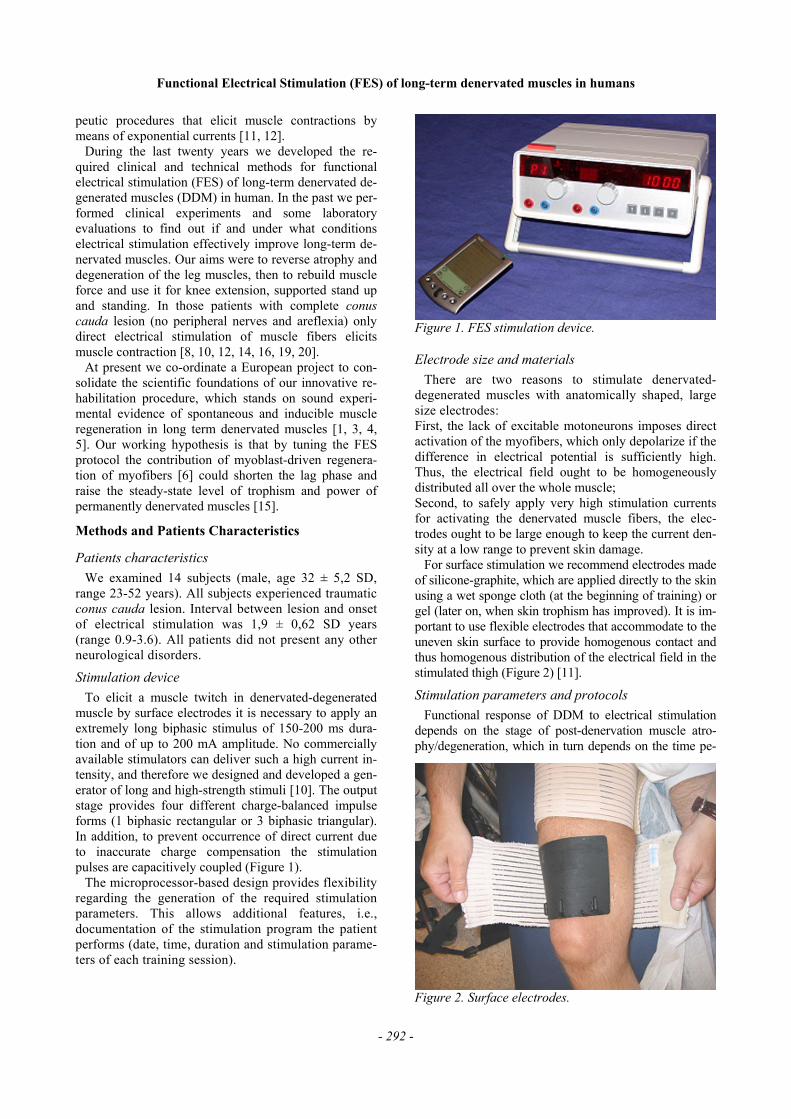
Stimulation device To elicit a muscle twitch in denervated-degenerated
muscle by surface electrodes it is necessary to apply an extremely long biphasic stimulus of 150-200 ms dura-tion and of up to 200 mA amplitude. No commercially available stimulators can deliver such a high current in-tensity, and therefore we designed and developed a gen-erator of long and high-strength stimuli [10]. The output stage provides four different charge-balanced impulse forms (1 biphasic rectangular or 3 biphasic triangular). In addition, to prevent occurrence of direct current due to inaccurate charge compensation the stimulation pulses are capacitively coupled (Figure 1).
The microprocessor-based design provides flexibility regarding the generation of the required stimulation parameters. This allows additional features, i.e., documentation of the stimulation program the patient performs (date, time, duration and stimulation parame-ters of each training session).
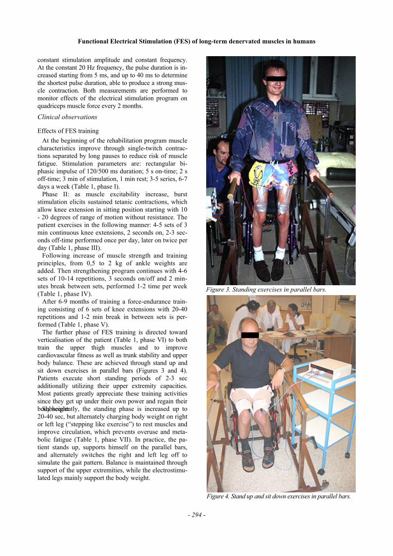
Electrode size and materials There are two reasons to stimulate denervated-
degenerated muscles with anatomically shaped, large size electrodes: First, the lack of excitable motoneurons imposes direct activation of the myofibers, which only depolarize if the difference in electrical potential is sufficiently high. Thus, the electrical field ought to be homogeneously distributed all over the whole muscle; Second, to safely apply very high stimulation currents for activating the denervated muscle fibers, the elec-trodes ought to be large enough to keep the current den-sity at a low range to prevent skin damage.
For surface stimulation we recommend electrodes made of silicone-graphite, which are applied directly to the skin using a wet sponge cloth (at the beginning of training) or gel (later on, when skin trophism has improved). It is im-portant to use flexible electrodes that accommodate to the uneven skin surface to provide homogenous contact and thus homogenous distribution of the electrical field in the stimulated thigh (Figure 2) [11].
Stimulation parameters and protocols Functional response of DDM to electrical stimulation
depends on the stage of post-denervation muscle atro-phy/degeneration, which in turn depends on the time pe-
Figure 1. FES stimulation device.
Figure 2. Surface electrodes.

Functional Electrical Stimulation (FES) of long-term denervated muscles in humans
- 293 -
riod between denervation event and stimulation onset. The minimal effective stimulation current depends on ex-tent of degeneration of the stimulated muscle. In these patients, from 1 year and up to 3 years after injury, at the beginning of the treatment we applied biphasic stimula-tion impulses with very long duration and high intensity. This strong stimulus is able to elicit single twitches of the degenerated-denervated muscle (DDM).
During the first several months of denervated-degenerated muscle training, a muscle twitch is elicited only by impulses lasting 120-150 msec. This is about 1500 times longer than in spastic paralysis patients, in which the motor neurones are preserved. With an inter-pulse interval of about 500 ms the resulting stimulation frequency is slightly less than 2 Hz (“single twitches” elicited every half second).
One effect of the electrical stimulation program is the increased excitability of the muscle fibers. Therefore the pulse duration and the inter-pulse interval could be ac-cordingly shortened to increase the number of stimuli per second delivered to the patient muscles. The change in excitability of muscle tissue slowly occurs in 3-6 months of electrical stimulation therapy, which patients perform at home five days a week (Table 1).
Once strength of muscle contraction increases in con-sequence of significant structural and metabolic im-provements of muscle tissue, the stimulation impulses are shortened to 50-35 ms (off-time 10 ms) to raise im-pulse frequency to 16-25 Hz [11, 12].
Daily therapy and training time The daily therapy program requires individual stimula-
tion of m. gluteus, m. quadriceps, hamstrings and m. tri-ceps surae for 15-20 minutes each muscle (in 3-5 series of 3- 4 minutes) once or twice a day. Patients use a stimu-lation device with two independent channels to simulta-neously stimulate left and right side muscles (Figure 1). The FES training takes approximately two hours a day (including time for donning and doffing the electrodes).
At the beginning, training is carried out in sitting posi-tion with extended legs (with or without foam roll) and
then with 90° knee flexion. The stimulation protocols with both single twitches and early tetanic contractions are done without added load. Later on, while knee ex-tension torque continues to increase, ankle weights are used to increase training intensity.
When the developed force in the leg muscles is suffi-cient to stabilize the knee joint in standing position, that is, the knee extension torque is higher than 20 Nm, the so-called functional training can start. The patient per-forms standing up exercises and simulates gait by alter-nately switching on and off muscle stimulation for the left and right leg in the upright position in parallel bars.
Usual outcome of our electrical stimulation program is the ability to extend the knee joint in sitting position after 4-6 months, and the ability to stabilize the knee joint in standing position after 12 months. Then standing up ex-ercises became possible, including initiation of gait simu-lation by manual control of the stimulation program.
Beside by clinical observations, effects of the stimula-tion program are measured by muscle cross sectional area with CT scans and by knee extension torque.
CT cross-section determination of thigh muscles Since thigh muscles are more or less spindle-shaped,
the CT-cut plan ought to be well defined, so that results could be compared. We use as reference points the tops of both trochanters, which are determined by CT scan. Preventing torsion of body axis, the reference line is es-tablished by linking the two trochanter tops. Results are reproducible within a single thigh, as well as in right vs. left leg comparison in each patient. All patients are lined supine on the table (“Feet-first position”) parallel to the table axis. The first body section is established at the tops of the trochanteres maiori. Three additional thigh sec-tions are performed distally every 100 mm. To clearly distinguish fat from skeletal muscle tissue a soft window frame (window 350, center 50) is used. Beside complete cross section area of the upper thigh the cross sectional areas of M. gluteus, M. quadriceps and hamstrings, as well as their density, are determined (Figure 5). Changes of muscle cross sectional area are expressed in absolute increase or decrease values (cm2) and/or as percent of base line value.
Force measurement Force of the quadriceps muscle is measured during
electrical stimulation as torque of extension movement of the knee. The measure is performed in sitting position using a purpose-designed chair where subjects sit with the legs in 90° knee flexion position. A dynamometer fixed between the chair and the leg measures force of the quadriceps muscle during electrical stimulation [11]. As an index of muscle trophism and of the efficacy of the training program, force of the thigh muscles is measured as extension torque and expressed in Nm, using increas-ing stimulation amplitudes from 0 to 160 Vpp in 10 V steps. The optimal stimulation parameters are determined in each patient by varying the impulse widths (msec) at
Table 1. Rehabilitation training program by surface-electrode FES of denervated-degenerated mus-cles after complete conus cauda lesion.
Phases Muscle Contraction Months of Training
Phase I single twitches 1-4 Phase II single twitches and 2-6 first tetanic contractions Phase III tetanic contractions and 4-12 knee extension Phase IV knee extension with 6-12 increasing ankle weight Phase V force endurance training 8-12 Phase VI verticalisation, standing up 12-18 Phase VII “stepping like” exercises 12-24
Functional Electrical Stimulation (FES) of long-term denervated muscles in humans
- 294 -
constant stimulation amplitude and constant frequency. At the constant 20 Hz frequency, the pulse duration is in-creased starting from 5 ms, and up to 40 ms to determine the shortest pulse duration, able to produce a strong mus-cle contraction. Both measurements are performed to monitor effects of the electrical stimulation program on quadriceps muscle force every 2 months.
Clinical observations
Effects of FES training At the beginning of the rehabilitation program muscle
characteristics improve through single-twitch contrac-tions separated by long pauses to reduce risk of muscle fatigue. Stimulation parameters are: rectangular bi-phasic impulse of 120/500 ms duration; 5 s on-time; 2 s off-time; 3 min of stimulation, 1 min rest; 3-5 series, 6-7 days a week (Table 1, phase I).
Phase II: as muscle excitability increase, burst stimulation elicits sustained tetanic contractions, which allow knee extension in sitting position starting with 10 - 20 degrees of range of motion without resistance. The patient exercises in the following manner: 4-5 sets of 3 min continuous knee extensions, 2 seconds on, 2-3 sec-onds off-time performed once per day, later on twice per day (Table 1, phase III).
Following increase of muscle strength and training principles, from 0,5 to 2 kg of ankle weights are added. Then strengthening program continues with 4-6 sets of 10-14 repetitions, 3 seconds on/off and 2 min-utes break between sets, performed 1-2 time per week (Table 1, phase IV).
After 6-9 months of training a force-endurance train-ing consisting of 6 sets of knee extensions with 20-40 repetitions and 1-2 min break in between sets is per-formed (Table 1, phase V).
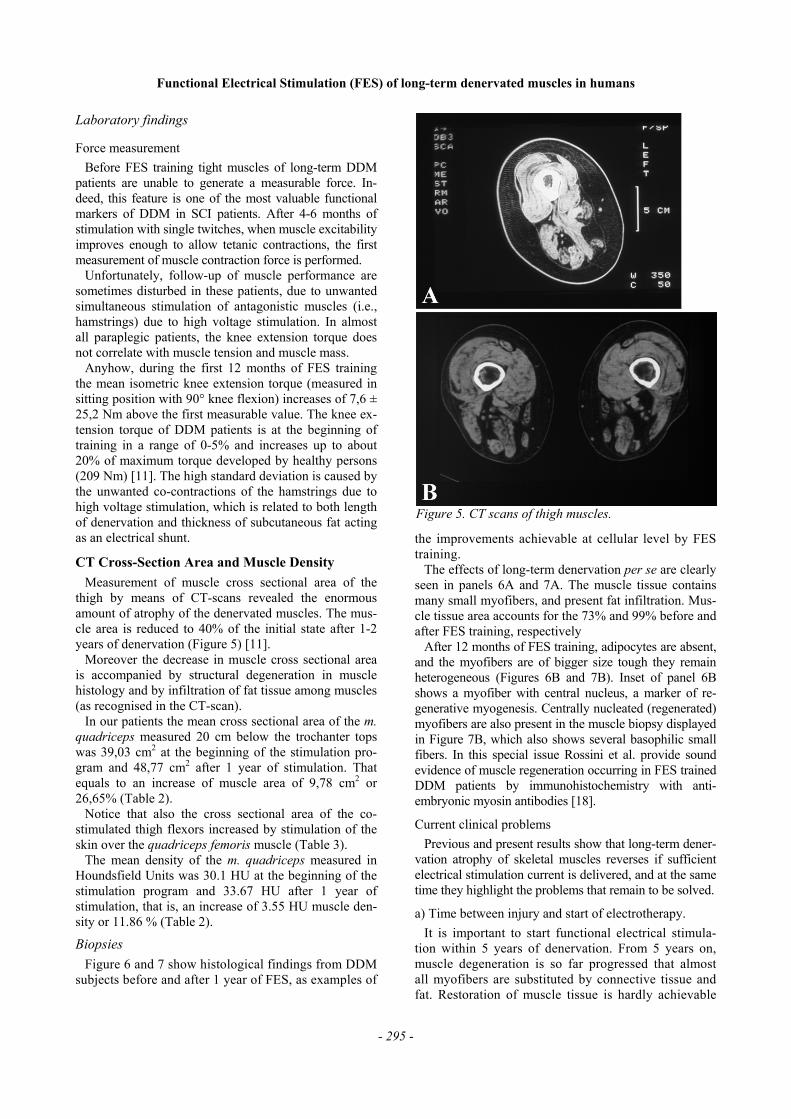
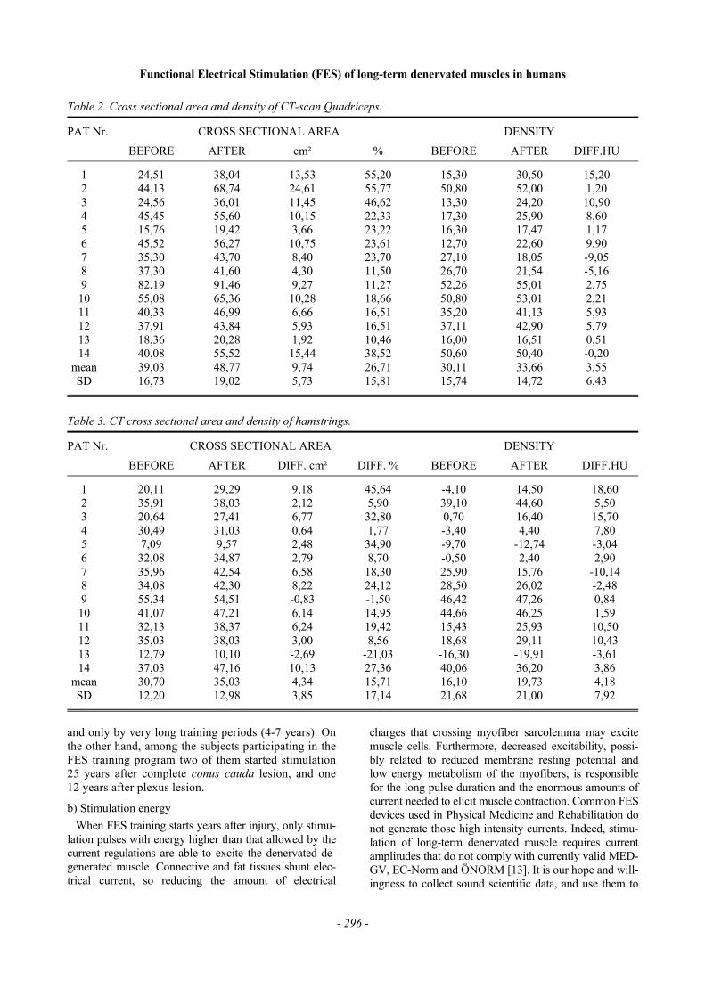
The further phase of FES training is directed toward verticalisation of the patient (Table 1, phase VI) to both train the upper thigh muscles and to improve cardiovascular fitness as well as trunk stability and upper body balance. These are achieved through stand up and sit down exercises in parallel bars (Figures 3 and 4). Patients execute short standing periods of 2-3 sec additionally utilizing their upper extremity capacities. Most patients greatly appreciate these training activities since they get up under their own power and regain their body height. Subsequently, the standing phase is increased up to 20-40 sec, but alternately charging body weight on right or left leg (“stepping like exercise”) to rest muscles and improve circulation, which prevents overuse and meta-bolic fatigue (Table 1, phase VII). In practice, the pa-tient stands up, supports himself on the parallel bars, and alternately switches the right and left leg off to simulate the gait pattern. Balance is maintained through support of the upper extremities, while the electrostimu-lated legs mainly support the body weight.
Figure 3. Standing exercises in parallel bars.
Figure 4. Stand up and sit down exercises in parallel bars.
Functional Electrical Stimulation (FES) of long-term denervated muscles in humans
- 295 -
Laboratory findings
Force measurement Before FES training tight muscles of long-term DDM
patients are unable to generate a measurable force. In-deed, this feature is one of the most valuable functional markers of DDM in SCI patients. After 4-6 months of stimulation with single twitches, when muscle excitability improves enough to allow tetanic contractions, the first measurement of muscle contraction force is performed.
Unfortunately, follow-up of muscle performance are sometimes disturbed in these patients, due to unwanted simultaneous stimulation of antagonistic muscles (i.e., hamstrings) due to high voltage stimulation. In almost all paraplegic patients, the knee extension torque does not correlate with muscle tension and muscle mass.
Anyhow, during the first 12 months of FES training the mean isometric knee extension torque (measured in sitting position with 90° knee flexion) increases of 7,6 ± 25,2 Nm above the first measurable value. The knee ex-tension torque of DDM patients is at the beginning of training in a range of 0-5% and increases up to about 20% of maximum torque developed by healthy persons (209 Nm) [11]. The high standard deviation is caused by the unwanted co-contractions of the hamstrings due to high voltage stimulation, which is related to both length of denervation and thickness of subcutaneous fat acting as an electrical shunt.
CT Cross-Section Area and Muscle Density Measurement of muscle cross sectional area of the
thigh by means of CT-scans revealed the enormous amount of atrophy of the denervated muscles. The mus-cle area is reduced to 40% of the initial state after 1-2 years of denervation (Figure 5) [11].
Moreover the decrease in muscle cross sectional area is accompanied by structural degeneration in muscle histology and by infiltration of fat tissue among muscles (as recognised in the CT-scan).
In our patients the mean cross sectional area of the m. quadriceps measured 20 cm below the trochanter tops was 39,03 cm2 at the beginning of the stimulation pro-gram and 48,77 cm2 after 1 year of stimulation. That equals to an increase of muscle area of 9,78 cm2 or 26,65% (Table 2).
Notice that also the cross sectional area of the co-stimulated thigh flexors increased by stimulation of the skin over the quadriceps femoris muscle (Table 3).
The mean density of the m. quadriceps measured in Houndsfield Units was 30.1 HU at the beginning of the stimulation program and 33.67 HU after 1 year of stimulation, that is, an increase of 3.55 HU muscle den-sity or 11.86 % (Table 2).
Biopsies Figure 6 and 7 show histological findings from DDM
subjects before and after 1 year of FES, as examples of
the improvements achievable at cellular level by FES training.
The effects of long-term denervation per se are clearly seen in panels 6A and 7A. The muscle tissue contains many small myofibers, and present fat infiltration. Mus-cle tissue area accounts for the 73% and 99% before and after FES training, respectively
After 12 months of FES training, adipocytes are absent, and the myofibers are of bigger size tough they remain heterogeneous (Figures 6B and 7B). Inset of panel 6B shows a myofiber with central nucleus, a marker of re-generative myogenesis. Centrally nucleated (regenerated) myofibers are also present in the muscle biopsy displayed in Figure 7B, which also shows several basophilic small fibers. In this special issue Rossini et al. provide sound evidence of muscle regeneration occurring in FES trained DDM patients by immunohistochemistry with anti-embryonic myosin antibodies [18].
Current clinical problems Previous and present results show that long-term dener-
vation atrophy of skeletal muscles reverses if sufficient electrical stimulation current is delivered, and at the same time they highlight the problems that remain to be solved.
a) Time between injury and start of electrotherapy. It is important to start functional electrical stimula-
tion within 5 years of denervation. From 5 years on, muscle degeneration is so far progressed that almost all myofibers are substituted by connective tissue and fat. Restoration of muscle tissue is hardly achievable
Figure 5. CT scans of thigh muscles.
Functional Electrical Stimulation (FES) of long-term denervated muscles in humans
- 296 -
and only by very long training periods (4-7 years). On the other hand, among the subjects participating in the FES training program two of them started stimulation 25 years after complete conus cauda lesion, and one 12 years after plexus lesion.
b) Stimulation energy When FES training starts years after injury, only stimu-
lation pulses with energy higher than that allowed by the current regulations are able to excite the denervated de-generated muscle. Connective and fat tissues shunt elec-trical current, so reducing the amount of electrical
charges that crossing myofiber sarcolemma may excite muscle cells. Furthermore, decreased excitability, possi-bly related to reduced membrane resting potential and low energy metabolism of the myofibers, is responsible for the long pulse duration and the enormous amounts of current needed to elicit muscle contraction. Common FES devices used in Physical Medicine and Rehabilitation do not generate those high intensity currents. Indeed, stimu-lation of long-term denervated muscle requires current amplitudes that do not comply with currently valid MED-GV, EC-Norm and ÖNORM [13]. It is our hope and will-ingness to collect sound scientific data, and use them to
Table 2. Cross sectional area and density of CT-scan Quadriceps.
PAT Nr. CROSS SECTIONAL AREA DENSITY BEFORE AFTER cm² % BEFORE AFTER DIFF.HU
1 24,51 38,04 13,53 55,20 15,30 30,50 15,20 2 44,13 68,74 24,61 55,77 50,80 52,00 1,20 3 24,56 36,01 11,45 46,62 13,30 24,20 10,90 4 45,45 55,60 10,15 22,33 17,30 25,90 8,60 5 15,76 19,42 3,66 23,22 16,30 17,47 1,17 6 45,52 56,27 10,75 23,61 12,70 22,60 9,90 7 35,30 43,70 8,40 23,70 27,10 18,05 -9,05 8 37,30 41,60 4,30 11,50 26,70 21,54 -5,16 9 82,19 91,46 9,27 11,27 52,26 55,01 2,75 10 55,08 65,36 10,28 18,66 50,80 53,01 2,21 11 40,33 46,99 6,66 16,51 35,20 41,13 5,93 12 37,91 43,84 5,93 16,51 37,11 42,90 5,79 13 18,36 20,28 1,92 10,46 16,00 16,51 0,51 14 40,08 55,52 15,44 38,52 50,60 50,40 -0,20 mean 39,03 48,77 9,74 26,71 30,11 33,66 3,55 SD 16,73 19,02 5,73 15,81 15,74 14,72 6,43
Table 3. CT cross sectional area and density of hamstrings.
PAT Nr. CROSS SECTIONAL AREA DENSITY BEFORE AFTER DIFF. cm² DIFF. % BEFORE AFTER DIFF.HU
1 20,11 29,29 9,18 45,64 -4,10 14,50 18,60 2 35,91 38,03 2,12 5,90 39,10 44,60 5,50 3 20,64 27,41 6,77 32,80 0,70 16,40 15,70 4 30,49 31,03 0,64 1,77 -3,40 4,40 7,80 5 7,09 9,57 2,48 34,90 -9,70 -12,74 -3,04 6 32,08 34,87 2,79 8,70 -0,50 2,40 2,90 7 35,96 42,54 6,58 18,30 25,90 15,76 -10,14 8 34,08 42,30 8,22 24,12 28,50 26,02 -2,48 9 55,34 54,51 -0,83 -1,50 46,42 47,26 0,84 10 41,07 47,21 6,14 14,95 44,66 46,25 1,59 11 32,13 38,37 6,24 19,42 15,43 25,93 10,50 12 35,03 38,03 3,00 8,56 18,68 29,11 10,43 13 12,79 10,10 -2,69 -21,03 -16,30 -19,91 -3,61 14 37,03 47,16 10,13 27,36 40,06 36,20 3,86 mean 30,70 35,03 4,34 15,71 16,10 19,73 4,18 SD 12,20 12,98 3,85 17,14 21,68 21,00 7,92

Functional Electrical Stimulation (FES) of long-term denervated muscles in humans
- 297 -
convince International Standardization Organizations to modify the rules (which are meant to ensure patient safety) according to the new clinical demands. We al-ready submitted a petition to extend these regulations, i.e., maximum energy per impulse up to 400-3000 mJ, to better serve the needs of DDM patients.
c) Long lag phase to achieve muscle hypertro-phy/hyperplasia and functionally significant amount of muscle contraction force
If FES training starts very late after injury, muscles are already degenerated, and thereafter the phase of muscle restoration takes from 1 to 2 years (depending on length of denervation) to achieve the muscle bulk and the con-traction force that allow the patient to stand. We estimate that from 1 to 2 millions of new muscle fibers have to be rebuilt in each quadriceps muscle. This equals to a syn-thesis rate of about 1000 new myofibers per day over a period of 3 years. The stimulation program and the pa-rameters applied to achieve such an improvement, i.e., to rebuild the surviving myofibers from 5-10 µm to 40-80 µm fiber diameter, and in meantime to avoid overuse and consequent muscle damage, are still based on prior clini-cal experiences. Primary focus is given to the actual daily muscle performance, which is often quite variable. Be-cause pain and muscle tension are not a reliable stress pa-rameter in these patients, sometimes muscle over-strain occurs. Signs of muscle over-training are muscle fatigue
during daily exercise, or more often a dramatic decrease of muscle performance the day after. In these cases, the FES training is discontinued 1-2 days to allow muscle healing, possibly by myofiber regeneration.
A second reason for the slow rate of muscle rebuild-ing could be related to the muscle structural changes that could be higher than the measurable gain of mus-cle force because of the denervation-induced replace-ment of myofibers with connective tissue and fat prior to electrostimulation.
All together, these constrains explain the slow rate of muscle hypertrophy and force gain, and thus of functional improvement.
Nevertheless, in long-term denervated patients positive effects of FES training are measured, as structural and metabolic improvements occur. This enables the patient to get up and to stand, but electrostimulated muscle en-durance is still not satisfying. The goal of our ongoing research is to understand the underlying mechanisms of muscle regeneration and to transform these structural and metabolic changes into major functional gains.
Discussion In DDM patients the improvements of muscle bulk,
force and endurance are achieved through force and force-endurance exercises in 6-12 months training. Af-terward, muscle are strengthened and endured enough to
Figures 6. Histological findings in muscle biopsies of DDM subject. H-E. A,C before and B,D after 12-month FES training.

Functional Electrical Stimulation (FES) of long-term denervated muscles in humans
- 298 -
perform functional training, i.e., standing up exercises, and standing in parallel bars gait simulation by switching the tetanic electrostimulation of left/right quadriceps al-ternately. Effects of these treatments are not only a better cosmetic aspect, but also a better perfusion of the para-lyzed legs. The continuous electrostimulation first changes the skin trophism (the skin is getting thicker and more resistant to electrical and mechanical stresses), and consequently eventual wound healing is faster. Thus, the risk of decubitus ulcer sharply decreases.
Both biopsies and thigh CT-scans show reduction of fat and connective tissue within and around the muscles. These findings indicate that degeneration of long-term denervated muscle is reversed by electrostimulation. Clinical observation of increased muscle mass and func-tion (standing up exercises) correlate well with the measurable laboratory findings.
High intensity stimulation training, i.e., with impulse parameters outside the current EU regulations, allows achieving within a relatively short time period (from 12 to 18 months) regeneration, hypertrophy and functional use of the denervated muscles. This high intensity stimu-lation program constitutes a risk only if the patients do not follow the instructions provided before he is enrolled in the stimulation program. Potential lesions are skin burns, if surface electrodes are not properly applied, and muscle over strain due to the absence of sensitivity (pain).
Though there are questions to be solved, we can say that risks for patients performing high intensity elec-trostimulation are low and that the FES training is effec-tive if high currents are used. In contrast to prior insuf-ficient treatments with exponential currents, high cur-rents maintain trophism of denervated muscle for pro-longed periods of time and even restore their structure and function after severe atrophy.
High intensity stimulation allows patients to sit longer in the wheel chair because of the increased perfusion, the better trophism of the skin and the bigger muscle mass, which, reducing the risk of pressure sores, permits activi-ties of daily living and thus a better social re-integration.
Still there are many open problems, such as optimal time for onset, duration, intensity and methods of appli-cation of the FES training, but we firmly believe that in co-operation with international partners we will solve most of them in the time course of the European re-search project “RISE”. Furthermore, we have the oppor-tunity to study in permanent denervated human muscles both regenerative myogenesis and myofiber plasticity induced by electrical stimulation without any influence of the nerve. This will lead to new knowledge to estab-lish better managements of elderly people and of pa-tients with peripheral nerve lesions.
Conclusions The therapeutic goals of FES training in DDM Patients
are not only to achieve muscle bulk, force and function, but also to improve overall trophism of the denervated leg (leg perfusion, muscle metabolism, and skin eutrophy).
These effects are of foremost importance because they concur in prevention of so-called secondary diseases, such as decubitus ulcera and cardiovascular diseases.
At present, the factors triggering the underlying mecha-nism of muscle regeneration of denervated-degenerated muscle are unknown. We need to know if the observed regenerative events of myofibers are either due to sponta-neous post-denervation activation of satellite cells, or to the electrostimulation-induced myoblast activity, or to both of them. Furthermore, we would like to establish the long-term potential of myoblast replications in dener-vated-degenerated muscles, since this could be crucial for the life-long FES therapy of denervated muscles.
The experience we made with pilot clinical studies will be re-examined, and FES therapeutic programs im-proved and optimized by animal experiments and clini-cal studies during the European research project RISE. The aim is an advanced rehabilitation methodology based on sound scientific data. To achieve the goal, we want all patients with complete conus cauda lesion throughout Europe are enrolled in the project trial.
Acknowledgements Supported by EU Commission Shared Cost Project
RISE (Contract n. QLG5-CT-2001-02191).
Address correspondence to: Dr. Helmut Kern, Institut für Physikalische Medizin
und Rehabilitation, Wilheminenspital Wien, Mont-leartstrasse 37, A-1171 Wien, tel. +43 1 49150 3401, fax +43 1 49150 3409, Email [email protected].
References [1] Billington L, Carlson BM: The recovery of long-
term denervated rat muscles after Marcaine treatment and grafting. Anat Rec 1996; 144 (1-2): 147-155.
[2] Brooke MH, Kaiser KK: Muscle fibre type: how many and what kind? Arch Neurol 1970; 23: 369-379.
[3] Carlson BM, Borisov AI, Dekov EI, Dow D, Kos-trominova TY: The biology and restorative capacity of long-term denervated skeletal muscle. Basic Appl Myol 2002; 12: xxx-xxx.
[4] Carraro U, Catani C, Degani A, Rizzi C: Myosin expression in denervated fast- and slow-twitch muscles: Fiber modulation and substitution, in Pette D (ed): The dynamic state of muscle fibers. Berlin, New York, Walter de Gruyter, 1990, pp 247-262.
[5] Carraro U, Morale D, Mussini I, Lucke S, Cantini M, Betto R, Catani C, Dalla Libera L, Danieli-Betto D, Noventa D: Chronic denervation of rat dia-phragm: maintenance of fiber heterogeneity with associated increasing uniformity of myosin iso-forms. J Cell Biol 1985; 100: 161-174.
[6] Carraro U: Modulation of trophism and fiber type ex-pression of denervated muscle by different patterns of electrical stimulation Basic Appl Myol 12: xxx-xxx.

Functional Electrical Stimulation (FES) of long-term denervated muscles in humans
- 299 -
[7] Cerrel-Bazo H, Rizzetto A, Pauletto D, Lucca L, Caldana L: Assisting paraplegic individuals to walk by means of electrically induced muscle contrac-tion: Gait performance and patient compliance. Eight World Cong Int Rehab Med Assoc, Kioto, Japan 1997; Session 91, Paper 66.
[8] Eichhorn K.F., Schubert W., David E.: Mainte-nance, training and functional use of denervated muscles. J Biomed Eng 1984; 6 (3): 205-11.
[9] Graupe D: An over view of the state of the art of non-invasive FES for independent deambulation by tho-racic level paraplegics. Neurol Res 2002: 24: 431-442.
[10] Hofer C, Mayr W, Stöhr H, Unger E, Kern H: A stimulator for functional activation of denervated muscles. Artif Organs 2002; 26 (3): 276-279.
[11] Kern H.: Funktionelle Elektrostimulation paraplegi-scher Patienten. Österr. Z. Phys. Med. 5, Heft 1 Supplementum, 1995.
[12] Kern H, Hofer C, Strohhofer M, Mayr W, Richter W, Stohr H: Standing up with denervated muscles in humans using functional electrical stimulation. Artif Organs 1999; 23 (5): 447-452.
[13] Kern H, Hofer C, Mödlin M, Forstner C, Raschka-Höger D, Mayr W, Stöhr H: Denervated muscles in humans: limitations and problems of currently used functional electrical stimulation training protocols. Artif Organs 2002; 26 (3): 216-218.
[14] Mayr W, Burian K, Carraro U, Gruber H, Kern H, Streinzer W, Szabolcs M, Thoma H, Zrunek M:: Functional electrical stimulation (FES) of dener-vated muscles, an experimental study on posterior cricoarytenoid muscle (PCM) of sheep. 32nd An-
nual Meeting of American Society for Artificial, In-ternational Organs, Anaheim, California, 1986. Ar-tificial Organs, Raven Press.
[15] Mayr W, Hofer C, Bijak M, Rafolt D, Reichel M, Unger E, Mödlin M, Kern H: FES of Denervated Muscles: Preliminary Experimental and Clinical Results and their Implications on the Actual EU-Project RISE. Proceedings of the 7th Annual Con-ference of the International Functional Electrical Stimulation Society, June 25-29, 2002.
[16] Mokrusch T, Engelhardt A, Eichhorn KF, Pris-chenk H, Sack G, Neundorfer B: Effects of long-impulse electrical stimulation on atrophy and fiber type composition of chronically denervated rabbit muscle. J Neurol 1990; 237: 29-34.
[17] Petrofsky JS, Stacy R, Laymon M: The relationship between exercise work intervals and duration of ex-ercise on lower extremity training induced by elec-trical stimulation in humans with spinal cord inju-ries. Eur J Appl Physiol 2000; 82 (5-6): 504-509.
[18] Rossini K, Zanin ML, Podhorska-Okolow M, Carraro U: Stage and quantify regenerative myo-genesis in FES-induced functional recovery of hu-man long-term permanent denervated muscle. Basic Appl Myol 2002; 12 (6): xxx-xxx
[19] Schubert W: Funktionelles Training schlaff gelähmter Muskulatur. Biomed Technik 1985; 30: 115.
[20] Valencic V, Vodovnik L, Stefancic M, Jelnikar T: Improved motor response due to chronic electrical stimulation of denervated tibialis anterior muscle in humans. Muscle Nerve 1986; 9: 612-617.