functionalized gold nanoparticles for sensing of
TRANSCRIPT
Volume 28 Issue 4 Article 4
2020
Functionalized gold nanoparticles for sensing of pesticides: A Functionalized gold nanoparticles for sensing of pesticides: A
review review
Follow this and additional works at: https://www.jfda-online.com/journal
Part of the Analytical Chemistry Commons, Apiculture Commons, Environmental Monitoring
Commons, and the Materials Chemistry Commons
This work is licensed under a Creative Commons Attribution-Noncommercial-No Derivative
Works 4.0 License.
Recommended Citation Recommended Citation Tseng, Wei-Bin; Hsieh, Ming-Mu; Chen, Che-Hsie; Chiu, Tai-Chia; and Tseng, Wei-Lung (2020) "Functionalized gold nanoparticles for sensing of pesticides: A review," Journal of Food and Drug Analysis: Vol. 28 : Iss. 4 , Article 4. Available at: https://doi.org/10.38212/2224-6614.1092
This Review Article is brought to you for free and open access by Journal of Food and Drug Analysis. It has been accepted for inclusion in Journal of Food and Drug Analysis by an authorized editor of Journal of Food and Drug Analysis.

Functionalized gold nanoparticles for sensing of pesticides: A review Functionalized gold nanoparticles for sensing of pesticides: A review
Cover Page Footnote Cover Page Footnote This work was financially supported by the Ministry of Science and Technology of Taiwan under contract number MOST 108-2113-M-017-003 and 109-2113-M-143-002.
This review article is available in Journal of Food and Drug Analysis: https://www.jfda-online.com/journal/vol28/iss4/4

Functionalized gold nanoparticles for sensing ofpesticides: A review
Wei-BinTseng a,Ming-MuHsieh b,Che-HsieChen c,Tai-ChiaChiu c,*,Wei-LungTseng d,e,**
a Department of College of Ecology and Resource Engineering, Wuyi University, Fujian, Chinab Department of Chemistry, National Kaohsiung Normal University, Taiwanc Department of Applied Science, National Taitung University, Taitung, Taiwand Department of Chemistry, National Sun Yat-sen University, No. 70, Lien-hai Road, Gushan District, Kaohsiung, 80424, Taiwane School of Pharmacy, Kaohsiung Medical University, No. 100, Shiquan 1st Road, Sanmin District, Kaohsiung, 80708, Taiwan
Abstract
Pesticides are a family of non-biodegradable chemical compounds which widely used in agriculture to control pestsand increase yield production. However, overuse or abuse of pesticides and their metabolites may cause potentialtoxicity for the environment as well as human health and all other living organisms, even at deficient concentrations.Consequently, the development of sensors for monitoring these compounds is significant. Recently, nanoparticles-basedsensors have been extensively employed as a potential alternative or complementary analytical tool to conventionaldetection methods for pesticides. Among them, gold nanoparticles (AuNPs) owing to their unique optical propertieshave been developed as smart sensors with high selectivity, sensitivity, simplicity, and portability. These comprehensivereviews have summarized various studies performed based on different detection strategies, i.e., colorimetric, fluores-cence, surface-enhanced Raman scattering, and electrochemical, using AuNPs as sensing probes for pesticide analysis invarious matrices. Additionally, the current challenges and future trends for developing novel AuNPs-based sensors forthe detection of pesticides are also discussed.
Keywords: Colorimetry, Electrochemical, Fluorescence, Gold nanoparticles, Pesticides, Surface-enhanced Ramanscattering
1. Introduction
P esticides are a large and heterogeneous groupof heavily employed chemicals in modern
agriculture for protecting crops, controlling insectpests, and improving productivity [1]. In terms oftheir chemical structures, pesticides can be clas-sified into four leading groups, including organ-ochlorines, organophosphorus, carbamate,chlorophenols, and synthetic pyrethroids [2]. Ifconcerned with pesticides' function, they can becategorized into five types that include in-secticides, herbicides, fungicides, rodenticide, andnematocides [3e5]. Many toxic pesticides havebeen extensively used in agriculture due to their
low cost and high effectiveness [1,2]. However,the overuse, abuse, and misuse of pesticides leadto their residues and metabolites' release, causingthe adverse effect on the ecological system andhuman health [6]. Even exposure to very lowpesticide levels could be highly involved in foodand water safety [7]. Additionally, some of thepesticides, identified as endocrine disruption,have been implicated with neurotoxicity, geno-toxicity, mutagenicity, and carcinogens [8e10].Based on the consideration of toxic pesticides'threats to human health and the environment, itis urgent to develop sensitive, selective, low-cost,and affordable analytical methods to monitortheir level in real-world samples.
Received 9 May 2020; revised 23 July 2020; accepted 6 August 2020.Available online 1 December 2020
* Corresponding author at: Department of Applied Science, National Taitung University, 369, Section 2, University Road, Taitung, 95092, Taiwan. Tel: +88689 517990; Fax: þ886 89 518108.
** Corresponding author at: Department of Chemistry, National Sun Yat-sen University, No. 70, Lien-hai Road, Gushan District, Kaohsiung, 80424,Taiwan. Fax: þ011 886 7 5254644.E-mail addresses: [email protected] (T.-C. Chiu), [email protected] (W.-L. Tseng).
https://doi.org/10.38212/2224-6614.10922224-6614/© 2020 Taiwan Food and Drug Administration. This is an open access article under the CC-BY-NC-ND license(http://creativecommons.org/licenses/by-nc-nd/4.0/).
REVIEW
ARTIC
LE

Current analytical methods used to determinepesticides and their metabolites mainly includethree parts of quick, easy, cheap, effective, ruggedand safe (QuEChERS) extraction, separation tech-niques (gas chromatography and high-performanceliquid chromatography), and mass spectrometry(e.g., quadrupole ion trap and time-of-flight in-struments) [11,12]. Although offering satisfactorysensitivity and excellent separation efficiency, mostmass spectrometry-related approaches still sufferfrom time-consuming sample preparation, sophis-ticated instrumentation, and required skilled oper-ators [13]. More importantly, these methods arehard to transform into a portable device for on-sitedetection. Recently, ligand-capped gold nano-particles (AuNPs) with a size range from 1 to 100 nmhave attracted tremendous interests since theyprovide high molar extinction coefficients(>108 M�1 cm�1), size-dependent optical properties,strong Rayleigh scattering, and easy functionaliza-tion. These features enable ligand-capped AuNPs tobe well-suited for the practical applications in pho-tonics [14e16], electronics [17,18], biosensing[18e20], bioimaging [21,22], and nanomedicine[23e26]. In addition to these applications mentionedabove, ligand-capped AuNPs also serve as a sensingplatform for colorimetric, fluorometric, and elec-trochemical detection of a broad range of analytesfrom metal ions and small biomolecules to proteinand DNA macromolecules. The sensing mechanismof ligand-capped AuNPs can be divided into threestrategies: (1) a target analyte induces the aggrega-tion of ligand-capped AuNPs via the coordinationinteraction between two ligands and a target ana-lyte; (2) a target analyte triggers the removal of acolloidal stabilizer from the nanoparticle surface viathe formation of AueS bonds, resulting in the salt-mediated assembly of ligand-capped gold nano-particles; (3) a target analyte drives the liberation ofa recognition molecule from the nanoparticle's sur-face through the complexation reaction. The aggre-gation of ligand-capped AuNPs can promote aredshift in surface Plasmon resonance, a restorationin fluorescence peak, and an enhancement inRaman scattering. Taking these advantages, re-searchers have developed different kinds of ligand-capped AuNPs for probing pesticides in real-worldsamples. In this review, we present a comprehen-sive discussion associated with the recent advance-ments in the use of unmodified and functionalizedAuNPs to detect pesticides. To the author's knowl-edge, there is no systematic review covering suchtopics. We especially emphasize integrating thecolorimetric, fluorescence, surface-enhanced Ramanscattering (SERS), and electrochemical strategies
(Fig. 1) with ligand-capped AuNPs for the determi-nation of pesticides.
2. Synthesis of AuNPs and their modification
Gold has different oxidation states that includeAuþ3 (auric), Auþ1 (aurous), and atomic Au0. SinceAuNPs consist of numerous Au0, the syntheticprocedure involves the reduction of Auþ3 or Auþ1 toAu0 with a reducing agent. Examples of reducingagents include citric acid [18,27,28], ascorbic acid[29,30], sodium borohydride [31], oxalic acids [32,33],sulfites [34,35], and hydrogen peroxide [27,36]. Atthe beginning of the process synthesis, chloroauricacid (HAuCl4) is frequently employed as a precursorchemical in the preparation of the AuNPs, and it canbe purchased from chemical companies or obtainedfrom the reaction of aqua regia and gold foil. Also,capping ligands are needed in a precursor solutionto prevent the formed AuNPs from aggregation andfurther growth during the synthetic process. Thecapping ligands enable the AuNPs to have sufficientelectrostatic repulsion, steric hindrance, or both tobe dispersed in an aqueous solution [37e39]. Thiol-terminated small molecules [37,40], trisodium citrate[41,42], surfactants [43,44], polymers [38,45], phos-phate-containing nucleotides [46,47], and phos-phorus ligands [48] are exemplified by cappingligands. It is interesting to emphasize that severalcapping ligands serve as a reducing agent and astabilizing agent [49]. After mixing HAuCl4 with aweak reducing agent in the presence of capping li-gands, the resultant solution is commonly treatedwith heating [50], microwave irradiation [51], andUV light exposure [52]. Once substituting a weakreducing agent with a strong one, the reactionsimply proceeds at ambient temperature [53,54].UVevisible and X-ray spectroscopies have beenimplemented for monitoring the nucleation andgrowth of the AuNPs [55]. Electron microscopic andoptical spectroscopic techniques can providedetailed information associated with the
Fig. 1. Schematic illustration for various AuNPs-based sensing strate-gies for the pesticide detection.
522 JOURNAL OF FOOD AND DRUG ANALYSIS 2020;28:521e538
REVIEW
ARTIC
LE

morphology, compositions, valence state, and opti-cal properties of the AuNPs [56,57]. Moreover, sin-gle-particle inductively coupled plasma massspectrometry (ICP-MS) has been adopted for accu-rate quantification of the molar and particle numberconcentrations of the AuNPs [58,59]. The measure-ment of zeta potential of the AuNPs can help theinvestigators to assess whether they are present inthe dispersed state [60]. The purification of theAuNPs can be performed by filtration, centrifuga-tion, dialysis, and separation techniques [61]. Amore detailed characterization of the AuNPs hasbeen reviewed in the recent literature [62].Faraday has pioneered the development of the
methodology for producing 5 nm-sized AuNPs fromthe reduction of HAuCl4 in the presence of thephosphorus-ether solution [63]. During the syn-thetic process, the mixed solution's color variedfrom brown, grey, purple, to deep red, which can beeasily visualized by the naked eye. This break-through opens the door to the synthesis of stableAuNPs and motivates the researchers to design newsynthetic routes. The Turkevich method is one of themost common ones used for the reproducible andsimple synthesis of approximately 10e50 nm AuNPswith a spherical shape [27]. The formed citrate-capped AuNPs and their surface modification areintensively implemented as a colorimetric reporterto sense organophosphate pesticides. It is worthemphasizing that the preparation of citrate-cappedgold nanoparticles is straightforward and simplewithout high-cost dedicated equipment and a greatexperience. Besides, the cost for 60 mL of 2.57 nMcitrate-capped gold nanoparticles is only ca. US$0.61. In comparison to the Turkevich method, theAuNPs produced from the seed-mediated growthmethod [64], Brust-Schiffrin approach [31], electro-chemical techniques [65e67], ionic liquid-relatedpreparation [68e70], and biological molecule-mediated synthesis [71e73] are rarely utilized as aplatform for the detection of organophosphate pes-ticides. Fig. 2 reveals the above-mentioned syntheticmethods. Accordingly, in the review article, wefocus on the detailed discussion of the Turkevichmethod. Briefly, the Turkevich method involvesinjecting trisodium citrate into a boiling solution ofHAuCl4 under reflux and vigorous stirring. Imme-diately, rapid nucleation occurs in the initial stage,followed by the aggregation of the nuclei. Next, theformed bigger particles undergo slow diffusiongrowth by reducing gold ion precursor and furthercoalescence. Finally, the resultant particles undergorapid growth to form a fixed size of the AuNPs. Theas-prepared citrate-capped AuNPs exhibit the sur-face plasmon band (SPR) in the visible regions. The
mechanism of gold nanoparticle formationmentioned above has been demonstrated usingsmall-angle X-ray scattering (SAXS) and X-ray ab-sorption near-edge spectroscopy (XANES) employ-ing synchrotron radiation [74]. The particle size andsize distribution of the AuNPs were reported to beheavily connected with the ratio of HAuCl4 to so-dium citrate [75,76], solution pH [77], and synthetictemperature [78]. Although the Turkevich methodprovides highly reproducible and straightforwardfor spherical particles' production, the resultant cit-rate-capped AuNPs are easy to aggregate in a high-ionic-strength solution. Thus, it is required tomodify the surface of citrate-capped AuNPs throughthe ligand-change reaction or stabilizers' adsorp-tion. Examples of colloidal stabilizers include DNAaptamer [79,80], proteins [81,82], neutral surfactants[83,84], adenosine triphosphate [85], cysteine [86],lipoic acid [87], p-nitroaniline dithiocarbamate [88].Among the stabilizers and capping ligandsmentioned above, the DNA aptamers-, adenosinetriphosphate-, cysteine-, lipoic acid- and p-nitro-aniline dithiocarbamate-modified AuNPs have beenutilized for colorimetric assay of organophosphatepesticides that will be discussed in the followingsection. In addition to using citrate as a reducingand stabilizing agent, D'Souza et al. reported thepreparation of ascorbic acid-capped AuNPs throughthe hydrothermal reduction of HAuCl4 with ascor-bic acid at 100 �C for 10 min [89]. The SPR band ofthe ascorbic acid-capped AuNPs was centered at525 nm with a particle size of 14.5 nm. Park et al.incubated heparin with HAuCl4 at 60 �C for 20 h,resulting in 20 nm-sized AuNPs with the SPR bandof 524 nm [90]. Likewise, heparin behaved as areducing and stabilized ligand for the synthesis ofthe AuNPs. Barman et al. synthesized highly stableAuNPs through trichloroacetic acid-triggeredreduction of HAuCl4 in the presence of cetyl-trimethylammonium bromide (CTAB) [91]. Theformed CTAB-capped AuNPs kept dispersed formore than six months. The ascorbic acid-, heparin-and CTAB-capped AuNPs mentioned above havebeen demonstrated to be powerful for sensingorganophosphate pesticides. The sensing procedurewill be discussed in the next section.
3. Detection strategies for pesticides based ongold nanoparticles
3.1. Colorimetric assays
The AuNP-related colorimetric assay is a powerfulmethod for the naked-eye detection of numerousanalytes due to their large molar extinction
JOURNAL OF FOOD AND DRUG ANALYSIS 2020;28:521e538 523
REVIEW
ARTIC
LE

coefficient [92]. The localized surface Plasmonresonance wavelength of AuNPs is highly suscepti-ble to their aggregation degree and particle size. Thepresence of the target analyte induces the change incolor and surface plasmon resonance wavelength ofAuNPs as a consequence of either the conversionfrom dispersed to aggregated nanoparticles or thedisassembly of aggregated nanoparticles, as shownin Fig. 3 [93]. Based on this sensing mechanism,AuNP-based colorimetric sensors have been well-developed for pesticide detection and showed theiradvantages of rapidity, simplicity, convenience,cost-effectiveness, and visualization by naked-eyessummarized in Table 1. Xu et al. [94] found thatacetamiprid molecules were capable of directlyattached on the surface of citrate-capped AuNPs,leading to the nanoparticle aggregation and colorchange. The visualized color change of citrate-cap-ped AuNPs from red to blue was well-suited fordetermining the concentration of acetamiprid in anaqueous solution. This method was successfullyapplied to detect acetamiprid in vegetables.Aptamers are short single-stranded oligonucleo-tides (RNA or DNA) with a specific binding affinitytowards their target analytes, and they developedthrough systematic evolution of ligands by expo-nential enrichment method [95e97]. Weerathungeet al. [98] demonstrated an aptamer-nanozyme forfast, highly selective, and sensitive detection ofacetamiprid based on the inhibition of the peroxi-dase-like activity of AuNPs. Pesticides-specificaptamer modified on AuNPs also has been devel-oped as apatsensors for colorimetric detection of
phorate [99], omethoate [80], malathion [100] andacetamiprid [101] with high selectivity and sensi-tivity. Dong et al. [102] developed a dual strategy forthe fluorescent and visual detection of cyanazinebased on the quantum dots (QDs)-AuNPs sensingsystem. In the presence of cyanazine can induce theaggregation of AuNPs with a color change from redto blue. Fahimi-Kashani et al. [103] demonstrated acolorimetric sensor array for monitoring fiveorganophosphate pesticides based on aggregationbehaviors of citrate-AuNPs at different pH valuesand ionic strengths, as shown in Fig. 4. Rana et al.[104] explored ligand exchange reactions on citrate-AuNPs with a similar concept for colorimetricdetection of acephate, phenthoate, profenofos,acetamiprid, chloronitrile, and cartap in water andvegetable samples. Baek et al. [105] designed aportable system for the detection of tebuconazolebased on the aggregation of citrate-AuNPs. Thecolorimetric sensors based on the aggregation ofcitrate-AuNPs was also used for the detection ofcarbendazim [106] and chlorothalonil [107]. Wuet al. [108] showed a colorimetric method for para-thion analysis based on the enzymatic hydrolysisreaction of acetylcholinesterase (AChE) and thedissolution of AuNPs in Au3þ-cetyl-trimethylammonium bromide solutions. The activ-ity of AChE is inhibited in the presence of parathionto induce the decreases in both the concentrationand size of the AuNPs with noticeable color changesfrom red to light pink or red to colorless. By loadingAuNPs on a cellulose paper, a dipstick could beused for the colorimetric detection of parathion by
Fig. 2. Summary of the classical methods for the synthesis of AuNPs with different size, shape, and capping ligands.
524 JOURNAL OF FOOD AND DRUG ANALYSIS 2020;28:521e538
REVIEW
ARTIC
LE

Fig. 3. Typical strategies of colorimetric detection mechanism with gold nanoparticles. Reproduced with permission from Ref. [93].
JOURNAL OF FOOD AND DRUG ANALYSIS 2020;28:521e538 525
REVIEW
ARTIC
LE

Table 1. AuNPs based colorimetric sensors for pesticides detection.
Pesticides Probes Linear ranges LODs Matrix Ref.
Acetamiprid Citrate-AuNPs 0.66e6.6 mM 0.044 mM Green vegetables, [94]6.6e66 mM Eggplant, Cucumber
Acetamiprid Apt-AuNPs 0.1e10 mg/mL 1.8 mg/mL e [98]Phorate Apt-AuNPs 0.01 nMe1.3 mM 0.01 nM. Apple [99]Omethoate Apt-AuNPs 0.1e10 mM 0.1 mM Soil [80]Malathion Apt-AuNPs 0.01e0.75 nM 1.94 pM Tap water,
Lake water, Apple[100]
Acetamiprid Apt-AuNPs 10e160 mg/mL 1.02 mg/mL Tomato, Wastewater [101]Cyanazine QDs-AuNPs 2.0e9.0 mM 0.2201 mM Tap water,
River water, Cabbage[102]
Azinphos-methyl Citrate-AuNPs 80 � 400 ng/mL 75 ng/mL Rice, Paddy water [103]Chlorpyrifos 12 � 80 ng/mL 118 ng/mLFenamiphos 80e400 ng/mL 75 ng/mLPirimiphos-methyl 40e800 ng/mL 30 ng/mLPhosalone 40e320 ng/mL 37 ng/mLAcephate Citrate-AuNPs 10e900 mM 0.346 mM Tap water, [104]Phenthoate 0.01e1.50 mM 3.0 nM Canal water,Profenofos 1.0e200 mM 0.6 mM River water,Acetamiprid 0.001e0.15 mM 0.624 nM Cabbage,Chloronitrile 1.0e1000 mM 0.375 mM Tomato,Cartap 0.05e1.50 mM 17 nM PotatoTebuconazole Citrate-AuNPs 0e1.0 mg/mL 52.0 ng/mL e [105]Carbendazim Citrate-AuNPs 10e600 ng/mL 3.4 ng/mL Cabbage, Apple [106]Chlorothalonil Citrate-AuNPs 5e100 ng/mL 3.6 ng/mL Cucumber [107]Parathion AChE-AuNPs-Au3þ-CTAB 15e65 ng/mL, 0.7 ng/mL (2.4 nM) Apple washing solution, [108]
140e1000 ng/mL Tap water, Sea waterZineb AuNPs 0.0008e0.020 mg/mL 0.00055 mg/mL River water, Tap water,
Well water, soil[109]
Ziram AuNPs 0.12e2.52 ng/mL 0.06 ng/mL Well water, River water,Soil, Potato, Carrot,
[110]
Wheat, Paddy soilPymetrozine Melamine-AuNPs 10e1000 nM 10 nM Tap water, Lake water, [111]
Green tea, Apple juiceDeltamethrin 2-mercapto-6-nitrobenzothiazole
-AuNPs0.005 e 1 mM 0.005 mM Cherry, Mini tomato [112]
Glyphosate Cysteamine-AuNPs 0.001e1000 mg/mL 0.026 mg/mL Spinach leaf, Corn leaf, [113]Apple peel
Pencycuron 6-aza-2-thiothymine-AuNPs 2.5e100 mM 0.42 mM Water, Rice, Potato, Cabbage [114]
AChE: acetylcholinesterase; Apt: aptamer; CTAB: cetyltrimethylammonium bromide; QD: quantum dots.
Fig. 4. Diagrams of colorimetric sensor array and detection principle of Organophosphorus pesticides based on unmodified AuNPs. Reproduced withpermission from Ref. [103].
526 JOURNAL OF FOOD AND DRUG ANALYSIS 2020;28:521e538
REVIEW
ARTIC
LE

naked eyes with a limit of detection of 35 ppb. Acombined analytical method by utilizing dispersiveliquid-liquid microextraction and AuNPs as acolorimetric sensor based on in situ formation ofAuNPs in carbon tetrachloride as an organic phasewas developed for the detection of zineb [109] andziram [110]. Modified small molecules on the sur-face of AuNPs can improve their selectivity andsensitivity. Thus, Kang et al. [111] have demon-strated pymetrozine-induced aggregation of mel-amine-AuNPs as a colorimetric sensor. The additionof pymetrozine caused the aggregation of mel-amine-AuNPs, indicating a color change from red toblue. Wang et al. [112] modified AuNPs with 2-mercapto-6-nitrobenzothiazole as colorimetricprobes for the detection of deltamethrin. Tu et al.[113] employed cysteamine-modified AuNPs toprobe glyphosate in aqueous solution. The presenceof glyphosate induced the aggregation of AuNPsaccompanied with the color change from red toblue. The glyphosate spiked spinach, apple, andcorn leaves were visualized by the naked eye. Kai-lasa et al. [114] developed 6-aza-2-thiothyminefunctionalized AuNPs to probe pencycuron. Thesensing mechanism involves the hydrogen bonding,p-p, and van der Waal interactions between pen-cycuron and 6-aza-2-thiothymine functionalizedAuNPs, leading to a change in color from red toblue. The designed AuNPs-based colorimetricsensor has been applied for quantitative determi-nation of pencycuron in rice, potato, cabbage, andwater samples. According to the above discussions,label-free AuNPs are sensitive to change in ionicstrength and pH of the buffer and other inorganicand organic analytes with high affinity to AuNPs.The labeling AuNPs provide more interactions be-tween target analytes and probe labeled AuNPswith high specificity and sensitivity.
3.2. Fluorescence assays
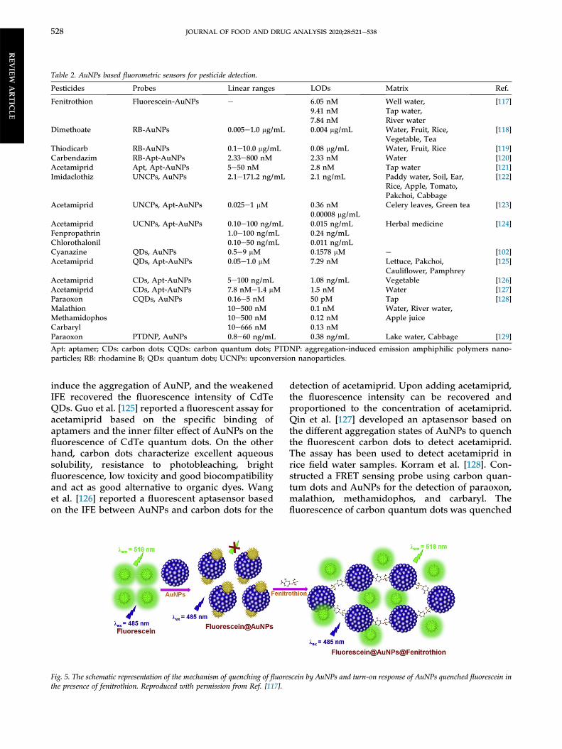
Fluorescence-based sensors are performed mainlybased on the quenching (turn-off) and enhancement(turn-on) of fluorescence intensity, or the fluores-cence resonance energy transfer (FRET) [115].AuNPs can act as highly efficient fluorescencequenchers, owing to the high molar extinction co-efficients and broad adsorption spectrum over-lapping [116]. The interactions between fluorescentmolecules and AuNPs cause the change of thefluorescence intensity. Thus, AuNPs can be utilizedas excellent fluorescence quenchers for FRET- andIFE-based assays for the determination of pesticides,as shown in Table 2. Nebu et al. [117] demonstratedthat fluorescein-capped AuNPs were sensitive for
fluorescence turn-on detection of fenitrothion. Asshown in Fig. 5, the fluorescence of fluorescein wasquenched by AuNPs and recovered in the presenceof fenitrothion. The approach was successfullydemonstrated on a paper strip with the detectionlimit in the nanomolar range. Hung et al. [118]developed a rhodamine B (RB)-functionalizedAuNPs as fluorescence “turn-on” probe for thedetermination of dimethoate. The sensing mecha-nism is based on the emission spectrum of RBsignificantly overlaps with the absorption spectrumof AuNPs. In the presence of dimethoate, the fluo-rescence was recovered owing to the competitiveadsorption between RB molecules and dimethoateon the surface of AuNPs. The approach has beenapplied to detect dimethoate in water and fruitsamples with satisfactory recoveries. Tseng et al.[119] used a similar strategy to detect thiodicarb inwater and food samples. Su et al. [120] used car-bendazim-specific aptamer as a sensing probe,AuNPs, and RB as the indicator to develop anaptasensor for the detection of carbendazim. Theaptamer can specifically be combined with carben-dazim to form a stable complex and desorbed fromthe surface of AuNPs, induced the aggregation ofAuNPs by NaCl. Bahreyni et al. [121] developed afluorometric aptasensor to detect acetamiprid basedon the use of an aptamer against acetamiprid,different complementary strands, and AuNPs.Except for organic dyes, upconversion nano-particles, quantum dots, and carbon dots have beenproved to be efficient fluorescence donors in recentyears. You et al. [122] constructed a competitiveimmunoassay for the detection of midacloprid byusing AuNPs as an absorber for the fluorescence ofupconversion nanoparticles through inner filter ef-fect. Yang et al. [123] demonstrated a colorimetricand fluorometric method based on the principle oftarget-triggered structure switch of aptamers, salt-induced AuNPs aggregation, and signal amplifica-tion from upconversion nanoparticles for thedetection of acetamiprid. Liu et al. [124] developed afluorescence turn-on method base on the lumines-cence resonance energy transfer between upcon-version nanoparticles, and AuNPs for the detectionof cyano-containing pesticides (acetamiprid, fen-propathrin, and chlorothalonil). The approach alsohas been applied to detect acetamiprid in Lanceolata,Angelica dahurica and Astragalus. Dong et al. [102]developed a dual strategy for the fluorescent andvisual detection of cyanazine based on the quantumdots (QDs)-AuNPs sensing system. The fluores-cence of CdTe QDs was remarkably quenched byAuNPs via the inner filter effect (IFE). Upon additionof cyanazine can adsorb on the surface of AuNPs to
JOURNAL OF FOOD AND DRUG ANALYSIS 2020;28:521e538 527
REVIEW
ARTIC
LE
induce the aggregation of AuNP, and the weakenedIFE recovered the fluorescence intensity of CdTeQDs. Guo et al. [125] reported a fluorescent assay foracetamiprid based on the specific binding ofaptamers and the inner filter effect of AuNPs on thefluorescence of CdTe quantum dots. On the otherhand, carbon dots characterize excellent aqueoussolubility, resistance to photobleaching, brightfluorescence, low toxicity and good biocompatibilityand act as good alternative to organic dyes. Wanget al. [126] reported a fluorescent aptasensor basedon the IFE between AuNPs and carbon dots for the
detection of acetamiprid. Upon adding acetamiprid,the fluorescence intensity can be recovered andproportioned to the concentration of acetamiprid.Qin et al. [127] developed an aptasensor based onthe different aggregation states of AuNPs to quenchthe fluorescent carbon dots to detect acetamiprid.The assay has been used to detect acetamiprid inrice field water samples. Korram et al. [128]. Con-structed a FRET sensing probe using carbon quan-tum dots and AuNPs for the detection of paraoxon,malathion, methamidophos, and carbaryl. Thefluorescence of carbon quantum dots was quenched
Table 2. AuNPs based fluorometric sensors for pesticide detection.
Pesticides Probes Linear ranges LODs Matrix Ref.
Fenitrothion Fluorescein-AuNPs e 6.05 nM Well water, [117]9.41 nM Tap water,7.84 nM River water
Dimethoate RB-AuNPs 0.005e1.0 mg/mL 0.004 mg/mL Water, Fruit, Rice, [118]Vegetable, Tea
Thiodicarb RB-AuNPs 0.1e10.0 mg/mL 0.08 mg/mL Water, Fruit, Rice [119]Carbendazim RB-Apt-AuNPs 2.33e800 nM 2.33 nM Water [120]Acetamiprid Apt, Apt-AuNPs 5e50 nM 2.8 nM Tap water [121]Imidaclothiz UNCPs, AuNPs 2.1e171.2 ng/mL 2.1 ng/mL Paddy water, Soil, Ear,
Rice, Apple, Tomato,Pakchoi, Cabbage
[122]
Acetamiprid UNCPs, Apt-AuNPs 0.025e1 mM 0.36 nM Celery leaves, Green tea [123]0.00008 mg/mL
Acetamiprid UCNPs, Apt-AuNPs 0.10e100 ng/mL 0.015 ng/mL Herbal medicine [124]Fenpropathrin 1.0e100 ng/mL 0.24 ng/mLChlorothalonil 0.10e50 ng/mL 0.011 ng/mLCyanazine QDs, AuNPs 0.5e9 mM 0.1578 mM e [102]Acetamiprid QDs, Apt-AuNPs 0.05e1.0 mM 7.29 nM Lettuce, Pakchoi,
Cauliflower, Pamphrey[125]
Acetamiprid CDs, Apt-AuNPs 5e100 ng/mL 1.08 ng/mL Vegetable [126]Acetamiprid CDs, Apt-AuNPs 7.8 nMe1.4 mM 1.5 nM Water [127]Paraoxon CQDs, AuNPs 0.16e5 nM 50 pM Tap
Water, River water,Apple juice
[128]Malathion 10e500 nM 0.1 nMMethamidophos 10e500 nM 0.12 nMCarbaryl 10e666 nM 0.13 nMParaoxon PTDNP, AuNPs 0.8e60 ng/mL 0.38 ng/mL Lake water, Cabbage [129]
Apt: aptamer; CDs: carbon dots; CQDs: carbon quantum dots; PTDNP: aggregation-induced emission amphiphilic polymers nano-particles; RB: rhodamine B; QDs: quantum dots; UCNPs: upconversion nanoparticles.
Fig. 5. The schematic representation of the mechanism of quenching of fluorescein by AuNPs and turn-on response of AuNPs quenched fluorescein inthe presence of fenitrothion. Reproduced with permission from Ref. [117].
528 JOURNAL OF FOOD AND DRUG ANALYSIS 2020;28:521e538
REVIEW
ARTIC
LE

in the presence of AuNPs and recovered by theaddition of acetylthiocholine iodide and AChE. Byevaluating the inhibition effect on the activity ofacetylthiocholine and fluorescence intensity, theconcentration of pesticides could be quantified.Chen et al. [129] demonstrated a sensing systemconsisted of aggregation-induced emission (AIE)NPs, AuNPs, and AChE for the detection of para-oxon. The sensing platform was proved to have awider linear range from 0.8 to 60 ng/mL with a LODat 0.38 ng/mL.
3.3. Surface-enhanced Raman scattering assays
Surface-enhanced Raman scattering (SERS) isbased on a significant enhancement of Ramansignal, typically up to 6 orders of magnitude, from arough metal surface owing to the electromagneticand chemical enhancement [130]. Nanostructuredsurfaces of gold, silver, or copper are the mostcommon SERS-active substrates. SERS provides thecharacteristic fingerprint of desirable analytes withhigh sensitivity at nanomolar to picomolar concen-tration, even at the single-molecule level [131e133].Thus, SERS has become a powerful analyticalmethod for detecting pesticides because of its highsensitivity, good selectivity, low cost, and rapidity[134,135].Recently, Xu et al. reviewed the development of
the SERS technique for pesticide detection in foodwith the advantages of high sensitivity, reproduc-ibility, selectivity, and low-cost [136]. The liquidsamples can be directly detected for pesticide resi-dues by SERS (Fig. 6a); the pesticide residues on thesurface of a solid or solid-liquid mixture can bedetected by SERS (Fig. 6b); the pesticide residuesinside solid sample also can be detected by SERSwith an extraction process (Fig. 6c). Thus, thefollowing are recent achievements in fabricatingappropriate SERS-active substrates for pesticidedetection. In recent year, several pesticides such asomethoate [137], chlorpyrifos [137], acetamiprid[138], clothianidin [138], phosmet [138], thiram [138],paraquat [139], carbendazim [140], pyrimethanil[141] was determined by SERS with easy-to-prepareAuNPs as shown in Table 3. Fernandes et al. [142]used dendrimer stabilized anisotropic AuNPs asSERS probes for the detection of sodium dieth-yldithiocarbamate, thiram, and paraquat in water.Tan et al. [143]. Demonstrated that osmiumcarbonyl-clusters binging on the AuNPs as a SERSprobe to enhance CO stretching vibration signal andavoid the interference of biomolecule. The probehas been used for highly sensitive detection ofglyphosate in spiked beer. Hong et al. [144] used a
suction method to fabricate a SERS substrate via theimmobilization of AuNPs on an ultrafiltrationmembrane. The thiabendazole standard solutionand orange peel extract can be concentrated on thesubstrate and analyzed by portable Raman spec-trometry. Luo et al. [145] performed AuNPs were insitu synthesized on pseudo-paper films and used asa SERS substrate for the analysis thiram and para-thion methyl with 3.02 � 106 enhancement. Yaseenet al. [146] demonstrated a simultaneously detectmulti-class pesticides (thiacloprid, profenofos andoxamyl) in peach with SERS on silver-coated AuNPswith 26 nm Au core size and 6 nm Ag shell
Fig. 6. Pesticide residues detection in liquid (a), on the surface (b), orinside solid foods by SERS. Reproduced with permission fromRef. [136].
JOURNAL OF FOOD AND DRUG ANALYSIS 2020;28:521e538 529
REVIEW
ARTIC
LE
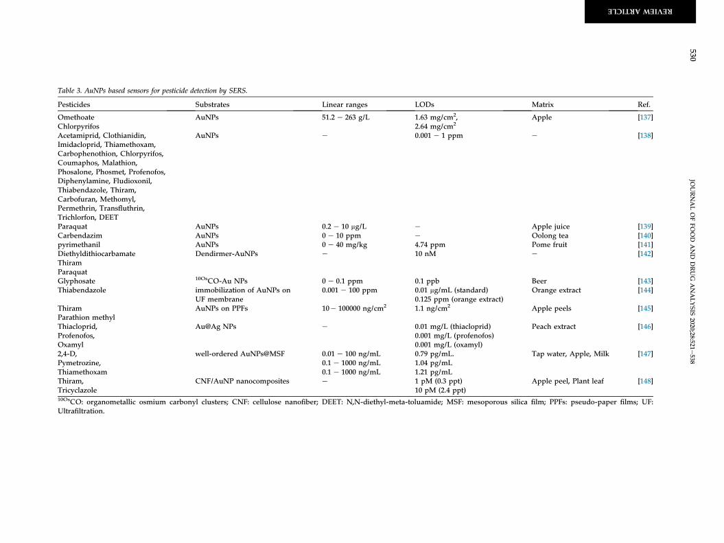
Table 3. AuNPs based sensors for pesticide detection by SERS.
Pesticides Substrates Linear ranges LODs Matrix Ref.
OmethoateChlorpyrifos
AuNPs 51.2 e 263 g/L 1.63 mg/cm2,2.64 mg/cm2
Apple [137]
Acetamiprid, Clothianidin,Imidacloprid, Thiamethoxam,Carbophenothion, Chlorpyrifos,Coumaphos, Malathion,Phosalone, Phosmet, Profenofos,Diphenylamine, Fludioxonil,Thiabendazole, Thiram,Carbofuran, Methomyl,Permethrin, Transfluthrin,Trichlorfon, DEET
AuNPs e 0.001 e 1 ppm e [138]
Paraquat AuNPs 0.2 e 10 mg/L e Apple juice [139]Carbendazim AuNPs 0 e 10 ppm e Oolong tea [140]pyrimethanil AuNPs 0 e 40 mg/kg 4.74 ppm Pome fruit [141]DiethyldithiocarbamateThiramParaquat
Dendirmer-AuNPs e 10 nM e [142]
Glyphosate 10OsCO-Au NPs 0 e 0.1 ppm 0.1 ppb Beer [143]Thiabendazole immobilization of AuNPs on
UF membrane0.001 e 100 ppm 0.01 mg/mL (standard)
0.125 ppm (orange extract)Orange extract [144]
ThiramParathion methyl
AuNPs on PPFs 10e 100000 ng/cm2 1.1 ng/cm2 Apple peels [145]
Thiacloprid,Profenofos,Oxamyl
Au@Ag NPs e 0.01 mg/L (thiacloprid)0.001 mg/L (profenofos)0.001 mg/L (oxamyl)
Peach extract [146]
2,4-D,Pymetrozine,Thiamethoxam
well-ordered AuNPs@MSF 0.01 e 100 ng/mL0.1 e 1000 ng/mL0.1 e 1000 ng/mL
0.79 pg/mL.1.04 pg/mL1.21 pg/mL
Tap water, Apple, Milk [147]
Thiram,Tricyclazole
CNF/AuNP nanocomposites e 1 pM (0.3 ppt)10 pM (2.4 ppt)
Apple peel, Plant leaf [148]
10OsCO: organometallic osmium carbonyl clusters; CNF: cellulose nanofiber; DEET: N,N-diethyl-meta-toluamide; MSF: mesoporous silica film; PPFs: pseudo-paper films; UF:Ultrafiltration.
530JO
URNALOFFO
OD
AND
DRUG
ANALYSIS
2020;28:521e538
REVIEWARTICLE

Table 4. AuNPs based electrochemical sensors for pesticide detection.
Pesticides Probes Linear ranges LODs Matrix Ref.
MalathionMethyl parathion
AChE/Nafion/AuNPs/rGO/GCE 0.0001e1 ng/mL 0.0278 pg/mL (0.084 pM) Tap water, Mineral water, Chinese cabbage [154]0.0217 pg/mL (0.0824 pM)
Paraoxon-ethyl AChE/MWCNTs-CS/AuNPs/SPCE 0.01e10 mg/mL 0.03 mg/mL Spinach [155]10e100 mg/mL
MalathionMethyl parathion
GCE/P-ABSA/DAR/AuNPs/DAR/AChE 0.003e30 pM 0.0016 pM Tap water, Well water, Chinese cabbage [156]0.0038e38 pM 0.0022 pM
Methyl parathion ITO/(GPDDA/GPSS)10ITO/(GPDDA/GPSS)1(AuNP/GPSS)10
0.95e152 mM (0.25e40 ppm) 0.859 mM (0.226 ppm) Tap water, Soil, Cabbage [157]1.90e228 mM (0.5e60 ppm) 2.930 mM (0.770 ppm)
Methyl parathion AuNPs/NR-BSA-graphene/Nafion/GCE 0.02e0.153 mM 6 nM Soil, Water, Potato juice [158]0.153e1.36 mM
Methyl parathionParathion
HAuNPs/rGO/GCE 0.3e10 mM 0.12 mM [159]0.11e50 mM 23 nM
Simazine MIP/ATP@AuNPs/ATP/Au electrode 0.03e140 mM 0.012 mM Tap water, River water, soil [160]Tebuconazole MIP/Au-PB/SH-G/AuNPs/GCE 50 nMe40 mM 12.5 nM Cucumber, Green vegetable, Strawberry [161]Diazinon Apt/AuNPs/SPGE 0.0304e304 ng/mL 0.005 ng/mL Rat plasma [163]Carbendazim MCH/Apt/AuNPs/1-AP-CNHs/GCE 0.001e1.0 ng/mL 0.5 pg/mL Lettuce, Orange juice [164]Malathion Apt/MCH/CP/AuNPs/PDA/GCE 0.5e650 pg/mL 0.5 pg/mL Cauliflower, Cabbage [165]Chlorpyrifos FTO-AuNPs-chlAb 1 fM - 1 mM 10 fM Apple, Pomegranate, Cabbage [166]Imidacloprid AuNPs-SPCE 50e10000 pM 22 pM Tap water, Watermelon, Tomato [167]
AP-CNHs: 1-aminopyrene modified carbon nanohorns; AChE: acetylcholinesterase; Apt: aptamer; ATP: o-aminothiophenol; AuNPs/rGO: gold nanoparticles/three-dimensionalgraphene; BSA: bovine serum albumin; chlAb: anti-chlorpyrifos antibodies; CP: capture probe; CS: chitosan; DAR: diazo-resins; FTO: fluorine doped tin-oxide; GCE: glassy carbonelectrode; HAuNPs: hollow gold nanoparticles; ITO: indium tin oxide; MCH: 6-mercapto-1-hexanol; MIP: molecularly imprinted polymer; MWCNT: multiwalled carbon nanotube;NR: neutral red; P-ABSA: p-aminobenzenesulfonic acid; PB: Prussian blue; PDA: polydopamine; SH-G: thiol graphene; SPCE: screen-printed carbon electrode.
JOURNALOFFO
OD
AND
DRUG
ANALYSIS
2020;28:521e538
531
REVIEW ARTICLE

thickness. Mesoporous silica-supported orderly-spaced AuNPs also used as the SERS substrate forthe detection of 2,4-D, pymetrozine and thiame-thoxam in food samples [147]. Kim et al. [148]fabricated a low-cost and flexible SERS substratebased on cellulose nanofiber/AuNPs nano-composites via vacuum-assisted filtration. The SERSsubstrate can effectively detect thiram and tricycla-zole with 4.5 � 109 enhancement and LODs weredown to 1 pM.
3.4. Electrochemical assays
Electrochemical sensors, because of their porta-bility, rapidity, low-cost, high sensitivity and selec-tivity, have become one of the most effectiveanalytical methods [149e151]. Electrochemical sen-sors can be basically divided into potentiometric,amperometric and conductometric methods ac-cording to the property of obtained response[152,153]. In recent years, AuNPs have been used forelectrochemical sensors for the detection of pesti-cides as shown in Table 4. Several AuNPs basedelectrochemical biosensors have been described inthe literature by using AChE enzyme [154,155].Dong et al. [154] developed an AChE sensor for thedetection of malathion and parathion methyl incabbage and water samples based on a film ofAuNPs/three-dimensional graphene. Hua et al.
[154] demonstrated a disposable amperometricsensor for parathion ethyl determination based on ascreen-printed carbon electrode consisted of AChEimmobilized onto the surface of multiwalled carbonnanotubes, chitson and AuNPs. Jiang et al. [156]demonstrated to construct stable covalentlyattached multilayer films by using layer-by-layerself-assembly of diazo-resins, AuNPs, and AChE.The films were immobilized on the surface of a p-aminobenzesulfonic acid-modified glassy carbonelectrode and used for quantitative detection ofmalathion and parathion methyl. Rodrigues et al.[157] performed the fabrication of layer-by-layerfilms composed of reduced graphene oxide andAuNPs for the detection of parathion methyl (Fig. 7).The differential pulse voltammetry was applied onthe layer-by-layer films modified on indium oxideelectrode, in the presence of AuNPs, a wider linearrange was achieved between 0.5 and 60 ppm, withLOD of 0.770 ppm for parathion methyl. Singh et al.have synthesized the AuNPs/neutral red-BSA-functionalized graphene nanocomposite andimmobilized on Nafion modified glassy carbonelectrode [158]. The prepared electrode exhibitshigher electrocatalytic ability towards methyl para-thion with the enlarged current. A sensitive vol-tammetric sensor for methyl parathion andparathion was developed by Lu et al. [159] based onreduced graphene oxide and hollow AuNPs
Fig. 7. Procedures to modify ITO electrodes with (GPPDA/GPSS)10 and (GPDDA/GPSS)1(AuNP/GPSS)10 LbL films (assembly): immersion time was15 min for both GPDDA and GPSS, and 6 min for AuNP. Reproduced with permission from Ref. [157].
532 JOURNAL OF FOOD AND DRUG ANALYSIS 2020;28:521e538
REVIEW
ARTIC
LE

immobilized on a glassy carbon electrode. In addi-tion, the molecularly imprinted electrochemicalsensors have also used to detect simazine [160] andtebuconazole [161] with AuNPs modified electrodes.Recently, Liu et al. reviewed the development ofaptasensor for pesticide detection [162]. Thus,aptasensors based on specific aptamers modified onthe AuNPs have been used for the detection ofdiazinon [163], carbendazim [164] and malathion[165]. Electrochemical immunosensor based onAuNPs have been used for the detection of chlor-pyrifos [166] and imidacloprid [167] with ultrahighsensitivity.
4. Conclusions and future trends
This review has given a brief overview aboutrecent advances of AuNPs-based colorimetric,fluorescence, SERS, and electrochemical sensors forthe determination of pesticides in environmentalsamples. In contrast to conventional chromato-graphic and mass spectrometry-based methods,AuNPs have been shown to be a very useful, alter-native tool for pesticide sensing owing to theirunique optical property, facile synthesis, easy sur-face functionalization, and satisfactory biocompati-bility. The selectivity of the AuNP-based probetoward a specific pesticide has been effectivelyimproved by modifying the nanoparticle surfacewith aptamer and integrating the enzyme-substratesystem. However, these reported AuNP-basedprobes still suffer from some limitations, includingthe adsorption of non-target molecules on thenanoparticle surface, uncontrolled nanoparticle ag-gregation in a high-ionic-strength solution, andpoor selectivity in complex matrices. Although themodification of AuNPs with neutral surfactants,such as Tween 20 and fluorosurfactant, enhancestheir colloidal stability in a high-ionic-strength so-lution, these modifiers provide an adverse effect onselectivity toward a specific pesticide. Additionally,the above-discussed detection methods (i.e., colori-metric, fluorescence, SERS, and electrochemicalspectroscopy) are unable to simultaneously sensemultiple pesticides with similar structural proper-ties. Therefore, enrichment or separation of thetarget pesticides would be inevitable prior to usingthe AuNP-based probe. It is suggested that theintegration of the AuNP-based probe with a recog-nition element-modified platform could be apromising way to allow the detection of targetanalytes despite of other possible interfering com-pounds. Examples of recognition elements includeaptamer, antibodies, and molecularly imprintedpolymers. Also, the integration of the novel
technologies such as handheld devices, microfluidicor paper chips and portable test strips with sensingsystem may promise a bright future for on-siteapplication. The recognition event can be deliveredimmediately to the servers through wire-lessnetworking and miniaturized device such as smartphone. The applications of AuNPs-based sensorsmay lead to a new generation in real-time and on-site monitoring pesticides.
Conflict of interest
The authors have declared no conflict of interest.
Acknowledgment
This work was financially supported by the Min-istry of Science and Technology of Taiwan, Taiwanunder contract number MOST 108-2113-M-017-003and 109-2113-M-143-002.
References
[1] Thuy PT, Van Geluwe S, Nguyen V-A, Van der Bruggen B.Current pesticide practices and environmental issues inVietnam: management challenges for sustainable use ofpesticides for tropical crops in (South-East) Asia to avoidenvironmental pollution. J Mater Cycles Waste Manag 2012;14:379e87.
[2] Marican A, Dur�an-Lara EF. A review on pesticide removalthrough different processes. Environ Sci Pollut Res 2018;25:2051e64.
[3] Gilden RC, Huffling K, Sattler B. Pesticides and health risks.J Obstet Gynecol Neonatal Nurs 2010;39:103e210.
[4] Gorell JM, Johnson C, Rybicki B, Peterson E, Richardson R.The risk of Parkinson's disease with exposure to pesticides,farming, well water, and rural living. Neurology 1998;50:1346e50.
[5] Tixier P, Chabrier C, Mal�ezieux E. Pesticide residues inheterogeneous plant populations, a model-based approachapplied to nematicides in banana (Musa spp.). J Agric FoodChem 2007;55:2504e8.
[6] Barra R, Vighi M, Maffioli G, Di Guardo A, Ferrario P.Coupling SoilFug model and GIS for predicting pesticidepollution of surface water at watershed level. Environ SciTechnol 2000;34:4425e33.
[7] Ko E, Choi M, Shin S. Bottom-line mechanism of organo-chlorine pesticides on mitochondria dysfunction linkedwith type 2 diabetes. J Hazard Mater 2020;393:122400.
[8] Naous GE-Z, Merhi A, Abboud MI, Mroueh M, Taleb RI.Carcinogenic and neurotoxic risks of acrylamide consumedthrough caffeinated beverages among the lebanese popu-lation. Chemosphere 2018;208:352e7.
[9] Legeay S, Billat P-A, Clere N, Nesslany F, Bristeau S,Faure S, et al. Two dechlorinated chlordecone derivativesformed by in situ chemical reduction are devoid of geno-toxicity and mutagenicity and have lower proangiogenicproperties compared to the parent compound. Environ SciPollut Res 2018;25:14313e23.
[10] Kim K-H, Kabir E, Jahan SA. Exposure to pesticides and theassociated human health effects. Sci Total Environ 2017;575:525e35.
[11] Schurek J, Portol�es T, Hajslova J, Riddellova K,Hern�andez F. Application of head-space solid-phasemicroextraction coupled to comprehensive two-
JOURNAL OF FOOD AND DRUG ANALYSIS 2020;28:521e538 533
REVIEW
ARTIC
LE

dimensional gas chromatographyetime-of-flight massspectrometry for the determination of multiple pesticideresidues in tea samples. Anal Chim Acta 2008;611:163e72.
[12] Harshit D, Charmy K, Nrupesh P. Organophosphoruspesticides determination by novel HPLC and spectropho-tometric method. Food Chem 2017;230:448e53.
[13] Bala R, Swami A, Tabujew I, Peneva K, Wangoo N,Sharma RK. Ultra-sensitive detection of malathion usingquantum dots-polymer based fluorescence aptasensor.Biosens Bioelectron 2018;104:45e9.
[14] Cormode DP, Roessl E, Thran A, Skajaa T, Gordon RE,Schlomka J-P, et al. Atherosclerotic plaque composition:analysis with multicolor CT and targeted gold nano-particles. Radiology 2010;256:774e82.
[15] El-Sayed IH, Huang X, El-Sayed MA. Surface plasmonresonance scattering and absorption of anti-EGFR antibodyconjugated gold nanoparticles in cancer diagnostics: appli-cations in oral cancer. Nano Lett 2005;5:829e34.
[16] Hutter E, Boridy S, Labrecque S, Lalancette-H�ebert M,Kriz J, Winnik FM, et al. Microglial response to goldnanoparticles. ACS Nano 2010;4:2595e606.
[17] Schmid G, Corain B. Nanoparticulated gold: syntheses,structures, electronics, and reactivities. Eur J Inorg Chem2003:3081e98.
[18] Daniel M-C, Astruc D. Gold nanoparticles: assembly, su-pramolecular chemistry, quantum-size-related properties,and applications toward biology, catalysis, and nanotech-nology. Chem Rev 2004;104:293e346.
[19] Mirkin CA, Letsinger RL, Mucic RC, Storhoff JJ. A DNA-based method for rationally assembling nanoparticles intomacroscopic materials. Nature 1996;382:607e9.
[20] Elghanian R, Storhoff JJ, Mucic RC, Letsinger RL,Mirkin CA. Selective colorimetric detection of poly-nucleotides based on the distance-dependent opticalproperties of gold nanoparticles. Science 1997;277:1078e81.
[21] Huang X, El-Sayed IH, Qian W, El-Sayed MA. Cancer cellimaging and photothermal therapy in the near-infraredregion by using gold nanorods. J Am Chem Soc 2006;128:2115e20.
[22] Mastrotto F, Caliceti P, Amendola V, Bersani S,Magnusson JP, Meneghetti M, et al. Polymer control ofligand display on gold nanoparticles for multimodalswitchable cell targeting. Chem Commun 2011;47:9846e8.
[23] Walkey CD, Olsen JB, Guo H, Emili A, Chan WC. Nano-particle size and surface chemistry determine serum pro-tein adsorption and macrophage uptake. J Am Chem Soc2012;134:2139e47.
[24] Walkey CD, Olsen JB, Song F, Liu R, Guo H, Olsen DWH,et al. Protein corona fingerprinting predicts the cellularinteraction of gold and silver nanoparticles. ACS Nano2014;8:2439e55.
[25] De Jong WH, Hagens WI, Krystek P, Burger MC, Sips AJ,Geertsma RE. Particle size-dependent organ distribution ofgold nanoparticles after intravenous administration. Bio-materials 2008;29:1912e9.
[26] Wilhelm S, Tavares AJ, Dai Q, Ohta S, Audet J, Dvorak HF,et al. Analysis of nanoparticle delivery to tumours. Nat RevMater 2016;1:1e12.
[27] Turkevich J, Stevenson PC, Hillier J. A study of the nucle-ation and growth processes in the synthesis of colloidalgold. Discuss Faraday Soc 1951;11:55e75.
[28] Frens G. Controlled nucleation for the regulation of theparticle size in monodisperse gold suspensions. Nat PhysSci (Lond) 1973;241:20e2.
[29] Busbee BD, Obare SO, Murphy CJ. An improved synthesisof high-aspect-ratio gold nanorods. Adv Mater 2003;15:414e6.
[30] Cao C, Park S, Sim SJ. Seedless synthesis of octahedral goldnanoparticles in condensed surfactant phase. J ColloidInterface Sci 2008;322:152e7.
[31] Brust M, Walker M, Bethell D, Schiffrin DJ, Whyman R.Synthesis of thiol-derivatised gold nanoparticles in a two-
phase liquideliquid system. J Chem Soc Chem Commun1994;801e2.
[32] Sun X, Dong S, Wang E. Large-scale, solution-phase pro-duction of microsized, single-crystalline, hexagonal goldmicroplates by thermal reduction of HAuCl4 with oxalicacid. Chem Lett 2005;34:968e9.
[33] Kumar DR, Kumavat S, Chamundeswari V, Patra PP,Kulkarni A, Prasad B. Surfactant-free synthesis of aniso-tropic gold nanostructures: can dicarboxylic acids alone actas shape directing agents? RSC Adv 2013;3:21641e7.
[34] Olenic L, Mihailescu G, Pruneanu S, Bratu I, Biris AR,Lupu D, et al. Nanoparticles from a gold complex withsulfite ion as ligand: preparation and characterization. PartSci Technol 2005;23:79e83.
[35] Yu H, Feng X, Chen X-x, Wang S-s, Jin J. A highly sensitivedetermination of sulfite using a glassy carbon electrodemodified with gold nanoparticles-reduced graphene oxidenano-composites. J Electroanal Chem 2017;801:488e95.
[36] Marișca OT, Leopold N. Anisotropic gold nanoparticle-cellinteractions mediated by collagen. Materials 2019;12:1131.
[37] L�evy R, Thanh NT, Doty RC, Hussain I, Nichols RJ,Schiffrin DJ, et al. Rational and combinatorial design ofpeptide capping ligands for gold nanoparticles. J Am ChemSoc 2004;126:10076e84.
[38] Rahme K, Chen L, Hobbs RG, Morris MA, O'Driscoll C,Holmes JD. PEGylated gold nanoparticles: polymer quan-tification as a function of PEG lengths and nanoparticledimensions. RSC Adv 2013;3:6085e94.
[39] Li X, Qin Y, Liu C, Jiang S, Xiong L, Sun Q. Size-controlledstarch nanoparticles prepared by self-assembly withdifferent green surfactant: the effect of electrostatic repul-sion or steric hindrance. Food Chem 2016;199:356e63.
[40] Chen Y, Xianyu Y, Jiang X. Surface modification of goldnanoparticles with small molecules for biochemical anal-ysis. Acc Chem Res 2017;50:310e9.
[41] Alkilany AM, Abulateefeh SR, Mills KK, Bani Yaseen AI,Hamaly MA, Alkhatib HS, et al. Colloidal stability of citrateand mercaptoacetic acid capped gold nanoparticles uponlyophilization: effect of capping ligand attachment and typeof cryoprotectants. Langmuir 2014;30:13799e808.
[42] Godoy-Reyes TM, Costero AM, Gavi~na P, Martínez-M�a~nez R, Sancen�on F. Colorimetric detection of normeta-nephrine, a pheochromocytoma biomarker, using bifunc-tionalised gold nanoparticles. Anal Chim Acta 2019;1056:146e52.
[43] Liu C-Y, Tseng W-L. Using polysorbate 40-stabilized goldnanoparticles in colorimetric assays of hydrogen cyanide incyanogenic glycoside-containing plants. Anal Methods2012;4:2537e42.
[44] Yoon Y-J, Kang S-H, Do C, Moon SY, Kim T-H. Water-redispersible and highly stable gold nanoparticles perma-nently capped by charge-controllable surfactants for po-tential medical applications. ACS Appl Nano Mater 2019;2:7924e32.
[45] Ribeiro CA, Albuquerque LJ, de Castro CE, Batista BL, deSouza AL, Albuquerque BL, et al. One-pot synthesis ofsugar-decorated gold nanoparticles with reduced cytotox-icity and enhanced cellular uptake. Colloids Surf A Phys-icochem Eng Asp 2019;580:123690.
[46] Green M, Smyth-Boyle D. Directed growth of gold nano-structures using a nucleoside/nucleotide. J Mater Chem2007;17:3588e90.
[47] Pu F, Ren J, Qu X. Nucleobases, nucleosides, and nucleo-tides: versatile biomolecules for generating functionalnanomaterials. Chem Soc Rev 2018;47:1285e306.
[48] Cano I, Chapman AM, Urakawa A, van Leeuwen PW.Air-stable gold nanoparticles ligated by secondaryphosphine oxides for the chemoselective hydrogenationof aldehydes: crucial role of the ligand. J Am Chem Soc2014;136:2520e8.
[49] Huang X, Wu H, Liao X, Shi B. One-step, size-controlledsynthesis of gold nanoparticles at room temperature usingplant tannin. Green Chem 2010;12:395e9.
534 JOURNAL OF FOOD AND DRUG ANALYSIS 2020;28:521e538
REVIEW
ARTIC
LE

[50] Sardar R, Shumaker-Parry JS. Spectroscopic and micro-scopic investigation of gold nanoparticle formation: ligandand temperature effects on rate and particle size. J AmChem Soc 2011;133:8179e90.
[51] Kundu S, Wang K, Liang H. Size-selective synthesis andcatalytic application of polyelectrolyte encapsulated goldnanoparticles using microwave irradiation. J Phys Chem C2009;113:5157e63.
[52] Agasti SS, Chompoosor A, You C-C, Ghosh P, Kim CK,Rotello VM. Photoregulated release of caged anticancerdrugs from gold nanoparticles. J Am Chem Soc 2009;131:5728e9.
[53] Jana NR, Gearheart L, Murphy CJ. Seeding growth for sizecontrol of 5�40 nm diameter gold nanoparticles. Langmuir2001;17:6782e6.
[54] Murphy CJ, Sau TK, Gole AM, Orendorff CJ, Gao J, Gou L,et al. Anisotropic metal nanoparticles: synthesis, assembly,and optical applications. J Phys Chem B 2005;109:13857e70.
[55] Ab�ecassis B, Testard F, Spalla O, Barboux P. Probing in situthe nucleation and growth of gold nanoparticles by small-angle X-ray scattering. Nano Lett 2007;7:1723e7.
[56] Nehl CL, Liao H, Hafner JH. Optical properties of star-shaped gold nanoparticles. Nano Lett 2006;6:683e8.
[57] Amendola V, Pilot R, Frasconi M, Marag�o OM, Iatì MA.Surface plasmon resonance in gold nanoparticles: a review.J Phys Condens Matter 2017;29:203002.
[58] Liu J, Murphy KE, MacCuspie RI, Winchester MR. Capa-bilities of single particle inductively coupled plasma massspectrometry for the size measurement of nanoparticles: acase study on gold nanoparticles. Anal Chem 2014;86:3405e14.
[59] Xu X, Chen J, Li B, Tang L, Jiang J. Single particle ICP-MS-based absolute and relative quantification of E. coli O15716S rRNA using sandwich hybridization capture. Analyst2019;144:1725e30.
[60] Stolarczyk EU, Stolarczyk K, Łaszcz M, Kubiszewski M,Maruszak W, Olejarz W, et al. Synthesis and characteriza-tion of genistein conjugated with gold nanoparticles and thestudy of their cytotoxic properties. Eur J Pharm Sci 2017;96:176e85.
[61] Sweeney SF, Woehrle GH, Hutchison JE. Rapid purificationand size separation of gold nanoparticles via diafiltration.J Am Chem Soc 2006;128:3190e7.
[62] Verma HN, Singh P, Chavan R. Gold nanoparticle: syn-thesis and characterization. Vet World 2014;7:72e7.
[63] De Souza CD, Nogueira BR, Rostelato MEC. Review ofthe methodologies used in the synthesis gold nano-particles by chemical reduction. J Alloys Compd 2019;798:714e40.
[64] Jana NR, Gearheart L, Murphy CJ. Evidence for seed-mediated nucleation in the chemical reduction of gold saltsto gold nanoparticles. Chem Mater 2001;13:2313e22.
[65] Yu Y-Y, Chang S-S, Lee C-L, Wang CC. Gold nanorods:electrochemical synthesis and optical properties. J PhysChem B 1997;101:6661e4.
[66] Mohamed MB, Ismail KZ, Link S, El-Sayed MA. Thermalreshaping of gold nanorods in micelles. J Phys Chem B1998;102:9370e4.
[67] Ma H, Yin B, Wang S, Jiao Y, Pan W, Huang S, et al. Syn-thesis of silver and gold nanoparticles by a novel electro-chemical method. ChemPhysChem 2004;5:68e75.
[68] Wei Z, Liu C-j. Synthesis of monodisperse gold nano-particles in ionic liquid by applying room temperatureplasma. Mater Lett 2011;65:353e5.
[69] Wei G-T, Yang Z, Lee C-Y, Yang H-Y, Wang CC. Aque-ous�organic phase transfer of gold nanoparticles and goldnanorods using an ionic liquid. J Am Chem Soc 2004;126:5036e7.
[70] Singh P, Kumari K, Katyal A, Kalra R, Chandra R. Synthesisand characterization of silver and gold nanoparticles inionic liquid. Spectrochim Acta Mol Biomol Spectrosc 2009;73:218e20.
[71] Song JY, Jang H-K, Kim BS. Biological synthesis of goldnanoparticles using Magnolia kobus and Diopyros kaki leafextracts. Process Biochem 2009;44:1133e8.
[72] Gardea-Torresdey J, Parsons J, Gomez E, Peralta-Videa J,Troiani H, Santiago P, et al. Formation and growth of Aunanoparticles inside live alfalfa plants. Nano Lett 2002;2:397e401.
[73] Iravani S. Green synthesis of metal nanoparticles usingplants. Green Chem 2011;13:2638e50.
[74] Polte JR, Ahner TT, Delissen F, Sokolov S, Emmerling F,Thünemann AF, et al. Mechanism of gold nanoparticleformation in the classical citrate synthesis method derivedfrom coupled in situ XANES and SAXS evaluation. J AmChem Soc 2010;132:1296e301.
[75] El-Sayed IH, Huang X, El-Sayed MA. Selective laser photo-thermal therapy of epithelial carcinoma using anti-EGFRantibody conjugated gold nanoparticles. Cancer Lett 2006;239:129e35.
[76] Kumar S, Gandhi K, Kumar R. Modeling of formation ofgold nanoparticles by citrate method. Ind Eng Chem Res2007;46:3128e36.
[77] Patungwasa W, Hodak JH. pH tunable morphology of thegold nanoparticles produced by citrate reduction. MaterChem Phys 2008;108:45e54.
[78] Li C, Li D, Wan G, Xu J, Hou W. Facile synthesis ofconcentrated gold nanoparticles with low size-distributionin water: temperature and pH controls. Nanoscale Res Lett2011;6:440.
[79] Bai W, Zhu C, Liu J, Yan M, Yang S, Chen A. Goldnanoparticleebased colorimetric aptasensor for rapiddetection of six organophosphorous pesticides. EnvironToxicol Chem 2015;34:2244e9.
[80] Wang P, Wan Y, Ali A, Deng S, Su Y, Fan C, et al. Aptamer-wrapped gold nanoparticles for the colorimetric detectionof omethoate. Sci China Chem 2016;59:237e42.
[81] Lin J-H, Huang K-H, Zhan S-W, Yu C-J, Tseng W-L,Hsieh M-M. Inhibition of catalytic activity of fibrinogen-stabilized gold nanoparticles via thrombin-induced inclu-sion of nanoparticle into fibrin: application for thrombinsensing with more than 104-fold selectivity. SpectrochimActa Mol Biomol Spectrosc 2019;210:59e65.
[82] Yeh P-R, Tseng W-L. Human serum albumin-coated goldnanoparticles for selective extraction of lysozyme from real-world samples prior to capillary electrophoresis.J Chromatogr A 2012;1268:166e72.
[83] Hung S-Y, Shih Y-C, Tseng W-L. Tween 20-stabilized goldnanoparticles combined with adenosine triphosphate-BODIPY conjugates for the fluorescence detection ofadenosine with more than 1000-fold selectivity. Anal ChimActa 2015;857:64e70.
[84] Shih Y-C, Ke C-Y, Yu C-J, Lu C-Y, Tseng W-L. CombinedTween 20-stabilized gold nanoparticles and reducedgraphite oxideeFe3O4 nanoparticle composites for rapidand efficient removal of mercury species from a complexmatrix. ACS Appl Mater Interfaces 2014;6:17437e45.
[85] Li X, Cui H, Zeng Z. A simple colorimetric and fluorescentsensor to detect organophosphate pesticides based onadenosine triphosphate-modified gold nanoparticles. Sen-sors 2018;18:4302.
[86] Satnami ML, Korram J, Nagwanshi R, Vaishanav SK,Karbhal I, Dewangan HK, et al. Gold nanoprobe for inhi-bition and reactivation of acetylcholinesterase: an applica-tion to detection of organophosphorus pesticides. SensActuator B-Chem 2018;267:155e64.
[87] Sun J, Guo L, Bao Y, Xie J. A simple, label-free AuNPs-based colorimetric ultrasensitive detection of nerve agentsand highly toxic organophosphate pesticide. Biosens Bio-electron 2011;28:152e7.
[88] Rohit JV, Basu H, Singhal RK, Kailasa SK. Development ofp-nitroaniline dithiocarbamate capped gold nanoparticles-based microvolume UVevis spectrometric method for facileand selective detection of quinalphos insecticide in
JOURNAL OF FOOD AND DRUG ANALYSIS 2020;28:521e538 535
REVIEW
ARTIC
LE

environmental samples. Sens Actuator B-Chem 2016;237:826e35.
[89] D'souza SL, Pati RK, Kailasa SK. Ascorbic acid functional-ized gold nanoparticles as a probe for colorimetric and vi-sual read-out determination of dichlorvos in environmentalsamples. Anal Methods 2014;6:9007e14.
[90] Park Y, Im A, Hong YN, Kim C-K, Kim YS. Detection ofmalathion, fenthion and methidathion by using heparin-reduced gold nanoparticles. J Nanosci Nanotechnol 2011;11:7570e8.
[91] Barman G, Maiti S, Laha JK. Trichloroacetic acid assistedsynthesis of gold nanoparticles and its application indetection and estimation of pesticide. J Anal Sci Technol2013;4:1e7.
[92] Ellman GL, Courtney KD, Andres Jr V, Featherstone RM.A new and rapid colorimetric determination of acetylcho-linesterase activity. Biochem Pharmacol 1961;7:88e95.
[93] Chen H, Zhou K, Zhao G. Gold nanoparticles: from syn-thesis, properties to their potential application as colori-metric sensors in food safety screening. Trends Food SciTechnol 2018;78:83e94.
[94] Xu Q, Du S, Li H, Hu XY. Determination of acetamiprid by acolorimetric method based on the aggregation of goldnanoparticles. Microchim Acta 2011;173:323e9.
[95] Ellington AD, Szostak JW. In vitro selection of RNAmolecules that bind specific ligands. Nature 1990;346:818e22.
[96] Ellington AD, Szostak JW. Selection in vitro of single-stranded DNA molecules that fold into specific ligand-binding structures. Nature 1992;355:850e2.
[97] Tuerk C, Gold L. Systematic evolution of ligands by expo-nential enrichment: RNA ligands to bacteriophage T4 DNApolymerase. Science 1990;249:505e10.
[98] Weerathunge P, Ramanathan R, Shukla R, Sharma TK,Bansal V. Aptamer-controlled reversible inhibition of goldnanozyme activity for pesticide sensing. Anal Chem 2014;86:11937e41.
[99] Bala R, Sharma RK, Wangoo N. Development of goldnanoparticles-based aptasensor for the colorimetric detec-tion of organophosphorus pesticide phorate. Anal BioanalChem 2016;408:333e8.
[100] Bala R, Dhingra S, Kumar M, Bansal K, Mittal S,Sharma RK, et al. Detection of organophosphoruspesticideeMalathion in environmental samples using pep-tide and aptamer based nanoprobes. Chem Eng J 2017;311:111e6.
[101] Yang W, Wu Y, Tao H, Zhao J, Chen H, Qiu S. Ultrasensi-tive and selective colorimetric detection of acetamipridpesticide based on the enhanced peroxidase-like activity ofgold nanoparticles. Anal Methods 2017;9:5484e93.
[102] Dong L, Hou C, Yang M, Fa H, Wu H, Shen C, et al. Highlysensitive colorimetric and fluorescent sensor for cyanazinebased on the inner filter effect of gold nanoparticles.J Nanopart Res 2016;18:164.
[103] Fahimi-Kashani N, Hormozi-Nezhad MR. Gold-nano-particle-based colorimetric sensor array for discriminationof organophosphate pesticides. Anal Chem 2016;88:8099e106.
[104] Rana K, Bhamore JR, Rohit JV, Park T-J, Kailasa SK. Ligandexchange reactions on citrate-gold nanoparticles for a par-allel colorimetric assay of six pesticides. New J Chem 2018;42:9080e90.
[105] Baek SH, Lee SW, Kim EJ, Shin D-H, Lee S-W, Park TJ.Portable agrichemical detection system for enhancing thesafety of agricultural products using aggregation of goldnanoparticles. ACS Omega 2017;2:988e93.
[106] Ma Y, Jiang H, Shen C, Hou C, Huo D, Wu H, et al.Detection of carbendazim residues with a colorimetricsensor based on gold nanoparticles. J Appl Spectrosc 2017;84:460e5.
[107] Liu Q, Han P, Gong W, Wang H, Feng X. Colorimetricdetermination of the pesticide chlorothalonil based on the
aggregation of gold nanoparticles. Microchim Acta 2018;185:354.
[108] Wu S, Li D, Wang J, Zhao Y, Dong S, Wang X. Goldnanoparticles dissolution based colorimetric method forhighly sensitive detection of organophosphate pesticides.Sens Actuator B-Chem 2017;238:427e33.
[109] Mohamadjafari S, Rastegarzadeh S. A sensing colorimetricmethod based on in situ formation of gold nanoparticlesafter dispersive liquid-liquid microextraction for determi-nation of zineb. Microchem J 2017;132:154e60.
[110] Hashemi F, Rastegarzadeh S, Pourreza N. A combination ofdispersive liquideliquid microextraction and surface plas-mon resonance sensing of gold nanoparticles for thedetermination of ziram pesticide. J Separ Sci 2018;41:1156e63.
[111] Kang J-y, Zhang Y-j, Li X, Dong C, Liu H-y, Miao L-j, et al.Rapid and sensitive colorimetric sensing of the insecticidepymetrozine using melamine-modified gold nanoparticles.Anal Methods 2018;10:417e21.
[112] Wang Z, Huang Y, Wang D, Sun L, Dong C, Fang L, et al.A rapid colorimetric method for the detection of delta-methrin based on gold nanoparticles modified with 2-mercapto-6-nitrobenzothiazole. Anal Methods 2018;10:1774e80.
[113] Tu Q, Yang T, Qu Y, Gao S, Zhang Z, Zhang Q, et al. In situcolorimetric detection of glyphosate on plant tissues usingcysteamine-modified gold nanoparticles. Analyst 2019;144:2017e25.
[114] Kailasa SK, Nguyen TP, Baek SH, Rafique R, Park TJ. As-sembly of 6-aza-2-thiothymine on gold nanoparticles forselective and sensitive colorimetric detection of pencycuronin water and food samples. Talanta 2019;205:120087.
[115] Jin Y, Gao X. Plasmonic fluorescent quantum dots. NatNanotechnol 2009;4:571e6.
[116] Huang C-C, Chang H-T. Selective gold-nanoparticle-based“turn-on” fluorescent sensors for detection of mercury(II) inaqueous solution. Anal Chem 2006;78:8332e8.
[117] Nebu J, Devi JA, Aparna R, Aswathy B, Lekha G, Sony G.Fluorescence turn-on detection of fenitrothion using goldnanoparticle quenched fluorescein and its separation usingsuperparamagnetic iron oxide nanoparticle. Sens ActuatorB-Chem 2018;277:271e80.
[118] Hung S-H, Lee J-Y, Hu C-C, Chiu T-C. Gold-nanoparticle-based fluorescent “turn-on” sensor for selective and sensi-tive detection of dimethoate. Food Chem 2018;260:61e5.
[119] Tseng M-H, Hu C-C, Chiu T-C. A fluorescence turn-onprobe for sensing thiodicarb using Rhodamine B function-alized gold nanoparticles. Dyes Pigments 2019;171:107674.
[120] Su L, Wang S, Wang L, Yan Z, Yi H, Zhang D, et al. Fluo-rescent aptasensor for carbendazim detection in aqueoussamples based on gold nanoparticles quenching Rhoda-mine B. Spectrochim Acta A Mol Biomol Spectros 2020;225:117511.
[121] Bahreyni A, Yazdian-Robati R, Ramezani M, Abnous K,Taghdisi SM. Fluorometric aptasensing of the neonicotinoidinsecticide acetamiprid by using multiple complementarystrands and gold nanoparticles. Microchim Acta 2018;185:272.
[122] You H, Hua X, Feng L, Sun N, Rui Q, Wang L, et al.Competitive immunoassay for imidaclothiz using upcon-version nanoparticles and gold nanoparticles as labels.Microchim Acta 2017;184:1085e92.
[123] Yang L, Sun H, Wang X, Yao W, Zhang W, Jiang L. Anaptamer based aggregation assay for the neonicotinoidinsecticide acetamiprid using fluorescent upconversionnanoparticles and DNA functionalized gold nanoparticles.Microchim Acta 2019;186:308.
[124] Liu M, Zhang L, Jiang S, Fu Z. A facile luminescenceresonance energy transfer method for detecting cyano-containing pesticides in herbal medicines. Microchem J2020;152:104451.
[125] Guo J, Li Y, Wang L, Xu J, Huang Y, Luo Y, et al. Aptamer-based fluorescent screening assay for acetamiprid via inner
536 JOURNAL OF FOOD AND DRUG ANALYSIS 2020;28:521e538
REVIEW
ARTIC
LE

filter effect of gold nanoparticles on the fluorescence ofCdTe quantum dots. Anal Bioanal Chem 2016;408:557e66.
[126] Wang J, Wu Y, Zhou P, Yang W, Tao H, Qiu S, et al. A novelfluorescent aptasensor for ultrasensitive and selectivedetection of acetamiprid pesticide based on the inner filtereffect between gold nanoparticles and carbon dots. Analyst2018;143:5151e60.
[127] Qin X, Lu Y, Bian M, Xiao Z, Zhang Y, Yuan Y. Influence ofgold nanoparticles in different aggregation states on thefluorescence of carbon dots and its application. Anal ChimActa 2019;1091:119e26.
[128] Korram J, Dewangan L, Nagwanshi R, Karbhal I, Ghosh KK,Satnami ML. A carbon quantum dotegold nanoparticlesystem as a probe for the inhibition and reactivation ofacetylcholinesterase: detection of pesticides. New J Chem2019;43:6874e82.
[129] Chen J, Chen X, Huang Q, Li W, Yu Q, Zhu L, et al.Amphiphilic polymer-mediated aggregation-inducedemission nanoparticles for highly sensitive organophos-phorus pesticide biosensing. ACS Appl Mater Interfaces2019;11:32689e96.
[130] Cai L, Dong J, Wang Y, Chen X. A review of developmentsand applications of thin-film microextraction coupled tosurface-enhanced Raman scattering. Electrophoresis 2019;40:2041e9.
[131] Laing S, Gracie K, Faulds K. Multiplex in vitro detectionusing SERS. Chem Soc Rev 2016;45:1901e18.
[132] Jiang X, Jiang Z, Xu T, Su S, Zhong Y, Peng F, et al. Surface-enhanced Raman scattering-based sensing in vitro: facileand label-free detection of apoptotic cells at the single-celllevel. Anal Chem 2013;85:2809e16.
[133] Dasary SS, Singh AK, Senapati D, Yu H, Ray PC. Goldnanoparticle based label-free SERS probe for ultrasensitiveand selective detection of trinitrotoluene. J Am Chem Soc2009;131:13806e12.
[134] Saute B, Premasiri R, Ziegler L, Narayanan R. Goldnanorods as surface enhanced Raman spectroscopysubstrates for sensitive and selective detection of ultra-low levels of dithiocarbamate pesticides. Analyst 2012;137:5082e7.
[135] Xu Q, Guo X, Xu L, Ying Y, Wu Y, Wen Y, et al. Template-free synthesis of SERS-active gold nanopopcorn for rapiddetection of chlorpyrifos residues. Sens Actuator B-Chem2017;241:1008e13.
[136] Xu M-L, Gao Y, Han XX, Zhao B. Detection of pesticideresidues in food using surface-enhanced Raman spectros-copy: a review. J Agric Food Chem 2017;65:6719e26.
[137] Chen J, Dong D, Ye S. Detection of pesticide residue dis-tribution on fruit surfaces using surface-enhanced Ramanspectroscopy imaging. RSC Adv 2018;8:4726e30.
[138] Dowgiallo A-M, Guenther DA. Determination of the limit ofdetection of multiple pesticides utilizing gold nanoparticlesand surface-enhanced Raman Spectroscopy. J Agric FoodChem 2019;67:12642e51.
[139] Luo H, Wang X, Huang Y, Lai K, Rasco BA, Fan Y. Rapidand sensitive surface-enhanced Raman spectroscopy(SERS) method combined with gold nanoparticles fordetermination of paraquat in apple juice. J Sci Food Agric2018;98:3892e8.
[140] Chen X, Lin M, Sun L, Xu T, Lai K, Huang M, et al.Detection and quantification of carbendazim in Oolong teaby surface-enhanced Raman spectroscopy and gold nano-particle substrates. Food Chem 2019;293:271e7.
[141] Mandrile L, Giovannozzi A, Durbiano F, Martra G, Rossi A.Rapid and sensitive detection of pyrimethanil residues onpome fruits by surface enhanced Raman scattering. FoodChem 2018;244:16e24.
[142] Fernandes T, Fateixa S, Nogueira H, Daniel-da-Silva A,Trindade T. Dendrimer-based gold nanostructures forSERS detection of pesticides in water. Eur J Inorg Chem2020:1153e62.
[143] Tan MJ, Hong Z-Y, Chang M-H, Liu C-C, Cheng H-F,Loh XJ, et al. Metal carbonyl-gold nanoparticle conjugates
for highly sensitive SERS detection of organophosphoruspesticides. Biosens Bioelectron 2017;96:167e72.
[144] Hong J, Kawashima A, Hamada N. A simple fabrication ofplasmonic surface-enhanced Raman scattering (SERS)substrate for pesticide analysis via the immobilization ofgold nanoparticles on UF membrane. Appl Surf Sci 2017;407:440e6.
[145] Luo W, Chen M, Hao N, Huang X, Zhao X, Zhu Y, et al. Insitu synthesis of gold nanoparticles on pseudo-paper filmsas flexible SERS substrate for sensitive detection of surfaceorganic residues. Talanta 2019;197:225e33.
[146] Yaseen T, Pu H, Sun D-W. Fabrication of silver-coated goldnanoparticles to simultaneously detect multi-class insecti-cide residues in peach with SERS technique. Talanta 2019;196:537e45.
[147] Xu Y, Kutsanedzie FY, Hassan M, Zhu J, Ahmad W, Li H,et al. Mesoporous silica supported orderly-spaced goldnanoparticles SERS-based sensor for pesticides detection infood. Food Chem 2020;315:126300.
[148] Kim D, Ko Y, Kwon G, Choo Y-M, You J. Low-cost, high-performance plasmonic nanocomposites for hazardouschemical detection using surface enhanced Raman scat-tering. Sens Actuator B-Chem 2018;274:30e6.
[149] Niu X, Yang W, Wang G, Ren J, Guo H, Gao J. A novelelectrochemical sensor of bisphenol A based on stackedgraphene nanofibers/gold nanoparticles composite modi-fied glassy carbon electrode. Electrochim Acta 2013;98:167e75.
[150] Xue C, Han Q, Wang Y, Wu J, Wen T, Wang R, et al.Amperometric detection of dopamine in human serumbyelectrochemical sensor based on gold nanoparticles dopedmolecularly imprinted polymers. Biosens Bioelectron 2013;49:199e203.
[151] Rezaei B, Boroujeni MK, Ensafi AA. Fabrication of DNA, o-phenylenediamine, and gold nanoparticle bioimprintedpolymer electrochemical sensor for the determination ofdopamine. Biosens Bioelectron 2015;66:490e6.
[152] Stradiotto NR, Yamanaka H, Zanoni MVB. Electrochemicalsensors: a powerful tool in analytical chemistry. J BrazChem Soc 2003;14:159e73.
[153] Pejcic B, De Marco R. Impedance spectroscopy: over 35years of electrochemical sensor optimization. ElectrochimActa 2006;51:6217e29.
[154] Dong P, Jiang B, Zheng J. A novel acetylcholinesterasebiosensor based on gold nanoparticles obtained by elec-troless plating on three-dimensional graphene for detectingorganophosphorus pesticides in water and vegetable sam-ples. Anal Methods 2019;11:2428e34.
[155] Hua QT, Ruecha N, Hiruta Y, Citterio D. Disposable elec-trochemical biosensor based on surface-modified screen-printed electrodes for organophosphorus pesticide analysis.Anal Methods 2019;11:3439e45.
[156] Jiang B, Dong P, Zheng J. A novel amperometric biosensorbased on covalently attached multilayer assemblies of goldnanoparticles, diazo-resins and acetylcholinesterase for thedetection of organophosphorus pesticides. Talanta 2018;183:114e21.
[157] Rodrigues GH, Miyazaki CM, Rubira RJ, Constantino CJ,Ferreira M. Layer-by-Layer films of graphene nanoplateletsand gold nanoparticles for Methyl Parathion Sensing. ACSAppl Nano Mater 2019;2:1082e91.
[158] Singh M, Kashyap H, Singh PK, Mahata S, Rai VK, Rai A.AuNPs/Neutral red-biofunctionalized graphene nano-composite for nonenzymatic electrochemical detection oforganophosphate via NO2 reduction. Sens Actuator B-Chem 2019;290:195e202.
[159] Lu J, Sun Y, Waterhouse GI, Xu Z. A voltammetric sensorbased on the use of reduced graphene oxide and hollowgold nanoparticles for the quantification of methyl para-thion and parathion in agricultural products. Adv PolymTechnol 2018;37:3629e38.
[160] Zhang J, Wang C, Niu Y, Li S, Luo R. Electrochemicalsensor based on molecularly imprinted composite
JOURNAL OF FOOD AND DRUG ANALYSIS 2020;28:521e538 537
REVIEW
ARTIC
LE

membrane of poly (o-aminothiophenol) with gold nano-particles for sensitive determination of herbicide simazinein environmental samples. Sens Actuator B-Chem 2017;249:747e55.
[161] Qi P, Wang J, Wang Z, Wang X, Wang X, Xu X, et al.Construction of a probe-immobilized molecularly imprin-ted electrochemical sensor with dual signal amplification ofthiol graphene and gold nanoparticles for selective detec-tion of tebuconazole in vegetable and fruit samples. Elec-trochim Acta 2018;274:406e14.
[162] Liu M, Khan A, Wang Z, Liu Y, Yang G, Deng Y, et al.Aptasensors for pesticide detection. Biosens Bioelectron2019;130:174e84.
[163] Hassani S, Akmal MR, Salek-Maghsoudi A, Rahmani S,Ganjali MR, Norouzi P, et al. Novel label-free electro-chemical aptasensor for determination of Diazinon usinggold nanoparticles-modified screen-printed gold electrode.Biosens Bioelectron 2018;120:122e8.
[164] Zhu C, Liu D, Chen Z, Li L, You T. An ultra-sensitiveaptasensor based on carbon nanohorns/gold nanoparticlescomposites for impedimetric detection of carbendazim atpicogram levels. J Colloid Interface Sci 2019;546:92e100.
[165] Xu G, Hou J, Zhao Y, Bao J, Yang M, Fa H, et al. Dual-signalaptamer sensor based on polydopamine-gold nanoparticlesand exonuclease I for ultrasensitive malathion detection.Sens Actuator B-Chem 2019;287:428e36.
[166] Talan A, Mishra A, Eremin SA, Narang J, Kumar A,Gandhi S. Ultrasensitive electrochemical immuno-sensingplatform based on gold nanoparticles triggering chlorpyri-fos detection in fruits and vegetables. Biosens Bioelectron2018;105:14e21.
[167] P�erez-Fern�andez B, Mercader JV, Abad-Fuentes A, Checa-Orrego BI, Costa-García A, de la Escosura-Mu~niz A. Directcompetitive immunosensor for Imidacloprid pesticidedetection on gold nanoparticle-modified electrodes. Talanta2020;209:120465.
538 JOURNAL OF FOOD AND DRUG ANALYSIS 2020;28:521e538
REVIEW
ARTIC
LE