fundamentals of fluorescence microscopy e. d. salmon university of north carolina at chapel hill...
TRANSCRIPT
Fundamentals of Fluorescence Microscopy
E. D. Salmon
University of North Carolina at Chapel Hill
References: Murphy Book; http://micro.magnet.fsu.edu/primer/techniques/
Fluorescence; and
www.chroma.com
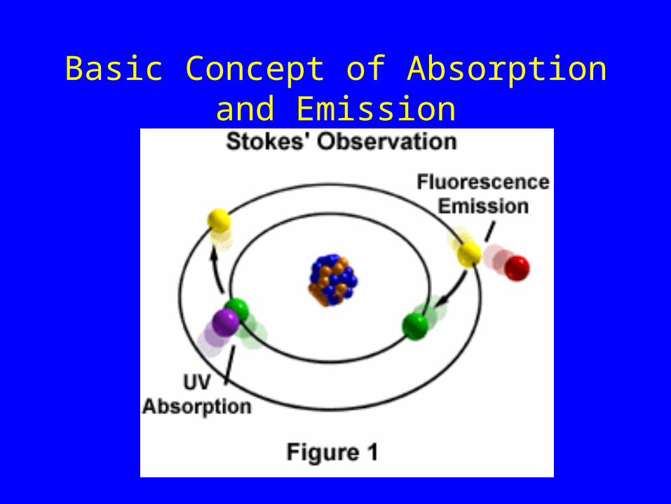
Basic Concept of Absorption and Emission
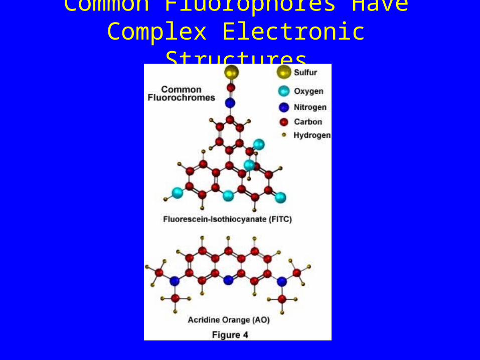
Common Fluorophores Have Complex Electronic Structures

Excitation and Emission Spectra
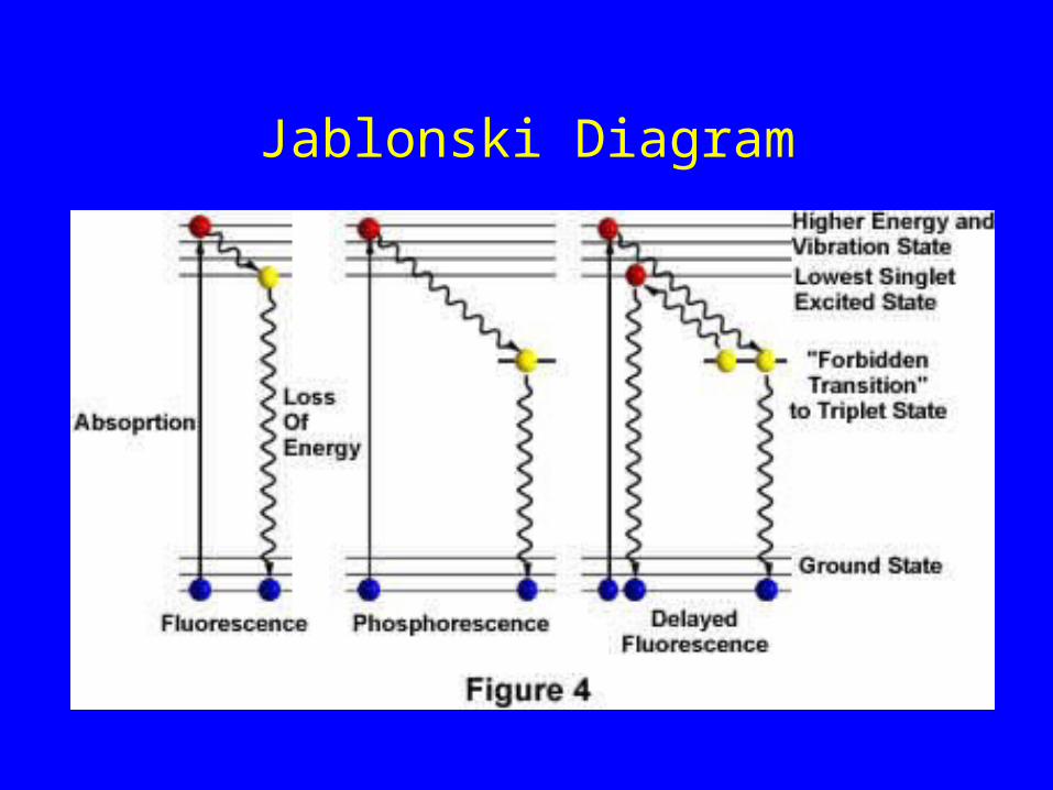
Jablonski Diagram
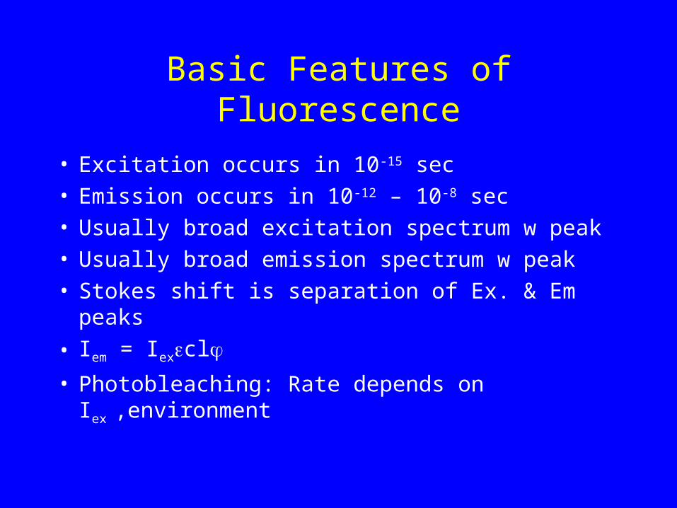
Basic Features of Fluorescence
• Excitation occurs in 10-15 sec• Emission occurs in 10-12 – 10-8 sec• Usually broad excitation spectrum w peak• Usually broad emission spectrum w peak• Stokes shift is separation of Ex. & Em peaks
• Iem = Iexcl
• Photobleaching: Rate depends on Iex ,environment
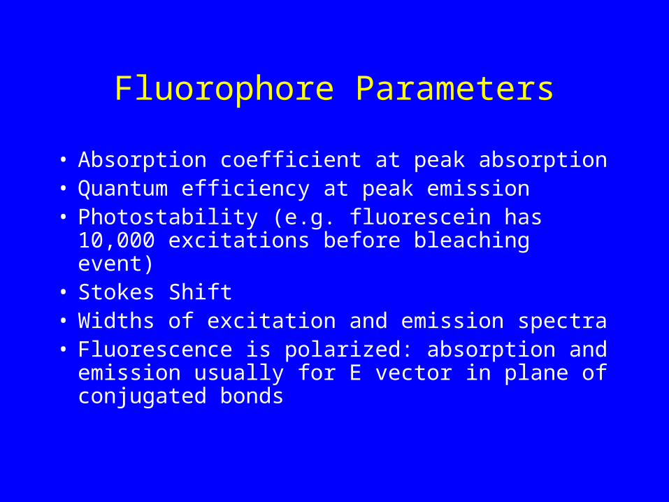
Fluorophore Parameters
• Absorption coefficient at peak absorption • Quantum efficiency at peak emission• Photostability (e.g. fluorescein has 10,000
excitations before bleaching event)• Stokes Shift• Widths of excitation and emission spectra• Fluorescence is polarized: absorption and
emission usually for E vector in plane of conjugated bonds
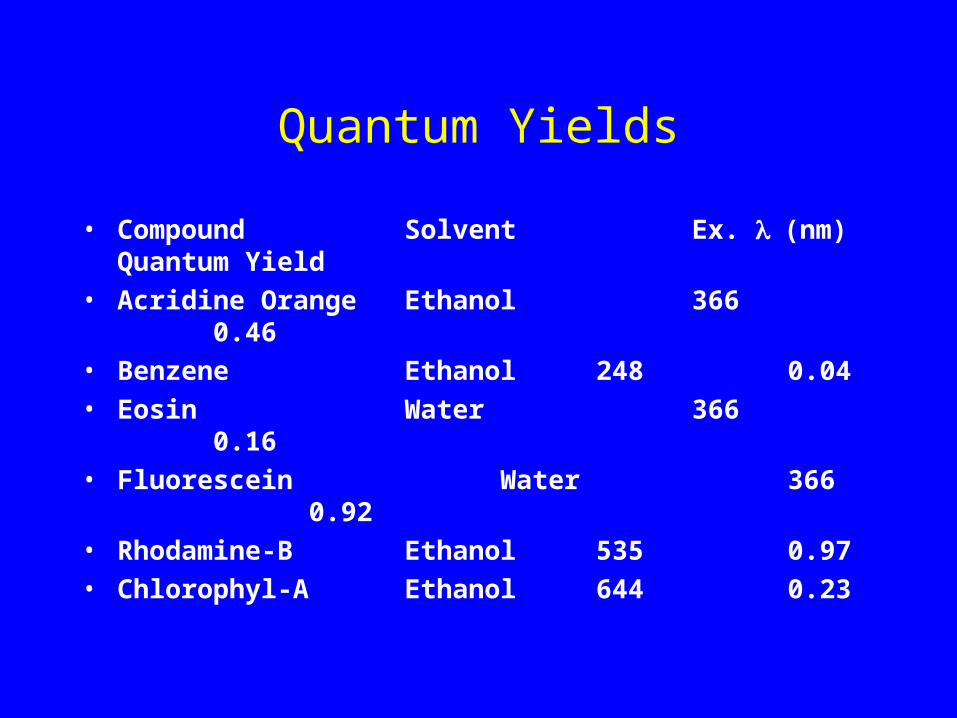
Quantum Yields
• Compound Solvent Ex. (nm) Quantum Yield
• Acridine Orange Ethanol 366 0.46
• Benzene Ethanol 248 0.04
• Eosin Water 366 0.16
• Fluorescein Water 366 0.92
• Rhodamine-B Ethanol 535 0.97
• Chlorophyl-A Ethanol 644 0.23
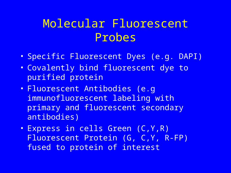
Molecular Fluorescent Probes
• Specific Fluorescent Dyes (e.g. DAPI)• Covalently bind fluorescent dye to purified protein• Fluorescent Antibodies (e.g immunofluorescent
labeling with primary and fluorescent secondary antibodies)
• Express in cells Green (C,Y,R) Fluorescent Protein (G, C,Y, R-FP) fused to protein of interest
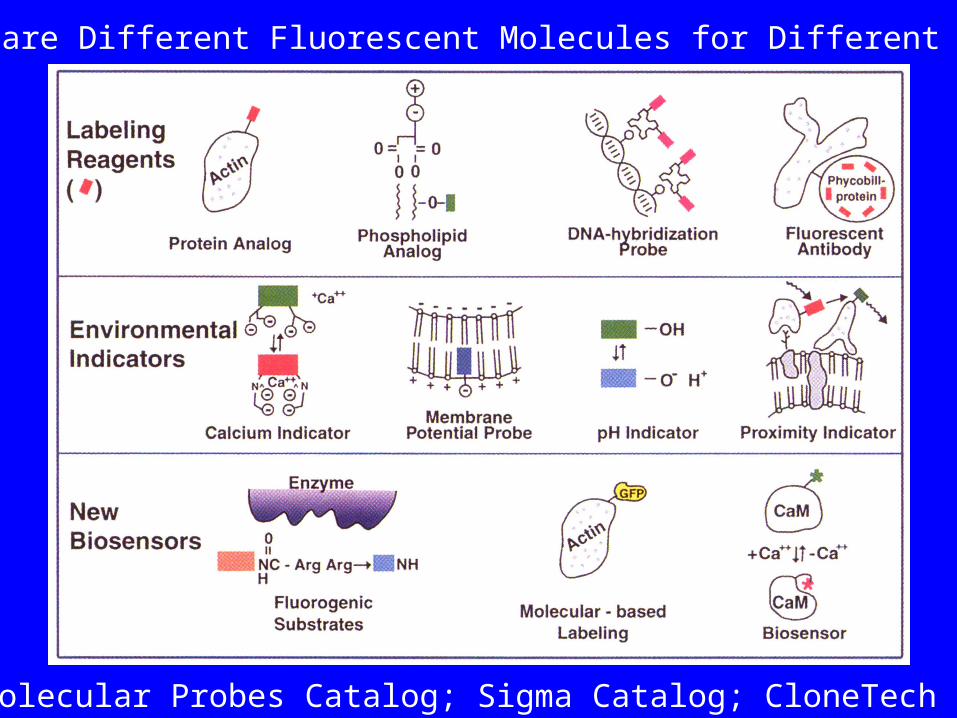
There are Different Fluorescent Molecules for Different Jobs
See Molecular Probes Catalog; Sigma Catalog; CloneTech for GFP
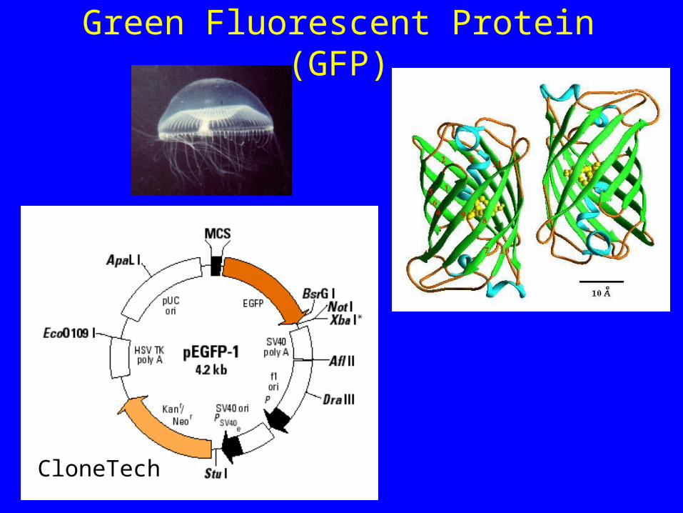
Green Fluorescent Protein (GFP)
CloneTech

Multi-Wavelength Fluorescence
Imaging

Basic Concept of Epi-Fluorescence Microscopy
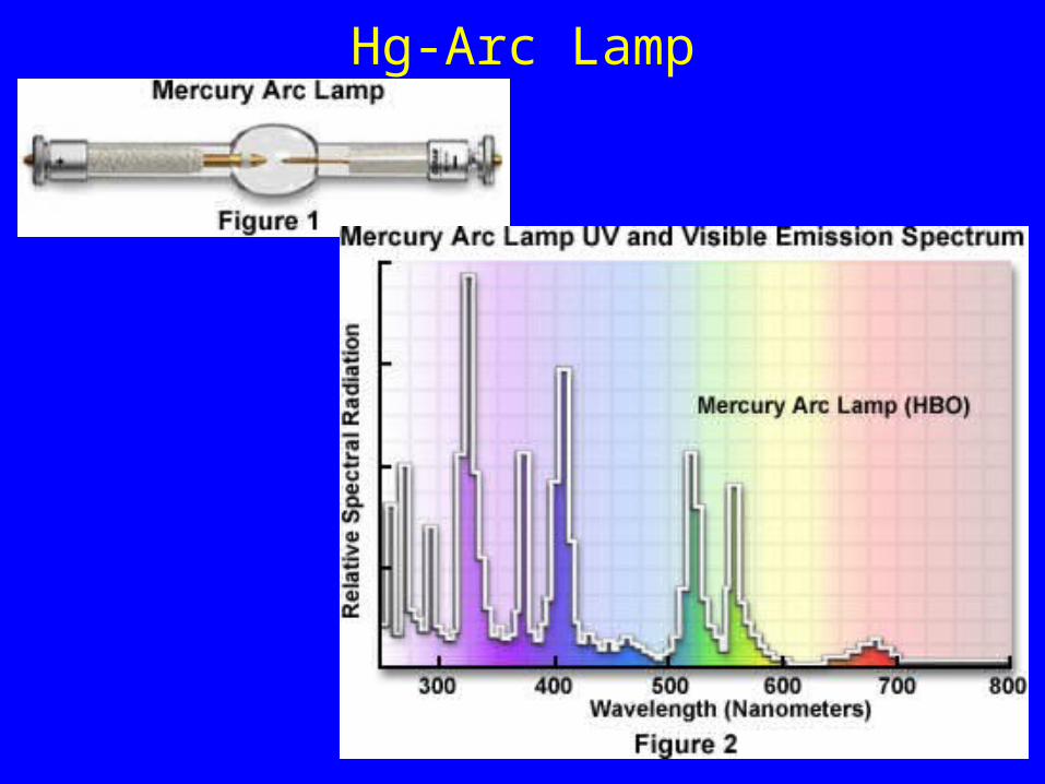
Hg-Arc Lamp
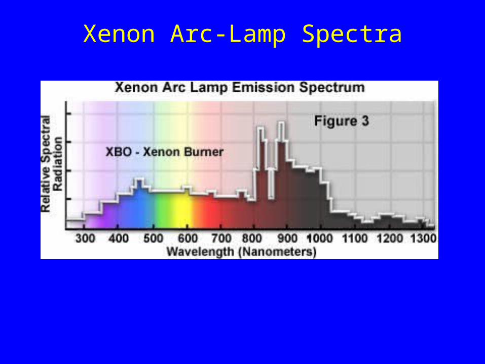
Xenon Arc-Lamp Spectra
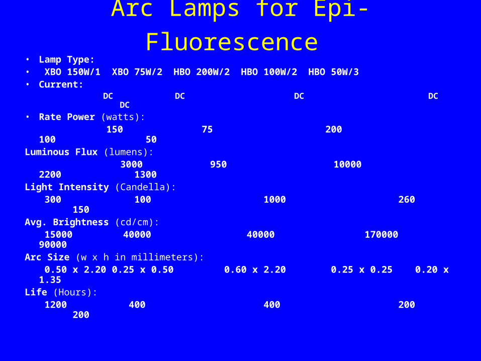
Arc Lamps for Epi-Fluorescence • Lamp Type:• XBO 150W/1 XBO 75W/2 HBO 200W/2 HBO 100W/2 HBO 50W/3• Current:
DC DC DC DC DC
• Rate Power (watts): 150 75 200 100 50
Luminous Flux (lumens): 3000 950 10000 2200 1300Light Intensity (Candella):
300 100 1000 260 150Avg. Brightness (cd/cm):
15000 40000 40000 170000 90000Arc Size (w x h in millimeters):
0.50 x 2.20 0.25 x 0.50 0.60 x 2.20 0.25 x 0.25 0.20 x 1.35Life (Hours):
1200 400 400 200 200
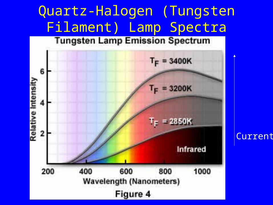
Quartz-Halogen (Tungsten Filament) Lamp Spectra
Current

Lasers Have Line Spectra
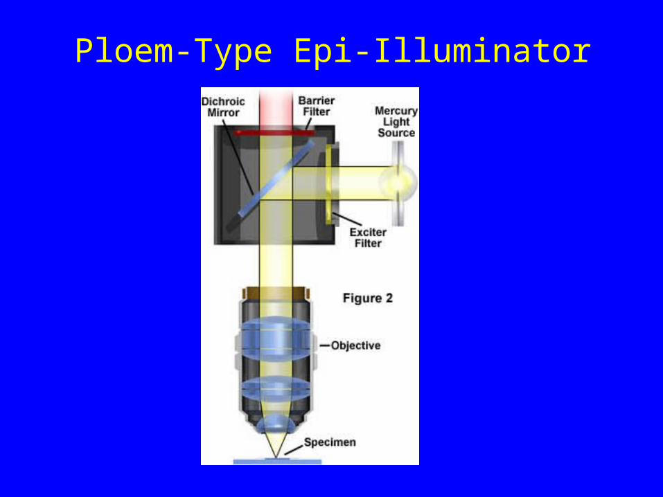
Ploem-Type Epi-Illuminator

Epi-Fluorescence Microscope
Objective Lens
Specimen
Objective BackFocal Plane
Eye
Eyepiece
Tube Lens
IntermediateImage Plane
Emission Filter
Filter Cube
Dichromatic Mirror
Excitation Filter
LampArc
Arc Image
Condenser Diaphragm
FieldDiaphragm
Arc image
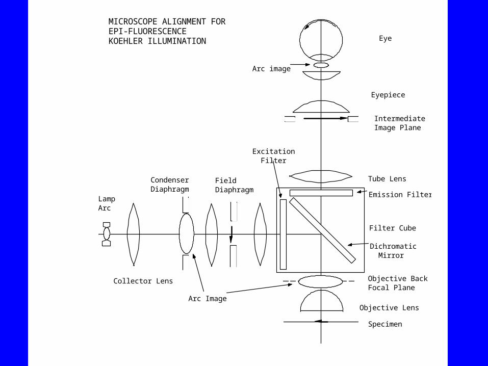
MICROSCOPE ALIGNMENT FOR EPI-FLUORESCENCE KOEHLER ILLUMINATION
Collector Lens
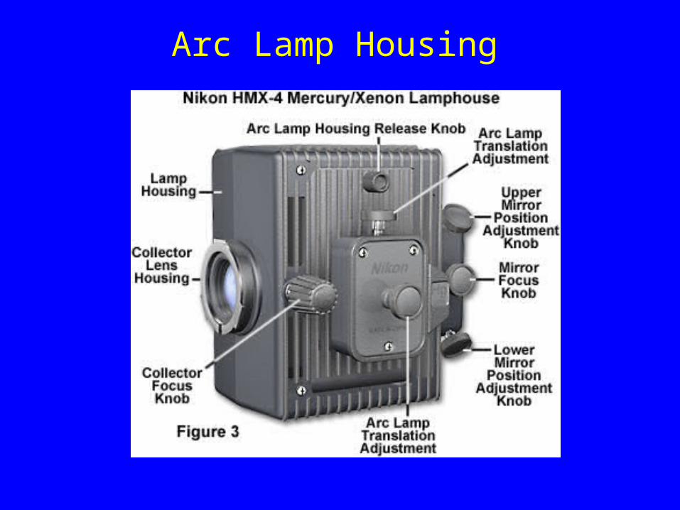
Arc Lamp Housing
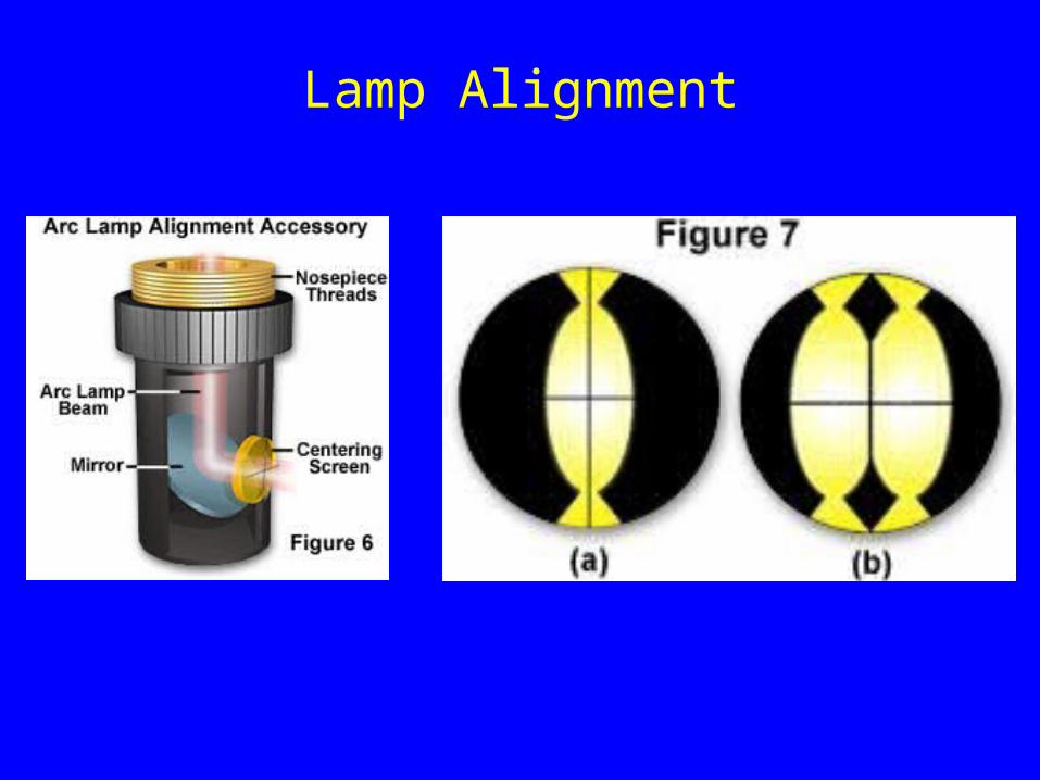
Lamp Alignment
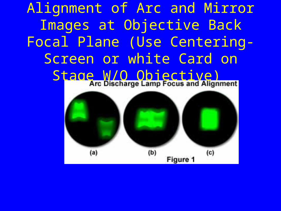
Alignment of Arc and Mirror Images at Objective Back Focal Plane (Use
Centering-Screen or white Card on Stage W/O Objective)
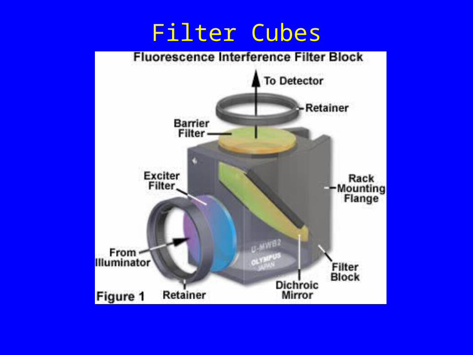
Filter Cubes

Filter Cubes Are Not Inter-Changeable Between Different Manufactures

Basic Design Features
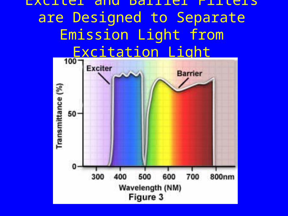
Exciter and Barrier Filters are Designed to Separate Emission Light from
Excitation Light
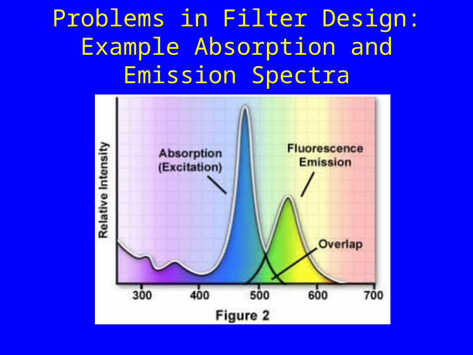
Problems in Filter Design: Example Absorption and Emission Spectra

The Dichromatic Mirror Further Isolates the Emission Light from the Excitation
Light
Modern Interference-Reflection filter Design Can Give Sharp Cut-Off with High Transmission Efficiency for the Pass Wavelengths.See web-sites for “Chroma Technology” and “Omega Optical”

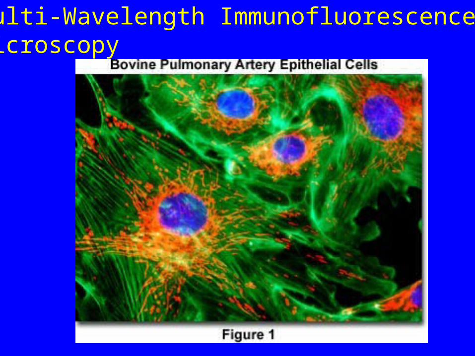
Multi-Wavelength Immunofluorescence Microscopy
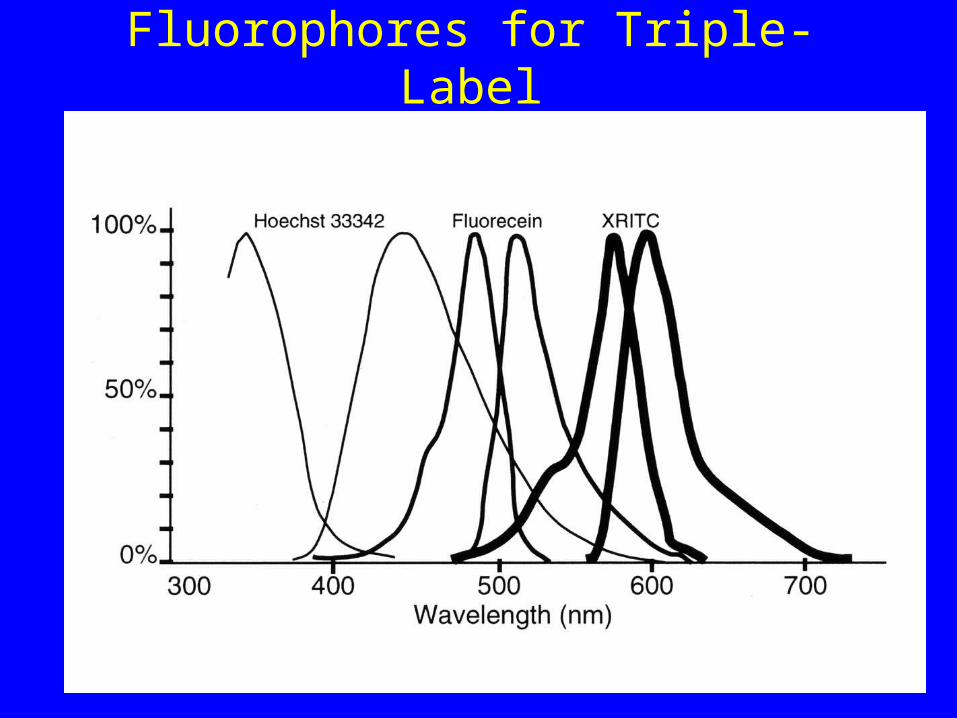
Fluorophores for Triple-Label

Multiple Band-Pass
Filters
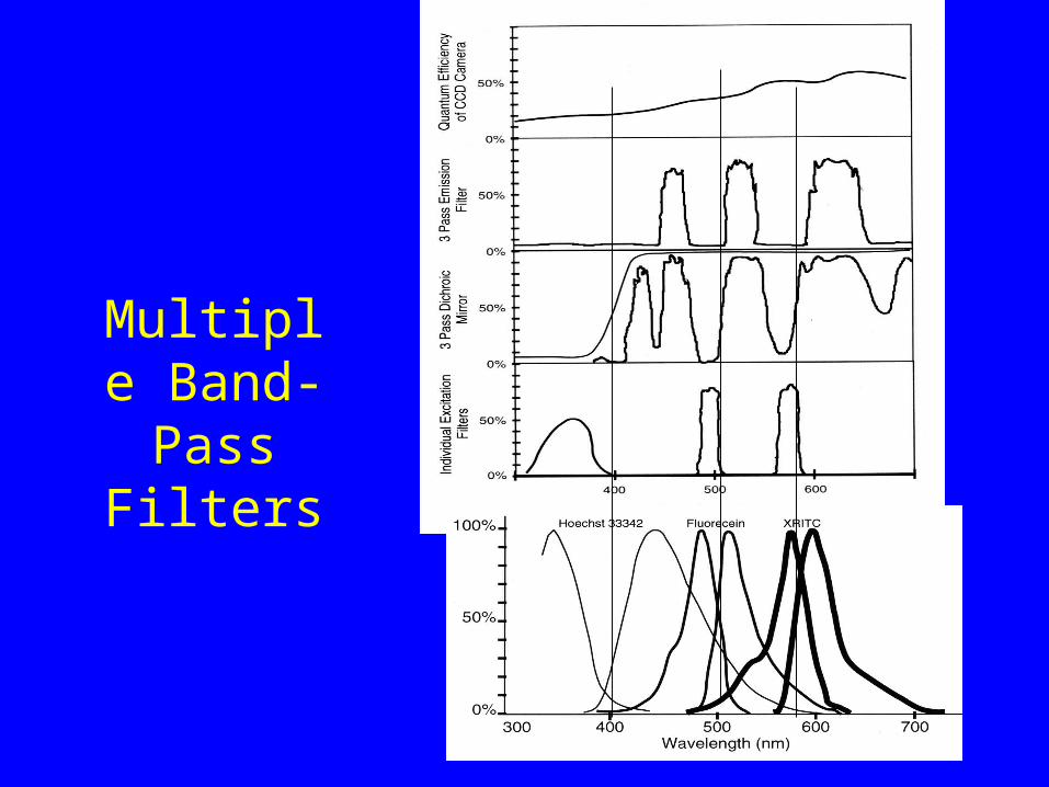
Multiple Band-Pass
Filters
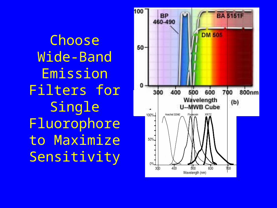
Choose Wide-Band Emission
Filters for Single Fluorophore to
Maximize Sensitivity
Chroma Technology Corp. is an employee- owned company that produces the world's finest optical filters and filter sets. The
company specializes in the design and manufacture of optical filters and coatings for applications which require the greatest
precision in color separation, optical quality and signal purity. For more about us, see our About Chroma page. Welcome to our new website! This site is under construction, so if you don't find what
you need please give us a call at (800) 824-7662.
Handbook of Optical Filters for Fluorescence Microscopy:
Download a copy of our "Handbook of Optical Filters for Fluorescence Microscopy"
in Adobe Acrobat PDF format.
www.chroma.com
Multi-Wavelength Immunofluorescence Microscopy
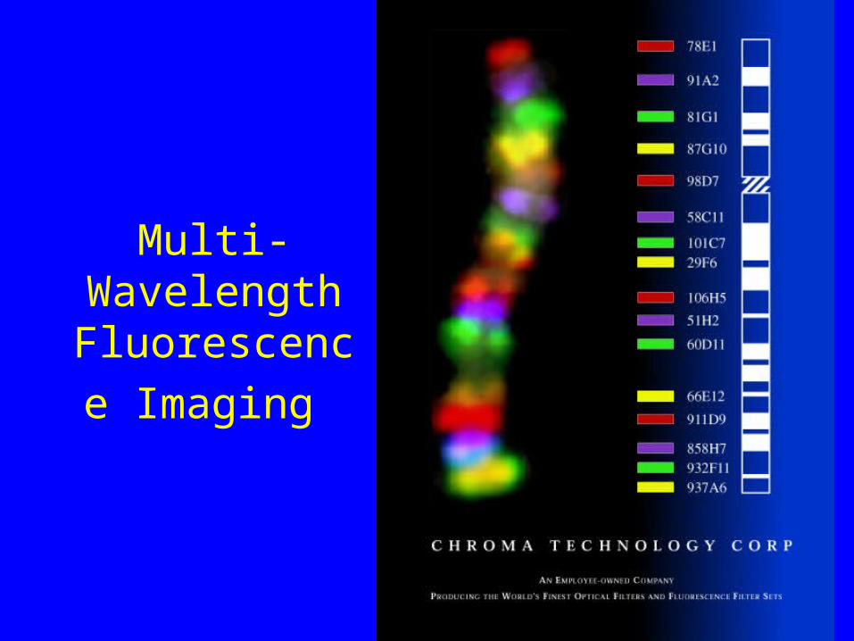
Multi-Wavelength Fluorescence
Imaging

Multi-wavelength Fluorescence Imaging

Multi-Wavelength Fluorescence
Imaging

Ploem-Type Epi-Illuminator
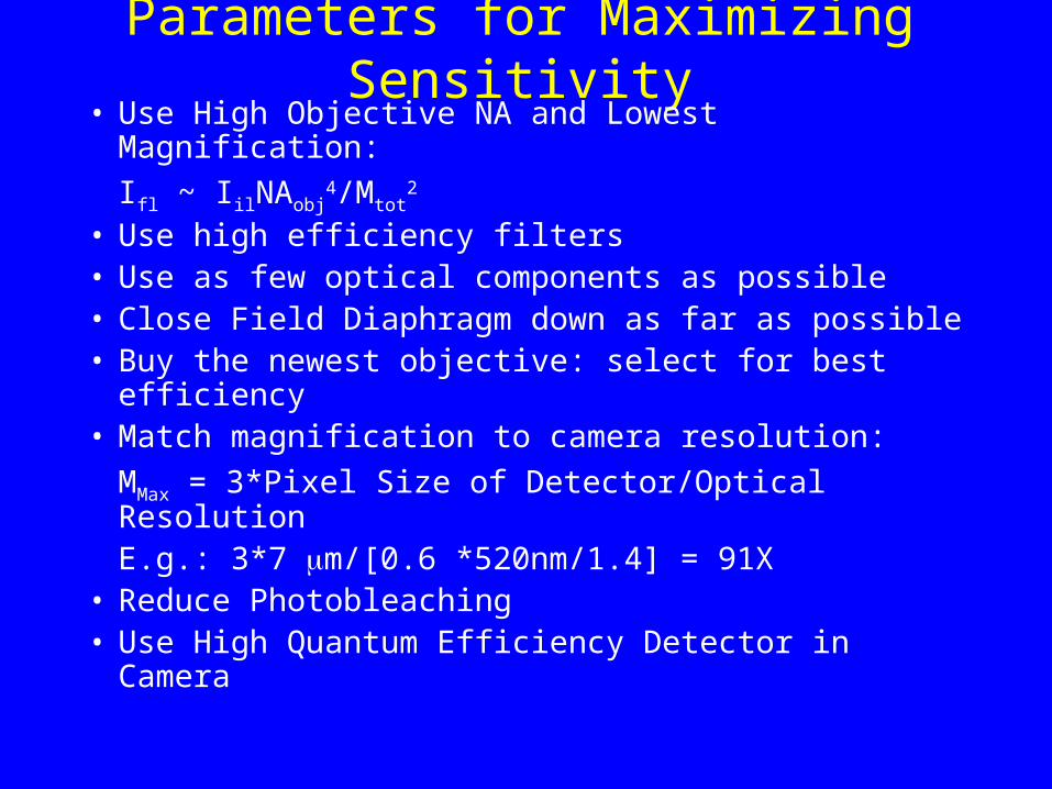
Parameters for Maximizing Sensitivity• Use High Objective NA and Lowest Magnification:
Ifl ~ IilNAobj4/Mtot
2
• Use high efficiency filters• Use as few optical components as possible• Close Field Diaphragm down as far as possible• Buy the newest objective: select for best efficiency• Match magnification to camera resolution:
MMax = 3*Pixel Size of Detector/Optical Resolution
E.g.: 3*7 m/[0.6 *520nm/1.4] = 91X• Reduce Photobleaching• Use High Quantum Efficiency Detector in Camera
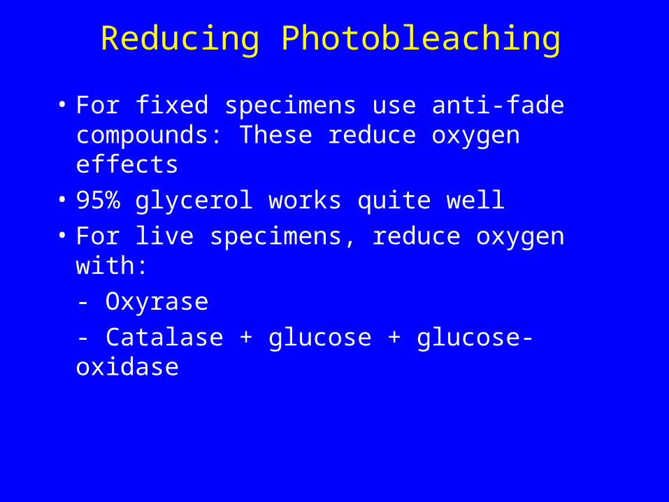
Reducing Photobleaching
• For fixed specimens use anti-fade compounds: These reduce oxygen effects
• 95% glycerol works quite well
• For live specimens, reduce oxygen with:
- Oxyrase
- Catalase + glucose + glucose-oxidase
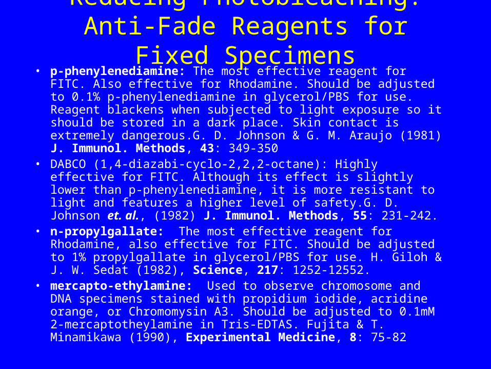
Reducing Photobleaching: Anti-Fade Reagents for Fixed Specimens
• p-phenylenediamine: The most effective reagent for FITC. Also effective for Rhodamine. Should be adjusted to 0.1% p-phenylenediamine in glycerol/PBS for use. Reagent blackens when subjected to light exposure so it should be stored in a dark place. Skin contact is extremely dangerous.G. D. Johnson & G. M. Araujo (1981) J. Immunol. Methods, 43: 349-350
• DABCO (1,4-diazabi-cyclo-2,2,2-octane): Highly effective for FITC. Although its effect is slightly lower than p-phenylenediamine, it is more resistant to light and features a higher level of safety.G. D. Johnson et. al., (1982) J. Immunol. Methods, 55: 231-242.
• n-propylgallate: The most effective reagent for Rhodamine, also effective for FITC. Should be adjusted to 1% propylgallate in glycerol/PBS for use. H. Giloh & J. W. Sedat (1982), Science, 217: 1252-12552.
• mercapto-ethylamine: Used to observe chromosome and DNA specimens stained with propidium iodide, acridine orange, or Chromomysin A3. Should be adjusted to 0.1mM 2-mercaptotheylamine in Tris-EDTAS. Fujita & T. Minamikawa (1990), Experimental Medicine, 8: 75-82
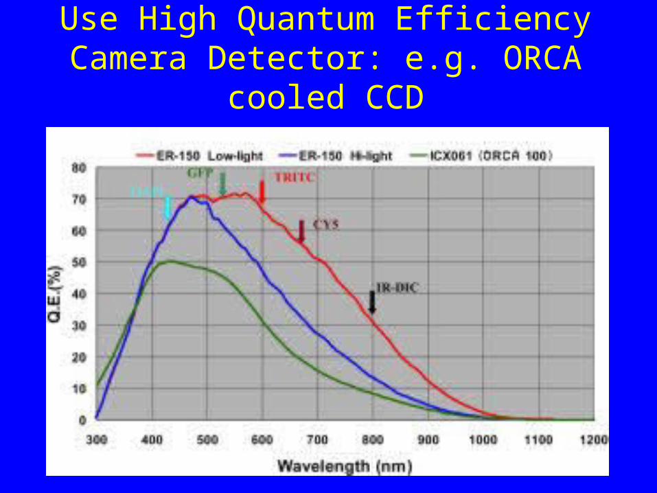
Use High Quantum Efficiency Camera Detector: e.g. ORCA cooled CCD
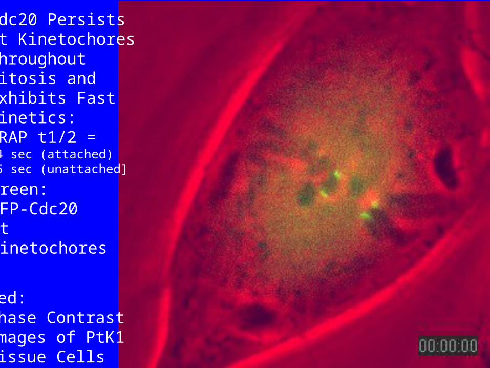
Green:GFP-Cdc20At Kinetochores
Red:Phase ContrastImages of PtK1Tissue Cells
Cdc20 PersistsAt KinetochoresThroughout Mitosis and Exhibits FastKinetics:FRAP t1/2 =[4 sec (attached)25 sec (unattached]