g model article in press - uzleuven.be · brugada syndrome hypothermia weight brugada loss...
TRANSCRIPT
J
C
I
MMLa
b
c
d
e
a
ARRA
KABHWI
I
t(aQSa
fl
e
A
(m
h1
ARTICLE IN PRESSG ModelCCASE-485; No. of Pages 4
Journal of Cardiology Cases xxx (2014) xxx–xxx
Contents lists available at www.sciencedirect.com
Journal of Cardiology Cases
journa l h om epage: www.elsev ier .com/ locate / j ccase
ase Report
ntermittent Brugada syndrome in an anorexic adolescent girl
artine K.F. Docx (MD)a,∗, Bart Loeys (MD, PhD)b, Annik Simons (MD)c,arc Gewillig (MD, PhD)d, Dorien Proost (PhD, student)b, Lut Van Laer (PhD)b,
uc Mertens (MD, PhD)e
Department of Paediatrics Queen Paola Children’s Hospital, Antwerp, BelgiumDepartment of Medical Genetics, University Hospital Antwerp, Antwerp, BelgiumDepartment of Child and Adolescent Psychiatry, AZM Middelheim Antwerp and University of Antwerp, Antwerp, BelgiumDepartment of Paediatric Cardiology, University Hospitals Leuven, Leuven, BelgiumDivision of Cardiology, The Hospital for Sick Children, Toronto, Canada
r t i c l e i n f o
rticle history:eceived 24 September 2013eceived in revised form 23 February 2014ccepted 15 March 2014
eywords:norexia nervosarugada syndromeypothermia
a b s t r a c t
We report an anorexic adolescent girl with an intermittent Brugada syndrome. A 14-year-old anorexicgirl with a body mass index (BMI) of 13.15 kg/m2 was admitted in the acute state of the disease withan ST elevation in V1 and V2, suggestive of Brugada syndrome. After 1 month of re-feeding, a controlelectrograph (ECG) was normal, but after an 8-month follow-up control with a nearly normal BMI, theECG was again suggestive of Brugada syndrome. A genetic analysis of the gene SNC5A established a geneticchange (p Leu 1582 pro), which provides the final explanation for the Brugada syndrome. Every rhythmproblem in the acute state or during the re-feeding procedure deserves a strict follow-up to distinguishiatrogenic from heritable rhythm problems.
eight lossatrogenic
<Learning objective: (i) We report the first case of a patient with anorexia nervosa with an intermittentBrugada syndrome. (ii) Moderate hypothermia can decrease the depolarization of pacemaker cells andcause ST-segment changes. (iii) Every rhythm problem in the acute state or during the re-feeding proce-dure deserves a strict follow-up to distinguish iatrogenic from heritable rhythm problems. (iv) A geneticanalysis can make the distinction and is necessary to give advice for the future lifestyle of the patient.>
© 2014 Japanese College of Cardiology. Published by Elsevier Ltd. All rights reserved.
ntroduction
The most common rhythm disorders in the anorexic popula-ion are extreme bradycardia <50 bpm with a prolonged QT interval15–40%) and sudden death in up to 10%. Other electrograph (ECG)bnormalities seen in these patients are low voltage of P waves andRS complexes, prolonged QTc, rightward QRS axis, non-specificT-T changes, and the presence of U waves. Less frequent are ectopictrial foci and second degree AV block Mobitz type I [1].
Minor T wave changes, such as T-wave inversion in V4 and T
Please cite this article in press as: Docx MKF, et al. Intermittent Brugadhttp://dx.doi.org/10.1016/j.jccase.2014.03.012
attening, are also noticed.ST depression is observed in patients with anorexia nervosa with
xtreme weight loss. The explanation of ST disturbances can be
∗ Corresponding author at: Queen Paola Children’s Hospital, Lindendreef 1, 2020ntwerp, Belgium. Tel.: +32 3 2803341; fax: +32 3 2802133.
E-mail addresses: [email protected], [email protected]. Docx), [email protected] (B. Loeys), [email protected] (A. Simons),
[email protected] (M. Gewillig), [email protected] (L. Mertens).
ttp://dx.doi.org/10.1016/j.jccase.2014.03.012878-5409/© 2014 Japanese College of Cardiology. Published by Elsevier Ltd. All rights re
found in the early repolarization and acidosis due to hypothermia.Moderate hypothermia can decrease the depolarization of pace-maker cells and cause ST-segment changes.
Case report
A 14-year-old girl with anorexia nervosa was admitted to thedepartment of eating disorders with a body mass index (BMI) of13.15 kg/m2 (weight: 35.8 kg and height: 165 cm) and a weight lossof 26.94% in less than 6 months.
In the personal antecedents, she had no episodes of palpitationsor suspicious syncopes. She once fainted following a tooth extrac-tion. In childhood she had atopic eczema and there were no surgicalprocedures or hospitalizations.
The girl has an older brother and a sister age 22 and 20 years,respectively, and both had a syncope during a peripheral venous
a syndrome in an anorexic adolescent girl. J Cardiol Cases (2014),
blood sample. No other suspicious episodes were noticed. Both par-ents are in good health. The father is an intense sportsman but neverhad any syncope. In the other family members we recorded neitherhad syncope nor sudden death.
served.
IN PRESSG ModelJ
2 ardiology Cases xxx (2014) xxx–xxx
1tcsw
pt(ta0
mh
e
t1E(
pvf
mb(
rt
Fe
ARTICLECCASE-485; No. of Pages 4
M.K.F. Docx et al. / Journal of C
During the physical examination, a cachectic girl (BMI:3.15 kg/m2, core temperature: 34.9 ◦C) presented with theypical features of anorexia nervosa namely lanugo hairs, acro-yanosis, hypotension (87/46 mm Hg) and bradycardia 39 bpm. Ayncope was not observed, neither chest pain and no medicationere taken.
Routine blood chemistry (creatinine, blood urea nitrogen,otassium, sodium, bicarbonate, magnesium and calcium) andhyroid hormones were normal. There was a high ferritin level258 ng/ml) (N: 5.0–122 ng/ml) and a slight elevation of the liverransaminases [aspartate aminotransferase (58 U/L) (N: 10–30 U/L),lanine aminotransferase (128 U/L) (N: 17–44 U/L)] and troponin I.0039 ng/ml (N: <0.0034 ng/ml).
Creatinine kinase and lactate dehydrogenase levels were nor-al. The levels of luteinizing hormone (LH), follicle-stimulating
ormone (FSH), and insulin-growth factor-1 were low.In the acute phase, an ECG together with a 2-D-
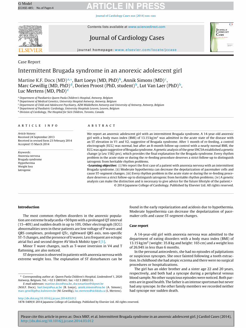
chocardiography with Doppler was obtained.The ECG showed ST elevation in V1, V2 and less in V3 (sugges-
ive of Brugada syndrome) (Fig. 1). The rhythm was 49 bpm, axis1◦, PR interval: 144 ms, QRS: 114 ms, QT: 422 ms, and QTc 384 ms.chocardiography also showed a pericardial effusion of 0.9–1.1 cmapex) and a decreased left ventricular mass/height2.7: 17.57 g/m2.7
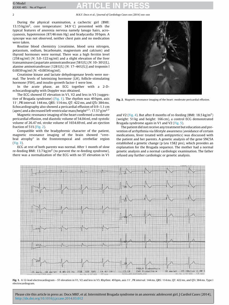
Magnetic resonance imaging of the heart confirmed a moderateericardial effusion, end diastolic volume of 54.04 ml, end-systolicolume of 26.47 ml, stroke volume of 1654.69 ml, and an ejectionraction of 51% (Fig. 2).
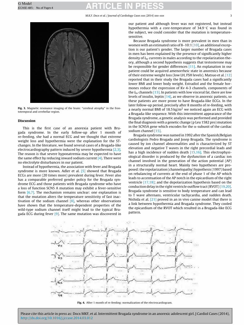
Compatible with the bradyphrenic character of the patient,agnetic resonance imaging of the brain showed “cere-
ral atrophy” in the frontotemporal and cerebellar region
Please cite this article in press as: Docx MKF, et al. Intermittent Brugadhttp://dx.doi.org/10.1016/j.jccase.2014.03.012
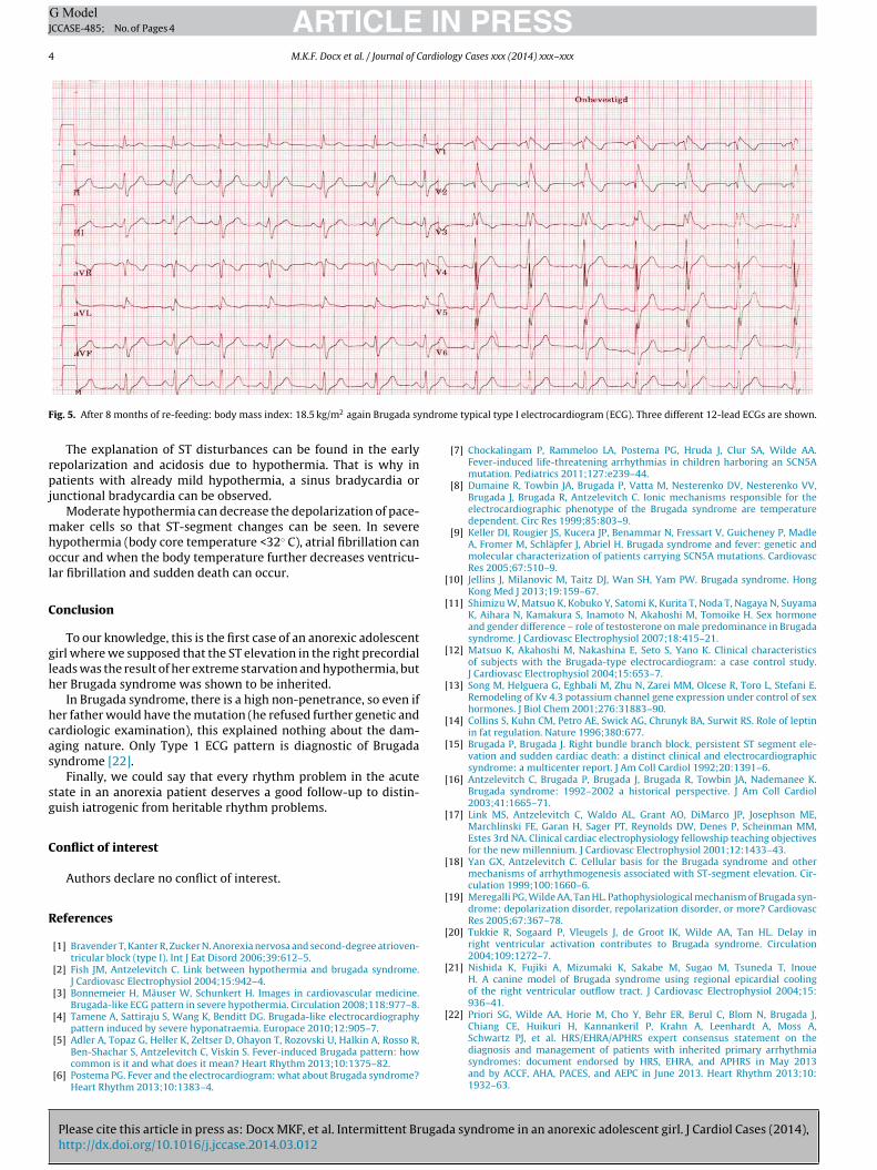
Fig. 3).ECG at rest of both parents was normal. After 1 month of slow
e-feeding BMI: 13.7 kg/m2 (to prevent the re-feeding syndrome),here was a normalization of the ECG with no ST elevation in V1
ig. 1. A 12-lead electrocardiogram – ST elevation in V1, V2 and less in V3. Rhythm: 49 blectrocardiogram.
Fig. 2. Magnetic resonance imaging of the heart: moderate pericardial effusion.
and V2 (Fig. 4). But after 8 months of re-feeding (BMI: 18.5 kg/m2)(weight: 51 kg and height: 166 cm), a control ECG demonstratedBrugada syndrome again in V1 and V2 (Fig. 5).
The patient did not receive any treatment but education and pre-vention of arrhythmia via lifestyle awareness (avoidance of certainmedications, fever treated with antipyretics) was discussed withthe patient and her parents. A genetic analysis of the gene SNC5Aestablished a genetic change (p Leu 1582 pro), which provides anexplanation for the Brugada sequence. The mother had a normal
a syndrome in an anorexic adolescent girl. J Cardiol Cases (2014),
genetic analysis and a normal cardiologic examination. The fatherrefused any further cardiologic or genetic analysis.
pm, axis 11◦ , PR interval: 144 ms, QRS: 114 ms, QT: 422 ms, and QTc 384 ms. Type I
ARTICLE ING ModelJCCASE-485; No. of Pages 4
M.K.F. Docx et al. / Journal of Cardiol
Ft
D
grwceTtn
sEhdaftthwg
ig. 3. Magnetic resonance imaging of the brain: “cerebral atrophy” in the fron-otemporal and cerebellar region.
iscussion
This is the first case of an anorexia patient with Bru-ada syndrome. In the early follow-up after 1 month ofe-feeding, she had a normal ECG and we thought that extremeeight loss and hypothermia were the explanation for the ST-
hanges. In the literature, we found several cases of a Brugada-likelectrocardiography pattern induced by severe hypothermia [2,3].he reason is that severe hyponatremia may be expected to havehe same effect by reducing inward sodium current [4]. There wereo electrolyte disturbances in our patient.
Instead of hypothermia, the association with fever and Brugadayndrome is more known. Adler et al. [5] showed that BrugadaCGs are more (20 times more) prevalent during fever. Fever alsoas a comparable preferred gender policy for the Brugada syn-rome ECG and those patients with Brugada syndrome who have
loss of function SCN5 A mutation may exhibit a fever-sensitiveorm [6,7]. The mechanism remains unclear: one explanation ishat the mutation alters the temperature sensitivity of fast inac-
Please cite this article in press as: Docx MKF, et al. Intermittent Brugadhttp://dx.doi.org/10.1016/j.jccase.2014.03.012
ivation of the sodium channel [8], whereas other observationsave shown that the temperature-dependent properties of theild-type sodium channel itself might lead to the typical Bru-
ada ECG during fever [9]. The same mutation was discovered in
Fig. 4. After 1 month of re-feeding: norm
PRESSogy Cases xxx (2014) xxx–xxx 3
our patient and although fever was not registered, but insteadhypothermia with a core-temperature of 34.9 ◦C was found inthe subject, we could consider that the mutation is temperature-sensitive.
Because Brugada syndrome is more prevalent in men than inwomen with an estimated ratio of 8–10:1 [10], an additional excep-tion is our patient’s gender. The larger number of Brugada casesin men has been explained by the presence of significantly higherdensity of Ito currents in males according to the repolarization the-ory, although a second hypothesis suggests that testosterone maybe responsible for gender differences [11]. An explanation in ourpatient could be acquired amenorrheic state in anorexics becauseof their extreme weight loss (low LH, FSH levels). Matsuo et al. [12]reported that in their study the Brugada cases had a significantlylower BMI and lower body weight. Estradiol and the female hor-mones reduce the expression of Kv 4-3 channels, components ofthe Ito channels [13]. In patients with low visceral fat, there are lowlevels of insulin, leptin [14], as we observe in anorexic patients, sothese patients are more prone to have Brugada-like ECGs. In thelater follow-up period, precisely after 8 months of re-feeding, witha nearly normal BMI of 18.5 kg/m2 we noticed again an ECG witha Brugada-like sequence. With this intermittent appearance of theBrugada syndrome, a genetic analysis was performed and providedthe final diagnosis with a genetic change (p Leu 1582 pro) mutationin the SCN5A gene which encodes for the �-subunit of the cardiacsodium channel [15].
Brugada syndrome was named in 1992 after the Spanish/Belgiancardiologists Pedro Brugada and Josep Brugada. The syndrome iscaused by ion channel abnormalities and is characterized by STelevation and negative T waves in the right precordial leads andhas a high incidence of sudden death [15,16]. This electrophysi-ological disorder is produced by the dysfunction of a cardiac ionchannel involved in the generation of the action potential (AP)in a structurally normal heart. Mainly two hypotheses are pro-posed: the repolarization (channelopathy) hypothesis (1997) basedon rebalancing of currents at the end of phase 1 of the AP whichleads to accentuation of the AP notch in the epicardium of the rightventricle [17,18]; and the depolarization hypothesis based on theconduction delay in the right ventricle outflow tract (RVOT) [19,20].Brugada syndrome is sensitive to body temperature and can leadto T wave alternans, ventricular tachycardia, and sudden death.
a syndrome in an anorexic adolescent girl. J Cardiol Cases (2014),
Nishida et al. [21] proved in an in vivo canine model that there isa link between hypothermia and Brugada syndrome. They cooledthe epicardium of the RVOT which resulted in a Brugada-like ECGpattern.
alization of the electrocardiogram.
ARTICLE IN PRESSG ModelJCCASE-485; No. of Pages 4
4 M.K.F. Docx et al. / Journal of Cardiology Cases xxx (2014) xxx–xxx
F ndrom
rpj
mhol
C
glh
hcas
sg
C
R
[
[
[
[
[
[
[
[
[
[
[
[
[
ig. 5. After 8 months of re-feeding: body mass index: 18.5 kg/m2 again Brugada sy
The explanation of ST disturbances can be found in the earlyepolarization and acidosis due to hypothermia. That is why inatients with already mild hypothermia, a sinus bradycardia or
unctional bradycardia can be observed.Moderate hypothermia can decrease the depolarization of pace-
aker cells so that ST-segment changes can be seen. In severeypothermia (body core temperature <32◦ C), atrial fibrillation canccur and when the body temperature further decreases ventricu-ar fibrillation and sudden death can occur.
onclusion
To our knowledge, this is the first case of an anorexic adolescentirl where we supposed that the ST elevation in the right precordialeads was the result of her extreme starvation and hypothermia, buter Brugada syndrome was shown to be inherited.
In Brugada syndrome, there is a high non-penetrance, so even ifer father would have the mutation (he refused further genetic andardiologic examination), this explained nothing about the dam-ging nature. Only Type 1 ECG pattern is diagnostic of Brugadayndrome [22].
Finally, we could say that every rhythm problem in the acutetate in an anorexia patient deserves a good follow-up to distin-uish iatrogenic from heritable rhythm problems.
onflict of interest
Authors declare no conflict of interest.
eferences
[1] Bravender T, Kanter R, Zucker N. Anorexia nervosa and second-degree atrioven-tricular block (type I). Int J Eat Disord 2006;39:612–5.
[2] Fish JM, Antzelevitch C. Link between hypothermia and brugada syndrome.J Cardiovasc Electrophysiol 2004;15:942–4.
[3] Bonnemeier H, Mäuser W, Schunkert H. Images in cardiovascular medicine.Brugada-like ECG pattern in severe hypothermia. Circulation 2008;118:977–8.
[4] Tamene A, Sattiraju S, Wang K, Benditt DG. Brugada-like electrocardiographypattern induced by severe hyponatraemia. Europace 2010;12:905–7.
Please cite this article in press as: Docx MKF, et al. Intermittent Brugadhttp://dx.doi.org/10.1016/j.jccase.2014.03.012
[5] Adler A, Topaz G, Heller K, Zeltser D, Ohayon T, Rozovski U, Halkin A, Rosso R,Ben-Shachar S, Antzelevitch C, Viskin S. Fever-induced Brugada pattern: howcommon is it and what does it mean? Heart Rhythm 2013;10:1375–82.
[6] Postema PG. Fever and the electrocardiogram: what about Brugada syndrome?Heart Rhythm 2013;10:1383–4.
e typical type I electrocardiogram (ECG). Three different 12-lead ECGs are shown.
[7] Chockalingam P, Rammeloo LA, Postema PG, Hruda J, Clur SA, Wilde AA.Fever-induced life-threatening arrhythmias in children harboring an SCN5Amutation. Pediatrics 2011;127:e239–44.
[8] Dumaine R, Towbin JA, Brugada P, Vatta M, Nesterenko DV, Nesterenko VV,Brugada J, Brugada R, Antzelevitch C. Ionic mechanisms responsible for theelectrocardiographic phenotype of the Brugada syndrome are temperaturedependent. Circ Res 1999;85:803–9.
[9] Keller DI, Rougier JS, Kucera JP, Benammar N, Fressart V, Guicheney P, MadleA, Fromer M, Schläpfer J, Abriel H. Brugada syndrome and fever: genetic andmolecular characterization of patients carrying SCN5A mutations. CardiovascRes 2005;67:510–9.
10] Jellins J, Milanovic M, Taitz DJ, Wan SH, Yam PW. Brugada syndrome. HongKong Med J 2013;19:159–67.
11] Shimizu W, Matsuo K, Kobuko Y, Satomi K, Kurita T, Noda T, Nagaya N, SuyamaK, Aihara N, Kamakura S, Inamoto N, Akahoshi M, Tomoike H. Sex hormoneand gender difference – role of testosterone on male predominance in Brugadasyndrome. J Cardiovasc Electrophysiol 2007;18:415–21.
12] Matsuo K, Akahoshi M, Nakashina E, Seto S, Yano K. Clinical characteristicsof subjects with the Brugada-type electrocardiogram: a case control study.J Cardiovasc Electrophysiol 2004;15:653–7.
13] Song M, Helguera G, Eghbali M, Zhu N, Zarei MM, Olcese R, Toro L, Stefani E.Remodeling of Kv 4.3 potassium channel gene expression under control of sexhormones. J Biol Chem 2001;276:31883–90.
14] Collins S, Kuhn CM, Petro AE, Swick AG, Chrunyk BA, Surwit RS. Role of leptinin fat regulation. Nature 1996;380:677.
15] Brugada P, Brugada J. Right bundle branch block, persistent ST segment ele-vation and sudden cardiac death: a distinct clinical and electrocardiographicsyndrome: a multicenter report. J Am Coll Cardiol 1992;20:1391–6.
16] Antzelevitch C, Brugada P, Brugada J, Brugada R, Towbin JA, Nademanee K.Brugada syndrome: 1992–2002 a historical perspective. J Am Coll Cardiol2003;41:1665–71.
17] Link MS, Antzelevitch C, Waldo AL, Grant AO, DiMarco JP, Josephson ME,Marchlinski FE, Garan H, Sager PT, Reynolds DW, Denes P, Scheinman MM,Estes 3rd NA. Clinical cardiac electrophysiology fellowship teaching objectivesfor the new millennium. J Cardiovasc Electrophysiol 2001;12:1433–43.
18] Yan GX, Antzelevitch C. Cellular basis for the Brugada syndrome and othermechanisms of arrhythmogenesis associated with ST-segment elevation. Cir-culation 1999;100:1660–6.
19] Meregalli PG, Wilde AA, Tan HL. Pathophysiological mechanism of Brugada syn-drome: depolarization disorder, repolarization disorder, or more? CardiovascRes 2005;67:367–78.
20] Tukkie R, Sogaard P, Vleugels J, de Groot IK, Wilde AA, Tan HL. Delay inright ventricular activation contributes to Brugada syndrome. Circulation2004;109:1272–7.
21] Nishida K, Fujiki A, Mizumaki K, Sakabe M, Sugao M, Tsuneda T, InoueH. A canine model of Brugada syndrome using regional epicardial coolingof the right ventricular outflow tract. J Cardiovasc Electrophysiol 2004;15:936–41.
22] Priori SG, Wilde AA, Horie M, Cho Y, Behr ER, Berul C, Blom N, Brugada J,Chiang CE, Huikuri H, Kannankeril P, Krahn A, Leenhardt A, Moss A,
a syndrome in an anorexic adolescent girl. J Cardiol Cases (2014),
Schwartz PJ, et al. HRS/EHRA/APHRS expert consensus statement on thediagnosis and management of patients with inherited primary arrhythmiasyndromes: document endorsed by HRS, EHRA, and APHRS in May 2013and by ACCF, AHA, PACES, and AEPC in June 2013. Heart Rhythm 2013;10:1932–63.