g16 osteoporotic fxs
TRANSCRIPT
Epidemiology, Diagnosis Prevention and Management of
Osteoporotic Fractures
Kenneth A. Egol, MDNYU-Hospital For Joint Diseases
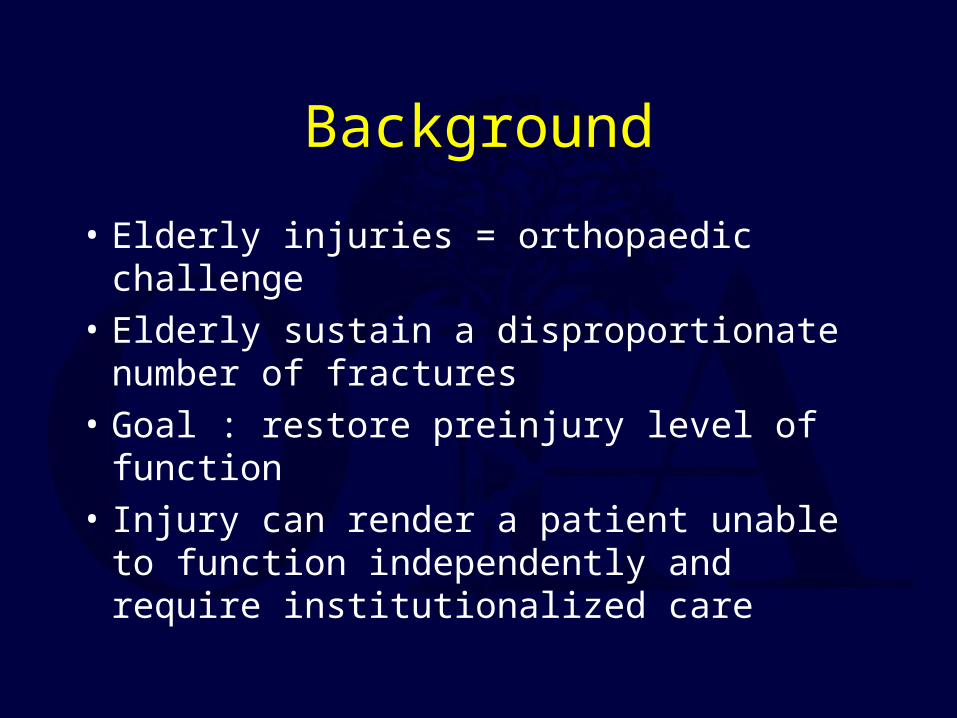
Background
• Elderly injuries = orthopaedic challenge• Elderly sustain a disproportionate number
of fractures• Goal : restore preinjury level of function• Injury can render a patient unable to
function independently and require institutionalized care
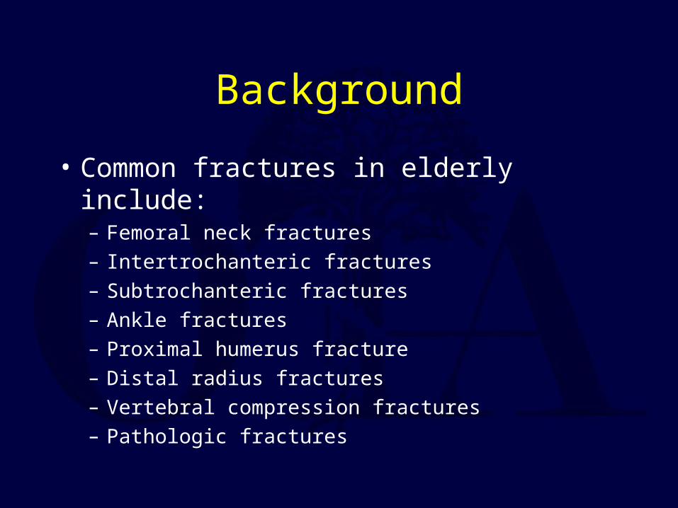
Background
• Common fractures in elderly include:– Femoral neck fractures– Intertrochanteric fractures– Subtrochanteric fractures– Ankle fractures– Proximal humerus fracture– Distal radius fractures– Vertebral compression fractures– Pathologic fractures
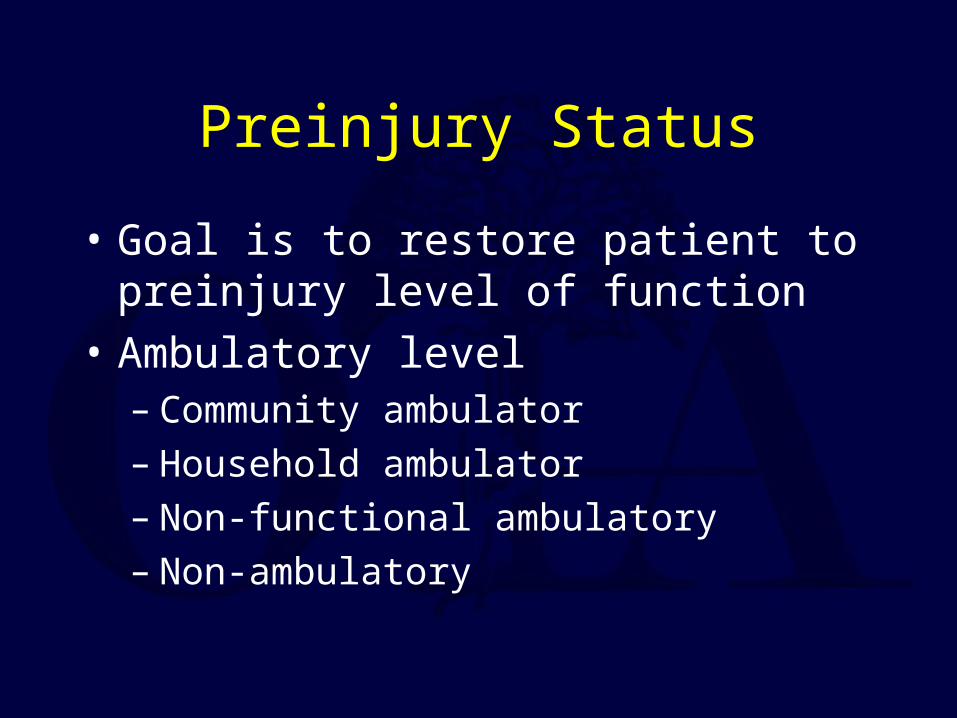
Preinjury Status
• Goal is to restore patient to preinjury level of function
• Ambulatory level– Community ambulator– Household ambulator– Non-functional ambulatory– Non-ambulatory
Preinjury Status
• Medical History• Cognitive History• Functional History
– Ambulatory status– Living arrangements
Preinjury Status
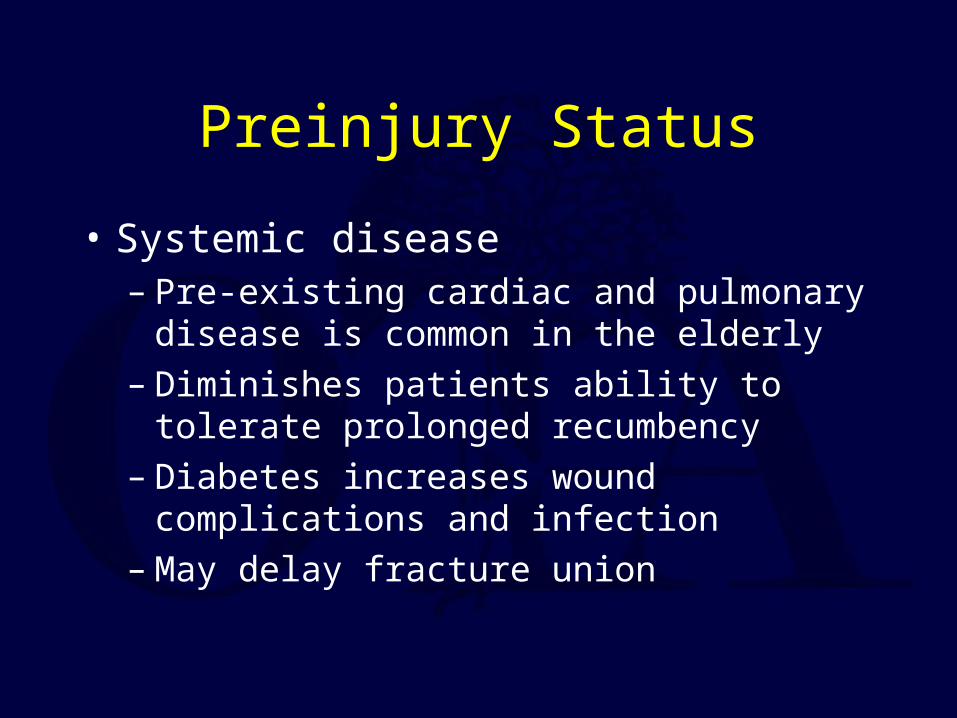
• Systemic disease– Pre-existing cardiac and pulmonary disease is
common in the elderly– Diminishes patients ability to tolerate
prolonged recumbency– Diabetes increases wound complications and
infection– May delay fracture union
Preinjury Status
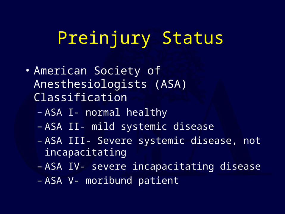
• American Society of Anesthesiologists (ASA) Classification– ASA I- normal healthy– ASA II- mild systemic disease– ASA III- Severe systemic disease, not
incapacitating– ASA IV- severe incapacitating disease– ASA V- moribund patient
Preinjury Status
• Systemic disease– Peripheral vascular disease– Thromboembolic disease
Preinjury Status
• Cognitive Status– Critical to outcome– Conditions may render patient unable to
participate in rehabilitation• Alzheimer’s• CVA• Parkinson's• Senile dementia
Osteopenia
• Osteoporosis is a decreased bone density with normal bone mineralization
• Osteomalacia is a decreased bone matrix mineralization with or without a change in bone density
• Some degree of osteopenia is found in virtually all healthy elderly patients
Osteopenia
• Senile osteoporosis common• Treatable causes should be investigated
– Nutritional deficiency– Malabsorption syndromes– Hyperparathyroidism– Cushings disease– Tumors
Osteopenia
• Risk factors– Female sex– European ancestry– Sedentary lifestyle– Multiple births– Excessive alcohol use
Osteopenia
• Complicates fracture treatment and healing• Internal fixation compromised
– Poor screw purchase– Increased risk of screw pull out– Augmentation with methylmethacrylate has been
advocated
• Increased risk of non-union– Bone augmentation (bone graft, substitutes) may be
indicated
Hip Fractures
• General principles– Approximately 250,000 hip fractures/ year– Cost approximately $8.7 billion annually– The number of hip fractures is expected to
double by the year 2050
Hip Fractures
• Epidemiology– Incidence in U.S is 80/100,000– Only 5.6/100,000 in S. African Bantus– 20% higher incidence in urban areas– 15% lifetime risk for white females who live to
age 80
Hip Fractures
• Epidemiology– Incidence increases after age 50– Female: Male ratio is 2:1– Femoral neck and intertrochanteric seen with
equal frequency
Hip Fractures
• Radiographic evaluation– Anterior-posterior view– Cross table lateral– internal rotation view will help delineate
fracture pattern
Hip Fractures
• Radiographic evaluation– Occult hip fracture
• Technetium bone scanning is a sensitive indicator, but may take 2-3 days to become positive
• Magnetic resonance imaging has been shown to be as sensitive as bone scanning and can be reliably performed within 24 hours
Hip Fractures
• Management– Prompt operative stabilization– Early mobilization– DVT prophylaxis
Hip Fractures
• Outcomes– Fracture related outcomes
• Healing• Quality of reduction
– Functional outcomes• Ambulatory ability• Mortality (25% at one year)• Return to prefracture activities of daily living
Hip Fractures
• Femoral neck fractures– Intracapsular location– Vascular Supply
• Medial and lateral circumflex vessels anastomose at the base of the neck and blood supply predominately from ascending arteries (90%)
• Artery of ligamentum teres (10%)

Hip Fractures
• Femoral neck fractures– Numerous classification schemes– Non-displaced and displaced most useful for
treatment and complications
Hip Fractures
• Femoral neck fractures• Treatment
– Non-displaced/ valgus impacted fractures• Non-operative 8-15% displacement rate• Operative with cannulated screws• Non-union 5% and osteonecrosis is approximately
8%
Hip Fractures
• Femoral neck fractures– Displaced fractures should be treated
operatively– Treatment: Open vs. Closed Reduction Internal
fixation• 30% non-union and 25%-30% osteonecrosis rate• Non-union requires reoperation 75% of the time
while osteonecrosis leads to 25% reoperation
Hip Fractures
• Femoral neck fractures• Treatment: Hemiarthroplasty
– Unipolar Vs Bipolar– Can lead to acetabular erosion, dislocation,
infection
Hip Fractures
• Femoral neck fractures• Treatment
– Displaced fractures can be treated non -operatively in certain situations
• Demented, non-ambulatory patient– Mobilize early
• Accept resulting non or malunion
Hip Fractures
• Intertrochanteric fractures– Extracapsular (well vascularized)– Region distal to the neck between the
trochanters– Calcar femorale– Posteromedial cortex– Important muscular insertions
Hip Fractures
• Intertrochanteric fractures– Numerous classifications exist
• Stable (posteromedial cortex intact) Vs unstable (posteromedial cortex off)
– Key to treatment is obtaining a stable reduction
Hip Fractures
• Intertrochanteric fractures– Treatment
• Usually treated surgically• Implant of choice is a hip compression screw that
slides in a barrel attached to a sideplate• The implant allows for controlled impaction upon
weightbearing
Hip Fractures
• Intertrochanteric fractures– Treatment
• Primary prosthetic replacement can be considered • For cases with significant comminution
Hip Fractures
• Subtrochanteric Fractures– Begin at or below the level of the lesser
trochanter– Typically higher energy injuries seen in
younger patients– far less common in the elderly

Hip Fractures
• Subtrochanteric Fractures– Treatment
• Intramedullary nail (high rates of union)• Plates and screws
Ankle Fractures
• Background– Common injury in the elderly– low energy injuries following twisting
reflecting the relative strength of the ligaments compared to osteopenic bone
Ankle Fractures
• Epidemiology– Age specific incidence in women older than 50
has increased over past 30 years– 187/100,000
Ankle Fractures
• Presentation– Follows twisting of foot relative to lower tibia– Patients present unable to bear weight– Ecchymosis, deformity– Careful neurovascular exam must be performed
Ankle Fractures
• Radiographic evaluation– Ankle trauma series includes:
• AP• Lateral• Mortise
– Examine entire length of the fibula
Ankle Fractures
• Classification– Lauge-Hansen– Danis-Weber
• Beyond the scope of this talk
Ankle Fractures
• Treatment– Isolated, non-displaced malleolar fracture
without evidence of disruption of syndesmotic ligaments treated non-operatively with full weight bearing
– My utilize walking cast or cast brace
Ankle Fractures
• Treatment– Unstable fracture patterns with bimalleolar
involvement, or unimalleolar fractures with talar displacement must be reduced
– Treatment closed requires a long leg cast to control rotation
• may be a burden to an elderly patient
Ankle Fractures
• Treatment– Reductions that are unable to be attained closed
require open reduction and internal fixation– The skin over the ankle is thin and prone to
complication– Await swelling reduction to achieve a tension
free closure
Ankle Fractures
• Treatment– Fixation may be suboptimal due to osteopenia– Reports in literature mixed
• Some no difference in operative Vs non-op treatment
• Some better outcomes in operatively treated group– Goal is return to preinjury functional status
Proximal Humerus
• Background– Very common in geriatric populations– 112/100,000 in men– 439/100,000 in women– Result of low energy trauma– Goal is to restore pain free range of shoulder
motion
Proximal Humerus
• Epidemiology– Incidence rises dramatically beyond the fifth
decade in women– 71% of all proximal humerus fractures occur in
patients older than 60– Associated with
• frail females• Poor neuromuscular control• Decreased bone mineral density
Proximal Humerus
• Background– Articulates with the glenoid portion of the
scapula to form the shoulder joint– Four parts– Combination of bony, muscular, capsular and
ligamentous structures maintains shoulder stability
– Rotator cuff key
Proximal Humerus
• Classification (Neer)– 4 part system
• Head• Shaft• Greater tuberosity• Lesser tuberosity
Proximal Humerus
• Radiographic evaluation– AP– Scapula Y– Axillary– CT scan can be helpful
Proximal Humerus
• Treatment– Minimally displaced (one part fractures)
usually stabilized by surrounding soft tissues
• Non operative: 91% good to excellent results
Proximal Humerus
• Treatment– Isolated lesser tuberosity fractures require
operative fixation only if the fragment contains a large articular portion or limits internal rotation
– Isolated greater tuberosity associated with longitudinal cuff tears and require ORIF
Proximal Humerus
• Treatment– Displaced surgical neck fractures can be treated
closed by reduction under anesthesia with X-ray guidance
• Anatomic neck fractures are rare but have a high rate of osteonecrosis
– If acceptable reduction is not attained open reduction should be undertaken
Proximal Humerus
• Treatment– Closed treatment of 3 and 4 part fractures have
yielded poor results– Failure of fixation is a problem in osteopenic
bone– Prosthetic replacement has been recommended
Proximal Humerus
• Treatment– Regardless of treatment all require prolonged,
supervised rehabilitation program– poor results are associated with rotator cuff
tears, malunion, nonunion– Prosthetic replacement can be expected to result
in relatively pain free shoulders– Functional recovery and ROM variable
Distal Radius
• Background– Very common in the elderly– Low energy injuries– Incidence increases with age, particularly in
women– Associated with dementia, poor eyesight and a
decrease in coordination
Distal Radius
• Epidemiology– Increasing in incidence
• Especially in women– Peak incidence in females 60-70– Lifetime risk is 15%– Most frequent cause: fall on outstretched arm– Decreased bone mineral density is a factor
Distal Radius
• Background– Distal radius and ulna articulate with each other
and the carpal bones– Many classifications based on fracture
geometry, degree of displacement , comminution, and articular involvement
Distal Radius
• Radiographic evaluIation– PA– Lateral– Oblique– Contralateral wrist
• Important to evaluate deformity
Distal Radius
• Treatment– Non-displaced fractures may be immobilized
for 6-8 weeks– Metacarpal-phalangeal and interphalangeal
joint motion must be started early
Distal Radius
• Treatment– Displaced fractures should be reduced with
restoration of radial length, inclination and tilt• Usually accomplished with longitudinal traction
under hematoma block– If satisfactory reduction is obtained treatment in
a long arm or short arm cast is undertaken• No statistical difference in method
– Weekly radiographs are required
Distal Radius
• Treatment: Operative– if acceptable reduction not obtained– regional or general anesthesia– Methods
• ORIF• Closed reduction and percutaneous pinning with
external fixation– Bone grafting for dorsal comminution
Distal Radius
• Treatment– Results are variable and depend on fracture
type and reduction achieved– Minimally displaced and fractures in which a
stable reduction has been achieved result in good functional outcomes
Distal Radius
• Treatment– Displaced fractures treated surgically produce
good to excellent results 70-90%– Functional limits include pain, stiffness and
decreased grip
Vertebral Compression Fractures
• Background– Nearly all post menopausal women over age 70
have sustained a vertebral compression fracture– Usually occur between T8 and L2– Kyphosis and scoliosis may develop
• markers for osteoporosis
Vertebral Compression Fractures
• Epidemiology– More common than hip fractures– 117/100,000– Twice as common in females– Lifetime risk in a 50 year old white female is
32%
Vertebral Compression Fractures
• Background– Present with acute back pain– Tender to palpation– Neurologic deficit is rare
Vertebral Compression Fractures
• Background• Patterns
– Biconcave (upper lumbar)– Anterior wedge (thoracic)– Symmetric compression (T-L junction)
Vertebral Compression Fractures
• Radiographic evaluation– AP and lateral radiographs of the spine– Symptomatic vertebrae 1/3 height of adjacent – Bone scan can differentiate old from new
fractures
Vertebral Compression Fractures
• Treatment– Simple osteoporotic vertebral compression
fractures are treated non-operatively and symptomatically
– Prolonged bedrest should be avoided– Progressive ambulation should be started early– Back exercises should be started after a few
weeks
Vertebral Compression Fractures
• Treatment– A corset may be helpful– Most fractures heal uneventfully
Prevention
• Strategies focus on controlling factors that predispose to fracture
• Fall prevention
Prevention
• Multidisciplinary programs– Medical adjustment– Behavior modification– Exercise classes– Controversial
Prevention and Treatment of Bone Fragility
• Well established link between decreasing bone mass and risk of fracture
• Treatment of osteoporosis– Estrogen– Ca supplements– Vit D– Calcitononin– Bisphosphonates
Prevention and Treatment of Bone Fragility
• Estrogen– 2-3% bone loss with menopause– Unopposed or combined therapy has been
shown to reduce hip fracture incidence in women aged 65-74 by 40-60% (Henderson et al. 1988)
– Risk of breast and endometrial cancer increased in unopposed therapy
Prevention and Treatment of Bone Fragility
• Fosmax– Shown to increase the bone density in femoral
neck in post menopausal women with osteoporosis (Lieberman et al. NEJM 1995)
– Reduced hip fracture rate by 50% in women who had sustained a previous vertebral fracture. (Black et al. Lancet 1996)

Conclusions
• Prevention is multifaceted• Cost containment also a joint effort between
orthopaedists, primary care physicians, PT and social work
• Functional outcome is maximized by early fixation and mobilization in operative cases
• Number of elderly is increasing all will have to work together in difficult economic times
Return to General Index