gabapentin receptor α2δ-1 is a neuronal thrombospondin ...tsp2 fragment (designated sd2 for...
TRANSCRIPT

Gabapentin Receptor a2d-1 Is a NeuronalThrombospondin Receptor Responsiblefor Excitatory CNS SynaptogenesisCagla Eroglu,1,2,* Nicola J. Allen,2 Michael W. Susman,2 Nancy A. O’Rourke,3 Chan Young Park,2 Engin Ozkan,3,4
Chandrani Chakraborty,2 Sara B. Mulinyawe,2 Douglas S. Annis,5 Andrew D. Huberman,2 Eric M. Green,2 Jack Lawler,8
Ricardo Dolmetsch,2 K. Christopher Garcia,3,4 Stephen J. Smith,3 Z. David Luo,6,7 Arnon Rosenthal,9 Deane F. Mosher,5
and Ben A. Barres21Duke University Medical Center, Cell Biology Department, Durham, NC 27710, USA2Department of Neurobiology3Department of Molecular and Cellular Physiology4Department of Structural Biology, Howard Hughes Medical InstituteStanford University School of Medicine, Stanford, CA 94305-5125, USA5Department of Medicine, Medical Sciences Center, University of Wisconsin, Madison, WI 53706, USA6Department of Anesthesiology and Perioperative Care7Department of PharmacologyUniversity of California Irvine Medical Center, Orange, CA 92868, USA8Department of Pathology, Beth Israel Deaconess Medical Center, Harvard Medical School, Boston, MA, USA9MazoRx Inc., P.O. Box 610098, Redwood City, CA 94061, USA*Correspondence: [email protected] 10.1016/j.cell.2009.09.025
SUMMARY
Synapses are asymmetric cellular adhesions that arecritical for nervous system development and func-tion, but the mechanisms that induce their formationare not well understood. We have previously identi-fied thrombospondin as an astrocyte-secreted pro-tein that promotes central nervous system (CNS)synaptogenesis. Here, we identify the neuronalthrombospondin receptor involved in CNS synapseformation as a2d-1, the receptor for the anti-epilepticand analgesic drug gabapentin. We show that theVWF-A domain of a2d-1 interacts with the epidermalgrowth factor-like repeats common to all thrombo-spondins. a2d-1 overexpression increases synapto-genesis in vitro and in vivo and is required postsynap-tically for thrombospondin- and astrocyte-inducedsynapse formation in vitro. Gabapentin antagonizesthrombospondin binding to a2d-1 and powerfullyinhibits excitatory synapse formation in vitro andin vivo. These findings identify a2d-1 as a receptorinvolved in excitatory synapse formation and suggestthat gabapentin may function therapeutically byblocking new synapse formation.
INTRODUCTION
Central nervous system (CNS) synapses are complex cell-celladhesions between neurons. Their establishment requires aninteraction between axons and dendrites, accompanied by the
appositional organization of pre- and postsynaptic specializa-tions. Several neuronal cell surface molecules and secretedsignals have been shown to be involved in processes that leadto synaptic organization and maturation (Fox and Umemori,2006), but molecules that regulate the formation of initialsynaptic adhesions remain poorly understood. Accumulatingevidence from our lab and others has shown that astrocytesplay active roles in the formation of synapses (Eroglu et al.,2008). We have previously identified thrombospondins (TSP)as a necessary and sufficient synaptogenic signal secreted byastrocytes that increases synapse number (Christophersonet al., 2005). TSP is present in astrocyte-conditioned media(ACM) and is responsible for the ability of astrocytes to increasesynapse number in vitro (Christopherson et al., 2005). TSPs arealso important for synapse formation in vivo. TSP1/2-deficientmice have a significant decrease in the number of excitatorysynapses. TSP1 and 2 are expressed during early postnatalages, when the majority of synapses are forming, and theseproteins are absent from the adult brain when the amount ofexcitatory synaptogenesis is significantly reduced (Christopher-son et al., 2005). Upon injury to the CNS, TSP1/2 levels are upre-gulated, and lack of TSP1/2 impairs synaptic and functionalrecovery from stroke (Liauw et al., 2008).TSP is able to promote synaptic adhesion and initiate the
events that lead to the establishment of pre- and postsynapticspecializations. Interestingly, these TSP-induced synapses areultrastructurally identical to fully developed synapses and arepresynaptically active but postsynaptically silent because ofthe lack of surface AMPA receptors. Astrocytes secrete a secondunrelated signal that is able to convert these silent synapses intofully active ones (Christopherson et al., 2005) (N.J.A. and B.A.B.unpublished data).
380 Cell 139, 380–392, October 16, 2009 ª2009 Elsevier Inc.

TSPs are large oligomeric, multidomain, extracellular matrixproteins that have been previously shown to play important rolesin cell attachment, cell migration, cytoskeletal dynamics, andangiogenesis (Bornstein et al., 2004). TSP mediates these func-tions via its interaction with various cell surface receptorsthrough specific domains (Adams and Lawler, 2004).We hypoth-esized that TSPs induce synapse formation by interacting witha neuronal cell-surface receptor. Here, we show that TSPsmediate synaptogenesis through their epidermal growth factor(EGF)-like domains, common to all TSP isoforms. Using thisdomain information, we identified the gabapentin receptor a2d-1as the TSP receptor involved in synapse formation.a2d-1 (Cacna2d1) was originally isolated as a nonessential
subunit of the L-type calcium channel complex from skeletalmuscle (Arikkath and Campbell, 2003), and it also binds toother proteins (Kaltenbach et al., 2007). a2d-1 is ubiquitouslyexpressed in many tissues and is highly expressed by manyCNS neurons (Cole et al., 2005), including retinal ganglion cells(RGCs). a2d-1 is translated from a single gene product, whichgets posttranslationally cleaved into a2 and d parts that remainassociated via disulfide bridges. The a2 part of the protein(!950 amino acids) is entirely extracellular, while the d part hasa small extracellular part that is attached to a2 and a transmem-brane domain with a very short cytoplasmic tail that tethers theprotein to the membrane (Davies et al., 2007).Much research on a2d-1 has focused on its role in the regula-
tion of calcium channel function and trafficking. However, thepresence of a large extracellular region containing a well-knownprotein-protein interaction fold, the Von Willebrand Factor A(VWF-A) domain, suggests that this protein could serve asa receptor for extracellular ligands. A recent study on skeletalmuscle cells, which express high levels of a2d-1, describedsuch a role for a2d-1 in myoblast attachment and extracellularsignaling that is independent of calcium channel function (Garciaet al., 2007).a2d-1 is the high-affinity receptor for two commonly
prescribed antiepileptic, antineuropathic pain medications,gabapentin (GBP, Neurontin) and pregabalin (Lyrica) (Gee et al.,1996). GBP and pregabalin were initially designed as hydro-phobic gamma amino butyric acid (GABA) analogs that couldcross the blood-brain barrier. Further studies have shown thateven though they posses anticonvulsant properties, they donot bind to GABA receptors or transporters. A recent study usinga knockin mouse that expresses amutant a2d-1 that cannot bindGBP or pregabalin has shown that a2d-1 is the in vivo target forthese drugs and that these drugs mediate their therapeuticaction through binding to a2d-1 (Field et al., 2006). GBP andpregabalin do not affect the single-channel kinetics of calciumchannels and have only modest effects on neurotransmission(Dooley et al., 2007). Thus, the cellular mechanisms underlyingthe mode of action of these drugs are unclear.In this study, we show that EGF-like domains of TSP directly
bind to a2d-1 and mediate its synapse-inducing activity via thisreceptor. These findings identify a2d-1 as a neuronal TSPreceptor that is required for CNS synapse formation. This func-tion of a2d-1 is likely to be independent of calcium channelfunction. We also show that GBP is a potent inhibitor of TSP/astrocyte-induced excitatory synapse formation in vitro and
in vivo. This function of GBP may be a central part of its mecha-nism of action.
RESULTS
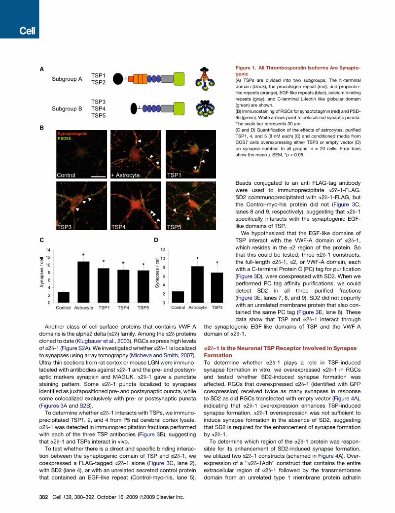
All TSP Isoforms Induce Synapse FormationThere are five TSP isoforms in mammals, which fall into twogroups according to their domain structure and oligomerizationstates (Figure 1A). Trimeric subgroup A TSPs, TSP1 and 2, aresynaptogenic (Christopherson et al., 2005). To determinewhether pentameric subgroup B TSPs are also synaptogenic,we cultured RGCs in the presence of astrocytes or with TSP 1,3, 4, or 5. All subgroup B TSPs increased synapse number signif-icantly to similar levels as TSP1 or astrocytes (Figures 1B–1D).These results suggest that the synaptogenic domain of TSP islocated in the conserved C-terminal portion of TSP, which iscommon to all isoforms spanning the EGF-like repeats, thecalcium-binding repeats, and C-terminal L-type lectin-like glob-ular domain.
The Synaptogenic Activity of TSP Maps to Its EGF-likeRepeatsTSPs interact with a number of known cell-surface receptorsthrough specific domains (Adams and Lawler, 2004). To identifythe synaptogenic domain of TSP, we treated RGCs with a panelof recombinant truncation constructs of TSP1 and 2. The TSPfragments that contained the EGF-like repeats mimicked theability of full-length TSP to induce synapses (Figures 2B and2C). A fragment containing the third EGF-like repeat togetherwith the C-terminal region of TSP2 also significantly increasedsynapse number; however, the third EGF like domain alonedid not induce a significant increase in synapse number(Figure 2C).We confirmed the importance of the EGF-like repeats in
synapse formation by functionally blocking synaptogenic effectof TSP on cultured RGCs, using monoclonal antibodies directedagainst different epitopes of TSP (Figure 2D). Monoclonal anti-bodies against the second (HB8432 and C6.7) (Annis et al.,2007) or third EGF-like repeats (A4.1) (Annis et al., 2006) blockedthe synaptogenic effect of TSP, whereas an antibody against theN-terminal domain (mAb200-1) did not (Figure 2E).To aid us in our efforts to identify the neuronal TSP receptor
involved in synapse formation, we expressed and purifiedamyc and 6-Histidine tagged TSP2 fragment containing all threeEGF-like repeats (Figures S1A and S1B available online). ThisTSP2 fragment (designated SD2 for synaptogenic domain 2)was strongly synaptogenic (Figures S1C and S1D). Collectively,these data suggest that TSP-induced synapse formation ismediated by an interaction involving EGF-like repeats of TSP.
a2d-1 Interacts with the Synaptogenic Domain of TSPThe EGF-like domains of TSP4 have been shown to bind to theVWF-A domain of integrin aM (Pluskota et al., 2005). Thus, weinvestigated whether integrin aM or other VWF-A domain con-taining integrins in RGCs were involved in TSP-induced synapseformation. None of the integrins that contained the VWF-Adomain andwere expressed by RGCswere crucial for the synap-togenic activity of TSP (data not shown).
Cell 139, 380–392, October 16, 2009 ª2009 Elsevier Inc. 381
Another class of cell-surface proteins that contains VWF-Adomains is the alpha2 delta (a2d) family. Among the a2d proteinscloned to date (Klugbauer et al., 2003), RGCs express high levelsof a2d-1 (Figure S2A). We investigated whether a2d-1 is localizedto synapses using array tomography (Micheva and Smith, 2007).Ultra-thin sections from rat cortex or mouse LGN were immuno-labeled with antibodies against a2d-1 and the pre- and postsyn-aptic markers synapsin and MAGUK. a2d-1 gave a punctatestaining pattern. Some a2d-1 puncta localized to synapsesidentified as juxtapositioned pre- and postsynaptic puncta, whilesome colocalized exclusively with pre- or postsynaptic puncta(Figures 3A and S2B).
To determine whether a2d-1 interacts with TSPs, we immuno-precipitated TSP1, 2, and 4 from P5 rat cerebral cortex lysate.a2d-1 was detected in immunoprecipitation fractions performedwith each of the three TSP antibodies (Figure 3B), suggestingthat a2d-1 and TSPs interact in vivo.
To test whether there is a direct and specific binding interac-tion between the synaptogenic domain of TSP and a2d-1, wecoexpressed a FLAG-tagged a2d-1 alone (Figure 3C, lane 2),with SD2 (lane 4), or with an unrelated secreted control proteinthat contained an EGF-like repeat (Control-myc-his, lane 5).
A
B
DC
02
46
810
1214
Syna
pses
/ ce
ll ** * *
0
2
4
6
8
10
12
Control Astrocyte TSP3
Syna
pses
/ ce
ll * *
TSP1 TSP5TSP4AstrocyteControl
+ AstrocyteControl
TSP5TSP4TSP3
TSP1
Synaptotagmin
PSD95
Subgroup A
Subgroup B
TSP1TSP2
TSP3TSP4TSP5
Figure 1. All Thrombospondin Isoforms Are Synapto-genic(A) TSPs are divided into two subgroups. The N-terminal
domain (black), the procollagen repeat (red), and properdin-
like repeats (orange), EGF-like repeats (blue), calcium binding
repeats (gray), and C-terminal L-lectin like globular domain
(green) are shown.
(B) Immunostaining of RGCs for synaptotagmin (red) and PSD-
95 (green). White arrows point to colocalized synaptic puncta.
The scale bar represents 30 mm.
(C and D) Quantification of the effects of astrocytes, purified
TSP1, 4, and 5 (8 nM each) (C) and conditioned media from
COS7 cells overexpressing either TSP3 or empty vector (D)
on synapse number. In all graphs, n = 20 cells. Error bars
show the mean ± SEM, *p < 0.05.
Beads conjugated to an anti FLAG-tag antibodywere used to immunoprecipitate a2d-1-FLAG.SD2 coimmunoprecipitated with a2d-1-FLAG, butthe Control-myc-his protein did not (Figure 3C,lanes 8 and 9, respectively), suggesting that a2d-1specifically interacts with the synaptogenic EGF-like domains of TSP.We hypothesized that the EGF-like domains of
TSP interact with the VWF-A domain of a2d-1,which resides in the a2 region of the protein. Sothat this could be tested, three a2d-1 constructs,the full-length a2d-1, a2, or VWF-A domain, eachwith a C-terminal Protein C (PC) tag for purification(Figure 3D), were coexpressed with SD2. When weperformed PC tag affinity purifications, we coulddetect SD2 in all three purified fractions(Figure 3E, lanes 7, 8, and 9). SD2 did not copurifywith an unrelated membrane protein that also con-tained the same PC tag (Figure 3E, lane 6). Thesedata show that TSP and a2d-1 interact through
the synaptogenic EGF-like domains of TSP and the VWF-Adomain of a2d-1.
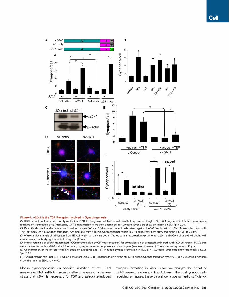
a2d-1 Is the Neuronal TSP Receptor Involved in SynapseFormationTo determine whether a2d-1 plays a role in TSP-inducedsynapse formation in vitro, we overexpressed a2d-1 in RGCsand tested whether SD2-induced synapse formation wasaffected. RGCs that overexpressed a2d-1 (identified with GFPcoexpression) received twice as many synapses in responseto SD2 as did RGCs transfected with empty vector (Figure 4A),indicating that a2d-1 overexpression enhances TSP-inducedsynapse formation. a2d-1 overexpression was not sufficient toinduce synapse formation in the absence of SD2, suggestingthat SD2 is required for the enhancement of synapse formationby a2d-1.To determine which region of the a2d-1 protein was respon-
sible for its enhancement of SD2-induced synapse formation,we utilized two a2d-1 constructs (schemed in Figure 4A). Over-expression of a ‘‘a2d-1Adh’’ construct that contains the entireextracellular region of a2d-1 followed by the transmembranedomain from an unrelated type 1 membrane protein adhalin
382 Cell 139, 380–392, October 16, 2009 ª2009 Elsevier Inc.

(Gurnett et al., 1996) mimicked the effect of full-length a2d-1 inenhancing SD2-induced synapse formation (Figure 4A), sug-gesting that the critical region of a2d-1 maps to the extracellularpart of the protein. We next overexpressed a ‘‘d-1 only’’construct (lacking the a2 region), which inhibited SD2-inducedsynapse formation (Figure 4A), indicating that the VWF-A-con-taining a2 region is necessary for enhancing synaptogenesisand that regions within d-1 may be involved in regulating down-stream interactions that are critical for TSP-induced synapseformation.Since the VWF-A domain of a2d-1 binds TSP, we investigated
whether antibodies against the VWF-A domain of a2d-1 wouldinterfere with TSP-induced synapse formation. Two monoclonalantibodies directed against the VWF-A domain of a2d-1, 5A5 and3B4, recognized a2d-1 in western blots and stained the surfaceof HEK293 cells overexpressing a2d-1 (Figures S3A and S3B).When RGCs were cultured with these antibodies in the presenceor absence of TSP and synapse number analyzed, both 5A5 and
A
B
C
E
321 1 2 3
N Type 1Properdin-
like
Type 3Ca Binding
Type 2EGF-likePC C
0 2 4 6 8 10 12 14
Control+Astrocytes
**
**
Synapses/cell
0 2 4 6 8 10
Control
+Astrocytes
***
*
Synapses/cell
N
o
PC
P1
P2
P3
E1
E3
Ca (wire)
CE2
A4.1
C6.7HB8432
mbc200-1
TSP - + - + - + - + - +plus
mbc200-1plus
HB8432plusC6.7
plusA4.1
Syna
pses
/cel
l
* *
D
*
inhibited
Figure 2. EGF-like Repeats of TSPs AreSynaptogenic(A) The domain structure of TSP1 and 2. N-terminal
domain (black), oligomerization domain and a pro-
collagen repeat (red, PC), three properdin-like
(TSP type 1, orange, P1–P3), three EGF-like (TSP
type 2, blue, E1–E3), and 13 calcium binding (TSP
type 3, gray) repeats [Ca(wire)] and a C-terminal
L-type lectin like globular domain (green, C).
(B and C) Quantification of the effect of TSP1 (B)
and TSP2 (C) fragments on synapse number.
RGCs were treated with astrocytes, full-length
TSP1, or a panel of TSP1 or TSP2 fragments (8
nM each).
(D) Location of epitopes targeted by TSP blocking
antibodies (modified from Carlson et al. [2008]).
The inset shows a magnified structure of EGF-
like repeats and the Ca-binding wire region and
the C-terminal L-lectin like domain. Highlighted
domains indicate putative synaptogenic domain
of TSP.
(E) Quantification of the effect of monoclonal anti-
TSP antibodies on TSP’s synaptogenic activity.
In all graphs, n = 20 cells. Error bars show the
mean ± SEM, *p < 0.05.
3B4 induced synapse formation similar toTSP. A control antibody (OX7) againstanother RGC surface receptor (Thy1) didnot affect synapse formation. The synap-togenic effect of 5A5 or 3B4 was notadditive with that of TSP (Figure 4B).These data show that antibody bindingto the VWF-A domain can mimic TSPssynaptogenic function and suggestthat the interaction of TSP with theVWF-A domain of a2d-1 is important forthe initiation of synapse formation. Suchligand-mimicking antibodies were alsodescribed for VWF-A domain-containingintegrins (Wilkins et al., 1996).
To determine whether a2d-1 is required for TSP-inducedsynapse formation, we used a small interfering RNA (siRNA)knockdown approach. An siRNA pool specific for rat a2d-1significantly reduced the expression of rat a2d-1 in transfectedHEK293 cells (Figure 4C). Knockdown of a2d-1 in RGCs withthis siRNA pool inhibited astrocyte or TSP-induced synapseformation in vitro (Figures 4D and 4E), whereas the nontargetingcontrol siRNA pool (siControl) did not affect synapse formation(Figures 4C and 4D).To show that the reduction in synapse formation by the a2d-1
siRNAs was due to the specific knockdown of a2d-1, we testedwhether the siRNA inhibition could be rescued by coexpressingan siRNA resistant a2d-1 construct. One of the siRNAs againstrat a2d-1, sia2d-1 Duplex 9, blocked overexpression of rata2d-1 but not human a2d-1 in HEK293 cells (Figure S4). Whenwe cotransfected RGCs with sia2d-1(9) and the human a2d-1construct, we rescued SD2 or astrocyte-induced synapseformation (Figure 4F), showing that siRNA knockdown of a2d-1
Cell 139, 380–392, October 16, 2009 ª2009 Elsevier Inc. 383

A
CB
D
Anti- 2 -1
Synapsin / Maguk / 2 1
Pre-
imm
une
sera
Anti-
TSP1
Anti-
TSP2
Anti-
TSP4
130
200 Cortic
allys
ate
Anti-
Cav1
.2An
ti-Ag
rin
Bead
s alon
e
HEK293
Lysates
Protein C Tag
Purification
1 2 3 4 5 6 7 8 9
Anti-
Prot
ein
CAn
ti-M
yc
2 -12
VWF-A
SD2
Anti FLAG IP
1 2 3 4 5 6
2 -1FLAG
SD2
Control-myc-hisAnti-His
HEK293lysates
7 8 9
***
**
E
Figure 3. Thrombospondins Interact with a2d-1(A) Array tomography analysis of synaptic localization of a2d-1 in cerebral cortex. RGCs were immunostained for synapsin I (blue) and MAGUK (green). a2d-1
puncta (red) associate both with synapses (white circles) and with isolated presynaptic (diamonds) or postsynaptic (squares) puncta. The scale bar represents
2 mm.
(B) Western-blot analysis of a2d-1 on the immunoprecipitation (IP) fractions performed with antibodies specific to TSP1, 2, or 4 as well as calcium channel a1C
(Cav1.2), or Agrin (positive and negative controls for IP, respectively).
(C) Western blot analysis of a2d-1 interaction with the synaptogenic domain of TSP2 (SD2). Left, HEK293 cell lysates from nontransfected (1), a2d-1-FLAG alone
(2), SD2 alone (3), a2d-1-FLAG and SD2 (4), and a1d-1FLAG and Control-myc-His construct (5) transfected cells. SD2 and Control-his-myc protein are marked
with redd. Anti-his antibody cross-reacts with several histidine rich proteins in HEK293 cell lysates (markedwith a blue *). Anti-a2d-1 antibody also weakly recog-
nizes the human a2d-1 expressed endogeneously in HEK293 cells at low levels (blueA) Right, anti-FLAG IP fractions from a2d-1-FLAG alone (6), SD2 alone (7),
a2d-1-FLAG and SD2 (8), and a2d-1FLAG and Control-myc-His construct (9) transfections.
(D) Domain structure of a2d-1 protein and scheme of a2d-1 protein C (PC) tagged constructs. SP, signal peptide; vWA_N and VGCC_a2, putative domains of
unknown structure. Yellow boxes indicate putative helical regions where no domain has yet been predicted. The red box shows the transmembrane (TM) region.
Orange hexagons indicate predicted N-glycosylation sites, and purple bars indicate positions of cysteines.
(E) SD2 interacts with the VWF-A domain of a2d-1. Lane 1 is nontransfected HEK293 cell lysate. SD2 was coexpressed with PC tagged full-length a2d-1, a2 only,
or VWF-A only constructs (lanes 3, 4, and 5) as well as CXCR4. SD2 coimmunopurified with the a2 only (8) and VWF-A only (9) constructs of a2d-1 as well as the
full-length protein (7) with anti-PC beads, (red arrows). SD2 did not copurify with CXCR4 (6).
384 Cell 139, 380–392, October 16, 2009 ª2009 Elsevier Inc.
blocks synaptogenesis via specific inhibition of rat a2d-1messenger RNA (mRNA). Taken together, these results demon-strate that a2d-1 is necessary for TSP and astrocyte-induced
synapse formation in vitro. Since we analyze the effect ofa2d-1 overexpression and knockdown in the postsynaptic cellsreceiving synapses, these data show a postsynaptic sufficiency
Syna
pses
/cel
lSy
naps
es/c
ell
2 -1-1 only
2 -1-Adh
Contro
lTS
P
OX7
5A5
5A5+
TSP
3B4
3B4+
TSP
Syna
pses
/cel
l **
*
**
siControl si 2 1
alon
e+
Astro
cyte
s
SD2 - + - + - + - + siControl siControlsi 2 1
(9)Empty Vector 2 1HUMAN
*
**
si 2 1(9)
si 2 1siControl2 1
actin
BA
C E
F
D
inhibited
siControl si 2 1+astros +astros+TSP +TSP
**
rescued
Syna
pses
/cel
l
-- - - ++++ pcDNA3 2 -1 -1 only 2 -1-Adh
*
SD2
**
Figure 4. a2d-1 Is the TSP Receptor Involved in Synaptogenesis(A) RGCs were transfected with empty vector (pcDNA3, Invitrogen) or pcDNA3 constructs that express full-length a2d-1, d-1 only, or a2d-1-Adh. The synapses
received by transfected cells (marked by GFP coexpression) were then quantified. n = 20 cells. Error bars show the mean ± SEM, *p < 0.05.
(B) Quantification of the effects of monoclonal antibodies 5A5 and 3B4 (mouse monoclonals raised against the VWF-A domain of a2d-1; Mazorx, Inc.) and anti-
Thy1 antibody OX7 in synapse formation. 5A5 and 3B7 mimic TSP’s synaptogenic function. n = 30 cells. Error bars show the mean ± SEM, *p < 0.05.
(C) Western blot analysis of cell lysates from HEK293 cells, which were cotransfected with an expression vector for rat a2d-1 and siControl or sia2d-1 pools, with
a monoclonal antibody against a2d-1 or against b-actin.
(D) Immunostaining of siRNA-transfected RGCs (marked blue by GFP coexpression) for colocalization of synaptotagmin (red) and PSD-95 (green). RGCs that
were transfected with sia2d-1 did not form many synapses even in the presence of astrocytes (see inset i versus ii). The scale bar represents 30 mm.
(E) Quantification of the effects of siRNA pools on astrocyte and TSP-induced synapse formation in RGCs. n = 20 cells. Error bars show the mean ± SEM,
*p < 0.05.
(F) Overexpression of human a2d-1, which is resistant to sia2d-1(9), rescues the inhibition of SD2-induced synapse formation by sia2d-1(9). n = 20 cells. Error bars
show the mean ± SEM, *p < 0.05.
Cell 139, 380–392, October 16, 2009 ª2009 Elsevier Inc. 385

and necessity for a2d-1 in astrocyte/TSP-induced synapseformation.
a2d-1-Mediated Synapse Formation Does Not Dependon Calcium Channel Surface Level or Functiona2d-1 is known to enhance calcium channel function and traf-ficking (Arikkath and Campbell, 2003). We therefore investigatedwhether the activity of a2d-1 in synapse formation is linked to itsrole in increasing calcium currents or calcium channel levels.Gene expression analysis of RGCs show that these cells expresspredominantly postsynaptic L-type and presynaptic N- and P/Q-type voltage gated calcium channels (VGCCs). To directly testwhether VGCC function was required for astrocyte-inducedsynapse formation, we added L-type calcium channel blockersto RGCs to block L-type channel function. These drugs had noeffect on SD2-induced synapse formation (Figure S5A). Similarlypresynaptic N- and P/Q-type channel blockers did not block
0
20
40
60
80
100
120
140
160
180 *
# VG
lut2
/PSD
95 S
ynap
ses
WT TG
WT TGA B
VG
lut2
/ P
SD
95 i ii
i ii
WT TG
*
10 p
A
0.2 secs
WT TG
10pA
0.0 2 secss
10 p
A
2 secs
WT TGFreq
uenc
y of
mEP
SCs,
Hz
0
1
2
3
4
WT TG
Ampl
itude
of m
EPSC
s, p
A
02468
1012
E*
D
C
Figure 5. a2d-1 Overexpression In Vivo IncreasesExcitatory Synapse Number(A) Immunolabeling of cortices from littermate wild-type (WT)
and a2d-1-overexpressing transgenic (TG) P21 mice for
VGlut2 and PSD95. The number of colocalized VGlut2/
PSD95 puncta (white arrows in insets i and ii) was higher in
the TGs then the WTs. The scale bar represents 20 mm.
(B) Quantification of VGlut2/PSD95 colocalization in brain
sections from WT and TG mice. Error bars show the mean ±
SEM, *p < 0.05.
(C) Representative raw data traces of mEPSCs from layer IV
cortical pyramidal neurons recorded from a WT and an a2d-1
TG mouse. Top, condensed trace. Bottom, expanded trace.
(D) Summary of the frequency of mEPSCs in layer IV cortical
pyramidal neurons of a2d-1 TG and WT. TG = 3.5 ± 0.3 Hz
(n = 11 cells); WT = 2.1 ± 0.2 Hz (n = 12 cells). Error bars
show the mean Hz ± SEM, p = 0.002.
(E) Summary of the amplitude of mEPSCs in layer IV cortical
pyramidal neurons of a2d-1 TG and WT. TG = 11.9 ± 0.3 pA,
WT = 11.6 ± 0.3 pA. Error bars show the mean ± SEM, p = 0.5.
TSP-induced synapse formation (data not shown).We next investigated whether increase of postsyn-aptic L-type calcium channel expression in RGCswould enhance synapse formation. Overexpressionof L-type a1C and b subunits in RGCs had noeffect on astrocyte-induced synapse formation(Figure S5B). Finally, we tested whether TSP treat-ment would lead to an increase in cytoplasmiccalcium levels in RGCs. Neither acute nor long-term TSP treatment led to a noticeable rise in spon-taneous calcium oscillations in RGCs (Figure S6).Taken together, these results show that the role ofa2d-1 in synapse formation cannot bedirectly linkedto calcium channel expression levels or function.
Overexpression of a2d-1 in NeuronsEnhances Synapse Formation In VivoTo determine whether a2d-1 plays a role in synapseformation in vivo,weexaminedsynapsenumberand
synaptic activity in transgenic mice that selectively overexpressa2d-1 in CNS neurons, under the control of the Thy1 promoter(Li et al., 2006). Sagittal brain sections from 21-day-old (P21)transgenic (TG) andwild-type (WT) littermatemicewere coimmu-nostained for PSD95 and either the presynaptic vesicular gluta-mate transporter 1 or 2 (VGlut1 and VGlut2). We quantified thenumber of colocalized pre- andpostsynaptic puncta to determinethe synaptic density in the cortices of these mice. The TG micehad significantly higher numbers of VGlut2-positive excitatorysynapses in the cortex than did the littermate WT controls (1.8-fold, Figures 5A and 5B); however, there was no difference inthe number of VGlut1-positive synapses between WT and TGmice (Figures S7A and S7B). The observation that a2d-1 overex-pression increases VGlut2-positive synapses provides evidencethat excitatory synapse formation is enhanced in the TGs.In the adult cortex thalamic neurons projecting onto layer IV
neurons form VGlut2-positive synapses, while synapses made
386 Cell 139, 380–392, October 16, 2009 ª2009 Elsevier Inc.

between cortical neurons contain VGlut1 (Fremeau et al., 2004).We confirmed that the increase in VGlut2-positive synapsenumber was not due to an increase in the number of neuronsin the cortex or thalamus, as the number of cells and neuronsin WT and TG brains were identical in these brain regions(Figures S8A and S8B).Excitatory synapses in the cortex are initially formed as VGlut2
positive, and there is an isoform switch to VGlut1 that happensaround the second week of postnatal development (Miyazakiet al., 2003). During this period, some synapses can transientlybe both VGlut1 and 2 positive (Nakamura et al., 2005). We deter-mined that the increase in VGlut2-positive synapses was not dueto a prolonged colocalization of VGlut1 and 2 at the samesynapse, since these proteins seldom colocalized at P21, andthere were no differences in the frequency of colocalization ofthese proteins between genotypes (Figures S9A and S9B).Taken together, these results show that the increase in VGlut2-positive synapses associated with a2d-1 overexpression is dueto neither an increase in the number of cortical or thalamicneurons nor a delay in the isoform switch from VGlut2 to 1 inthe cortex.In addition to analyzing synapse number by immunohisto-
chemistry, we performed whole-cell patch-clamp recordings inlayer IV cortical pyramidal neurons and assayed the number ofactive synapses by analyzing the frequency and amplitude ofminiature excitatory postsynaptic currents (mEPSCs). Recordedcells were dye filled, and their identity was verified (Figures S10Aand S10B). We targeted layer IV pyramidal neurons bothbecause these cells receive VGlut2-positive synapses andbecause array tomography revealed an increase in a2d-1 immu-nostaining in TG animals in this layer (Figure S10C). There wasa highly significant increase in the frequency of mEPSCs ina2d-1 TGmice comparedwithWTmice (1.63-fold), with no effecton the amplitude of mEPSCs (Figures 5C–5E). The increase inthe frequency of mEPSCs in TG mice is very consistent withthe increased excitatory synapse number found by the immuno-histochemical analysis described above. Taken together, thesedata show that a2d-1 plays a role in promoting excitatorysynapse formation in the brain.
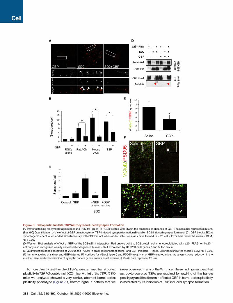
Gabapentin, the High-Affinity Ligand for a2d-1, StronglyInhibits TSP-Induced Synapse FormationIn order to determine whether GBP, the high-affinity ligand fora2d-1 affects TSP or astrocyte-induced synapse formation, wecultured RGCs with TSP, SD2, or ACM in the presence orabsence of GBP (32 mM). GBP strongly inhibited TSP, SD2, orastrocyte-induced synapse formation (Figures 6A–6C andS1C). To determine whether GBP could dissolve already estab-lished synapses, we cultured RGCs with SD2 for 5 days to allowsynapses to formand then addedGBP for an additional day.GBPhadno effect on synapse numberwhen added after the synapseswere formed (Figure 6C). Thus, GBP blocks new synapseformation induced by TSP and astrocytes but does not dissolveestablished synapses. Interestingly, GABA, an inhibitory neuro-transmitter that binds to a2d-1 with much lower affinity (IC50 =650 mM)(Suman-Chauhan et al., 1993), also blocked SD2-induced synapse formation when used at high concentrations(Figure S11).
To determine whether GBP blocks TSP-induced synapseformation by inhibiting the a2d-1-TSP interaction, we coculturedtwo populations of HEK293 cells, one expressing a2d-1FLAGand the other expressing SD2, in the presence or absence ofGBP. Immunoprecipitation with anti-FLAG antibodies revealedthat the SD2-a2d-1 interaction was diminished in the presenceof GBP (Figure 6D), suggesting that GBP blocks TSP-inducedsynapse formation by interfering with the interaction betweena2d-1 and TSP.To test whether GBP similarly blocks synapse formation
in vivo, we injected neonatal mice with either GBP or saline forthe first postnatal week, which coincides with the initiation ofsynapse formation in the brain. At this age, glutamatergicsynapses in the cortex are predominantly VGlut2 positive (Miya-zaki et al., 2003). Therefore, we coimmunostained sagital brainsections from P7 saline- or GBP-injected mice with antibodiesagainst VGlut2 and PSD95 and quantified the number of colocal-ized pre- and postsynaptic puncta in the cortex of these mice.There were significantly fewer excitatory synapses in the cere-bral cortex of the GBP-injected mice relative to control mice(Figure 6E). This difference was mainly due to a severe decreasein synapse number in half of the GBP-injected animals. In themice that responded strongly to GBP, the VGlut2/PSD95synaptic densities went down profoundly, to less than 10% ofthe saline-injected values, although the number of neurons didnot change. GBP injection affected both VGlut2 and PSD95puncta by reducing their number, size, and colocalization(Figure 6E), similar to its effect on synaptic puncta in vitro. Thesefindings show that GBP is a powerful inhibitor of new synapseformation both in vitro and in vivo.
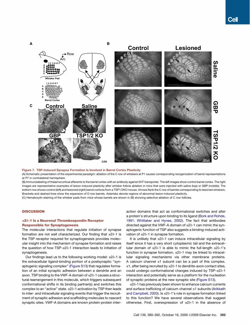
Inhibition of TSP-Induced Synapse Formation Interfereswith Lesion-Induced Barrel Cortex PlasticityTo determine whether astrocyte-induced synapse formation isinvolved in remodeling neural circuits during development, weutilized a well-established developmental plasticity paradigm,the ‘‘barrel cortex plasticity’’ assay. The nerves that innervatethemajor whiskers on the snout of the mouse project to the brainas a topographically ordered ‘‘somatotopic’’ map (Erzurumluet al., 2006). In the primary somatosensory cortex, this map isorganized as ‘‘barrels’’ (Figure 7A) that exhibit structural changesin response to peripheral whisker manipulations.To test whether TSP-induced synapse formation is involved in
mechanisms of experience-dependent plasticity, we injectedtwo groups of neonatal mice either with GBP or saline dailystarting at P0 until P7. On P1, five whiskers from the C row onone side of each mouse were lesioned. The mice were sacrificedat P7, and barrel cortex organization in both the unlesioned‘‘control’’ and the lesioned hemisphere was analyzed. Bothsaline- and GBP-injected mice had typical barrel organizationformed on the control side (Figure 7B, top two left panels). Onthe lesioned side, although all saline-injected mice displayeda normal barrel cortex plasticity pattern, 50% of the GBP-injected mice displayed an atypical plasticity response(Figure 7B, right panels), where the A and B rows as well asthe C row lost form and fused, even though the whisker folliclesfor these rows were undisturbed in all mice (Figures 7B, 7C,and S12).
Cell 139, 380–392, October 16, 2009 ª2009 Elsevier Inc. 387
Tomoredirectly test the role of TSPs,we examinedbarrel cortexplasticity inTSP1/2double-null (KO)mice.A thirdof theTSP1/2KOmice we analyzed showed a very similar, aberrant barrel cortexplasticity phenotype (Figure 7B, bottom right), a pattern that we
never observed in any of theWTmice. These findings suggest thatastrocyte-secreted TSPs are required for rewiring of the barrelspost injury and that themain effect ofGBP inbarrel cortex plasticityis mediated by its inhibition of TSP-induced synapse formation.
A
B
C
D
# VG
lut2
/PSD
95 s
ynap
ses
Saline GBP
*
E
VGlu
t2/P
SD95
i ii
Salinei
GBPii
2 -1Flag
HEK293
LysatesAnti Flag IPsAnti- 2 1
Anti-His
Anti- 2 1
Anti-His
+ - -+ + +
- + ++ - +
- - +- + +
SD2
GBP
F
RGCsalone
GBPRat ACM Mouse
ACM
Syna
pses
/cel
l
- + - + - + - +TSP
*
* *
0
1
2
3
4
5
6
7
8
Control GBP
SD2
+GBP6 days
+GBPlast day
Syna
pses
/cel
l
*
GBP SD2 SD2+GBP
PSD
95Sy
ntag
Mer
ged
Figure 6. Gabapentin Inhibits TSP/Astrocyte-Induced Synapse Formation(A) Immunostaining for synaptotagmin (red) and PSD-95 (green) in RGCs treated with SD2 in the presence or absence of GBP The scale bar represents 30 mm.
(B and C) Quantification of the effect of GBP on astrocyte- or TSP-induced synapse formation (B) and on SD2-induced synapse formation (C). GBP blocks SD2’s
synaptogenic effect when added simultaneously with SD2 but not when added after synapses have formed. n = 20 cells. Error bars show the mean ± SEM,
*p < 0.05.
(D) Western Blot analysis of effect of GBP on the SD2-a2d-1 interaction. Red arrows point to SD2 protein coimmunoprecipitated with a2d-1FLAG. Anti-a2d-1
antibody also recognizes weakly expressed endogenous human a2d-1 expressed by HEK293 cells (lanes 2 and 5, top blots).
(E) Quantification of colocalization of VGlut2 and PSD95 in brain sections from saline- and GBP-injected P7 mice. Error bars show the mean ± SEM, *p < 0.05.
(F) Immunolabeling of saline- and GBP-injected P7 cortices for VGlut2 (green) and PSD95 (red). Half of GBP-injected mice had a very strong reduction in the
number, size, and colocalization of synaptic puncta (white arrows, inset i versus ii). Scale bars represent 20 mm.
388 Cell 139, 380–392, October 16, 2009 ª2009 Elsevier Inc.
DISCUSSION
a2d-1 Is a Neuronal Thrombospondin ReceptorResponsible for SynaptogenesisThe molecular interactions that regulate initiation of synapseformation are not well characterized. Our finding that a2d-1 isthe TSP receptor required for synaptogenesis provides molec-ular insight into the mechanism of synapse formation and raisesthe question of how TSP-a2d-1 interaction leads to initiation ofsynaptogenesis.Our findings lead us to the following working model: a2d-1 is
the extracellular ligand-binding portion of a postsynaptic ‘‘syn-aptogenic signaling complex’’ (Figure S13) that regulates forma-tion of an initial synaptic adhesion between a dendrite and anaxon. TSP binding to the VWF-A domain of a2d-1 causes a struc-tural rearrangement in this molecule, which triggers subsequentconformational shifts in its binding partner(s) and switches thiscomplex to an ‘‘active’’ state. a2d-1 activation by TSP then leadsto inter- and intracellular signaling events that trigger the recruit-ment of synaptic adhesion and scaffolding molecules to nascentsynaptic sites. VWF-A domains are known protein-protein inter-
action domains that act as conformational switches and altera protein’s structure upon binding to its ligand (Bork and Rohde,1991; Whittaker and Hynes, 2002). The fact that antibodiesdirected against the VWF-A domain of a2d-1 can mimic the syn-aptogenic function of TSP also suggests a binding-induced acti-vation of a2d-1 in synapse formation.It is unlikely that a2d-1 can induce intracellular signaling by
itself since it has a very short cytoplasmic tail and the extracel-lular domain of a2d-1 is able to mimic the full-length a2d-1’sfunction in synapse formation. a2d-1 may be linked to intracel-lular signaling mechanisms via other membrane proteins.A calcium channel a1 subunit can be a part of this complex.a1, after being recruited by a2d-1 to dendrite-axon contact sites,could undergo conformational changes induced by TSP-a2d-1interaction and potentially serve as a platform for the nucleationof synaptic proteins at the new synaptic site (Figure S13).a2d-1 has previously been shown to enhance calcium currents
and surface trafficking of calcium channel a1 subunits (Arikkathand Campbell, 2003). Is a2d-1’s role in synapse formation linkedto this function? We have several observations that suggestotherwise. First, overexpression of a2d-1 in the absence of
Figure 7. TSP-Induced Synapse Formation Is Involved in Barrel Cortex Plasticity(A) Schematic presentation of the experimental paradigm: ablation of the C row of whiskers at P1 causes corresponding reorganization of barrel representations
at P7 in contralateral hemisphere.
(B) Immunolabeling of thalamocortical afferents to the barrel cortex with an antibody against 5HT transporter. The left images show control barrel cortex. The right
images are representative examples of lesion-induced plasticity after whisker follicle ablation in mice that were injected with saline (top) or GBP (middle). The
bottom row shows control (left) and lesioned (right) barrel cortices from a TSP1/2KOmouse. Arrows flank the C row of barrels corresponding to lesionedwhiskers.
Brackets and dashed lines show the expansion of D row barrels. Asterisks denote regions of abnormal lesion-induced plasticity.
(C) Hematoxylin staining of the whisker pads from mice whose barrels are shown in (B) showing selective ablation of C row follicles.
Cell 139, 380–392, October 16, 2009 ª2009 Elsevier Inc. 389

TSP enhances calcium channel surface expression (Gurnettet al., 1996) but does not lead to an increase in synapse number.Second, the a2d-1Adh protein can mimic the effect of full-lengtha2d-1 in enhancing synapse formation, but it does not induce anincrease in calcium currents like the full-length protein (Gurnettet al., 1996). Third, neither the overexpression nor the pharmaco-logical blocking of calcium channels interferedwith TSP-inducedsynapse formation. Similarly, acute or long-term TSP treatmentdid not increase cytoplasmic calcium levels in RGCs; thus, it isunlikely that TSP triggers activation of a homeostatic mechanismthat can activate synapse formation. Taken together, our resultsshow that global changes in calcium channel numbers orcurrents are not involved in TSP-induced synapse formation.However, since the a2d-1Adh construct, which enhancessynapse formation, can interact with the a1 subunit, and sinced-1 construct, which inhibits synapse formation, can interferewith the a2d-1 and a1 interaction, a physical interaction betweena2d-1 and the calcium channel a1 subunits might be importantfor synapse formation. Future studies exploring whether knock-ing down expression of a1 subunits affects TSP-inducedsynapse formation are necessary to verify this possibility.
a2d-1 might also interact with other proteins that are involvedin organization of synaptic contacts. Such dual functions havebeen described for the g subunits of VGCCs also known as star-gazins. Theywere initially isolated as a component of the calciumchannel complex but are now known to play primary roles inAMPA receptor regulation (Chen et al., 2000). Identification ofthe relevant a2d-1 interacting molecules promises to providenew molecular insight into the process of synapse formation.In addition, there could be other CNS molecules that shareTSP’s and GBP’s abilities to bind to a2d-1 and trigger or inhibitsynapse formation.
Our findings have a number of important implications for futurestudies. First, TSP and a2d-1 are also highly concentrated at theneuromuscular junction; thus, it is likely that these molecules areinvolved in formation of this synapse (Arber and Caroni, 1995;Arikkath and Campbell, 2003). Second, other a2d familymembers might also regulate synapse formation. In fact, disrup-tion of the a2d-4 gene in mice leads to a severe loss of ribbonsynapses in the photoreceptor cells (Wycisk et al., 2006), andmutations in a2d-3 cause defects in synaptic transmission anda morphological defect in presynaptic organization at theDrosophila neuromuscular junction (Dickman et al., 2008; Kur-shan et al., 2009). These observations suggest that the functionof a2d subunits in promoting synaptogenesis may be evolution-arily conserved and can be exerted presynaptically as well aspostsynaptically.
Gabapentin Is a Powerful Blocker of Synapse FormationOur findings suggest that GBP blocks TSP-induced synapseformation by interfering with TSP-a2d-1 interaction. GBP bindingto a2d-1 involves a region just upstream of the VWF-A domain ina2 (Wang et al., 1999). Therefore, it is unlikely that TSP and GBPcompete for the same binding site. It is known for integrins thatconformational changes in VWF-A domains can be constrainedby interactions made by regions flanking this domain (Bork andRohde, 1991; Whittaker and Hynes, 2002). We propose thatGBP binding to a2d-1 restricts the conformation of the VWF-A
domain and keeps a2d-1 in its ‘‘inactive conformation.’’ This per-turbs the TSP-a2d-1 interaction and inhibits activation of the syn-aptogenic signaling complex (Figure S13).GABA, leucine, and isoleucine can also bind to a2d-1, albeit at
lower affinity thanGBP (Dooley et al., 2007), and thus they can bephysiological ligands for a2d-1 and regulate excitatory synapseformation. In agreement with this, we found that high concentra-tions of GABA inhibited synapse formation in culture. Such highconcentrations of GABA are present in the CNS right next toa GABAergic axon. Dendritic filopodia in the developing brainactively seek for synaptic partners and establish exclusivelyglutamatergic contacts. Interestingly, dendritic filopodia thatcontact a GABAergic axon never stabilize the contact and retract(Lohmann and Bonhoeffer, 2008; Wierenga et al., 2008). In futurestudies, it will be interesting to explore whether a2d-1 functionsas a physiologically relevant GABA receptor that enables initialselectivity for the formation of excitatory synapses by dendriticfilopodia.
a2d-1-TSP Interaction Regulates Synapse Formationduring Development and after InjuryThe ability of GBP to strongly decrease synapse formation inwild-type mouse brains points to a critical role for TSP-a2d-1interaction and astrocytes in driving synaptogenesis in vivo. Inaddition, the correct execution of barrel cortex plasticitydepends on TSP-induced synapse formation. Since the unle-sioned barrel cortices are formed normally both in GBP injectedand TSP1/2 KO mice, TSPs might specifically play a role insynaptic remodeling plasticity upon injury in this system. Thesefindings add to the growing data that astrocytes not only activelycontribute to normal synaptogenesis but also mediate synapticremodeling events after injury.It is interesting that the effect of GBP in vivo is an ‘‘all or none’’
effect rather than a fractional decrease in synapse number, andonly 50% of the mice responded strongly to GBP injections. It ispossible that a critical threshold concentration of GBP in thecerebrospinal fluid is required to be effective in blocking synapseformation, which is only achieved in half of the mice. Gendercould be critical in GBP responsiveness by affecting in GBPdelivery to neural tissues and can explain the 50% penetrancewe have observed. In fact, a recent study demonstrated thatintraperitoneal GBP injections were not as effective at blockingseizures in female mice as in males (Traa et al., 2008).Since GBP strongly blocks TSP-induced synapse formation
within its therapeutic concentration, it is possible that inhibitionof excitatory synapse formation is an important mode of its ther-apeutic action in epilepsy and pain. Reactive astrocytosis isprominent both in epileptic lesions and in the spinal cord afterperipheral nerve injury that leads to neuropathic pain (Liu et al.,2000; Ridet et al., 1997). Reactive astrocytes express high levelsof TSP1 and 2 (Lin et al., 2003). Similarly, upon injury in the spinalnerve, both a2d-1 and TSP4 genes are upregulated in the spinalcord (Valder et al., 2003; Wang et al., 2002). Increased a2d-1levels were shown to lead to enhanced excitatory synaptic trans-mission and elevated neuropathic pain states (Li et al., 2004,2006). Similarly, there is increased excitation in the epilepticbrain (Prince, 1999). All these observations point to the possi-bility that aberrant excitatory synaptogenesis may contribute to
390 Cell 139, 380–392, October 16, 2009 ª2009 Elsevier Inc.

the pathophysiology of neuropathic pain and epilepsy. ThusGBP may act by limiting these excess synapses from forming,a possibility which can now be directly tested in animal modelsof these diseases. In conclusion, by identifying a2d-1 as areceptor for TSP mediated glial-induced synapse formation,we have gained molecular understanding not only of astrocytes’role in synapse formation in health and disease, but also of theprocess of synapse formation itself.
EXPERIMENTAL PROCEDURES
Purification and Culture of RGCs and AstrocytesRGCs were purified with greater than 99.5% purity from P5 Sprague-Dawley
rats (Charles Rivers) and cultured in serum-free medium as previously
described (Christopherson et al., 2005; Meyer-Franke et al., 1995; Ullian
et al., 2001). Cortical astrocyte inserts and ACM were prepared as described
in (Christopherson et al., 2005). RCGs were cultured for 3–4 days to allow
robust process outgrowth and then cultured with astrocyte inserts, ACM, or
TSPs for an additional 6 days.
MiceTSP1/2 double-null mice on an FVB background were used (n = 12). WT FVB
mice were purchased from Charles River Laboratories. Brains from P21, a2d-
1-overexpressing, TG mice and their littermate WT controls (n = 8) were
provided by Li and colleagues and are described in Li et al. (2006).
Quantification of Synapse NumbersFor synapse quantification of RGCs, we followed a previously developed
immunohistochemistry (IHC)-based method described and validated in Chris-
topherson et al. (2005) and Ullian et al. (2001). For quantification of excitatory
synapse number in mouse brain, three sagital brain sections per animal were
stained with pre- and postsynaptic markers, and 5 mm confocal scans were
performed (optical section width 0.38 mm, 14 optical sections each) at the
cortex. The parameters for scanning were always set up for WT (or saline-
injected) brain sections, and the same imaging parameters were used for
TG (or GBP-injected) animals. Merged single optical section images at 1 mm
intervals were analyzed with the ImageJ puncta analyzer option to count for
number of colocalized pre- and postsynaptic puncta (five optical sections/
section, 15 images/brain). Average synaptic density per imaged area was
calculated for each condition. Details on IHC conditions, image acquisition,
and quantification can be found in Supplemental Data.
Electrophysiological RecordingsExperiments were carried out on littermate WT and a2d-1 transgenic mice
aged P21–P25, and recordings and analysis were both carried out blind to
genotype. Whole-cell voltage-clamp recordings of layer IV pyramidal neurons
in the visual cortex were carried out at room temperature in flowing isotonic
saline containing 1 mM tetrodotoxin (TTX) and 40 mM bicuculline to isolate
mEPSCs. mEPSCs were recorded for one minute and analyzed with Minianal-
ysis software from Synaptosoft.
Saline and Gabapentin InjectionsMice were given daily intraperitoneal injections of either a single dose of
400 mg/kg of GBP (Sigma-Aldrich) or a matching volume of saline solution
(PBS). Pups were weighed just before injections to determine the dose admin-
istered and to follow weight gain and general health, which showed no differ-
ences between GBP- and saline-injected mice.
Whisker Lesions and Barrel Cortex ImmunohistochemistryNeonatal mice were held on their left side under a dissecting scope and
received two parallel incisions with a surgical blade flanking the C row of
whiskers to be removed. The skin between the incisions was pulled back
with forceps. Follicles were individually removed with forceps at the opening.
The lesion site was then cauterized with silver nitrate using flexible caustic
applicators (Tech-Med). Mice were allowed to recover in their home cage.
P7 mice were sacrificed, and brains were harvested. Samples were blinded
during rest of the analysis of the barrel cortex plasticity. Tangential cortical
sections were stained with anti-serotonin (5-HT) transporter rabbit polyclonal
antibody (Calbiochem, 1:400) Barrels were imaged with a Nikon Eclipse
E800 fluorescent microscope, and images were digitally acquired with an
SPOT camera (Diagnostic Instruments). The complete maps of the barrel
cortex were reassembled from 5-HTT-stained images of serial sections by
reconstruction in Photoshop (Adobe Systems). Details on the immunohisto-
chemistry conditions, image acquisition, and data analysis can be found in
Supplemental Data.
SUPPLEMENTAL DATA
Supplemental Data include Supplemental Experimental Procedures and 13
figures and can be found with this article online at http://www.cell.com/
supplemental/S0092-8674(09)01185-4.
ACKNOWLEDGMENTS
We thank Maria L. Fabian, Navid Nouri, and Mark Duquette for excellent tech-
nical assistance, and Beth Stevens, Junryo Watanabe, and Alissa Winzeler for
critical reading of the manuscript. This work was supported by grants from the
National Institute of Drug Addiction (DA15043 to B.A.B.), the National Heart,
Lung and Blood Institute (HL49081 to J.L.), and the National Institute of Neuro-
logical Disorders and Stroke and National Institute of Dental and Craniofacial
Research (NS40135 and DE14545 to Z.D.L.). C.E. and N.J.A. were supported
by the Human Frontiers Scientific Program long-term fellowships and A.D.H by
the Helen Hay Whitney postdoctoral fellowship. Arnon Rosenthal is founder
and president of MazoRx Inc.
Received: February 16, 2008
Revised: February 4, 2009
Accepted: August 26, 2009
Published online: October 8, 2009
REFERENCES
Adams, J.C., and Lawler, J. (2004). The thrombospondins. Int. J. Biochem. Cell
Biol. 36, 961–968.
Annis, D.S., Murphy-Ullrich, J.E., and Mosher, D.F. (2006). Function-blocking
antithrombospondin-1 monoclonal antibodies. J. Thromb. Haemost. 4,
459–468.
Annis, D.S., Gunderson, K.A., and Mosher, D.F. (2007). Immunochemical
analysis of the structure of the signature domains of thrombospondin-1 and
thrombospondin-2 in low calcium concentrations. J. Biol. Chem. 282,
27067–27075.
Arber, S., and Caroni, P. (1995). Thrombospondin-4, an extracellular matrix
protein expressed in the developing and adult nervous system promotes
neurite outgrowth. J. Cell Biol. 131, 1083–1094.
Arikkath, J., and Campbell, K.P. (2003). Auxiliary subunits: essential compo-
nents of the voltage-gated calcium channel complex. Curr. Opin. Neurobiol.
13, 298–307.
Bork, P., and Rohde, K. (1991). More von Willebrand factor type A domains?
Sequence similarities with malaria thrombospondin-related anonymous
protein, dihydropyridine-sensitive calcium channel and inter-alpha-trypsin
inhibitor. Biochem. J. 279, 908–910.
Bornstein, P., Agah, A., and Kyriakides, T.R. (2004). The role of thrombospon-
dins 1 and 2 in the regulation of cell-matrix interactions, collagen fibril forma-
tion, and the response to injury. Int. J. Biochem. Cell Biol. 36, 1115–1125.
Carlson, C.B., Lawler, J., andMosher, D.F. (2008). Structures of thrombospon-
dins. Cell. Mol. Life Sci. 65, 672–686.
Chen, L., Chetkovich, D.M., Petralia, R.S., Sweeney, N.T., Kawasaki, Y., Went-
hold, R.J., Bredt, D.S., andNicoll, R.A. (2000). Stargazin regulates synaptic tar-
geting of AMPA receptors by two distinct mechanisms. Nature 408, 936–943.
Cell 139, 380–392, October 16, 2009 ª2009 Elsevier Inc. 391

Christopherson, K.S., Ullian, E.M., Stokes, C.C., Mullowney, C.E., Hell, J.W.,
Agah, A., Lawler, J., Mosher, D.F., Bornstein, P., and Barres, B.A. (2005).
Thrombospondins are astrocyte-secreted proteins that promote CNS synap-
togenesis. Cell 120, 421–433.
Cole, R.L., Lechner, S.M., Williams, M.E., Prodanovich, P., Bleicher, L.,
Varney, M.A., and Gu, G. (2005). Differential distribution of voltage-gated
calcium channel alpha-2 delta (alpha2delta) subunit mRNA-containing cells
in the rat central nervous system and the dorsal root ganglia. J. Comp. Neurol.
491, 246–269.
Davies, A., Hendrich, J., Van Minh, A.T., Wratten, J., Douglas, L., and Dolphin,
A.C. (2007). Functional biology of the alpha(2)delta subunits of voltage-gated
calcium channels. Trends Pharmacol. Sci. 28, 220–228.
Dickman, D.K., Kurshan, P.T., and Schwarz, T.L. (2008). Mutations in
a Drosophila alpha2delta voltage-gated calcium channel subunit reveal
a crucial synaptic function. J. Neurosci. 28, 31–38.
Dooley, D.J., Taylor, C.P., Donevan, S., and Feltner, D. (2007). Ca2+ channel
alpha2delta ligands: novel modulators of neurotransmission. Trends Pharma-
col. Sci. 28, 75–82.
Eroglu, C., Barres, B.A., and Stevens, B. (2008). Glia as active paricipants in
the development and function of synapses. In Structural and Functional Orga-
nization of the Synapse, J. Hell and M. Ehlers, eds. (New York: Springer),
pp. 683–714.
Erzurumlu, R.S., Chen, Z.F., and Jacquin, M.F. (2006). Molecular determinants
of the face map development in the trigeminal brainstem. Anat. Rec. 288,
121–134.
Field, M.J., Cox, P.J., Stott, E., Melrose, H., Offord, J., Su, T.Z., Bramwell, S.,
Corradini, L., England, S., Winks, J., et al. (2006). Identification of the alpha2-
delta-1 subunit of voltage-dependent calcium channels as a molecular target
for pain mediating the analgesic actions of pregabalin. Proc. Natl. Acad. Sci.
USA 103, 17537–17542.
Fox, M.A., and Umemori, H. (2006). Seeking long-term relationship: axon and
target communicate to organize synaptic differentiation. J. Neurochem. 97,
1215–1231.
Fremeau, R.T., Jr., Voglmaier, S., Seal, R.P., and Edwards, R.H. (2004).
VGLUTs define subsets of excitatory neurons and suggest novel roles for
glutamate. Trends Neurosci. 27, 98–103.
Garcia, K., Nabhani, T., and Garcia, J. (2007). The calcium channel alpha2/
delta1 subunit is involved in extracellular signaling. J. Physiol. 586, 727–738.
Gee, N.S., Brown, J.P., Dissanayake, V.U., Offord, J., Thurlow, R., and
Woodruff, G.N. (1996). The novel anticonvulsant drug, gabapentin (Neurontin),
binds to the alpha2delta subunit of a calcium channel. J. Biol. Chem. 271,
5768–5776.
Gurnett, C.A., De Waard, M., and Campbell, K.P. (1996). Dual function of the
voltage-dependent Ca2+ channel alpha 2 delta subunit in current stimulation
and subunit interaction. Neuron 16, 431–440.
Kaltenbach, L.S., Romero, E., Becklin, R.R., Chettier, R., Bell, R., Phansalkar, A.,
Strand,A., Torcassi, C., Savage, J., Hurlburt, A., et al. (2007).Huntingtin interact-
ing proteins are genetic modifiers of neurodegeneration. PLoS Genet. 3, e82.
Klugbauer, N., Marais, E., and Hofmann, F. (2003). Calcium channel
alpha2delta subunits: differential expression, function, and drug binding. J.
Bioenerg. Biomembr. 35, 639–647.
Kurshan, P.T., Matos, A.O., and Schwarz, T.L. (2009). The presynaptic Ca2+-
channel subunit! 200-3 is required for synaptic morphogenesis independent of
its Ca2+-channel functions. Nat. Neurosci. 12, in press. 10.1038/nn.2417.
Li, C.Y., Song, Y.H., Higuera, E.S., and Luo, Z.D. (2004). Spinal dorsal horn
calcium channel alpha2delta-1 subunit upregulation contributes to peripheral
nerve injury-induced tactile allodynia. J. Neurosci. 24, 8494–8499.
Li, C.Y., Zhang, X.L., Matthews, E.A., Li, K.W., Kurwa, A., Boroujerdi, A., Gross,
J., Gold, M.S., Dickenson, A.H., Feng, G., et al. (2006). Calcium channel
alpha2delta1 subunit mediates spinal hyperexcitability in pain modulation.
Pain 125, 20–34.
Liauw, J., Hoang, S., Choi, M., Eroglu, C., Choi, M., Sun, G.H., Percy, M.,
Wildman-Tobriner, B., Bliss, T., Guzman, R.G., et al. (2008). Thrombospondins
1 and 2 are necessary for synaptic plasticity and functional recovery after
stroke. J. Cereb. Blood Flow Metab. 28, 1722–1732.
Lin, T.N., Kim, G.M., Chen, J.J., Cheung, W.M., He, Y.Y., and Hsu, C.Y. (2003).
Differential regulation of thrombospondin-1 and thrombospondin-2 after focal
cerebral ischemia/reperfusion. Stroke 34, 177–186.
Liu, L., Rudin, M., and Kozlova, E.N. (2000). Glial cell proliferation in the spinal
cord after dorsal rhizotomy or sciatic nerve transection in the adult rat. Exp.
Brain Res. 131, 64–73.
Lohmann, C., and Bonhoeffer, T. (2008). A role for local calcium signaling in
rapid synaptic partner selection by dendritic filopodia. Neuron 59, 253–260.
Meyer-Franke, A., Kaplan, M.R., Pfrieger, F.W., and Barres, B.A. (1995). Char-
acterization of the signaling interactions that promote the survival and growth
of developing retinal ganglion cells in culture. Neuron 15, 805–819.
Micheva, K.D., and Smith, S.J. (2007). Array tomography: a new tool for
imaging the molecular architecture and ultrastructure of neural circuits.
Neuron 55, 25–36.
Miyazaki, T., Fukaya, M., Shimizu, H., and Watanabe, M. (2003). Subtype
switching of vesicular glutamate transporters at parallel fibre-Purkinje cell
synapses in developing mouse cerebellum. Eur. J. Neurosci. 17, 2563–2572.
Nakamura,K.,Hioki,H., Fujiyama, F., andKaneko, T. (2005). Postnatal changes
of vesicular glutamate transporter (VGluT)1 andVGluT2 immunoreactivities and
their colocalization in the mouse forebrain. J. Comp. Neurol. 492, 263–288.
Pluskota, E., Stenina, O.I., Krukovets, I., Szpak, D., Topol, E.J., and Plow, E.F.
(2005). Mechanism and effect of thrombospondin-4 polymorphisms on
neutrophil function. Blood 106, 3970–3978.
Prince, D.A. (1999). Epileptogenic neurons and circuits. Adv. Neurol. 79,
665–684.
Ridet, J.L., Malhotra, S.K., Privat, A., and Gage, F.H. (1997). Reactive astro-
cytes: cellular and molecular cues to biological function. Trends Neurosci.
20, 570–577.
Suman-Chauhan, N., Webdale, L., Hill, D.R., and Woodruff, G.N. (1993). Char-
acterisation of [3H]gabapentin binding to a novel site in rat brain: homogenate
binding studies. Eur. J. Pharmacol. 244, 293–301.
Traa, B.S., Mulholland, J.D., Kadam, S.D., Johnston, M.V., and Comi, A.M.
(2008). Gabapentin neuroprotection and seizure suppression in immature
mouse brain ischemia. Pediatr. Res. 64, 81–85.
Ullian, E.M., Sapperstein, S.K., Christopherson, K.S., and Barres, B.A. (2001).
Control of synapse number by glia. Science 291, 657–661.
Valder, C.R., Liu, J.J., Song, Y.H., and Luo, Z.D. (2003). Coupling gene chip
analyses and rat genetic variances in identifying potential target genes that
may contribute to neuropathic allodynia development. J. Neurochem. 87,
560–573.
Wang, H., Sun, H., Della Penna, K., Benz, R.J., Xu, J., Gerhold, D.L., Holder,
D.J., and Koblan, K.S. (2002). Chronic neuropathic pain is accompanied by
global changes in gene expression and shares pathobiology with neurodegen-
erative diseases. Neuroscience 114, 529–546.
Wang, M., Offord, J., Oxender, D.L., and Su, T.Z. (1999). Structural require-
ment of the calcium-channel subunit alpha2delta for gabapentin binding.
Biochem. J. 342, 313–320.
Whittaker, C.A., and Hynes, R.O. (2002). Distribution and evolution of von
Willebrand/integrin A domains: widely dispersed domains with roles in cell
adhesion and elsewhere. Mol. Biol. Cell 13, 3369–3387.
Wierenga, C.J., Becker, N., and Bonhoeffer, T. (2008). GABAergic synapses
are formed without the involvement of dendritic protrusions. Nat. Neurosci.
11, 1044–1052.
Wilkins, J.A., Li, A., Ni, H., Stupack, D.G., and Shen, C. (1996). Control of beta1
integrin function. Localization of stimulatory epitopes. J. Biol. Chem. 271,
3046–3051.
Wycisk, K.A., Budde, B., Feil, S., Skosyrski, S., Buzzi, F., Neidhardt, J., Glaus,
E., Nurnberg, P., Ruether, K., and Berger, W. (2006). Structural and functional
abnormalities of retinal ribbon synapses due to Cacna2d4 mutation. Invest.
Ophthalmol. Vis. Sci. 47, 3523–3530.
392 Cell 139, 380–392, October 16, 2009 ª2009 Elsevier Inc.