gadgets, devices & tools for endoscopy anthony a. starpoli
TRANSCRIPT

NYSGE First Year Fellows
Endoscopy Course 2018
Gadgets, Devices & Toolsfor Endoscopy
Anthony A. Starpoli, MD NYSGEF-AGAFAssociate Director of Esophageal Endotherapy
Lenox Hill Hospital - Northwell Health

Learning Objectives
Endoscope design
Endoscopic accessories
Applications & Techniques

Endoscopic Imaging
1. Send out light to illuminate the mucosa
2. Have a “camera” to detect image

Earliest flexible endoscope, optical fibers
Described in a 1954 Nature paper by Hopkins & Kapany
Presented by Basil I. Hirschowitz in May 1957 at the annual meeting of the American Gastroscopy Society
Endoscope History
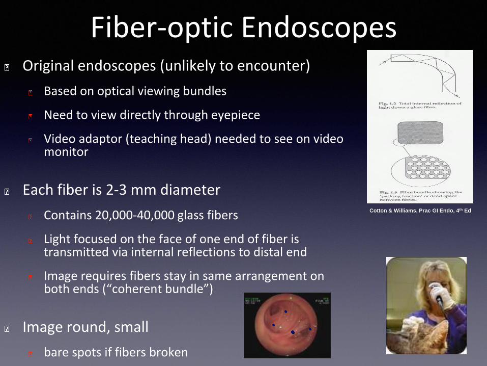
Fiber-optic EndoscopesOriginal endoscopes (unlikely to encounter)
Based on optical viewing bundles
Need to view directly through eyepiece
Video adaptor (teaching head) needed to see on video monitor
Each fiber is 2-3 mm diameter
Contains 20,000-40,000 glass fibers
Light focused on the face of one end of fiber is transmitted via internal reflections to distal end
Image requires fibers stay in same arrangement on both ends (“coherent bundle”)
Image round, small
bare spots if fibers broken
Cotton & Williams, Prac GI Endo, 4th Ed
. ...

Video-Endoscopes “Modern Endoscopes”
Video chips
Individual photocells (pixels)
Each cell receives photons reflected from white light reflecting from mucosa→black/white signal
Sensor that converts light into electrical
charges

Image Generation• Insertion tube tip contains a digital imager for color image
generation
- Charge- coupled device (CCD)
- Complementary Metal-Oxide Semiconductor (CMOS)
• CCD and CMOS image sensors convert light into
electrons
- Light converted to electricity
• Standard resolution of 400,000 pixels
• High-resolution endoscopes with high-density CCD sensors (600,000–1,400,000+ pixels)
- Provide image enlargement up to 100+ times compared with 30 times of standard endoscopes

Video-Endoscopes (“Modern Endoscopes”)
LIGHT SOURCE and VIDEO PROCESSOR
High-Definition signal and monitors
Pure digital signals

Video-Endoscopes (“Modern Endoscopes”)
Color Video
RGB sequential videoscope
Light source has a pulsed or strobed light with color filter wheel with red, green, blue (RGB) filters
Color chip videoscope
Color filters bonded to surface of black/white CCD allow detection of red, green or blue (RGB) colors

Processor and Light Source

What is “White Balancing”?
Light emitted from endoscope varies based on lamps and fiber bundle
“White Balancing” allows the processor to adjust signal interpretation for accurate color imaging
Point endoscope tip towards white object and press “white balance” button

Scope Anatomy
Umbilicus /
Connectors
Insertion Tube
Handle

Scope Tip in Cross Section

Endoscope Tip Deflection
EGD scopes
Up/Down 210º/90º
Left/Right 100º/100º
Colonoscopes
Up/Down 180º/ 180º
Left/Right 160º/ 160º

Air – Water - Suction
FYI: May use CO2 instead of room air.
Cleared from colon 100 x faster than room air - absorbed and exhaled
May result in improved patient satisfaction and fewer complications

Irrigation Channel
Auxiliary Water Inlet

Irrigation Pump

CO2 Insufflation

Check list before starting each endoscopy…
Air
Water
Suction
Image on screen (brightness)
Scope white balanced
Irrigation available

General GI Endoscopes
Esophago-gastro-duodenoscope (EGD)
Diagnostic
Therapeutic
Single Channel
Double channel
Extra large Channel
Ultra-thin
Colonoscopes
Diagnostic
Therapeutic
Pediatric
Variable stiffness

Specialized GI Endoscopes
Duodenoscope (ERCP)
Side viewing to see ampulla
Elevator to move accessories
Enteroscopes
Long scopes (250 cm)
Double Balloon scope
Endoscopic Ultrasound
Radial scanning
Linear array (FNA)
• Choledochoscope
Fits through duodenoscope
Scope and Biopsy Channel Diameters
Scope ModelOuter
Diameter
Accessory
Channel
Diameter
Accessory
Diameter
EGD
(110-135 cm)
Ultrathin
(trans-nasal)
4.9-5.4
mm2.0 mm
1.8 mm
(peds)
Diagnostic 9.5 mm 2.8 mm2.4 mm
(regular)
Therapeutic
(single channel)11.3 3.7 mm
3.4 mm
(jumbo)
Therapeutic
(double channel)12.6 3.7 mm
3.4 mm
(jumbo)
Giant Therapeutic
(vacuum cleaner)12.9 6 mm
5.6 mm
(giant jumbo)
Colonoscope
(133-168 cm)Pediatric 11.5 3.2 mm
2.4 mm
(regular)
Diagnostic 12.8 3.7 mm3.4 mm
(jumbo)
Therapeutic 13.7 4.2 mm3.4 mm
(jumbo)

Accessories
• Biopsy forceps
• Polypectomy Snares
• Retrieval baskets
• Endoclips
• EndoLoops
• Injection Needles
• Tattoo
• Overtube
• Contrast Dyes

Biopsy Forceps
Giant Jumbo/Jumbo/Regular/Pediatric
Jumbo give greater amount of mucosa, but not necessarily deeper
“bigger is better if tissue is the issue”
“Bite-on-bite”/”multi-bite” versus individual bites?
Spike or no-spike?

Biopsy Forceps
Giant Jumbo Jumbo Regular Pediatric
Jaw
Opening12 mm 9 mm 7 mm 5 mm
Diameter 5.5 mm 3.2 mm 2.3 mm 1.8 mm

These are NOT Biopsy Forceps
“Rat Tooth” Forceps for Object Removal – NOT BIOPSY
(These can perforate the GI tract!)

Snare
Polyfilament, braided wire
Generally use “hot” monopolar snare to cauterize the polyp stalk/base to help remove and prevent bleeding
“Cold” or “guillotine” snare can be used for smaller polyps
‣ Usually for polyps < 6 mm

Retrieval Net
Allows object to be grabbed in net for removal (i.e. polyp or foreign body)

Endoclips

EndoLoops
PolyLoop

Open Closed
Ovesco: Over The Scope Clip Clipl

Ovesco: Over The Scope Clip Clipl

Sclerotherapy Needle
Used for submucosal injections
Tattoo, endoscopic mucosal resection, epinephrine for bleeding
Injection of varices
Usually 22g-25g
Needle protrudes 5 mm

Mucosal Tattoo
Goal to identify an area or lesion at a later time either by endoscopy or surgery
Permanent ink (i.e. SPOT® tattoo or India Ink)
Must be sterile – otherwise risk of infection
Submucosal injection using sclerotherapy needle
Ideally place small marks circumferentially around lesion
Well placed injection should be visible from serosal side
Avoid injecting too deeply or else will have dye throughout the peritoneal cavity
tattoo
bleeding tic

Biopsy/Suction Channel in Lower Half of Screen6-7 o’clock corner of the screen
*Position lesions to suction or biopsy in this area*
7
1
3
6
9

OvertubeLarge tube placed into UGI tract through which scope can pass
Esophageal or gastric lengths
Use special care not to perforate esophagus when placing
Consider placing over 54 Fr. Maloney or Guardus overtube
Indications
Protects airway and esophagus when removing foreign bodies
Allows frequent passage of scope during EMR
Help keep stomach straight during enteroscopy

Disposable versus Reusable Accessories
Disposable accessories
‣ Specified by manufactures to be “single use only”
‣ Easy to use for staff
‣ Guaranteed sterile
Reusable accessories
‣ Should be labeled such by company
‣ Potentially less expensive
‣ Environmentally friendly
‣ Difficult to clean
‣ Potential risk to staff when cleaning
‣ Don’t throw away after using!

Improved Lens Positioning
Endoscopic Image EnhancementImage processing improves a given image
Image AnalysisAnalysis of a stored image and extraction of
characteristics in numerical parameters for subsequent reconstruction
How Can We See More?

Pentax Retroview®

Third Eye Panoramic®

“Full Spectrum”

Spreading of the Folds
EndoCuff EndoRings
G-Eye Balloon
Cap
Third-Eye

Image Enhancement Techniques to Improve Visualization of Mucosal Surface
High-resolution and high-definition endoscopy
Magnification endoscopy
Chromoendoscopy
Magnification chromoendoscopy
Narrow band imaging
Confocal Laser Endomicroscopy
Image Enhancement Techniques to Improve Visualization of Mucosal Surface

1
Microvascular Architecture
40x 80x
Enhanced microvascular architecture
Magnification Endoscopy
1 mm 1 mm

Contrast stainsIndigo carmine - pools in mucosal grooves, pits, and depressed
areas
Absorbed (Vital) stains Lugol solution - stains glycogen in squamous epithelium
Methylene blue - actively absorbed by intestinal epithelium
Cresyl violet - stains gastric and intestinal epithelium
Acetic acidOpacifies the mucosal layer
Chromoendoscopy

Magnification Chromoendoscopy
• Gastrointestinal epithelium is translucent
• Light beam reaches the subepithelial vascular network
• Absorption spectrum of hemoglobin creates red appearance
Opacification of mucosal surface allows visualization of mucosal pit pattern.

Kudo Pit Patterns
Diagnosis of colorectal
tumorous lesions by
magnifying endoscopy.
Kudo S, Tamura S,
Nakajima T, Yamano H,
Kusaka H, Watanabe H

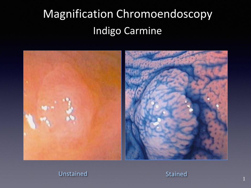
Indigo Carmine (0.4%)
Pools in mucosal grooves, pits, and depressed areas
Highlights details of mucosal surface
Conventional Chromoendoscopy
Unstained Stained
Contrast stain not absorbed by intestinal mucosa

Lugol Solution (1% - 2%)
Conventional Chromoendoscopy
Squamous-columnar junction
• Negative staining on nodular mucosa
• Biopsy consistent with Barrett’s esophagus with mild dysplasia

Methylene Blue Facilitates Diagnosis of Barrett’s Esophagus
Canto MI et al. Gastrointest Endoscopy 1996
Conventional Chromoendoscopy
Unstained Stained

Colonic Adenoma + MB

Eleview
Sustained Lifting and Staining
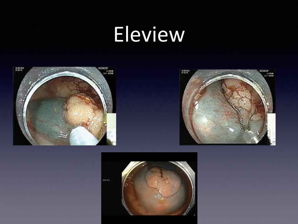
Eleview
1
Indigo Carmine
Magnification Chromoendoscopy
Unstained Stained

Cresyl Violet (0.25% - 0.5%)
Normal gastric mucosa Intestinal metaplasia in Barrett’s esophagus
Magnification Chromoendoscopy
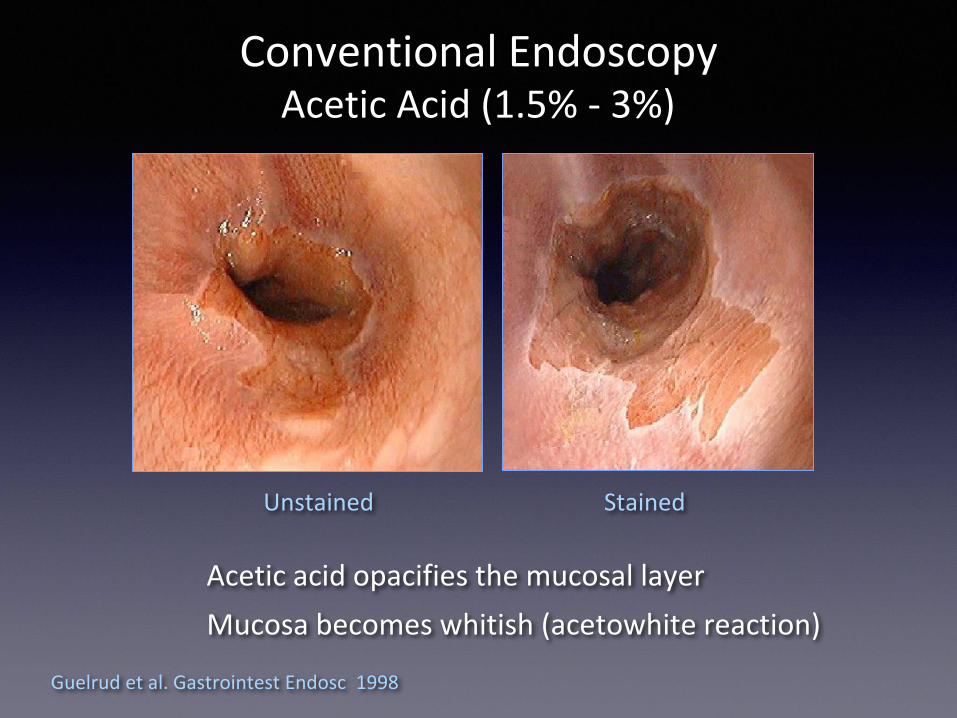
Acetic Acid (1.5% - 3%)
Unstained Stained
Guelrud et al. Gastrointest Endosc 1998
Conventional Endoscopy
Acetic acid opacifies the mucosal layer
Mucosa becomes whitish (acetowhite reaction)

Acetic Acid with Magnification(Enhanced Magnification Endoscopy)
Magnification Chromoendoscopy
Unstained
Stained
Mag 40 x Mag 80x
Mucosal Pattern
SurfaceVascular
ArchitectureNo magnification
Unstained

Narrow Band Imaging (NBI)
Gono, K et al. J Biomed Opt 2004Machida H et al. Endoscopy 2004
NBI
Conventional
• Filters decrease the red light, allowing only narrow band of blue light and green light to illuminate the mucosal surface
• The system of NBI uses blue narrow band light (390-445 nm) and green narrow band light (530-550 nm)
•Blue light (short wavelength) that penetrates most superficially
•Green light has deeper penetration
•NBI improves the image of the mucosal surface patterns and highlights vasculature
400 450 500 550

•Hemoglobin absorbs blue light
•Superficial vessels appear black
•Deeper subepithelial veins appear cyan
Narrow Band Imaging (NBI)
Blue light penetrates
only superficially
Green light has deep
penetration
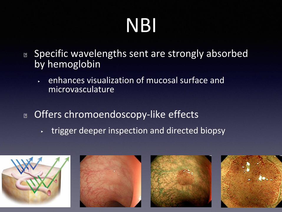
NBISpecific wavelengths sent are strongly absorbed by hemoglobin
‣ enhances visualization of mucosal surface and microvasculature
Offers chromoendoscopy-like effects
‣ trigger deeper inspection and directed biopsy

NBI Helps to Define Pallisade Vessels*
Magnification NBI Endoscopy
*landmark of the esophagogastric junction

Narrow Band Imaging
Magnification Magnification with NBI
Magnification NBI Endoscopy

OE Optical Enhancement
How does it work?
▪ Optical filters create band limited light that focuses on the color
spectrum associated with hemoglobin absorption. This provides
an enhanced view of fine vascular and mucosal structures
▪ OE Mode 1 designed to improve visualization of microvessels
with a sufficient amount of light
▪ OE Mode 1 focuses on a similar light spectrum to NBI, but
allows more white light to improve brightness
▪ OE mode 2 is designed to improve contrast of white-light
observation by bringing the color tone of the overall image
closer to that of natural color
OE Optical Enhancement modes
i-SCAN and OE image enhancement technologies are intended to be used as an optional adjunct following traditional white light endoscopy and is not intended to replace
histopathological sampling. i-SCAN and OE are compatible with PENTAX Medical video gastrointestinal endoscopes

Flexible spectral Imaging Color Enhancement (FICE)

Mucosal Pattern Enhancement
• NBI, OE & FICE
• Specific color wavelengths and light frequencies enhance blood vessels & fine capillary patterns in the mucous membranes

Probe Based Confocal Laser Endomicroscopy
• pCLE: miniprobe (GastroFlex UHD, Cellvizio; Mauna Kea Technologies, Paris, France)
• Low-power laser is focused onto a single point within the tissue
• Light emanating from this point is focused through a pinhole to a detector
• Laser raster scans the two-dimensional imaging plane
• Field of view of 240 μm, lateral resolution of 1 μm
• Imaging depth of 60 μm below the tissue surface
• Injection of sodium fluorescein (2.5 mL, 10%)
• Targeted biopsySharma 2011 et al. Gastrointestinal Endoscopy 2011 74: 3, 465-472

Source: Gastrointestinal Endoscopy 2011; 74:465-472 (DOI:10.1016/j.gie.2011.04.004 )
Copyright © 2011 American Society for Gastrointestinal Endoscopy Terms and Conditions
Probe-based
Confocal Laser
Endomicroscopy
BE with early esophageal
adenocarcinoma
Nondysplastic Barrett’s esophagus
(BE)

Nvision VLE OCT* Imaging System• Evaluation of the esophageal
tissue microstructure
• Analogous to B-mode ultrasound, using light instead of sound
• Low coherence near-infrared light pointed at tissue
• Cross sectional scanning through a 6cm length and 3mm depth
• Visualizes the squamous mucosa, submucosa, and muscularis propria
• Improved biopsy targeting*Volumetric Laser Endomicroscopy
Optical Coherence Tomography

Nvision VLE OCT Imaging System
• Real time image capture, 3mm beneath the mucosa at a 7 micron resolution
• Full-field view (~10,000mm2) vs. “point” image typically obtained with confocal microscopy (0.25mm2)
• 25X higher resolution than endoscopic ultrasound

Conclusions
You are responsible for understanding how to use your equipment and accessories
‣ Sole dependency on your nurses or techs is risky
Constantly educate yourself and your staff
Keep current and stay sharp!

Good Luck!