gait training after stroke with a wearable robotic device
TRANSCRIPT
1
Key Words: electromyography; gait endurance; Honda Walking Assist Device®; robot assisted gait training
INTRODUCTION
Improving walking ability is a high priority for individuals with stroke; it is also one of the main goals of rehabilita-tion.1,2) Conventional rehabilitation is known to improve walking ability, but its effectiveness is often limited3,4)
because the recovery of walking ability in stroke patients reaches a plateau at 115) to 176) weeks after onset. Despite having completed rehabilitation, many stroke patients have residual gait disturbance that seriously limits their activities of daily living (ADL) and their social involvement 7,8) It is, therefore, necessary to develop ways to further improve the
Received: May 11, 2021, Accepted: August 25, 2021, Published online: September 16, 2021aDepartment of Physical Therapy, Ibaraki Prefectural University of Health Sciences Hospital, Ibaraki, JapanbDepartment of Physical Therapy, School of Health Sciences, Ibaraki Prefectural University of Health Sciences, Ibaraki, JapancCenter for Medical Sciences, Ibaraki Prefectural University of Health Sciences, Ibaraki, JapandDepartment of Cognitive Behavioral Physiology, Chiba University Graduate School of Medicine, Chiba, Japan.Correspondence: Yutaka Kohno, MD, PhD, 4669-2 Ami, Ami-machi, Inashiki-gun, Ibaraki 300-0394, Japan, E-mail: [email protected] © 2021 The Japanese Association of Rehabilitation Medicine
This is an open-access article distributed under the terms of the Creative Commons Attribution Non-Commercial No Derivatives (CC BY-NC-ND) 4.0 License. http://creativecommons.org/licenses/by-nc-nd/4.0/
Progress in Rehabilitation Medicine 2021; Vol. 6, 20210037doi: 10.2490/prm.20210037
Background: Conventional rehabilitation is known to improve walking ability after stoke, but its effectiveness is often limited. Recent studies have shown that gait training combining conventional rehabilitation and robotic devices in stroke patients provides better results than conventional rehabilitation alone, suggesting that gait training with a robotic device may lead to further improvements in the walking ability recovered by conventional rehabilitation. Therefore, the aim of this report was to highlight the changes in kinematic and electromyographic data recorded during walking before and after gait training with the Honda Walking Assist Device® (HWAT) in a male patient whose walking speed had reached a recovery plateau under conven-tional rehabilitation. Case: The patient was a 42-year-old man with severe hemiplegia caused by right putaminal hemorrhage. He underwent conventional rehabilitation for 20 weeks following the onset of stroke, after which his walking speed reached a recovery plateau. Subsequently, we added robotic rehabilitation using HWAT to his regular rehabilitation regimen, which resulted in improved step length symmetry and gait endurance. We also noted changes in muscle activity pat-terns during walking. Discussion: HWAT further improved the walking ability of a patient who had recovered with conventional rehabilitation; this improvement was accompanied by changes in muscle activity patterns during walking. The improvement in gait endurance exceeded the smallest meaningful change in stroke patients, suggesting that this improvement represented a noticeable enhancement in the quality of life in relation to mobility in the community. Further clinical trials are needed to confirm the results of the present case study.
CASE REPORTGait Training after Stroke with a Wearable Robotic Device: A Case Report of Further Improvements in Walking Ability
after a Recovery PlateauKiyoshige Ishibashi, PT, MSc a Kenichi Yoshikawa, PT, PhD a Kazunori Koseki, PT, MSc a
Toshiyuki Aoyama, PT, PhD b Daisuke Ishii, OT, PhD c,d Satoshi Yamamoto, PT, PhD b Tomoyuki Matsuda, PT, PhD b Kazuhide Tomita, PT, PhD b Hirotaka Mutsuzaki, MD, PhD c
and Yutaka Kohno, MD, PhD c
Copyright © 2021 The Japanese Association of Rehabilitation Medicine
walking ability that has been recovered by conventional rehabilitation. Not only that, but it has also been shown that gait training that combines conventional rehabilitation and robotic devices in stroke patients provides better results than conventional rehabilitation alone.9,10)
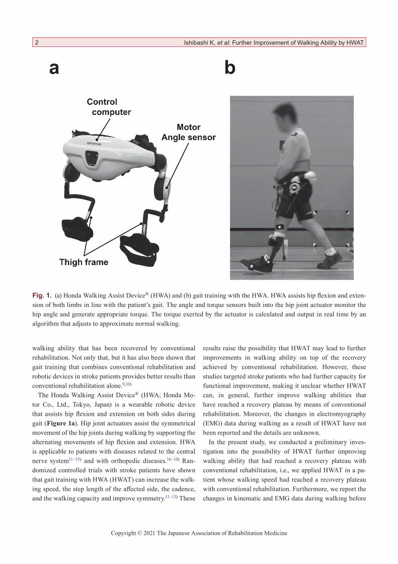
The Honda Walking Assist Device® (HWA; Honda Mo-tor Co., Ltd., Tokyo, Japan) is a wearable robotic device that assists hip flexion and extension on both sides during gait (Figure 1a). Hip joint actuators assist the symmetrical movement of the hip joints during walking by supporting the alternating movements of hip flexion and extension. HWA is applicable to patients with diseases related to the central nerve system11–15) and with orthopedic diseases.16–18) Ran-domized controlled trials with stroke patients have shown that gait training with HWA (HWAT) can increase the walk-ing speed, the step length of the affected side, the cadence, and the walking capacity and improve symmetry.11–13) These
results raise the possibility that HWAT may lead to further improvements in walking ability on top of the recovery achieved by conventional rehabilitation. However, these studies targeted stroke patients who had further capacity for functional improvement, making it unclear whether HWAT can, in general, further improve walking abilities that have reached a recovery plateau by means of conventional rehabilitation. Moreover, the changes in electromyography (EMG) data during walking as a result of HWAT have not been reported and the details are unknown.
In the present study, we conducted a preliminary inves-tigation into the possibility of HWAT further improving walking ability that had reached a recovery plateau with conventional rehabilitation, i.e., we applied HWAT in a pa-tient whose walking speed had reached a recovery plateau with conventional rehabilitation. Furthermore, we report the changes in kinematic and EMG data during walking before
2 Ishibashi K, et al: Further Improvement of Walking Ability by HWAT
Fig. 1. (a) Honda Walking Assist Device® (HWA) and (b) gait training with the HWA. HWA assists hip flexion and exten-sion of both limbs in line with the patient’s gait. The angle and torque sensors built into the hip joint actuator monitor the hip angle and generate appropriate torque. The torque exerted by the actuator is calculated and output in real time by an algorithm that adjusts to approximate normal walking.
Copyright © 2021 The Japanese Association of Rehabilitation Medicine
and after HWAT.
CASE
A 42-year-old man with sudden weakness of his left limbs and gait disturbance was diagnosed with right putaminal hemorrhage based on a head computed tomography image in an emergency hospital. He received conservative treat-ment and was admitted to our hospital for rehabilitation 41 days after stroke onset. The patient’s physical function was characterized by severe motor paralysis of the left upper and lower limbs, with motor function classified as 0 on the Stroke Impairment Assessment Set (SIAS) and 16 on Fugl-Meyer Assessment (FMA) of the lower limbs. The deep tendon re-flexes were enhanced in the left upper and lower limbs. The sensory function on SIAS was 3 for the position of the upper extremity and 2 for the others, which indicated mild sensory disturbance. The patient also had difficulty walking, and the Functional Ambulation Categories showed 0 and his ADL ability was 82 on the Functional Independence Measure.
The patient performed 60–120 min of physical therapy and 40–60 min of occupational therapy each day. The former was carried out with the purpose of achieving independence
of ADL by attaining a stabilized mobility capability, and included stretching of the affected lower limb, gait train-ing using an orthosis, pedaling exercises, outdoor walking exercises, gait training without waking aids, and stair climb-ing exercises. The latter exercises were aimed at achieving independence of ADL and at the functional reconstruction of the affected upper limb; these exercises included ADL re-acquisition training, stretching of the affected upper limb, and a functional reconstruction program (i.e., peripheral nerve electrical stimulation therapy for the reconstruction of finger function and repetitive arm movement under active assistance).
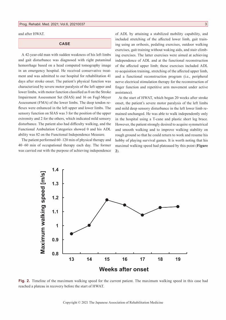
At the start of HWAT, which began 20 weeks after stroke onset, the patient’s severe motor paralysis of the left limbs and mild deep sensory disturbance in the left lower limb re-mained unchanged. He was able to walk independently only in the hospital using a T-cane and plastic short leg brace. However, the patient strongly desired to acquire symmetrical and smooth walking and to improve walking stability on rough ground so that he could return to work and resume his hobby of playing survival games. It is worth noting that his maximal walking speed had plateaued by this point (Figure 2).
Prog. Rehabil. Med. 2021; Vol.6, 20210037 3
Fig. 2. Timeline of the maximum walking speed for the current patient. The maximum walking speed in this case had reached a plateau in recovery before the start of HWAT.
Copyright © 2021 The Japanese Association of Rehabilitation Medicine
The present study was conducted in accordance with the Declaration of Helsinki and was approved by the Ethics Committee of the Ibaraki Prefectural University of Health Sciences (approval number: e204). Written informed consent was obtained from the patient for the publication of this case study and for the use of the accompanying images.
HWAT InterventionHWAT was conducted in parallel with conventional reha-
bilitation as part of physical therapy. The HWAT intervention was provided as an alternative to outdoor walking exercise. Other regimens, including occupational therapy, remained constant during the intervention. Each HWAT session in-volved a maximum net walking time of 20 min, excluding rest time, and was carried out once a day, 5 times a week for 4 weeks (20 sessions in total). At the start of each session, the torque assistance of HWAT was adjusted by the physiothera-pist based on patient feedback and visual motion analysis to ensure that walking was as comfortable and symmetrical as possible. HWAT was performed in the physical therapy room or in the corridors of the hospital with the patient using a T-cane and a plastic short leg brace (Figure 1b).
Outcome MeasurementThe FMA for motor paralysis of the affected lower limb,
the Modified Ashworth Scale (MAS) for spasticity, the 6-minute walking test (6MWT) for gait endurance, and the 10-m walking test (10mWT) for assessing overall walking ability were recorded before and after 20 sessions of HWAT. The 10mWT was performed at a comfortable speed three times at each time point; the walking speed and cadence were calculated as the average values of these three trials. In addition, the subject was asked to carry an activity meter from 2 weeks before the start of HWAT to assess changes in the daily stepping activity during the HWAT intervention.
The patient’s movements during 10mWT were recorded in the sagittal plane with a digital video camera (Handy-cam, Sony, Tokyo, Japan) to facilitate analysis of changes in gait pattern. The sampling frequency was 60 Hz, and two-dimensional motion analysis software (Frame Dias V, DKH, Tokyo, Japan) was used to analyze two consecutive gait cycles per trial (a total of six gait cycles at each time point). To ensure accurate analysis, we placed markers on the patient’s acromion, greater trochanter, lateral knee fissure, lateral malleolus (on the plastic short leg brace), and the fifth metatarsophalangeal base (on the shoe). We defined the trunk axis as the line that connects the acromion to the greater tro-chanter, the thigh axis as the line linking the greater trochan-
ter to the lateral knee fissure, the lower leg axis as the line connecting the lateral knee fissure to the lateral malleolus, and the plantar axis as the line connecting the lateral malleo-lus to the fifth metatarsophalangeal base. The angle between these axes was used to define the angle in the sagittal plane of the hip, knee, and ankle joints. However, calculating the correct angle for the ankle joint was challenging because the marker, which was placed on the shoe at the assumed loca-tion of the fifth metatarsophalangeal base, moved because of twisting between the foot and the brace and/or between the brace and the shoe during walking. To address this problem, we calibrated the obtained ankle joint angles by defining the ankle joint angle at the point when the lower leg axis was perpendicular to the floor in the mid-stance phase as 0°. Furthermore, the step length of the affected and nonaffected lower limbs was calculated as the distance between the rear end of the shoe on the opposite limb and the rear end of the shoe on the target limb. We also calculated the symmetry of the step lengths (step length of the nonaffected limb/step length of the affected limb).
EMG data were recorded during 10mWT with a sampling frequency of 2000 Hz and with bandpass filtering of 20–450 Hz from the gluteus maximus (Gmax), the proximal portion of the rectus femoris (RF-p), the distal portion of the rectus femoris (RF-d), the biceps femoris (BF), and the tibialis anterior (TA) of the affected lower limb (Trigno Lab; Delsys Inc., Boston, MA, USA). Surface EMG data have shown that RF-p is selectively activated during hip flexion; as a result, we employed RF-p as the muscle to indicate hip flexion.19,20) For analysis, we used the EMG data obtained for the same gait that was the target of the video analysis described above. The software program MATLAB (ver. R2020b; MathWorks, Natick, MA, USA) was used offline for the analysis, and the EMG data were debiased, rectified, integrated with a time constant of 200 ms, and then normalized to the EMG value at maximum voluntary contraction (MVC). Furthermore, the EMG data were segmented for each gait cycle, and each data set was interpolated to 200 data points.
Muscle synergy analysis has recently been applied to detect changes in the nervous system underlying the func-tional improvement achieved during rehabilitation after a stroke.21–24) Muscle synergy is a hypothesis for the manage-ment of the redundancy issue in multi-joint movement,25) and the physiological validity and robustness of muscle synergy analysis based on this hypothesis have been veri-fied in previous studies.26,27) Compared with previous EMG studies, which compare individual muscle activity patterns, muscle synergy analysis is able to provide a comprehensive
4 Ishibashi K, et al: Further Improvement of Walking Ability by HWAT
Copyright © 2021 The Japanese Association of Rehabilitation Medicine
assessment of movement coordination.28) Muscle synergy analysis was carried out in reference to the procedures used in previous studies relating to stroke patients.29–31) The EMG data were debiased, high-pass filtered (40 Hz) with a zero lag fourth-order Butterworth filter, rectified, and smoothed with a zero lag fourth-order low-pass (4 Hz) Butterworth filter. The post-filter EMG data were normalized to the EMG value at MVC. Furthermore, this EMG data was segmented ac-cording to each gait cycle, and each data set was interpolated to 200 data points. The nonnegative matrix factorization (NNMF) algorithm that we applied generates a matrix of muscle weightings (which represent coactive muscle group-ings) and a matrix of activation timing profiles that indicate at what time in the gait cycle the muscle groupings were active. The optimum number of modules was determined based on the variability accounted for (VAF) and is calcu-lated by the equation below, which uses the original EMG (EMGo) and the reconstructed EMG (EMGr) that employs the muscle weightings and the activation timing profiles that were calculated using the NNMF algorithm:
VAF = 1 − (EMGo − EMGr)2/EMGo2
The optimum number of modules was defined as the number of modules in which the calculated VAF exceeded 90% for the first time when the NNMF was performed by increasing the number of modules in order from 1.23,30,32)
Statistical AnalysesWe obtained data relating to the step length for both sides,
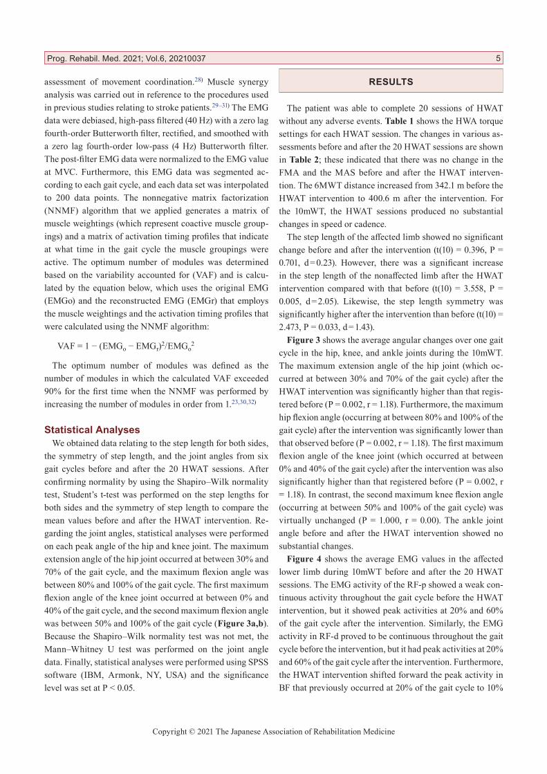
the symmetry of step length, and the joint angles from six gait cycles before and after the 20 HWAT sessions. After confirming normality by using the Shapiro–Wilk normality test, Student’s t-test was performed on the step lengths for both sides and the symmetry of step length to compare the mean values before and after the HWAT intervention. Re-garding the joint angles, statistical analyses were performed on each peak angle of the hip and knee joint. The maximum extension angle of the hip joint occurred at between 30% and 70% of the gait cycle, and the maximum flexion angle was between 80% and 100% of the gait cycle. The first maximum flexion angle of the knee joint occurred at between 0% and 40% of the gait cycle, and the second maximum flexion angle was between 50% and 100% of the gait cycle (Figure 3a,b). Because the Shapiro–Wilk normality test was not met, the Mann–Whitney U test was performed on the joint angle data. Finally, statistical analyses were performed using SPSS software (IBM, Armonk, NY, USA) and the significance level was set at P < 0.05.
RESULTS
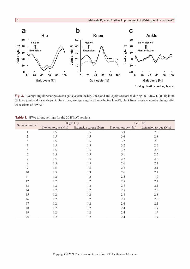
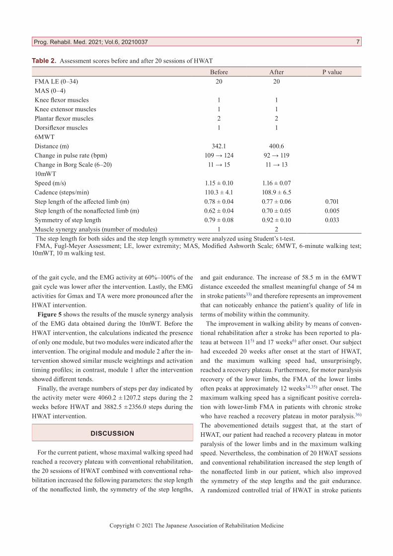
The patient was able to complete 20 sessions of HWAT without any adverse events. Table 1 shows the HWA torque settings for each HWAT session. The changes in various as-sessments before and after the 20 HWAT sessions are shown in Table 2; these indicated that there was no change in the FMA and the MAS before and after the HWAT interven-tion. The 6MWT distance increased from 342.1 m before the HWAT intervention to 400.6 m after the intervention. For the 10mWT, the HWAT sessions produced no substantial changes in speed or cadence.
The step length of the affected limb showed no significant change before and after the intervention (t(10) = 0.396, P = 0.701, d = 0.23). However, there was a significant increase in the step length of the nonaffected limb after the HWAT intervention compared with that before (t(10) = 3.558, P = 0.005, d = 2.05). Likewise, the step length symmetry was significantly higher after the intervention than before (t(10) = 2.473, P = 0.033, d = 1.43).
Figure 3 shows the average angular changes over one gait cycle in the hip, knee, and ankle joints during the 10mWT. The maximum extension angle of the hip joint (which oc-curred at between 30% and 70% of the gait cycle) after the HWAT intervention was significantly higher than that regis-tered before (P = 0.002, r = 1.18). Furthermore, the maximum hip flexion angle (occurring at between 80% and 100% of the gait cycle) after the intervention was significantly lower than that observed before (P = 0.002, r = 1.18). The first maximum flexion angle of the knee joint (which occurred at between 0% and 40% of the gait cycle) after the intervention was also significantly higher than that registered before (P = 0.002, r = 1.18). In contrast, the second maximum knee flexion angle (occurring at between 50% and 100% of the gait cycle) was virtually unchanged (P = 1.000, r = 0.00). The ankle joint angle before and after the HWAT intervention showed no substantial changes.
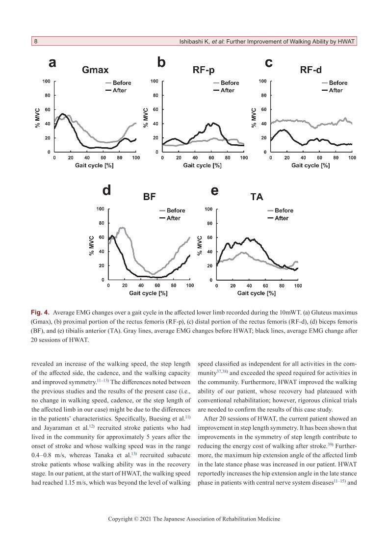
Figure 4 shows the average EMG values in the affected lower limb during 10mWT before and after the 20 HWAT sessions. The EMG activity of the RF-p showed a weak con-tinuous activity throughout the gait cycle before the HWAT intervention, but it showed peak activities at 20% and 60% of the gait cycle after the intervention. Similarly, the EMG activity in RF-d proved to be continuous throughout the gait cycle before the intervention, but it had peak activities at 20% and 60% of the gait cycle after the intervention. Furthermore, the HWAT intervention shifted forward the peak activity in BF that previously occurred at 20% of the gait cycle to 10%
Prog. Rehabil. Med. 2021; Vol.6, 20210037 5
Copyright © 2021 The Japanese Association of Rehabilitation Medicine
6 Ishibashi K, et al: Further Improvement of Walking Ability by HWAT
Fig. 3. Average angular changes over a gait cycle in the hip, knee, and ankle joints recorded during the 10mWT. (a) Hip joint, (b) knee joint, and (c) ankle joint. Gray lines, average angular change before HWAT; black lines, average angular change after 20 sessions of HWAT.
Table 1. HWA torque settings for the 20 HWAT sessions
Session numberRight Hip Left Hip
Flexion torque (Nm) Extension torque (Nm) Flexion torque (Nm) Extension torque (Nm)1 1.5 1.5 3.3 2.62 1.5 1.5 3.6 2.83 1.5 1.5 3.2 2.64 1.5 1.5 3.2 2.65 1.5 1.5 3.2 2.66 1.5 1.5 3.1 2.57 1.5 1.5 2.8 2.28 1.5 1.5 2.6 2.19 1.5 1.5 2.6 2.110 1.3 1.3 2.6 2.111 1.2 1.2 2.5 1.912 1.2 1.2 2.8 2.113 1.2 1.2 2.8 2.114 1.2 1.2 2.8 2.815 1.2 1.2 2.8 2.816 1.2 1.2 2.8 2.817 1.2 1.2 2.6 2.118 1.2 1.2 2.4 1.919 1.2 1.2 2.4 1.920 1.2 1.2 2.4 1.9
Copyright © 2021 The Japanese Association of Rehabilitation Medicine
of the gait cycle, and the EMG activity at 60%–100% of the gait cycle was lower after the intervention. Lastly, the EMG activities for Gmax and TA were more pronounced after the HWAT intervention.
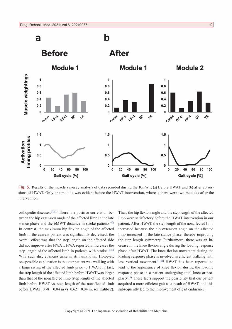
Figure 5 shows the results of the muscle synergy analysis of the EMG data obtained during the 10mWT. Before the HWAT intervention, the calculations indicated the presence of only one module, but two modules were indicated after the intervention. The original module and module 2 after the in-tervention showed similar muscle weightings and activation timing profiles; in contrast, module 1 after the intervention showed different tends.
Finally, the average numbers of steps per day indicated by the activity meter were 4060.2 ± 1207.2 steps during the 2 weeks before HWAT and 3882.5 ± 2356.0 steps during the HWAT intervention.
DISCUSSION
For the current patient, whose maximal walking speed had reached a recovery plateau with conventional rehabilitation, the 20 sessions of HWAT combined with conventional reha-bilitation increased the following parameters: the step length of the nonaffected limb, the symmetry of the step lengths,
and gait endurance. The increase of 58.5 m in the 6MWT distance exceeded the smallest meaningful change of 54 m in stroke patients33) and therefore represents an improvement that can noticeably enhance the patient’s quality of life in terms of mobility within the community.
The improvement in walking ability by means of conven-tional rehabilitation after a stroke has been reported to pla-teau at between 115) and 17 weeks6) after onset. Our subject had exceeded 20 weeks after onset at the start of HWAT, and the maximum walking speed had, unsurprisingly, reached a recovery plateau. Furthermore, for motor paralysis recovery of the lower limbs, the FMA of the lower limbs often peaks at approximately 12 weeks34,35) after onset. The maximum walking speed has a significant positive correla-tion with lower-limb FMA in patients with chronic stroke who have reached a recovery plateau in motor paralysis.36) The abovementioned details suggest that, at the start of HWAT, our patient had reached a recovery plateau in motor paralysis of the lower limbs and in the maximum walking speed. Nevertheless, the combination of 20 HWAT sessions and conventional rehabilitation increased the step length of the nonaffected limb in our patient, which also improved the symmetry of the step lengths and the gait endurance. A randomized controlled trial of HWAT in stroke patients
Prog. Rehabil. Med. 2021; Vol.6, 20210037 7
Table 2. Assessment scores before and after 20 sessions of HWAT
Before After P valueFMA LE (0–34) 20 20MAS (0–4)Knee flexor muscles 1 1Knee extensor muscles 1 1Plantar flexor muscles 2 2Dorsiflexor muscles 1 16MWTDistance (m) 342.1 400.6Change in pulse rate (bpm) 109 → 124 92 → 119Change in Borg Scale (6–20) 11 → 15 11 → 1310mWTSpeed (m/s) 1.15 ± 0.10 1.16 ± 0.07Cadence (steps/min) 110.3 ± 4.1 108.9 ± 6.5Step length of the affected limb (m) 0.78 ± 0.04 0.77 ± 0.06 0.701Step length of the nonaffected limb (m) 0.62 ± 0.04 0.70 ± 0.05 0.005Symmetry of step length 0.79 ± 0.08 0.92 ± 0.10 0.033Muscle synergy analysis (number of modules) 1 2The step length for both sides and the step length symmetry were analyzed using Student’s t-test.FMA, Fugl-Meyer Assessment; LE, lower extremity; MAS, Modified Ashworth Scale; 6MWT, 6-minute walking test;
10mWT, 10 m walking test.
Copyright © 2021 The Japanese Association of Rehabilitation Medicine
revealed an increase of the walking speed, the step length of the affected side, the cadence, and the walking capacity and improved symmetry.11–13) The differences noted between the previous studies and the results of the present case (i.e., no change in walking speed, cadence, or the step length of the affected limb in our case) might be due to the differences in the patients’ characteristics. Specifically, Buesing et al.11) and Jayaraman et al.12) recruited stroke patients who had lived in the community for approximately 5 years after the onset of stroke and whose walking speed was in the range 0.4–0.8 m/s, whereas Tanaka et al.13) recruited subacute stroke patients whose walking ability was in the recovery stage. In our patient, at the start of HWAT, the walking speed had reached 1.15 m/s, which was beyond the level of walking
speed classified as independent for all activities in the com-munity37,38) and exceeded the speed required for activities in the community. Furthermore, HWAT improved the walking ability of our patient, whose recovery had plateaued with conventional rehabilitation; however, rigorous clinical trials are needed to confirm the results of this case study.
After 20 sessions of HWAT, the current patient showed an improvement in step length symmetry. It has been shown that improvements in the symmetry of step length contribute to reducing the energy cost of walking after stroke.39) Further-more, the maximum hip extension angle of the affected limb in the late stance phase was increased in our patient. HWAT reportedly increases the hip extension angle in the late stance phase in patients with central nerve system diseases11–15) and
8 Ishibashi K, et al: Further Improvement of Walking Ability by HWAT
Fig. 4. Average EMG changes over a gait cycle in the affected lower limb recorded during the 10mWT. (a) Gluteus maximus (Gmax), (b) proximal portion of the rectus femoris (RF-p), (c) distal portion of the rectus femoris (RF-d), (d) biceps femoris (BF), and (e) tibialis anterior (TA). Gray lines, average EMG changes before HWAT; black lines, average EMG change after 20 sessions of HWAT.
Copyright © 2021 The Japanese Association of Rehabilitation Medicine
orthopedic diseases.17,18) There is a positive correlation be-tween the hip extension angle of the affected limb in the late stance phase and the 6MWT distance in stroke patients.40) In contrast, the maximum hip flexion angle of the affected limb in the current patient was significantly decreased; the overall effect was that the step length on the affected side did not improve after HWAT. HWA reportedly increases the step length of the affected limb in patients with stroke.11,13) Why such discrepancies arise is still unknown. However, one possible explanation is that our patient was walking with a large swing of the affected limb prior to HWAT. In fact, the step length of the affected limb before HWAT was larger than that of the nonaffected limb (step length of the affected limb before HWAT vs. step length of the nonaffected limb before HWAT: 0.78 ± 0.04 m vs. 0.62 ± 0.04 m, see Table 2).
Thus, the hip flexion angle and the step length of the affected limb were satisfactory before the HWAT intervention in our patient. After HWAT, the step length of the nonaffected limb increased because the hip extension angle on the affected limb increased in the late stance phase, thereby improving the step length symmetry. Furthermore, there was an in-crease in the knee flexion angle during the loading response phase after HWAT. The knee flexion movement during the loading response phase is involved in efficient walking with less vertical movement.41,42) HWAT has been reported to lead to the appearance of knee flexion during the loading response phase in a patient undergoing total knee arthro-plasty.16) These facts support the possibility that our patient acquired a more efficient gait as a result of HWAT, and this subsequently led to the improvement of gait endurance.
Prog. Rehabil. Med. 2021; Vol.6, 20210037 9
Fig. 5. Results of the muscle synergy analysis of data recorded during the 10mWT. (a) Before HWAT and (b) after 20 ses-sions of HWAT. Only one module was evident before the HWAT intervention, whereas there were two modules after the intervention.
Copyright © 2021 The Japanese Association of Rehabilitation Medicine
The pattern of muscle activity before and after the HWAT intervention showed substantial changes in RF-p, RF-d, and BF toward the muscle activity patterns normally seen in gait, i.e., the hip flexor muscles are active at about 10% and 70% of the gait cycle, the knee extensor muscles are active at about 10% and 60% of the gait cycle, and the knee flexor muscles are active at the beginning and after about 80% of the gait cycle.43,44) Furthermore, the muscle synergy analysis performed on EMG data from the 10mWT indicated that the number of modules during walking increased from 1 to 2 after HWAT. The change in the number of modules reflects a change in the relevant neural network,23) and there is an association between an increase in the number of modules and an improvement in walking ability in stroke patients.21) In healthy subjects, there are approximately four modules during walking, 45) whereas the modules are merged because of the co-contractions of several muscles in stroke patients, resulting in a decreased number of modules.30,46) In our case, the mechanism through which the muscle activities were able to change and the number of modules to increase after the intervention is unknown. However, HWA assisted the hip movements symmetrically and alternately during walk-ing, and the muscle activity patterns shifted to the patterns observed in normal gait without motor paralysis improve-ment. The central pattern generator (CPG) of the spinal cord, which is deeply involved in the generation of walking-specific muscle activities, is activated by hip movement.47) In particular, the activity of CPG is amplified by alternate hip movement rather than unilateral hip movement.48)
The HWA torques were set to decrease as the number of sessions progressed, although some variations were observed, as shown in Table 1. This setup was conducted according to the hypothesis that excessive assistance by a robotic device would reduce the patient’s effort and could be detrimental to motor recovery after stroke.49) However, the optimality of this setup remains uncertain. Nevertheless, in our case, HWAT was performed using this setup, and the abovementioned results were achieved.
There are several limitations to this case study. First, this is the report of a single case without a control group, which implies that the results of our case can in no way be general-ized, and future validation with similar patients is necessary. Second, it is unclear how long the changes obtained through HWAT intervention will be sustained from the follow-up on-wards. Third, the number of EMG channels recorded in our case was small because a brace was worn at the time of EMG recording. Consequently, we were not able to record data for the lower leg muscles, such as the soleus and gastrocnemius
muscle, which have an important function in walking.29)
In conclusion, 20 treatment sessions that included HWAT were completed by a patient whose walking speed had reached a recovery plateau after conventional rehabilitation. Consequently, we noted improvements in the maximum hip extension angle of the affected limb during the late stance phase, in the knee flexion angle of the affected limb during the loading response phase, in the symmetry of step length, and in gait endurance. These improvements were accompa-nied by changes in muscle activity patterns during walking. The improvement of gait endurance exceeded the smallest meaningful change in patients with stroke, indicating a sig-nificant enhancement in the patient’s quality of life in terms of mobility in the community. However, further clinical trials should be conducted to confirm the results of this case study.
ACKNOWLEDGMENTS
We would like to thank Keiko Miyamoto, robotic technical assistant, and all the physical therapists in the Department of Physical Therapy, Ibaraki Prefectural University of Health Sciences Hospital. This research was supported in part by grants from the Japan Society for the Promotion of Science (KAKENHI) (nos. 20H01140, 21H04305, 20H01147, and 19K19835) and a Grant-in-Aid for Project Research (no. 1962-1) from the Ibaraki Prefectural University of Health Sciences.
CONFLICTS OF INTEREST
The authors declare the following potential conflict of in-terest relating to the research, authorship, and/or publication of this article: Honda Motor Corporation lent us the HWA free of charge for this research.
REFERENCES
1. Eng JJ, Tang PF: Gait training strategies to optimize walking ability in people with stroke: a synthesis of the evidence. Expert Rev Neurother 2007;7:1417–1436. DOI:10.1586/14737175.7.10.1417, PMID:17939776
2. Lindquist AR, Prado CL, Barros RM, Mattioli R, da Costa PH, Salvini TF: Gait training combining par-tial body-weight support, a treadmill, and functional electrical stimulation: effects on poststroke gait. Phys Ther 2007;87:1144–1154. DOI:10.2522/ptj.20050384, PMID:17609334
10 Ishibashi K, et al: Further Improvement of Walking Ability by HWAT
Copyright © 2021 The Japanese Association of Rehabilitation Medicine
3. Langhorne P, Bernhardt J, Kwakkel G: Stroke reha-bilitation. Lancet 2011;377:1693–1702. DOI:10.1016/S0140-6736(11)60325-5, PMID:21571152
4. Dobkin BH: Clinical practice. Rehabilitation after stroke. N Engl J Med 2005;352:1677–1684. DOI:10.1056/NEJMcp043511, PMID:15843670
5. Jørgensen HS, Nakayama H, Raaschou HO, Olsen TS: Recovery of walking function in stroke patients: The Copenhagen Stroke Study. Arch Phys Med Rehabil 1995;76:27–32. DOI:10.1016/S0003-9993(95)80038-7, PMID:7811170
6. Smith J, Brotheridge S, Young J: Patterns of hemipare-sis recovery in lacunar and partial anterior circulation infarct stroke syndromes. Clin Rehabil 2001;15:59–66. DOI:10.1191/026921501668563820, PMID:11237163
7. Schaechter JD: Motor rehabilitation and brain plasticity after hemiparetic stroke. Prog Neurobiol 2004;73:61–72. DOI:10.1016/j.pneurobio.2004.04.001, PMID:15193779
8. Stamm BJ, Burke JF, Lin CC, Price RJ, Skolarus LE: Disability in community-dwelling older adults: exploring the role of stroke and dementia. J Prim Care Community Health 2019;10:2150132719852507. DOI:10.1177/2150132719852507, PMID:31185786
9. Bruni MF, Melegari C, De Cola MC, Bramanti A, Bramanti P, Calabrò RS: What does best evidence tell us about robotic gait rehabilitation in stroke patients: a systematic review and meta-analysis. J Clin Neu-rosci 2018;48:11–17. DOI:10.1016/j.jocn.2017.10.048, PMID:29208476
10. Hobbs B, Artemiadis P: A review of robot-assisted lower-limb stroke therapy: unexplored paths and future directions in gait rehabilitation. Front Neu-rorobot 2020;14:19. DOI:10.3389/fnbot.2020.00019, PMID:32351377
11. Buesing C, Fisch G, O’Donnell M, Shahidi I, Thomas L, Mummidisetty CK, Williams KJ, Takahashi H, Rymer WZ, Jayaraman A: Effects of a wearable exo-skeleton stride management assist system (SMA®) on spatiotemporal gait characteristics in individuals after stroke: a randomized controlled trial. J Neuroeng Rehabil 2015;12:69. DOI:10.1186/s12984-015-0062-0, PMID:26289955
12. Jayaraman A, O’Brien MK, Madhavan S, Mum-midisetty CK, Roth HR, Hohl K, Tapp A, Brennan K, Kocherginsky M, Williams KJ, Takahashi H, Rymer WZ: Stride management assist exoskeleton vs functional gait training in stroke: a randomized trial. Neurology 2019;92:e263–e273. DOI:10.1212/WNL.0000000000006782, PMID:30568009
13. Tanaka N, Matsushita S, Sonoda Y, Maruta Y, Fu-jitaka Y, Sato M, Simomori M, Onaka R, Harada K, Hirata T, Kinoshita S, Okamoto T, Okamura H: Effect of stride management assist gait training for poststroke hemiplegia: a single center, open-label, randomized controlled trial. J Stroke Cerebrovasc Dis 2019;28:477–486. DOI:10.1016/j.jstrokecerebrovasdis.2018.10.025, PMID:30420315
14. Kawasaki S, Ohata K, Yoshida T, Yokoyama A, Yamada S: Gait improvements by assisting hip move-ments with the robot in children with cerebral palsy: a pilot randomized controlled trial. J Neuroeng Re-habil 2020;17:87. DOI:10.1186/s12984-020-00712-3, PMID:32620131
15. Yoshikawa K, Mutsuzaki H, Koseki K, Endo Y, Hashi-zume Y, Nakazawa R, Aoyama T, Yozu A, Kohno Y: Gait training using a wearable robotic device for non-traumatic spinal cord injury: a case report. Geri-atr Orthop Surg Rehabil 2020;11:2151459320956960. DOI:10.1177/2151459320956960, PMID:33194254
16. Koseki K, Mutsuzaki H, Yoshikawa K, Endo Y, Kanaza-wa A, Nakazawa R, Fukaya T, Aoyama T, Kohno Y: Gait training using a hip-wearable robotic exoskeleton after total knee arthroplasty: a case report. Geriatr Orthop Surg Rehabil 2020;11:2151459320966483. DOI:10.1177/2151459320966483, PMID:33194256
17. Koseki K, Mutsuzaki H, Yoshikawa K, Endo Y, Maeza-wa T, Takano H, Yozu A, Kohno Y: Gait training using the Honda Walking Assistive Device® in a patient who underwent total hip arthroplasty: a single-subject study. Medicina (B Aires) 2019;55:69. DOI:10.3390/medicina55030069, PMID:30875846
18. Koseki K, Yozu A, Takano H, Abe A, Yoshikawa K, Maezawa T, Kohno Y, Mutsuzaki H: Gait training us-ing the Honda Walking Assist Device® for individuals with transfemoral amputation: a report of two cases. J Back Musculoskeletal Rehabil 2020;33:339–344. DOI:10.3233/BMR-191726, PMID:31929139
Prog. Rehabil. Med. 2021; Vol.6, 20210037 11
Copyright © 2021 The Japanese Association of Rehabilitation Medicine
19. Watanabe K, Kouzaki M, Moritani T: Task-dependent spatial distribution of neural activation pattern in hu-man rectus femoris muscle. J Electromyogr Kinesiol 2012;22:251–258. DOI:10.1016/j.jelekin.2011.11.004, PMID:22153052
20. Watanabe K, Kouzaki M, Moritani T: Regional neu-romuscular regulation within human rectus femoris muscle during gait. J Biomech 2014;47:3502–3508. DOI:10.1016/j.jbiomech.2014.09.001, PMID:25246002
21. Routson RL, Clark DJ, Bowden MG, Kautz SA, Neptune RR: The influence of locomotor rehabilita-tion on module quality and post-stroke hemiparetic walking performance. Gait Posture 2013;38:511–517. DOI:10.1016/j.gaitpost.2013.01.020, PMID:23489952
22. Ferrante S, Chia Bejarano N, Ambrosini E, Nardone A, Turcato AM, Monticone M, Ferrigno G, Pedrocchi A: A personalized multi-channel FES controller based on muscle synergies to support gait rehabilitation af-ter stroke. Front Neurosci 2016;10:425. DOI:10.3389/fnins.2016.00425, PMID:27695397
23. Hashiguchi Y, Ohata K, Kitatani R, Yamakami N, Sakuma K, Osako S, Aga Y, Watanabe A, Yamada S: Merging and fractionation of muscle synergy indicate the recovery process in patients with hemiplegia: the first study of patients after subacute stroke. Neural Plast 2016;2016:1–7. DOI:10.1155/2016/5282957, PMID:28090358
24. Tan CK, Kadone H, Watanabe H, Marushima A, Hada Y, Yamazaki M, Sankai Y, Matsumura A, Suzuki K: Differences in muscle synergy symmetry between subacute post-stroke patients with bioelectrically-controlled exoskeleton gait training and conventional gait training. Front Bioeng Biotechnol 2020;8:770. DOI:10.3389/fbioe.2020.00770, PMID:32850696
25. d’Avella A, Saltiel P, Bizzi E: Combinations of muscle synergies in the construction of a natural motor be-havior. Nat Neurosci 2003;6:300–308. DOI:10.1038/nn1010, PMID:12563264
26. Ivanenko YP, Cappellini G, Dominici N, Poppele RE, Lacquaniti F: Coordination of locomotion with volun-tary movements in humans. J Neurosci 2005;25:7238–7253. DOI:10.1523/JNEUROSCI.1327-05.2005, PMID:16079406
27. Muceli S, Jiang N, Farina D: Extracting signals robust to electrode number and shift for online simultaneous and proportional myoelectric control by factorization algorithms. IEEE Trans Neural Syst Rehabil Eng 2014;22:623–633. DOI:10.1109/TNSRE.2013.2282898, PMID:24132017
28. Safavynia S, Torres-Oviedo G, Ting L: Muscle synergies: implications for clinical evaluation and rehabilitation of movement. Top Spinal Cord Inj Rehabil 2011;17:16–24. DOI:10.1310/sci1701-16, PMID:21796239
29. Allen JL, Kautz SA, Neptune RR: The influence of merged muscle excitation modules on post-stroke hemiparetic walking performance. Clin Biomech (Bristol, Avon) 2013;28:697–704. DOI:10.1016/j.clin-biomech.2013.06.003, PMID:23830138
30. Clark DJ, Ting LH, Zajac FE, Neptune RR, Kautz SA: Merging of healthy motor modules predicts reduced locomotor performance and muscle coordination com-plexity post-stroke. J Neurophysiol 2010;103:844–857. DOI:10.1152/jn.00825.2009, PMID:20007501
31. Kautz SA, Bowden MG, Clark DJ, Neptune RR: Comparison of motor control deficits dur-ing treadmill and overground walking poststroke. Neurorehabil Neural Repair 2011;25:756–765. DOI:10.1177/1545968311407515, PMID:21636831
32. Barroso FO, Torricelli D, Molina-Rueda F, Alguacil-Diego IM, Cano-de-la-Cuerda R, Santos C, Moreno JC, Miangolarra-Page JC, Pons JL: Combining muscle synergies and biomechanical analysis to as-sess gait in stroke patients. J Biomech 2017;63:98–103. DOI:10.1016/j.jbiomech.2017.08.006, PMID:28882330
33. Perera S, Mody SH, Woodman RC, Studenski SA: Meaningful change and responsiveness in common physical performance measures in older adults. J Am Geriatr Soc 2006;54:743–749. DOI:10.1111/j.1532-5415.2006.00701.x, PMID:16696738
34. Verheyden G, Nieuwboer A, De Wit L, Thijs V, Dobbelaere J, Devos H, Severijns D, Vanbeveren S, De Weerdt W: Time course of trunk, arm, leg, and functional recovery after ischemic stroke. Neurorehabil Neural Repair 2008;22:173–179. DOI:10.1177/1545968307305456, PMID:17876069
35. Duncan PW, Goldstein LB, Horner RD, Landsman PB, Samsa GP, Matchar DB: Similar motor recovery of upper and lower extremities after stroke. Stroke 1994;25:1181–1188. DOI:10.1161/01.STR.25.6.1181, PMID:8202977
12 Ishibashi K, et al: Further Improvement of Walking Ability by HWAT
Copyright © 2021 The Japanese Association of Rehabilitation Medicine
36. Nadeau S, Arsenault AB, Gravel D, Bourbonnais D: Analysis of the clinical factors determining natural and maximal gait speeds in adults with a stroke. Am J Phys Med Rehabil 1999;78:123–130. DOI:10.1097/00002060-199903000-00007, PMID:10088586
37. Perry J, Garrett M, Gronley JK, Mulroy SJ: Classifi-cation of walking handicap in the stroke population. Stroke 1995;26:982–989. DOI:10.1161/01.STR.26.6.982, PMID:7762050
38. Schmid A, Duncan PW, Studenski S, Lai SM, Richards L, Perera S, Wu SS: Improvements in speed-based gait classifications are meaningful. Stroke 2007;38:2096–2100. DOI:10.1161/STROKEAHA.106.475921, PMID:17510461
39. Awad LN, Palmer JA, Pohlig RT, Binder-Macleod SA, Reisman DS: Walking speed and step length asymmetry modify the energy cost of walking after stroke. Neurorehabil Neural Repair 2015;29:416–423. DOI:10.1177/1545968314552528, PMID:25288581
40. Awad LN, Binder-Macleod SA, Pohlig RT, Reisman DS: Paretic propulsion and trailing limb angle are key determinants of long-distance walking function after stroke. Neurorehabil Neural Repair 2015;29:499–508. DOI:10.1177/1545968314554625, PMID:25385764
41. Inman VT: Human locomotion. Can Med Assoc J 1966;94:1047–1054. PMID:5942660
42. Lehmann JF, de Lateur BJ, Price R: Biomechan-ics of abnormal gait. Phys Med Rehabil Clin N Am 1992;3:125–138. DOI:10.1016/S1047-9651(18)30668-5
43. Winter DA, Yack HJ: EMG profiles during normal human walking: stride-to-stride and inter-subject variability. Electroencephalogr Clin Neurophysiol 1987;67:402–411. DOI:10.1016/0013-4694(87)90003-4, PMID:2444408
44. Ivanenko YP, Poppele RE, Lacquaniti F: Five basic muscle activation patterns account for muscle activity during human locomotion. J Physiol 2004;556:267–282. DOI:10.1113/jphysiol.2003.057174, PMID:14724214
45. Ting LH, Chiel HJ, Trumbower RD, Allen JL, McKay JL, Hackney ME, Kesar TM: Neuromechanical prin-ciples underlying movement modularity and their implications for rehabilitation. Neuron 2015;86:38–54. DOI:10.1016/j.neuron.2015.02.042, PMID:25856485
46. Van Criekinge T, Vermeulen J, Wagemans K, Schröder J, Embrechts E, Truijen S, Hallemans A, Saeys W: Lower limb muscle synergies during walk-ing after stroke: a systematic review. Disabil Rehabil 2020;42:2836–2845. DOI:10.1080/09638288.2019.1578421, PMID:30905215
47. Dietz V, Müller R, Colombo G: Locomotor activ-ity in spinal man: significance of afferent input from joint and load receptors. Brain 2002;125:2626–2634. DOI:10.1093/brain/awf273, PMID:12429590
48. Kawashima N, Nozaki D, Abe MO, Akai M, Nakazawa K: Alternate leg movement amplifies locomotor-like muscle activity in spinal cord injured persons. J Neuro-physiol 2005;93:777–785. DOI:10.1152/jn.00817.2004, PMID:15385590
49. Hornby TG, Campbell DD, Kahn JH, Demott T, Moore JL, Roth HR: Enhanced gait-related improvements after therapist- versus robotic-assisted locomotor train-ing in subjects with chronic stroke: a randomized con-trolled study. Stroke 2008;39:1786–1792. DOI:10.1161/STROKEAHA.107.504779, PMID:18467648
Prog. Rehabil. Med. 2021; Vol.6, 20210037 13