galanin stimulates ca2+mobilization, inositol...
TRANSCRIPT
[CANCER RESEARCH 5l. 1674-1679. March 15. 1
Galanin Stimulates Ca2+Mobilization, Inositol Phosphate Accumulation, and
Clonal Growth in Small Cell Lung Cancer CellsTariq Sethi and Enrique Rozengurt'Imperial Cancer Research Fund, P. O. Box 123, Lincoln's Inn Fields. London H'C2.4 3P\. L'nited Kingdom
ABSTRACT
Addition of the neuropeptidc galanin to small cell lung cancer (SCI.C)cells loaded with the fluorescent Ca2+ indicator fura-2-tetraacetoxyme-
thylester causes a rapid and transient increase in the intracellular concentration of Ca2+(|Ca!*|¡)followed by homologous desensitization. Galanin increased |<'a''], in a concentration-dependent fashion with half-
maximum effect (ECso) at 20-22 n.M in H69 and H510 SCLC cells.Galanin mobilized Ca!+ from intracellular stores since its effects on |Ca:+|¡were not blocked by dictation of extracellular Ca!*. Pretreatment withpertussis toxin (200 ng/ml for 4 h) did not prevent galanin-induced <;i ''
mobilization. In contrast, direct activation of protein kinase C withphorbol esters attenuated the Ca!+ response induced by galanin. The
effects of galanin could be dissociated from changes in membrane potential: galanin did not increase membrane potential in SCLC cells loadedwith bis(l,3-diethyltiobarbiturate)-trimethineoxonol and induced Ca2+
mobilization in depolarized SCLC cells, i.e., in cells suspended in asolution containing 145 HIMk' instead of Na+. Galanin also caused an
increase in the formation of inositol phosphates in a time- and dose-dependent manner (ECso 10 n.M).A rapid increase in the inositol tris-phosphate fraction was followed by a slower increase in the inositolmonophosphate fraction. Galanin stimulated clonal growth of both (169and 11510 cells in semisolid (agarose-containing) medium. This growth-promoting effect was sharply dependent on galanin concentration (ECso20 nM) and markedly inhibited by |Arg6,D-Trp''',MePhe*|substance P, a
recently identified broad spectrum neuropeptide antagonist. The resultsshow for the first time that galanin receptors are coupled to inositolphosphate and |( '¡i''|,responses in SCLC cells and, in particular, that
this neuropeptide can act as a direct growth factor for these human cancercells.
INTRODUCTION
It is increasingly recognized that neuropeptides act as molecular messengers in a complex network of information exchangeby cells throughout the body. Galanin, a 29-amino acid peptide(1), has widespread distribution and occurs in central andperipheral neurones (2). It elicits a variety of rapid biologicalresponses including modulation of the release of several hormones (3), stimulation of smooth muscle contractility, andinhibition of neuronal excitability (4). Since galanin may playan important role in the regulation of endocrine, neuronal, andsmooth muscle function, its mechanism of action is attractingconsiderable attention.
In the endocrine pancreas and in pancreatic /3-cell models invitro, galanin inhibits the release of insulin (for review see Ref.5). Galanin activates an ATP-sensitive K+ channel, hyperpolar-
izes the plasma membrane (6, 7), and thereby inhibits theactivity of voltage-dependent Ca2+ channels (8, 9). In this manner, galanin reduces Ca2+ influx and blocks the activity of
various agents that increase the intracellular concentration ofCa2+ ([Ca:+]¡)in the pancreatic /3-cell. These effects are induced
via a pertussis toxin-sensitive G protein (7). In myenteric neurons, galanin also hyperpolarizes the plasma membrane and
Received 8/24/90: accepted 1/9/91.The costs of publication of this article were defrayed in part by the payment
of page charges. This article must therefore be hereby marked advertisement inaccordance with 18 U.S.C. Section 1734 solely to indicate this fact.
' To whom requests for reprints should be addressed.
blocks Ca2+ influx via voltage-gated Ca2+ channels (2, 10).
Furthermore, galanin inhibits muscarinic agonist-stimulatedbreakdown of inositol phospholipids in tissue slices of ventralhippocampus (11). To date, galanin has been found to neitherstimulate inositol phosphate production or Ca2+ mobilization
from internal stores in target cells nor act as a direct regulatorof proliferation in any cell type.
Evidence is rapidly accumulating that neuropeptides actingthrough distinct receptors and signal transduction pathwayscan control the proliferation of a variety of cell types (12-14).SCLC2 cells are known to produce and respond to a variety ofneuropeptides (15-19). Bombesin-like peptides including GRPinduce a rapid increase in [Ca2+]¡and act as autocrine growth
factors for certain SCLC cell lines (15, 16). Recently, it hasbeen shown that multiple neuropeptides stimulate Ca:+ mobi
lization in a variety of SCLC cell lines (17, 18, 20). Althoughthe precise role of [Ca2+]¡in the control of cell proliferation
remains undefined, this ionic response is part of a mitogenicsignaling cascade identified in Swiss 3T3 cells (12-14), a cellline that has provided a model system for the response of SCLCto neuropeptides. In view of the fact that galanin opposes Ca2+
signals and modulates the action of other neuropeptides invarious cellular systems (see above), it was important to determine whether galanin could reduce [Ca2+]¡and antagonize theCa:+-mobilizing effects of other neuropeptides in SCLC cell
lines. Surprisingly, a preliminary result indicated that galaninincreased rather than decreased [Ca2+]¡in certain SCLC cell
lines (17). Hence, the elucidation of the signal transductionpathways activated by galanin in SCLC cell lines warrantedfurther experimental work.
In the present study we demonstrate that galanin stimulatesa rapid mobilization of Ca2+ from intracellular stores and
induces an increase in the production of inositol phosphates inSCLC cell lines. Furthermore, we also show that galanin is agrowth factor for responsive SCLC cell lines, stimulating clonalgrowth in semisolid medium.
MATERIALS AND METHODS
SCLC Cell Culture. SCLC cell lines H345 and H510 were the kindgift of Dr. Adi Gazdar (National Cancer Institute. Bethesda, MD). H69was purchased from the American Type Culture Collection. Stockswere maintained in RPMI 1640 medium supplemented with 10% (v/v)fetal bovine serum (heat inactivated at 57°Cfor 1 h) in a humidifiedatmosphere of 10% CO2:90% air at 37°C.They were passaged every 7
days. For experimental purposes, the cells were grown in RPMI 1640medium with HITESA (21).
Determination of |Ca2*|¡Concentration. Aliquots of 4-5 x 10" SCLC
cells, cultured in HITESA for 3-5 days, were washed and incubated for2 h at 37°Cin 10 ml fresh HITESA medium. Then, l ¿IMfura-2 AME
: The abbreviations used are: SCLC. small cell lung cancer: GRP. gastrin-
releasing peptide: HITESA. 10 n\t hydrocortisone. 5 pg/ml insulin. 10 jig/mltransferrin, 10 n\t estradici. 30 nM selenium, and 0.25rÃbovine serum albumin:AME, tetraacetoxymethylester; Hepes. 4-(2-hydro\yethyl)-l-pipera/ineethane-sulfonic acid; EGTA. ethyleneglycol bis(/i-aminoethyl cther)-iV,iV,A",A''-tetraa-cetic acid: bis-oxonol. bis(1.3-diethyltiobarblturatc)-trimelhineoxonol: TCA, tri-chloroacetic acid: FPLC. fast protein liquid chromatography: InsP. inositolphosphate: EC,,,, half-maximum stimulation: PBt>. 12.13-dibutyrate.
1674
Research. on August 21, 2018. © 1991 American Association for Cancercancerres.aacrjournals.org Downloaded from

GALANIN STIMI LATES EARLY SIGNALS AND GROWTH
from a stock of 1 HIMin dimethyl sulfoxide was added, and the cellswere incubated for a further 5 min. The cell suspension was centrifugedat 2000 rpm for 15 s, and the cells were resuspended in 2 ml ofelectrolyte solution containing 140 mM NaCl, 5 irmi KCI, 0.9 IHMMgCl;, 1.8 HIMCaCI;, 25 mM glucose. 16 niM Hepes, 6 mM Tris, anda mixture of amino acids at pH 7.2. transferred to a quartz cuvette, andstirred continuously. Fluorescence was recorded continuously in aPerkin-Elmer LS5 luminescence spectrometer with an excitation wavelength of 336 nm and an emission wavelength of 510 nm. [Ca2+)¡was
calculated using the formula (22. 23):
n\i = - nwhere F is the fluorescence at the unknown [Ca:*]¡,fmaxis the fluores
cence after the trapped fluorescence is released by the addition of 0.02%Triton-\-100. and F„„„is the fluorescence remaining after the Ca2+ inthe solution is chelated with 10 mM EGTA. The value of A"was 220
for fura-2 (22).Measurement of Membrane Potential. Membrane potential was mon
itored with the lipophilic fluorescent dye bis-oxonol (22). Cells culturedin HITESA were washed and incubated for 2 h in fresh H1TESA. Thecells were then resuspended in 2 ml electrolyte solution (see above),placed in a quartz cuvette, and stirred continuously. Bis-oxonol wasadded at a final concentration of 100 nM from a stock solution of 1mM in dimethyl sulfoxide to the cell suspension in electrolyte solutionat 37°Cfor 5 min before starting the experiment. Fluorescence wasmonitored in a Perkin-Elmer Ls5 luminescence spectrometer at 37°C.
Excitation and emission wavelengths were 540 and 580 nM. respectively(22).
Accumulation of Inositol Phosphates. The SCLC cell line H69 wasmaintained in culture as previously described. Cells (2 x IO7) werelabeled in 20 ml HITESA with 10 MCi/ml myo-['H]inositol for 24 h.
For determination of the production of total inositol phosphates, cellswere washed twice in HITESA 0.02 M-Hepes-Na. pH 7.2. at 37°C.Approximately 1.5 x IO*cells were resuspended in 1 ml HITESA plus
Hepes and incubated with 20 niM LiCl for 20 min prior to the additionof galanin at the concentrations and times as indicated. Following theincubation at 37°Cthe cells were lysed using 200 p\ 18% perchloricacid and left at 4°Cfor 30 min. The supernatant was collected after
centrifugation and neturalized with 0.5 M KOH-25 mM Hepes-10 m.MEDTA using 0.01 '"<phenol red as an indicator. The precipitated salts
were removed by centrifugation. The supernatant was diluted in waterand loaded on Dowex columns which were subsequently washed fourtimes with water, and the total inositol phosphates were eluted using 5ml 0.1 M formic acid and l Mammonium formate (24). Aliquots ( 1 ml)of eluates were transferred to scintillation vials containing 10 mlPicofluor and radioactivity was determined in a Beckman ¿-Counter.
Separation of Inositol Phosphate by FPLC. Cells were maintained inculture as previously described, washed at 3-5 days postpassage, andlabeled in HITESA with 50 nCi/ml myo['H]inositol for 24 h. Cellswere then washed twice in HITESA at 37°C,pH 7.2, and 3-5 x 10"
cells were resuspended in I ml electrolyte solution and 20 m\i LiCl for20 min before addition of galanin (100 nM). The cells were thenincubated at 37°Cfor various times as indicated. The cells were thenlysed with 250 ^1 25% TCA. cooled rapidly on ice, and left at 4°Cfor
30 min. The extract was centrifuged and the TCA in the supernatantwas removed by six extractions with water-saturated ether. Excess etherwas blown off under nitrogen and the sample was diluted to 10 ml withbuffer A (10 mM Hepes-100 ^M EDTA. pH 7.4). The ¡nositolphosphates were separated by anion-exchange chromatography using aMono Q column fitted in a Pharmacia FPLC system (24). The inositolphosphates were eluted with a gradient of sodium sulfate at a flow rateof 1 ml/min at pH 7.4. The gradient used was from 0 (buffer A) to 0.5M sodium sulfate (buffer B) as follows: 25 min buffer A 100%, a 20-min gradient to 20% buffer B: a 25-min gradient to 32.5% buffer B: a15-min gradient to 50% buffer B: and a 25-min elution at 100% bufferB. Fractions (1 ml) were collected and counted in 4.5 ml Picofluor in aBeckman ¿-Counter. Separation of ['H]lns(l,4.5)P> from ['H]
lns(1.3.4)Pi was carried out by the same method except that the
gradient used was 25-min buffer A (100%). a 17-min gradient to 15%buffer B, and then an isocratic elution at 15% for 40 min followed by25-min elution at 100% buffer B. Radioactivity peaks were identifiedby use of ['Hjinositol standards added to controls not pretreated withmyo-l'Hlinositol which were then lysed and treated as previously described or by coelution of Pl;-labeled inositol standards with samples.
Clonogenic Assay. SCLC cells, 3-5 days postpassage, were washedand resuspended in HITESA. Cells were then disaggregated by twopasses through a 19-gauge needle into an essentially single cell suspension as judged by microscopy. Cell number was determined using aCoulter Counter and IO4 cells were mixed with HITESA containing
0.3% agarose and galanin, at the concentrations indicated, and layeredover a solid base of 0.5% agarose in HITESA with galanin at the sameconcentration, in 33-mm plastic dishes. The cultures were incubated inhumidified 10% CO2:90% air at 37°Cfor 21 days and then stained with
the vital stain nitro-blue tetrazolium. Colonies of > 120 pm in diameter(16 cells) were counted using a microscope.
Materials. Radiochemicals were obtained from Amersham International (Amersham. United Kingdom). Galanin was purchased from-Sigma Chemical Co. (St. Louis, MO), antagonist [Arg6, D-Trp7-1*,MePhe*]substance P from Peninsula Laboratories (Belmont, CA), fura-2-AME from Calbiochem Corp. (La Jolla. CA), agarose from Seakem(Rockland. ME), Dowex (mesh size, 200-400) from Bio-Rad Laboratories (Richmond. CA). and pertussis toxin from List Biological Laboratories (Campbell. CA). Bis-oxonol was obtained from MolecularProbes (Eugene. OR). Fetal bovine serum was from Gibco Europe(Paisley. United Kingdom). All other reagents were of the highest gradecommercially available.
RESULTSGalanin Increases [Ca^^in SCLC. Addition of 100 nM galanin
to either H69 or H510 cells loaded with the fluorescent Ca2+indicator fura-2 AME increased [Ca:+]¡without any measurabledelay (Fig. 1). At this concentration, galanin increased [Ca2+]¡from 81 ±4.5 (SEM) (n = 35) to 115 ±6.3 (n = 10) nM inH69 cells and from 107 ±5.9 (n = 27) to 152 ±9 (n = 10) nMin H510 cells. [Ca:*]¡reached peak values at 20-30 s and
subsequently declined toward the basal level. The magnitudeand kinetics of the [Ca:+]¡response induced by galanin were
H510
WA 'Gal
1 MinKig. I. Effect of galanin »n[Ca:*|, ¡nSCLC cells. SCLC cell lines H69 (left)
and H510 (right) were cultured in HITESA for 3-5 days. Aliquots of 4-5 x 10*cells were washed and incubated in 10 ml fresh HITEÕSÕmedium for 2 h at 37°C.Then, l ¿IMfura-2-AME was added for 5 min. The cells were washed andresuspended in 2 ml of electrolyte solution. This cell suspension was placed in aquart/ cuvette. Fluorescence was monitored and |Ca!*]i was calculated as describedin "Materials and Methods." Agonists and antagonists were added either inde
pendently (lop) or sequentially (middle and bottom) at the following final concentrations: BK. 10n\i bradykinm; BA'+. 100 nM bradykinin: IP. 10 nM vasopressin:\'P+. 100 n\i vasopressin: BKA. 10 ^M [n-Arg°.Hyp-1.Thi'-".n-Phe|bradykinin:PIP, 10(1 n%t (Pmp'.OMc.Tyr.Arg*]\asopressin: galanin (dal) was added at
either 100 n\T (top) or 25 n\t (middle and lower).
1675
Research. on August 21, 2018. © 1991 American Association for Cancercancerres.aacrjournals.org Downloaded from

GALANIN STIMULATES EARLY SIGNALS AND GROWTH
120
r—• 100
80
H69 H510
10 1 0 100
H69 Control EGTA P.Tx.150
1 30
100
Galanin (nM)Fig. 2. Dose-dependent effect of galanin on [Ca2*]¡in SCLC cells. Left. H69:
righi. H510. Cells (4-5 x 10") were loaded with fura-2-AME (1 ^M) and resus-
pended in 2 ml electrolyte solution. Galanin was added at the concentrationsindicated. Fluorescence was monitored continuously as described in "Materialsand Methods." Basal [CV*]¡and peak [Ca2*]¡»erecalculated at the concentrationsindicated. The results represent peak [Ca2*j¡values obtained at the given concen
trations of 3-5 independent experiments. Point, mean; bar, ±SEM.
comparable to those induced by other Ca2+-mobilizing neuro-
peptides such as bradykinin (H69) or vasopressin (H510).Repeated additions of galanin (at 25 n\i) caused homologous
desensitization of Ca2+ mobilization but did not prevent theincrease in [Ca2+]¡induced through other neuropeptide recep
tors such as bradykinin and vasopressin (Fig. 1). Accordingly,addition of specific bradykinin and vasopressin antagonistsblocked the effect of the corresponding neuropeptides but didnot interfere with the rapid increase in [Ca2+]¡induced by
galanin (Fig. 1).Galanin increased the peak level of [Ca2+]¡in a concentration-
dependent manner in both H69 and H510 cells (Fig. 2). Theconcentrations of galanin required to induce ECso of [Ca2+]¡
increase were 22 and 20 nM in H69 and H510 cells, respectively.Maximum stimulation was achieved at 100 nM galanin in bothSCLC cell lines.
Effect of EGTA, Pertussis Toxin, and Phorbol 12,13-Dibutyr-ate. Since the effect of galanin on [Ca2+]¡in the SCLC cell lines
was entirely different from that observed in other cellular systems (see "Introduction"), we characterized the Ca2+ response
to galanin in more detail (Fig. 3; Table 1). The increase of[Ca2+]¡results from Ca2+ mobilization from internal stores since
it still occurred after the addition of 1.8 m\i EGTA to chelateextracellular Ca2* just prior to the addition of galanin (Fig. 3).In pancreatic ^-cells, galanin receptors are coupled to the ATP-sensitive K+ channel via a pertussis toxin-sensitive G protein
(7). Treatment with pertussis toxin (200 ng/ml for 4 h) did notprevent galanin-induced Ca2+ mobilization in SCLC cells (Fig.
3). In other cellular systems activation of protein kinase Cattenuates Ca2+ mobilization from intracellular stores (25).
Table I Effect of EGTA, pertussis toxin, and membrane depolarization on theincrease in /C'a2*/ induced by galanin
Experimental conditions are identical to those described in the legend to Figs.3 and 4. The increase in [Ca2*]¡caused by 25 nM galanin in the absence orpresence of various additions was calculated by subtracting the basal |Ca2*], fromthe [Ça2*],peak.
Increase in (Ca2*|, (n.M)
AdditionEGTAPertussis
toxinK*. 145 mMH69
cells31±3.5°
24 ±1.830 ±2.831 ±3.9H5
10cells37
±3.826 ±237 ±3.641 ±3.8
" Means ±SEM of 3-6 independent determinations.
H510 cal Gal
Fig. 3. Effect of EGTA, pertussis toxin, and PBt2 on galanin-induced Ca2*
mobilization in SCLC cell lines H69 (top) and H510 (bottom). Cells werepreloaded with fura-2-AME and fluorescence was monitored continuously aspreviously described. EGTA: The Ca2* chelator EGTA was added to a final
concentration of 1.8 m\i 1-2 min priorie the addition of galanin. Pertussis toxin:Cells cultured in HITESA for 3-5 days were washed and incubated in 10 ml freshH1TESA. Pertussis toxin (P.Tx.) was added to a final concentration of 200 ng/ml and incubated for 4 h at 37°C.Fura-2-AME (l /IM) was then added for 5 min
and the cells were then washed and the fluorescence was monitored as previouslydescribed. PBt2: Cells were pretreatcd with PBt2 at a final concentration of 500nsi for 3 min prior to the addition of galanin. In all cases, the final concentrationof galanin (dal) was 25 nM. The results obtained in 3-6 independent experimentsare shown in Table 1.
Similarly, direct activation of protein kinase C with phorbolPBt2 inhibited the Ca2+ response to galanin in both H69 and
H510 cells (Fig. 3).Dissociation of Ca2+-mobilizing Effects of Galanin from
Changes in Membrane Potential. It has been suggested that theinhibitory effects of galanin on pancreatic secretion and neuronal excitability are mediated by increases in membrane potential (2, 10). Therefore, we examined whether galanin exertsany effect on membrane potential of SCLC cell lines using cellsloaded with the membrane potential sensitive dye bis-oxonol.Fig. 4 shows that 25 nM galanin did not cause any detectablechange in membrane potential, as judged by bis-oxonol fluorescence. As expected, addition of 80 mM K+ caused a striking
depolarization of the cells. We next determined whether galanincan induce Ca2+ mobilization in depolarized cells. Galaninincreased [Ca2+]¡in either H69 or H510 cells suspended inmedium in which the extracellular Na+ was substituted by K+(Fig. 4; Table 1). Thus, the effects of galanin on [Ca2+]iin SCLC
cells can be dissociated from changes in membrane potential.
H69 K+145mM Na+ 140m M
91-
'Gal
1 Min Gal Gal
Fig. 4. Effect of galanin on membrane potential and |Ca2*l¡in SCLC cells.
Top. 1169: bottom. H510. Left: Cells cultured in HITESA were washed andincubated for 2 h in fresh HITESA. The cells were then resuspended in 2 mlelectrolyte solution and placed in a quartz cuvette. Bis-oxonol at a final concentration of 100 nM was then added to the cell suspension which was continuouslystirred for 5 min prior to the sequential addition of galanin 25 nM (Gal) and 80mM KCI (K*). Fluorescence was monitored as described in "Materials andMethods." Right: Cells preloaded with fura-2-AME were resuspended in electrolyte solution (140 m.M. Na*) or in a modified electrolyte solution in which Na*was replaced by K*. giving a concentration of 145 mM K*. [Ca2*|j was calculatedas described in "Materials and Methods."
1676
Research. on August 21, 2018. © 1991 American Association for Cancercancerres.aacrjournals.org Downloaded from
GALANIN STIMULATES EARLY SIGNALS AND GROWTH
Galanin Stimulates Accumulation of Inositol Phosphates. Thebinding of a variety of ligands to their specific receptors causesbreakdown of phosphatidylinositol 4,5-bisphosphate by a phos-pholipase C, yielding Ins(l,4,5)P3 which is released into cytosoland mobilizes Ca2+ from intracellular stores (for review see Ref.
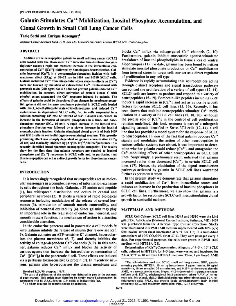
26). Consequently, we determined whether galanin stimulatesthe formation of inositol phosphates in SCLC cell lines. Theinositol phosphate response was amplified by adding LiCl for20 min prior to the termination of the incubation (26). Asshown in Fig. 5. addition of galanin to H69 cells labeled withmyo-pHjinosito! stimulated the accumulation of total inositolphosphates in a time- and dose-dependent manner. The response can be detected at a concentration of 5 n\i and the EC50value was 10 nivi. Similar results were obtained when H510cells were used instead of H69 cells.
In order to characterize the inositol response in more detail,the major inositol phosphate fractions were separated by FPLCafter various times of galanin treatment. As shown in Fig. 6,an increase in InsP3 and InsP2 was detectable within seconds ofgalanin addition. This was followed by a marked and slowerincrease in the InsP, fraction. A marked increase in [3H]
lns(l,4,5)P.i was observed after 30 s of galanin treatment,whereas ['H]Ins(l,3,4)P3 was predominant after 20 min of
incubation (Fig. 6, bottom). In both H69 and H510 there was arapid increase in InsP4, which was detectable at 30 s (increaseof 40-60% above control) and maintained for up to 20 min(increase 50-190% above control) (data not shown). The resultsshown in Figs. 5 and 6 demonstrate that galanin stimulates aninositol phosphate response in SCLC cell lines.
Galanin Stimulates Clonal Growth in SCLC. The rapid stimulation of Ca2+ mobilization and inositol phosphate production
induced by galanin in SCLC cells prompted us to test the effectof this neuropeptide on the growth of these cells. Transformed
100
Galanin (nM)Fig. 5. Effect of galanin on the accumulation of inositol phosphates. H69
SCLC cells incubated in HITESA for 3-5 days were »ashed and labeled inHITESA with 10 >jCi/ml myo-[3H]inositol for 24 h. Cells were then »ashedtwicein HITESA at 37°C.Approximately 1.5 x 10' cells were resuspended in 1 mlHITESA containing 0.02 M Hepes and incubated at 37°Cwith 20 HIMLiCl for
20 min before the addition of galanin at the concentrations indicated. Cells wereincubated »ithgalanin for 20 min. The accumulation of total inositol phosphateswas determined as described in "Materials and Methods." The increase in inositol
phosphate at a particular galanin concentration is expressed as a percentageincrease above the control (i.e., cultures incubated in the presence of LiCl for 40min) and represents the mean of 4 independent experiments. Point, mean: bar, ±SEM. Average control value. 1405 cpm (n = 10): average 100 nM galanin value.2141 cpm (n = 10). Inset, time course of the accumulation of inositol phosphates.Galanin (100 nM. •)was added for the times indicated in the presence of LiCl.Percentage of increase in total inositol phosphates from control is shown. Thecontrol (O) (LiCl only for 40 min) value. 2194 cpm: 100 nM galanin for 20 minvalue. 3330 cpm. All other details were as described in "Materials and Methods."
or tumor cells, including SCLC, are able to form colonies insemisolid media (27, 28). Consequently, the ability of H69 andH510 cells to form colonies in this assay was tested in thepresence of increasing concentrations of galanin. Galanincaused a marked stimulation of colony formation in a concentration-dependent fashion (Fig. 7). The concentrations requiredto promote half-maximum stimulation were approximately 20nM for H69 and H510 cells. The maximum effect was achievedwithin a narrow range of galanin concentration (about 50 nM).At higher concentrations, the growth-promoting effect of galanin was sharply reduced, presumably due to homologous de-sensitization. The half-maximum concentrations required toinduce clonal growth were similar to those required to stimulateCa2+ mobilization (Fig. 2) or inositol phosphate accumulation
(Fig. 5). As reported previously (17), galanin did not increase[Ça-*];in H345, a SCLC cell line responsive to GRP. In this
cell line, galanin failed to promote clonal growth, whereas GRP,added to parallel assays, caused a marked stimulation (resultsnot shown). Thus, the growth-promoting effects of galanin areclearly associated with the ability of this neuropeptide to induceearly signaling events.
Recently, [Arg6,D-Trp7MVlePhe8]substance P (6-11) has been
identified as a broad spectrum neuropeptide antagonist (29).
03m(0
o
150 H69 H510100
InsP
50InsP
60lnsP)
1 10 20 1 10 20
Time (Min)
Q.
U
2000
1000
lns(1,4,5)Pj
25 30
Fraction NumberFig. 6. Top, changes in the level of InsPi, InsP2, and InsPj in galanin-stitnulated
H69 and H510 SCLC cells as a function of time. H69 and H510 cells wereprelabeled with myo-|3H]inositol and incubated in HITESA containing 20 mM
LiCl for 20 min. Then. 100 nM galanin was added for various times. Parallelcultures were incubated in the presence of LiCl but without galanin (controls).Incubations were stopped by addition of 250 /il ice cold TCA. The samples wereanalyzed for their composition of inositol phosphates by aniónexchange chro-tnatography on a Mono Q column. All other experimental conditions »ereasdescribed in "Materials and Methods." Each point has an appropriate control.Point, mean percentage change from the control of 3-5 experiments: bar, ±SEM.Bottom, elution profile of Ins(l,4.5)P, and Ins(1.3,4)P, in H69 cells stimulatedby galanin. H69 cells were prelabeled with myo-[3H]inositol as described aboveand incubated ¡nHITESA containing 20 mM LiCl for 40 min. Galanin (100 n.vi)was added either for 30 s (•)or for 20 min (A) before the termination of theexperiment. Parallel cultures were incubated for 40 min in the presence of LiClwithout galanin (D). [3H]Ins(l,4,5)Pj was separated from [3H]lns(1.3,4)P, asdescribed in "Materials and Methods." The peak of radioactivity corresponding
to lns( 1,4,5)P.i ¡nthe sample was assigned on the basis of coelution with standards[3HJIns(1.4,5)P, and ("PJInsf 1,4.5)?,. In this system, the peak eluting immedi
ately prior to lns(l,4.5)P, is ascribed to Ins(l,3.4)P3 (25). A similar profile wasobtained in 3 independent experiments.
1677
Research. on August 21, 2018. © 1991 American Association for Cancercancerres.aacrjournals.org Downloaded from

GALANIN STIMULATES EARLY SIGNALS AND GROWTH
£ 300in
D
2 200
_0
O 100o
H 69 H 510A300
10 100 O 10
Galanin (riM)
100
Fig. 7. Effect of galanin on colony formation in H69 (left) and H510 (right)SCLC cells. Cells 3-5 days postpassage were washed, resuspended in HITESA,and then disaggregated into an essentially single cell suspension. Cell numberwas determined using a Coulter Counter and IO4 cells in Q.K agarose werelayered on top of 0.5('c agarose, both layers containing galanin at the same
concentration in 33-mm plastic dishes. Colonies represent aggregates of cells >I6counted under a microscope after 21 days. H69: point, mean of 7 experiments(each with 5 replicates): bar, ±SEM.H510: point, mean of 5 experiments (eachwith 5 replicates); bar, ±SEM.
Fig. 8 shows that addition of this antagonist, at 20 /tM, prevented the increase in [Ca:+]¡caused by a subsequent addition
of galanin in H69 cells. This prompted us to determine whetherthis antagonist could also prevent galanin from stimulatingclonal growth in these cells. As shown in Fig. 8 (bottom),[Arg6,D-Trp7-9,MePhe8]substance P, added at 20 /IM, caused a
profound inhibition of colony formation. This inhibitory effectwas reversed by high concentrations of galanin.
DISCUSSION
The results presented here demonstrate that the neuropeptidegalanin induces a rapid and transient increase in [Ca2+]¡and an
accumulation of inositol phosphates and stimulates clonalgrowth of SCLC cell lines. The findings demonstrate thatgalanin can act as a direct growth factor for cultured humancells.
Galanin is widely distributed and elicits a multiplicity ofphysiological responses (2). However, the only model systemin which the signal transduction pathways activated by galaninhave been studied in detail is the pancreatic ß-cell(5). In thesecells, galanin stimulates an ATP-sensitive K* channel which
increases the plasma membrane potential, blocks the influx ofCa2+ through voltage-gated Ca2+ channels, and thereby decreases [Ca2+]¡(8). These effects are mediated by a pertussistoxin-sensitive G protein (7). The results presented here demonstrate that galanin initiates an entirely different set of earlyevents in SCLC cell lines. Galanin stimulates a rapid increasein [Ca2+]i from internal stores through a pertussis toxin-insensitive pathway. This Ca2+-mobilizing action of galanin can be
completely dissociated from changes in membrane potential.Furthermore, galanin stimulates the production of inositolphosphates, consistent with the hypothesis that galanin-inducedCa2+ mobilization is mediated by Ins(l,4,5)Pj. This is the first
time that galanin has been shown to evoke inositol phosphateand Ca2+ mobilization responses in any cell type.
Pharmacological and molecular cloning studies provided evidence that neuropeptide receptors are frequently expressed inmultiple molecular forms (30). We propose that a galaninreceptor exists in at least two different molecular subtypes. Onecouples to K+ channels via a pertussis toxin-sensitive G protein,e.g., in the pancreatic /i-cell. A second subtype, present in certainSCLC cells, is coupled to phospholipase C which generatesIns(l,4,5)P, and thereby leads to Ca2+ mobilization from inter-
^ 133-+ ^
3 101--
400
147-
97-
'Gal TAnt. *Gal '
M
b
300
200
coö 100Ü
1 Min
0 10 25 SO 100 200
Galanin (nM)Fig. 8. Top, effect of antagonist [Arg6.n-Trp7',MePhe8)substance P on galanin-
stimulated Ca:* mobilization in H69 SCLC cells. Additions: galanin. 25 nM(Gal): galanin I /IM (Gal+); [Arg6.n-Trp7-',MePhe8|substance P. 20 /IM (Ant).Bottom, effect of [Arg".D-Trp''-',MePhe8]substance P on galanin-induced colonyformation. Cells (IO4) in 0.3?; agarose containing galanin at concentrations areindicated either in the absence (•)or in the presence (ED)of 20 /IM (Arg'.D-Trp7',MePhe8]substance P. Colonies of >16 cells were counted after 21 days
under a microscope. Column, mean of 2 experiments (each with 5 replicates); bar±SD.
nal stores. In this context, H69 and H510 SCLC cells mayprovide a useful model to study these novel effects of galanin.
Lung cancer remains the commonest fatal malignancy in thedeveloped world. SCLC constitutes nearly 25% of all pulmonary cancers and follows a rapid and aggressive clinical coursedespite initial chemosensitivity (31). Increased understandingof the signal-transduction pathways that regulate SCLC growthmay identify novel targets for therapeutic intervention. Recentwork from this (17) and other (18) laboratories has shown thata variety of neuropeptides, acting through distinct receptors,induces Ca2+ mobilization in SCLC cell lines. However, the
precise relationship between a rapid and transient increase in[Ca2+]¡and long-term in SCLC growth remains undefined. Inview of the Ca2+-mobilizing actions of galanin shown in this
study, it was important to determine whether this neuropeptideinfluences the growth of responsive SCLC. Tumor and transformed cells including SCLC are able to form colonies inagarose medium. Indeed, there is a positive correlation betweencloning efficiency of the cells and the histological involvementand invasiveness of the tumor in specimens taken from SCLC(27, 28). Consequently, we determined the effect of galanin onthe ability of H69 and H510 cells to form colonies in semisolidmedium.
In the present study we demonstrate that galanin markedlystimulates the clonal growth of either H69 or H510 cells insemisolid medium. The EC5(>values for promoting colony formation are in excellent agreement with the EC50values for Ca2+
mobilization and inositol phosphate accumulation. Furthermore, [Arg6,D-Trp79,MePhe(t]substance P (6-11), recently iden
tified as a broad spectrum neuropeptide antagonist (29), prevented Ca2+ mobilization induced by galanin and strikingly-
inhibited basal and galanin-stimulated colony formation. Thisis the first time that galanin has been shown to act as a growthfactor for any cell type. Galanin is widely distributed (2) and inhuman lung is associated with other peptides in neuroendocrine
1678
Research. on August 21, 2018. © 1991 American Association for Cancercancerres.aacrjournals.org Downloaded from
GALANIN STIMULATES EARLY SIGNALS AND GROWTH
cells (32) from which it is presumed that SCLCs derive (33). Inview of its widespread distribution, it is likely that galaninregulates the proliferation of other cell types, a possibility thatwarrants further experimental work. The finding that galanincan act as a direct growth factor for SCLC cells supports theproposition that the growth of these tumors may be regulatedin a complex manner by multiple autocrine/paracrine interactions involving neuropeptides.
REFERENCES
1. Tatemolo. K.. Rokaeus. A.. Jornvall, H„McDonald. T. J.. and Mutt. V.Galanin—a novel biologically active peptide from porcine intestine. FEBSLett.. 164: 124-128. 1983.
2. Rokaeus. A. Galanin—a newly isolated biologically-active neuropeptide.Trends Neurosa.. IO: 158-164." 1987.
3. Fisone. G.. Wu, C. F.. Consolo. S., Nordstrom. O.. Brynne. N.. Bartfai. T..Melander, T., and Hokfelt. T. Galanin inhibits acetylcholine release in theventral hippocampus of the rat: histochemical, autoradiographic. in viro, andin rirro studies. Proc. Nati. Acad. Sci. USA., 84: 7339-7343. 1987.
4. Ekblad. E.. Hakanson. R.. Sundler. F.. and Wahlestedt, C. Galanin: neuro-modulatory and direct contractile effects on smooth muscle preparations. Br.J. Pharmacol.. 86: 241-246. 1985.
5. Ahren. B., Rorsman. P.. and Berggren. P-O. Galanin and the endocrinepancreas. FEBS Lett.. 229: 233-237. 1988.
6. de-Weille. J.. Schmid-Antomarchi, H.. Fosset. M.. and Lazdunski. M. ATP-sensitive K* channels that arc blocked by hypoglycemia-inducing sulfonylu-reas in insulin-secreting cells are activated by galanin, a hyperglycemia-inducing hormone. Proc. Nati. Acad. Sci. USA. 85: 1312-1316. 1988.
7. Dunne. M. J.. Bulle«.M. J.. Li. G. D.. \Vollheim. C. B.. and Petersen. O.H. Galanin activates nucleotide-dependent K* channels in insulin-secretingcells via a pertussis toxin-sensitive G-protein. EMBO J.. 8: 413-420. 1989.
8. Nilsson. T.. Arkhammar. P.. Rorsman. P.. and Berggren. P. O. Suppressionof insulin release by galanin and somatostatin is mediated by a G-protein: aneffect involving repolarization and reduction in cytoplasmic free Ca2* concentration. J. Biol. Chem.. 264: 973-980. 1989.
9. Sharp. G. W.. Le-Marchand-Brustel. Y., Yada. T.. Russo. L. L.. Bliss, C. R.,Cormont. M., Monge, L.. and Van-Obberghcn. E. Galanin can inhibit insulinrelease by a mechanism other than membrane hypcrpolarization or inhibitionof adenyiate cyclasc. J. Biol. Chem.. 264: 7302-7309. 1989.
10. Tamura. K., Palmer, J. M.. Winkelmann. C. K., and Wood. J. D. Mechanismof action of galanin on myenteric neurons. J. Neurophysiol.. 60: 966-979.1988.11. Palazzi. E.. Fisonc. G.. Hokfclt. T.. Bartfai. T.. and C'onsolo. S. Galanin
inhibits the muscarinic stimulation of phosphoinositide turnover in rat ventral hippocampus. Eur. J. Pharmacol.. 148: 479-480. 1988.
12. Rozengurt. E. Early signals in the mitogenic response. Science (WashingtonDC). 234: 161-166. 1986.
13. Zachary, I.. Woll. P.. and Rozengurt. E. A role for neuropeptides in thecontrol of cell proliferation. Dev. Biol.. 124: 295-308. 1987.
14. Rozengurt, E.. and Sinnett-Smith. J. Bombesin stimulation of fibroblastmitogcnesis: specific receptors, signal transduction and early events. Philos.Trans. R. Soc. Lond-Biol. Sci., 327: 209-221. 1990.
15. Trepel, J. B., Mover. J. D.. Heikkila. R.. and Sausville. E. A. Modulation ofbombesin-induced phosphatidylinositol hydrolysis in a small-cell lung cancer
cell line. Biochem. J., 255:403-410. 1988.16. Cuttitta, F., Carney, D. N., Mulshine. J.. Moody, T. W.. Fedorko, J.. Fischler,
A., and Minna. J. D. Bombesin-like peptides can function as autocrine growthfactors in human small cell lung cancer. Nature (Lond.), 316:823-826, 1985.
17. Woll, P. J., and Rozengurt, E. Multiple neuropeptides mobilise calcium insmall cell lung cancer: effects of vasopressin, bradykinin, cholecystokin,galanin and neurotcnsin. Biochem. Biophys. Res. Commun.. 164: 66-73,1989.
18. Bunn. P. A. Jr.. Dienhart. D. G.. Chan. D.. Puck. T. T.. Tagawa. M.. Jewett.P. B.. and Braunschweiger. E. Neuropeptide stimulation of calcium flux inhuman lung cancer cells: delineation of alternative pathways. Proc. Nail.Acad. Sci. USA, 87: 2162-2166. 1990.
19. Abe, K., Kameya. T.. Yamaguchi. K.. Kikuchi. K.. Adachi. !.. Tanaka. M.,Kiiiiui ,i. S., Kodama. T.. Shimosato. Y.. and Ishikawa. S. Hormone-producing lung cancers. Endocrinologie and morphologic studies. In: K. L. Beckerand A. F. Gazdar (eds.). The Endocrine Lung in Health and Disease, pp.549-595. London: W. B. Saunders. 1984.
20. Heikkila. R.. Trepel. J. B.. Cuttitta. F.. Neckers. L. M., and Sausville. E. A.Bombesin-related peptides induce calcium mobilization in a subset of humansmall cell lung cancer cell lines. J. Biol. Chem.. 262: 16456-16460. 1987.
21. Simms, E., Gazdar, A. F., Abrams, P. G., and Minna. J. D. Growth ofhuman small cell (oat cell) carcinoma of the lung in serum-free growth factor-supplemented medium. Cancer Res., 40: 4356-4363. 1980.
22. Tsien, R. Y., Pozzan, T., and Rink. T. J. T-cell mitogens cause early changesin cytoplasmic free Ca2* and membrane potential in lymphocytes. Nature(Loñd.).295:68-71. 1982.
23. Mendoza. S. A.. Schneider, J. A.. Lopez-Rivas. A.. Sinnett-Smith. J. W..and Rozengurt, E. Early events elicited by bombesin and structurally relatedpeptides in quiescent Swiss 3T3 cells. II. Changes in Na* and Ca2* fluxes,Na*/K* pump activity and intracellular pH. J. Cell Biol., 102: 2223-2233,
1986.24. Nanbcrg. E.. and Rozengurt. E. Temporal relationship between inositol
polyphosphate formation and increases in cytosolic Ca2* in quiescent 3T3cells stimulated by platelet-derived growth factor, bombesin and vasopressin.EMBO J., 7: 2741-2748. 1988.
25. Lopez-Rivas. A.. Mendoza. S. A., Nánberg,E., Sinnett-Smith. J.. and Rozengurt. E. The Ca!*-mobilizing actions of platelet-derived growth factor
differ from those of bombesin and vasopressin in Swiss 3T3 cells. Proc. Nati.Acad. Sci. USA. 84: 5768-5772, 1987.
26. Berridge. M. J.. and Irvine. R. F. Inositol phosphates and cell signalling.Nature (Lond.). 341: 197-205. 1990.
27. Carney. D. N.. Gazdar. A. F., and Minna. J. D. Positive correlation betweenhistológica! tumor involvement and generation of tumor cell colonies inagarose in specimens taken directly from patients with small cell carcinomaof the lung. Cancer Res.. 40: 1820-1823. 1980.
28. Carney, D. N.. Cuttitta. F., Moody, T. W., and Minna, J. D. Selectivestimulation of small eell lung cancer clonal growth by bombesin and gastrin-releasing peptide. Cancer Res.. 47: 821-825, 1987.
29. Woll, P. J.. and Rozengurt. E. A neuropeptide antagonist that inhibits thegrowth of small cell lung cancer. Cancer Res.. 50: 3968-3973. 1990.
30. Woll. P. J.. and Rozengurt. E. Neuropeptides and growth regulators. Br.Med. Bull., 45: 492-505. 1989.
31. Smyth, J. F.. Fowlie, S. M.. Gregor. A.. Crompton. G. K., Busuttil, A.,Leonard. R. C.. and Grant. I. W. The impact of chemotherapy on small cellcarcinoma of the bronchus. Quant. J. Med.. 61: 969-973. 1986.
32. Uddman, R.. and Sundler. F. Neuropeptide in the airways: a review. Am.Rev. Respir. Dis.. 136: S3-8. 1987.
33. Gazdar. A. F., and McDowell, M. Pathobiology of lung cancer. In: S. T.Rosen, J. L. Mulshine, F. Cuttitta. and P. G. Abrams (eds.). Biology of LungCancer: Diagnosis and Treatment, pp. 1-42. New York: Marcel Dekker Inc.,1988.
1679
Research. on August 21, 2018. © 1991 American Association for Cancercancerres.aacrjournals.org Downloaded from

1991;51:1674-1679. Cancer Res Tariq Sethi and Enrique Rozengurt CellsAccumulation, and Clonal Growth in Small Cell Lung Cancer
Mobilization, Inositol Phosphate2+Galanin Stimulates Ca
Updated version
http://cancerres.aacrjournals.org/content/51/6/1674
Access the most recent version of this article at:
E-mail alerts related to this article or journal.Sign up to receive free email-alerts
Subscriptions
Reprints and
To order reprints of this article or to subscribe to the journal, contact the AACR Publications
Permissions
Rightslink site. Click on "Request Permissions" which will take you to the Copyright Clearance Center's (CCC)
.http://cancerres.aacrjournals.org/content/51/6/1674To request permission to re-use all or part of this article, use this link
Research. on August 21, 2018. © 1991 American Association for Cancercancerres.aacrjournals.org Downloaded from