galen courses 2014 - esor · by an online self-assessment test. ... 2nd or 3rd year of training in...
TRANSCRIPT
GALEN Courses 2014
GALEN Foundation CourseNeuroradiology June 26–28, Wroclaw/Poland
GALEN Advanced CoursesCardio-Thoracic Cross-Sectional Imaging June 5–6, Palermo/Italy
Abdominal Oncologic Imaging September 5–6, Moscow/Russia
Oncologic Imaging September 18–19, Geneva/Switzerland
Musculoskeletal Cross-Sectional Imaging October 30–31, Amsterdam/Netherlands
availab
le on-lin
e
myESR
.org/es
or

Date of printing: February 2014While the greatest care is taken in compiling this information, the editor does not take responsibility for any printing errors or omissions.
European School of RadiologyThe European School of Radiology (ESOR) is an integrated and multi-dynamicinstitution, fulfilling the mission of the European Society of Radiology (ESR) in the field of education. Its main goal is to assist in harmonising radiological education in Europe. With its wide range of activities, ESOR additionally aims to raise standards in the field of scientific radiology, to extend and coordinate teaching resources worldwide and to help young radiologists to obtain the knowledge and skills to fulfil tomorrow’s requirements.
Table of contents
1 Welcome1 ESOR Committee 4 GALEN Foundation Course6 Neuroradiology12 GALEN Advanced Courses14 Cardio-Thoracic Cross-Sectional Imaging18 Abdominal Oncologic Imaging22 Oncologic Imaging30 Musculoskletal Cross-Sectional Imaging
All ESOR activities are exclusively for ESR members. Further information about ESR membership is available on the ESR website myESR.org.
Please note that programmes are marked with a logo to indicate their classification according to the European Training Curriculum.
First three years of training
Fourth and fi�h years of training (general radiologist standard)
Subspecialty training standard
1
Welcome
Dear Colleagues,
The European School of Radiology has become an integrated and multi-dynamic initiative that aspires to extend teaching resources throughout Europe and worldwide, to assist in harmonising radiological education and to enhance the scientific profile of radiology. One of the main ESOR activities is the GALEN project currently in partnership with Bracco, offering foundation and advanced courses. All courses have been designed around an established course format, combining lectures and interactive workshops, offered by internationally renowned European experts. The courses have been specifically structured with the trainee radiologist in mind and deal with subjects of practical importance; they have an interactive character, encouraging informal and free communication between participants and lecturers. They are also supported by an online self-assessment test.
The GALEN Foundation Courses aimed at residents in their 1st, 2nd or 3rd year of training in radiology, are primarily assigned to the geographic area of Central and South-eastern Europe. One such course will be offered in 2014, focused on Neuroradiology.
The GALEN Advanced Courses are aimed at 4th and 5th year residents and recently board-certified radiologists from all around Europe. Four such courses are organised in 2014, structured along the lines of the most recent advances in cross-sectional imaging. These are focused on Abdominal, Cardio-Thoracic, Musculoskeletal and Oncologic Imaging.
In this brochure you will find the relevant programmes and venues. We would like to emphatically encourage you to participate. Since the courses are very popular, you should book your place quickly.
Professor Nicholas GourtsoyiannisESOR Scientific/Educational Director
ESOR Committee March 2014 – March 2015
Committee Chairman Scientific/Educational Director
Nicholas Gourtsoyiannis, Athens/GR
Committee Members
Regina Beets-Tan, Maastricht/NL
Hans-Ulrich Kauczor, Heidelberg/DE
Celso Matos, Brussels/BE
Malgorzata Szczerbo-Trojanowska, Lublin/PL
Ex-officio Members
Paul M. Parizel, Antwerp/BE
Boris Brkljačić, Zagreb/HR
Birgit Ertl-Wagner, Munich/DE
Peter Baierl, Vienna/AT
Brigitte Lindlbauer, Vienna/AT
Programme Planning Committee Members
Maria Argyropoulou, Ioannina/GR
Luis Donoso, Barcelona/ES
Thomas Helbich, Vienna/AT
Christian Loewe, Vienna/AT
Mario Maas, Amsterdam/NL
Borut Marincek, Cleveland/US
Luis Marti-Bonmati, Valencia/ES
Paul M. Parizel, Antwerp/BE ESNR European Course of Neuroradiology
Wolfgang Steinbrich, Basel/CH ESMRMB School of MRI
Harriet Thoeny, Bern/CH

2
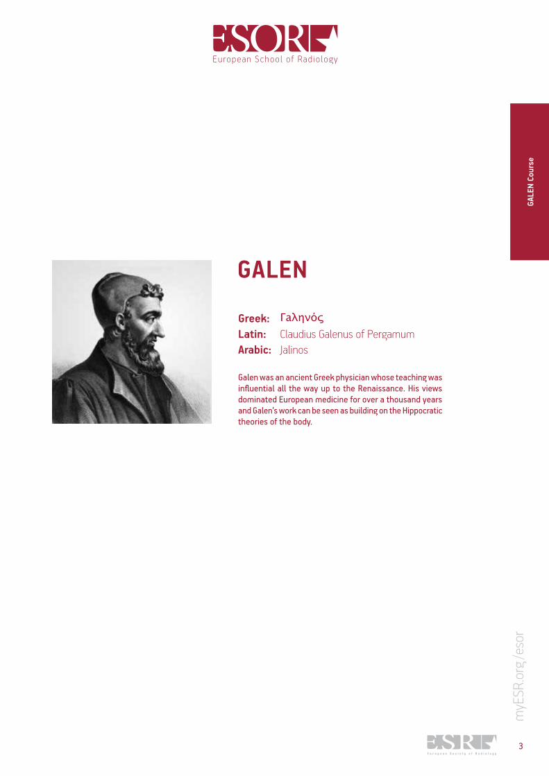
Greek:Latin: Claudius Galenus of PergamumArabic: Jalinos
Galen was an ancient Greek physician whose teaching was influential all the way up to the Renaissance. His views dominated European medicine for over a thousand years and Galen’s work can be seen as building on the Hippocratic theories of the body.
GALEN
3
GALE
N Co
urse
GALE
N Fo
unda
tion
Cour
ses
GALEN Foundation Courses
4
AimThe GALEN Foundation Courses have been designed to familiarise young radiologists with the established approaches and most recent achievements in diagnostic imaging, related to topics across the modalities. The courses are aimed at residents in their 1st, 2nd or 3rd year of training in radiology and are primarily assigned to the geographic area of south, central and eastern Europe. Each programme is divided into organ-oriented lecture series and interactive workshops, offered by an internationally renowned European faculty. All programmes have been specifically structured with the trainee radiologist in mind and are meant to deal with subjects of practical importance; they have an interactive character to encourage informal and free communication between participants and lecturers.
Course programmeEach programme is divided into organ-oriented lecture series and interactive workshops, offered by an internationally renowned European faculty. All programmes have been specifically structured with the trainee radiologist in mind and are meant to deal with subjects of practical importance; they have an interactive character to encourage informal and free communication between participants and lecturers.Each course is accompanied by an online self-assessment test consisting of multiple-choice questions.
ESR/ESOR reserves the right to amend course programmes.
Course languageAll courses are held in English.
EligibilityFoundation courses are intended for residents in their 1st, 2nd or 3rd year of training in radiology. Participants must be proficient in English. Furthermore, ESR membership fees for 2014 must be settled in advance. Further information about ESR membership is available atwww.myESR.org/membership
RegistrationOnline registration is available at www.myESR.org/esor.
The registration fees for the GALEN Foundation Courses vary. For the current registration fees for the courses please refer to the website or relevant course in this brochure. It is possible to attend more than one course. As the number of participants per course is limited to 60, registrations will be considered on a first come, first serve basis. The registration fee includes lectures and workshops attendance, teaching material for the course (printed syllabus), coffee breaks and lunch as per programme. Kindly note that the fee for residents applies to radiologists in training only. Resident registration must therefore be accompanied by a confirmation of this status, signed by the head of department. When making your travel arrangements, please make sure that you will be able to stay for the entire course.

Terms of cancellationCancellation by the course organiser: • In case the minimum number of participants has not
been reached four weeks prior to the course, the organiser reserves the right to cancel the course. Registration fees will be refunded in full.
Cancellation by the participant: • No refund from ESR/ESOR is possible in the event
of cancellation or no-show by the participant. We recommend taking out the cancellation insurance offered by Europäische Reiseversicherung.
Hotel accommodationParticipants are responsible for their travel and hotel arrangements. Recommendations for hotels at the venues or nearby are available online at www.myESR.org/esor under the respective course information. Please book early in order to ensure availability.
CME accreditationAn application has been made to the European Accreditation Council for Continuing Medical Education (EACCME) for CME accreditation of all ESOR courses.The EACCME is an institution of the European Union of Medical Specialists (UEMS), www.uems.net. European accreditation is granted by the EACCME in order to allow participants to validate the credits obtained at this activity in their home European country. The maximum number of CME credits designated for each event is announced on the ESOR website as soon as the application process of the specific event has been finalised.
CopyrightAll material supplied or used as part of an ESOR course is protected by copyright and remains the intellectual property of the speaker. This material is only for use by those participants who have attended an ESOR course; copying or use by others of such material is prohibited.
Videotaping and photography policyESOR does not allow any unauthorised videotaping or photography of any lectures or workshops.
For up-to-date information please visit www.myESR.org/esor
5
Course information
This course is for young radiologists in training, who desire a comprehensive and concise review of neuroradiology. Basic neuroanatomy, general disease categories of traumatic, infectious, neoplastic, vascular, congenital, demyelinating diseases of the brain and spine will be covered. Internationally renowned European experts will conduct a series of didactic lectures and dedicated interactive case review discussions. The course encompasses the broad spectrum of neuroradiology practice, including comprehensive reviews, updates in new trends and relevant imaging techniques.
Learning objectives
• to learn the essential imaging fi ndings of the most common brain and spine pathologies
• to prioritise imaging studies that will guide patient management
• to select the best and most cost-eff ective protocols and imaging techniques available
Neuroradiology June 26–28, 2014Wroclaw/Poland
GALEN Foundation Course
GALE
N Fo
unda
tion
Cour
seNe
uror
adio
logy
For further informationon the programme andregistration please visit
Neuroradiology June 26–28, 2014
Wroclaw/Poland
GALE
N Fo
unda
tion
Cour
seNe
uror
adio
logy
Thursday, June 26, 2014
15:00–16:00 Registration
16:00–16:15 Welcome and introduction
16:15–17:00 Imaging of spine trauma – vertebral and extramedullary spinal canal injuries J. Walecki, Warsaw/PL
17:00–17:45 Imaging of spine trauma – spinal cord injuries M. Stajgis, Poznan/PL
17:45–18:30 Imaging in degenerative disease of the spine M. Sasiadek, Wroclaw/PL
Friday, June 27, 2014
09:00–09:30 Pattern recognition in neuroradiology L. Van den Hauwe, Antwerp/BE
09:30–10:00 MRI of the brain: why, how, when R. Gasparotti, Brescia/IT
10:00–10:30 Congenital abnormalities of the brain A. Rossi, Genova/IT
10:30–10:50 Coff ee break
10:50–13:00 Workshops (L. Van den Hauwe, R. Gasparotti, A. Rossi)
13:00–14:00 Lunch break
14:00–14:30 Brain injury P. Parizel, Antwerp/BE
14:30–15:00 CNS infections E. Papadaki, Heraklion/GR
15:00–15:30 CNS malignancies C. Calli, Izmir/TR
15:30–15:50 Coff ee break
15:50–18:00 Workshops (P. Parizel, E. Papadaki, C. Calli)
Saturday, June 28, 2014
09:00–09:30 Imaging in epilepsy P. Sundgren, Lund/SE
09:30–10:00 Stroke: diagnosis and therapy P. Vilela, Lisbon/PT
10:00–10:30 White matter diseases M. Thurnher, Vienna/AT
10:30–10:50 Coff ee break
10:50–13:00 Workshops (P. Sundgren, P. Vilela, M. Thurnher)
13:00 Certifi cate of attendance
Programme
Local Organiser
M. SasiadekWroclaw/PL
VenueUniversity of Wroclaw – Law Department
ul. Uniwersytecka 7–1050–137 Wroclaw
Poland
Registration fee Residents
Early fee € 130(until eight weeks prior to the course)
Late fee € 175(aft er eight weeks prior to the course)
Board-certifi ed radiologistsEarly fee € 230
(until eight weeks prior to the course)Late fee € 275
(aft er eight weeks prior to the course)
6
City information
Wroclaw is the fourth largest city in Poland and is located in the west of the country. With an average temperature of -0,5 degrees Celsius in the winter, it is one of the warmest cities in the country. One of its most famous sights is the Centennial Hall, a UNESCO World Heritage site that is frequently hosting cultural and sport events, such as the Euro Basket tournament. Further tourist attractions are the main station, the Wroclaw Zoo as well as the Multimedia Fountain, which offers a special night programme. A popular way of getting to know the city is by sailing small passenger vessels on the river Oder. In 2016, Wroclaw will be the European Capital of Culture.
Course information
This course is for young radiologists in training, who desire a comprehensive and concise review of neuroradiology. Basic neuroanatomy, general disease categories of traumatic, infectious, neoplastic, vascular, congenital, demyelinating diseases of the brain and spine will be covered. Internationally renowned European experts will conduct a series of didactic lectures and dedicated interactive case review discussions. The course encompasses the broad spectrum of neuroradiology practice, including comprehensive reviews, updates in new trends and relevant imaging techniques.
Learning objectives
• to learn the essential imaging fi ndings of the most common brain and spine pathologies
• to prioritise imaging studies that will guide patient management
• to select the best and most cost-eff ective protocols and imaging techniques available
Neuroradiology June 26–28, 2014Wroclaw/Poland
GALEN Foundation Course
GALE
N Fo
unda
tion
Cour
seNe
uror
adio
logy
For further informationon the programme andregistration please visit
Neuroradiology June 26–28, 2014
Wroclaw/Poland
GALE
N Fo
unda
tion
Cour
seNe
uror
adio
logy
Thursday, June 26, 2014
15:00–16:00 Registration
16:00–16:15 Welcome and introduction
16:15–17:00 Imaging of spine trauma – vertebral and extramedullary spinal canal injuries J. Walecki, Warsaw/PL
17:00–17:45 Imaging of spine trauma – spinal cord injuries M. Stajgis, Poznan/PL
17:45–18:30 Imaging in degenerative disease of the spine M. Sasiadek, Wroclaw/PL
Friday, June 27, 2014
09:00–09:30 Pattern recognition in neuroradiology L. Van den Hauwe, Antwerp/BE
09:30–10:00 MRI of the brain: why, how, when R. Gasparotti, Brescia/IT
10:00–10:30 Congenital abnormalities of the brain A. Rossi, Genova/IT
10:30–10:50 Coff ee break
10:50–13:00 Workshops (L. Van den Hauwe, R. Gasparotti, A. Rossi)
13:00–14:00 Lunch break
14:00–14:30 Brain injury P. Parizel, Antwerp/BE
14:30–15:00 CNS infections E. Papadaki, Heraklion/GR
15:00–15:30 CNS malignancies C. Calli, Izmir/TR
15:30–15:50 Coff ee break
15:50–18:00 Workshops (P. Parizel, E. Papadaki, C. Calli)
Saturday, June 28, 2014
09:00–09:30 Imaging in epilepsy P. Sundgren, Lund/SE
09:30–10:00 Stroke: diagnosis and therapy P. Vilela, Lisbon/PT
10:00–10:30 White matter diseases M. Thurnher, Vienna/AT
10:30–10:50 Coff ee break
10:50–13:00 Workshops (P. Sundgren, P. Vilela, M. Thurnher)
13:00 Certifi cate of attendance
Programme
Local Organiser
M. SasiadekWroclaw/PL
VenueUniversity of Wroclaw – Law Department
ul. Uniwersytecka 7–1050–137 Wroclaw
Poland
Registration fee Residents
Early fee € 130(until eight weeks prior to the course)
Late fee € 175(aft er eight weeks prior to the course)
Board-certifi ed radiologistsEarly fee € 230
(until eight weeks prior to the course)Late fee € 275
(aft er eight weeks prior to the course)
7
GALE
N Fo
unda
tion
Cour
se
Neur
orad
iolo
gy
Learning Objectives
8
Imaging of spine trauma – vertebral and extramedullary spinal canal injuriesJ. Walecki, Warsaw/PL
• to illustrate vertebral injuries• to illustrate value of understanding biomechanics• to stress multidisciplinary approach
Imaging of spine trauma – spinal cord injuriesM. Stajgis, Poznan/PL
• to understand the mechanisms of spinal cord injury
• to learn about MR imaging findings in different traumatic abnormalities of the spinal cord
• to gain knowledge about new imaging techniques used in evaluation of spinal cord injury
Imaging in degenerative disease of the spineM. Sasiadek, Wroclaw/PL
• to gain knowledge about the full spectrum of the degenerative changes in the spine
• to learn about the imaging findings of the particular degenerative changes
• to understand the advanced imaging techniques in the degenerative disease of the spine
Pattern recognition in neuroradiology L. Van den Hauwe, Antwerp/BE
• to learn how to use anatomic location in CNS lesion characterisation (intra- vs. extra-axial, supra- vs. infratentorial)
• to learn how to correlate signal intensity changes with biochemical and pathological findings
• to learn how to integrate these findings in a pattern analysis approach to establish the (differential) diagnosis
MRI of the brain: why, how, when R. Gasparotti, Brescia/IT
• to learn about the commonest neurological disorders that require MR investigation
• to understand the specific role of MRI in the assessment of brain neurological disorders and to learn the appropriate protocols
• to become familiar with typical MR imaging findings in main neurological disorders
• to consolidate which MR imaging techniques should be used to answer the clinical question based on the patient’s clinical/neurological symptoms
Congenital abnormalities of the brain A. Rossi, Genova/IT
• to understand the key morphological features of the different categories of malformations
• to recognise the most important entities• to be able to use a simplified diagnostic imaging
approach
Neuroradiology June 26–28, 2014 Wroclaw/Poland
Learning Objectives
Brain injury P. Parizel, Antwerp/BE
• to present a pattern-based diagnostic approach to the patient with acute traumatic brain injury
• to review different types of traumatic intracranial lesions, and explain the difference between primary and secondary traumatic brain lesions
• to illustrate how the brain can be severely damaged in closed head injuries (deceleration trauma, diffuse axonal injuries)
• to demonstrate how advanced MRI techniques, such as DWI and DTI, can reveal evidence for micro-structural brain damage
CNS infections E. Papadaki, Heraklion/GR
• to discuss the pathophysiology and etiology (bacterial, viral, fungal) of CNS infections, the risk factors and the imaging findings
• to become familiar with the advanced neuroimaging MR techniques, such as DWI and MRS and learn how to differentiate CNS infections from tumours
• to learn about new concepts in infectious diseases of the brain, particularly in immunosuppressed patients (IRIS)
CNS malignancies C. Calli, Izmir/TR
• to give a brief information about “what is a CNS malignancy?”
• to describe the radiological imaging methods for the CNS malignancies
• to inform the audience about the common radiological classification concepts and findings of CNS tumours
• to give the notion to the audience about “how to approach these malignancies on imaging?”
Imaging in epilepsy P. Sundgren, Lund/SE
• to have knowledge about the different causes of epilepsy
• to understand how to perform the correct imaging in the work-up of patients with epilepsy
• to have knowledge about the imaging characteristics of mesial temporal sclerosis and malformations of cortical development
• to be aware of a proper work-up approach for patients with refractory epilepsy
Stroke: diagnosis and therapy P. Vilela, Lisbon/PT
• to become familiar with the most common ethiologies and pathophysiologic mechanisms of stroke in the adult population
• to recognise the imaging signs of early infarct on CT and MRI
• to understand the importance of multimodal CT and/or MR imaging protocols in stroke
• to overview the current treatment strategies of stroke treatment and its implication on stroke imaging
White matter diseases M. Thurnher, Vienna/AT
• to learn the classification of white matter diseases (WMD) of the brain
• to review the MRI findings in multiple sclerosis (MS)
• to learn how to distinguish MS from other white matter diseases and MS mimics
Neuroradiology June 26–28, 2014
Wroclaw/Poland
GALE
N Fo
unda
tion
Cour
se
Neur
orad
iolo
gy
9

GALE
N Fo
unda
tion
Cour
ses
10

12
GALE
N Ad
vanc
ed C
ours
es
GALEN Advanced Courses
Aim Advanced CoursesThe GALEN Advanced Courses are focused on recent advances in radiological imaging, related to topics across cross-sectional imaging. The courses are aimed at residents in their 4th and 5th year of training in radiology and recently board-certified radiologists. Each programme is divided into lectures and interactive workshops, offered by an internationally renowned European faculty. The programme has been specifically designed with the trainee radiologist in mind and is meant to deal with subjects of practical importance; it has an interactive character so as to encourage informal and free communication between participants and lecturers.
Course programmeEach programme is divided into organ-oriented lecture series and interactive workshops, offered by an internationally renowned European faculty. All programmes have been specifically structured with the trainee radiologist in mind and are meant to deal with subjects of practical importance; they have an interactive character to encourage informal and free communication between participants and lecturers. Each course is accompanied by an online self-assessment test consisting of multiple-choice questions.
ESR/ESOR reserves the right to amend course programmes.
Course languageAll courses are held in English.
EligibilityAdvanced Courses are intended for residents in their 4th and 5th year of training in radiology and recently board-certified radiologists, who desire a comprehensive review of diagnostic radiology. Participants must be proficient in English. Furthermore, ESR membership fees for 2014 must be settled in advance. Further information about ESR membership is available at
www.myESR.org/membership
Registration
Online registration is available at www.myESR.org/esor.
The registration fees for the GALEN Advanced Courses vary. For the current registration fees for the courses please refer to the website or relevant course in this brochure. It is possible to attend more than one course. As the number of participants per course is limited to 60, registrations will be considered on a first come, first serve basis. The registration fee includes lectures and workshops attendance, teaching material for the course (printed syllabus), coffee breaks and lunch as per programme. Kindly note that the fee for residents applies to radiologists in training only. Resident registration must therefore be accompanied by a confirmation of this status, signed by the head of department.When making your travel arrangements, please make sure that you will be able to stay for the entire course.

Terms of cancellationCancellation by the course organiser: • In case the minimum number of participants has not
been reached four weeks prior to the course, the organiser reserves the right to cancel the course. Registration fees will be refunded in full.
Cancellation by the participant: • No refund from ESR/ESOR is possible in the event
of cancellation or no-show by the participant. We recommend taking out the cancellation insurance offered by Europäische Reiseversicherung.
Hotel accommodationParticipants are responsible for their travel and hotel arrangements. Recommendations for hotels at the venues or nearby are available online at www.myESR.org/esor under the respective course information. Please book early in order to ensure availability.
CME accreditationAn application has been made to the European Accreditation Council for Continuing Medical Education (EACCME) for CME accreditation of all ESOR courses.The EACCME is an institution of the European Union of Medical Specialists (UEMS), www.uems.net. European accreditation is granted by the EACCME in order to allow participants to validate the credits obtained at this activity in their home European country. The maximum number of CME credits designated for each event is announced on the ESOR website as soon as the application process of the specific event has been finalised.
CopyrightAll material supplied or used as part of an ESOR course is protected by copyright and remains the intellectual property of the speaker. This material is only for use by those participants who have attended an ESOR course; copying or use by others of such material is prohibited.
Videotaping and photography policyESOR does not allow any unauthorised videotaping or photography of any lectures or workshops.
For up-to-date information please visit www.myESR.org/esor
Course information
This course is aimed at senior residents and recently board-certifi ed radiologists. For the fi rst time, this course will be dedicated to cardio-thoracic imaging and to provide an integrated view into modern state-of-the-art cardio-thoracic cross-sectional imaging. During these one and a half days recent advances and already established applications in cardiac CT and MR will be presented as well as recent advances in chest imaging and integrated views on cardio-thoracic diseases. The faculty consists of internationally renowned European experts; and they will share their expertise and their cases. The spectrum of topics treated during this course ranged from technical basics to modern applications, from examination protocols to image interpretation, and from acute to chronic. Acute chest pain will be an additional focus as well as ischemic heart disease. The combination of didactic lectures with interactive workshops guarantees the high educational value of this teaching programme. So take the chance and be part of this course and learn about recent advances in cardio-thoracic cross-sectional imaging.
Learning objectives
• to review methodology and clinical aspects of cardio-thoracic cross-sectional imaging
• to refresh knowledge on cardiac morphology and function as related to most common and most important cardiac diseases
• to be familiar with current best practice indications and integrated imaging techniques for the evaluation of cardiac diseases
• to learn about an integrated approach to cardio-thoracic diseases
Cardio-ThoracicCross-SectionalImagingJune 5–6, 2014Palermo/Italy
GALEN Advanced Course
GALE
N Ad
vanc
ed C
ours
esCa
rdio
-Tho
raci
c Im
agin
g
Local Organiser
M. MidiriPalermo/IT
VenueNH Hotel Palermo
Foro Italico 22b90133 Palermo
Italy
Registration fee Residents
Early fee € 150(until eight weeks prior to the course)
Late fee € 195(aft er eight weeks prior to the course)
Board-certifi ed radiologistsEarly fee € 250
(until eight weeks prior to the course)Late fee € 295
(aft er eight weeks prior to the course)
For further informationon the programme andregistration please visit
Cardio-ThoracicCross-Sectional
ImagingJune 5–6, 2014
Palermo/Italy
Programme
GALE
N Ad
vanc
ed C
ours
esCa
rdio
-Tho
raci
c Im
agin
g
Thursday, June 5, 2014
13:00–13:45 Registration
13:45–14:00 Welcome and introduction
14:00–14:10 State of the art CT cardiac imaging protocolsH.-C. Becker, Munich/DE
14:10–14:20 Most important cardiac MRI protocolsA. de Roos, Leiden/NL
14:20–14:50 CT diagnosis/rule out of stable coronary heart diseaseV. Sinitsyn, Moscow/RU
14:50–15:20 Diagnosis of coronary heart disease beyond stenosisdetection: imaging the ischemic myocardium (CT/MR)M. Francone, Rome/IT
15:20–15:50 Non ischemic cardiomyopathiesA. de Roos, Leiden/NL
15:50–16:20 Coff ee Break
16:20–18:30 Workshops(V. Sinitsyn, M. Francone, A. de Roos)
Friday, June 6, 2014
09:00–09:10 My top 5 cardiac CT indicationsC. Loewe, Vienna/AT
09:10–09:20 My top 5 cardiac MR indicationsM. Francone, Rome/IT
09:20–09:50 Valvular heart diseaseH.-C. Becker, Munich/DE
09:50–10:20 Congenital heart disease in children and adultsA. Madureira, Porto/PT
10:20–10:50 Current and future role of imaging in acute chest pain(CT/MRI)C. Loewe, Vienna/AT
10:50–11:20 Coff ee break
11:20–13:30 Workshops(H.-C. Becker, A. Madureira, C. Loewe)
13:30–14:30 Lunch break
14:30–15:00 Imaging of pulmonary embolism andpulmonary hypertensionC. Schaefer-Prokop, Amersfoort/NL
15:00–15:30 HRCT of interstitial lung diseasesA. Larici, Rome/IT
15:30–16:00 Management of intrapulmonary nodulesH. Prosch, Vienna/AT
16:00–16:20 Coff ee break
16:20–18:30 Workshops(C. Schaefer-Prokop, A. Larici, H. Prosch)
18:30 Certifi cate of attendance
14
City information
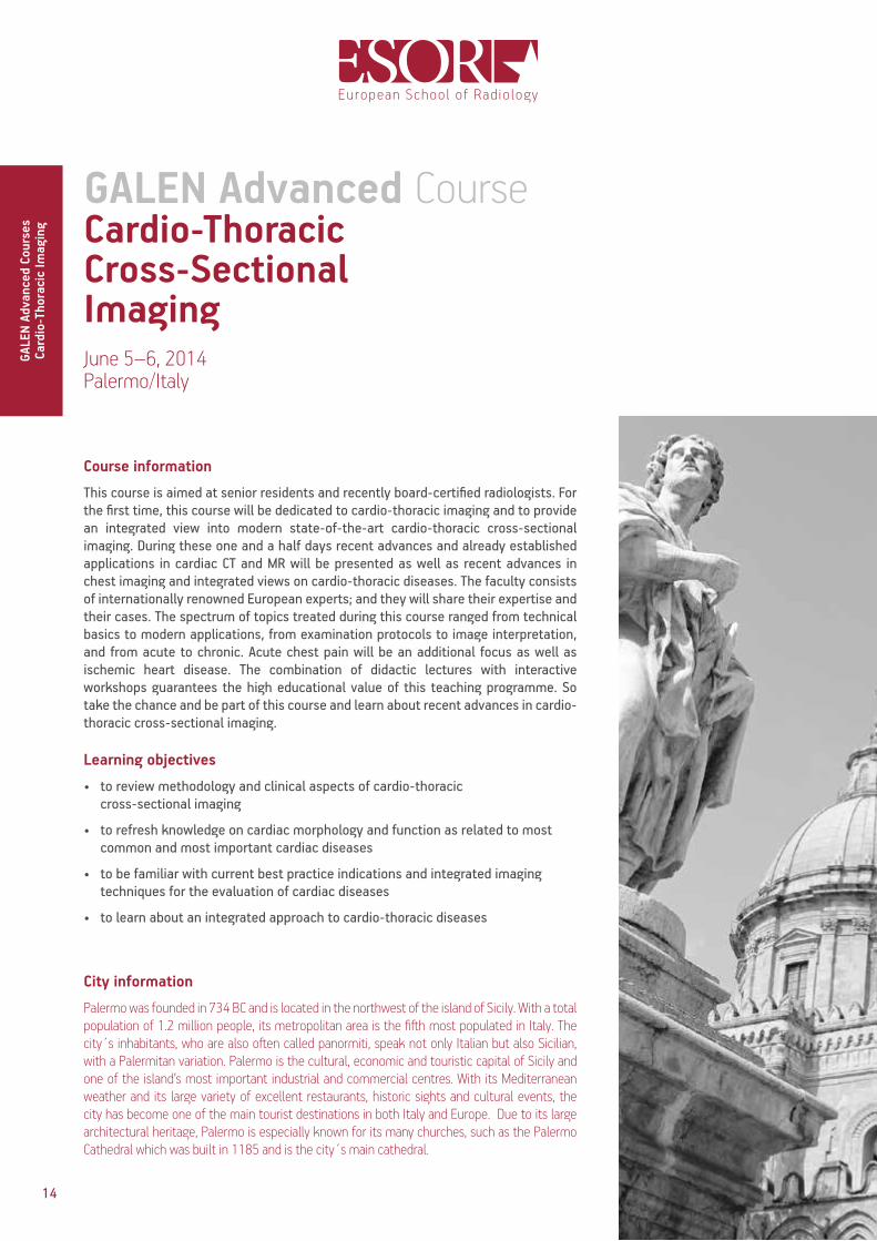
Palermo was founded in 734 BC and is located in the northwest of the island of Sicily. With a total population of 1.2 million people, its metropolitan area is the fifth most populated in Italy. The city´s inhabitants, who are also often called panormiti, speak not only Italian but also Sicilian, with a Palermitan variation. Palermo is the cultural, economic and touristic capital of Sicily and one of the island’s most important industrial and commercial centres. With its Mediterranean weather and its large variety of excellent restaurants, historic sights and cultural events, the city has become one of the main tourist destinations in both Italy and Europe. Due to its large architectural heritage, Palermo is especially known for its many churches, such as the Palermo Cathedral which was built in 1185 and is the city´s main cathedral.
Course information
This course is aimed at senior residents and recently board-certifi ed radiologists. For the fi rst time, this course will be dedicated to cardio-thoracic imaging and to provide an integrated view into modern state-of-the-art cardio-thoracic cross-sectional imaging. During these one and a half days recent advances and already established applications in cardiac CT and MR will be presented as well as recent advances in chest imaging and integrated views on cardio-thoracic diseases. The faculty consists of internationally renowned European experts; and they will share their expertise and their cases. The spectrum of topics treated during this course ranged from technical basics to modern applications, from examination protocols to image interpretation, and from acute to chronic. Acute chest pain will be an additional focus as well as ischemic heart disease. The combination of didactic lectures with interactive workshops guarantees the high educational value of this teaching programme. So take the chance and be part of this course and learn about recent advances in cardio-thoracic cross-sectional imaging.
Learning objectives
• to review methodology and clinical aspects of cardio-thoracic cross-sectional imaging
• to refresh knowledge on cardiac morphology and function as related to most common and most important cardiac diseases
• to be familiar with current best practice indications and integrated imaging techniques for the evaluation of cardiac diseases
• to learn about an integrated approach to cardio-thoracic diseases
Cardio-ThoracicCross-SectionalImagingJune 5–6, 2014Palermo/Italy
GALEN Advanced Course
GALE
N Ad
vanc
ed C
ours
esCa
rdio
-Tho
raci
c Im
agin
g
Local Organiser
M. MidiriPalermo/IT
VenueNH Hotel Palermo
Foro Italico 22b90133 Palermo
Italy
Registration fee Residents
Early fee € 150(until eight weeks prior to the course)
Late fee € 195(aft er eight weeks prior to the course)
Board-certifi ed radiologistsEarly fee € 250
(until eight weeks prior to the course)Late fee € 295
(aft er eight weeks prior to the course)
For further informationon the programme andregistration please visit
Cardio-ThoracicCross-Sectional
ImagingJune 5–6, 2014
Palermo/Italy
Programme
GALE
N Ad
vanc
ed C
ours
esCa
rdio
-Tho
raci
c Im
agin
g
Thursday, June 5, 2014
13:00–13:45 Registration
13:45–14:00 Welcome and introduction
14:00–14:10 State of the art CT cardiac imaging protocolsH.-C. Becker, Munich/DE
14:10–14:20 Most important cardiac MRI protocolsA. de Roos, Leiden/NL
14:20–14:50 CT diagnosis/rule out of stable coronary heart diseaseV. Sinitsyn, Moscow/RU
14:50–15:20 Diagnosis of coronary heart disease beyond stenosisdetection: imaging the ischemic myocardium (CT/MR)M. Francone, Rome/IT
15:20–15:50 Non ischemic cardiomyopathiesA. de Roos, Leiden/NL
15:50–16:20 Coff ee Break
16:20–18:30 Workshops(V. Sinitsyn, M. Francone, A. de Roos)
Friday, June 6, 2014
09:00–09:10 My top 5 cardiac CT indicationsC. Loewe, Vienna/AT
09:10–09:20 My top 5 cardiac MR indicationsM. Francone, Rome/IT
09:20–09:50 Valvular heart diseaseH.-C. Becker, Munich/DE
09:50–10:20 Congenital heart disease in children and adultsA. Madureira, Porto/PT
10:20–10:50 Current and future role of imaging in acute chest pain(CT/MRI)C. Loewe, Vienna/AT
10:50–11:20 Coff ee break
11:20–13:30 Workshops(H.-C. Becker, A. Madureira, C. Loewe)
13:30–14:30 Lunch break
14:30–15:00 Imaging of pulmonary embolism andpulmonary hypertensionC. Schaefer-Prokop, Amersfoort/NL
15:00–15:30 HRCT of interstitial lung diseasesA. Larici, Rome/IT
15:30–16:00 Management of intrapulmonary nodulesH. Prosch, Vienna/AT
16:00–16:20 Coff ee break
16:20–18:30 Workshops(C. Schaefer-Prokop, A. Larici, H. Prosch)
18:30 Certifi cate of attendance
15
GALE
N Ad
vanc
ed C
ours
es
Card
io-T
hora
cic
Imag
ing
Learning Objectives
16
CT diagnosis/rule out of stable coronary heart disease V. Sinitsyn, Moscow/RU
• to become familiar with protocols of cardiac CTA, image analysis and reporting
• to learn about results of clinical application of coronary CTA
• to know about current indications to CTA in stable coronary artery disease
Diagnosis of coronary heart disease beyond stenosis detection: imaging the ischemic myocardium (CT/MR) M. Francone, Rome/IT
• to review the scientific evidence of CT and MRI role in the evaluation of acute and chronic ischemic myocardium
• to present the different CT/MR stress imaging acquisition techniques for depiction of myocardial ischemia including rest-stress perfusion imaging and inducible wall motion abnormalities
• to understand the importance of CT-MRI imaging before coronary revascularisation for better patient selection and risk stratification
• to understand the relevant prognostic information provided by CT-MR ischemic rest and stress techniques
Non ischemic cardiomyopathies A. de Roos, Leiden/NL
• to learn techniques for evaluating cardiomyopathy
• to know causes for ischemic and non-ischemic cardiomyopathy
• to identify the role of MRI for differentiating various causes
Valvular heart disease H.-C. Becker, Munich/DE
• to identify strengths and weaknesses of CT and MRI in the imaging of patients with acute chest pain
• to learn about acquisition strategies and protocols for CT and MRI in this specific patient cohort
• to discuss future developments and the potential future role of these techniques in patients with chest pain
Congenital heart disease in children and adults A. Madureira, Porto/PT
• to understand the basic embryology of the heart and its implications in the most important congenital heart diseases (CHD)
• to understand the role of the different imaging modalities in the assessment of CHD, their relative strengths and weaknesses
• to review the major imaging characteristics of the most important CHD
• to understand the important role of MR in the post-operative assessment of patients with CHD
Cardio-Thoracic Cross-Sectional ImagingJune 5–6, 2014 Palermo/Italy
Learning Objectives
Current and future role of imaging in acute chest pain (CT/MRI) C. Loewe, Vienna/AT
• to become familiar with the definition of acute coronary syndrome (ACS) and the role of imaging in the management of patients suspected to suffer from ACS
• to learn about the most important non cardiac differential diagnoses of acute coronary syndromes, mainly including pulmonary embolism and acute aortic syndromes
• to become familiar with the most important cardiac differential diagnoses of the acute coronary syndrome including myocarditis and tako tsubo cardiomyopathy
• to discuss the current and potential future role of CT and MRT for the early triage of chest pain patients in the emergency room as well as for the follow up of patients after ACS
Imaging of pulmonary embolism and pulmonary hypertension C. Schaefer-Prokop, Amersfoort/NL
• to get familiar with CT features of acute and chronic embolism
• to learn about the variable signs of pulmonary hypertension on CT and their reliability
• to learn how to optimise the examination protocol dependant on scanner technique available and the clinical indication
• to discuss options and limitations of CT versus MR imaging for pulmonary embolism and grading of pulmonary hypertension
HRCT of interstitial lung diseases A. Larici, Rome/IT
• to become familiar with technical aspects and normal findings on HRCT scans
• to learn how to analyse HRCT findings of interstitial lung diseases on the basis of a pattern approach
• to know the typical appearances of the most important interstitial lung diseases
• to learn about clinical and radiologic key features for diagnosing interstitial lung diseases
Management of intrapulmonary nodules H. Prosch, Vienna/AT
• to be able to estimate the pretest probability of malignancy for pulmonary nodules based on their CT characteristics
• to comprehend the indications and limitations of different radiological techniques used to characterise pulmonary nodules
• to become familiar with the recent guidelines for the evaluation of pulmonary nodules
Cardio-Thoracic Cross-Sectional
ImagingJune 5–6, 2014
Palermo/Italy
GALE
N Ad
vanc
ed C
ours
es
Card
io-T
hora
cic
Imag
ing
17

Course information
The course is aimed at senior residents, board-certifi ed radiologists and fellows in oncologic and abdominal imaging. It is designed to provide an in-depth view into modern state-of-the-art oncologic imaging of the abdomen. The spectrum of topics treated during the course ranges from functional MRI to CEUS applications in abdominal oncology to structure imaging reporting of abdominal neoplasia, including gastroesophageal, liver, pancreatic, bile ducts, colon and anorectal cancer. European experts will present the benefi ts of multimodality imaging ensuring a high-quality teaching programme, combining lectures and interactive case discussions.
Learning objectives
• to present recent advances in diagnostic imaging of the most frequent abdominal malignancies, including CEUS and functional MRI
• to explain clinically relevant protocols and imaging strategies in the early detection, characterisation, staging and follow-up of main abdominal malignancies
• to get familiar with structured reporting of cross-sectional imaging features and their signifi cance in the management of patients with such neoplasms
AbdominalOncologicImagingSeptember 5–6, 2014Moscow/Russia
GALEN Advanced Course
GALE
N Ad
vanc
ed C
ours
esAb
dom
inal
Onc
olog
ic Im
agin
g
For further informationon the programme andregistration please visit
AbdominalOncologic
ImagingSeptember 5–6, 2014
Moscow/Russia
Programme
GALE
N Ad
vanc
ed C
ours
esAb
dom
inal
Onc
olog
ic Im
agin
g
Friday, September 5, 2014
08:00–08:45 Registration
08:45–09:00 Welcome and introduction
09:00–09:30 Structured reporting of esophageal and GOJ neoplasms S. Jackson, Plymouth/UK
09:30–10:00 CEUS in abdominal oncologic imaging O. Lucidarme, Paris/FR
10:00–10:30 Functional MRI in abdominal oncology V. Vandecaveye, Leuven/BE
10:30–10:50 Coff ee break
10:50–13:00 Workshops (S. Jackson, O. Lucidarme, V. Vandecaveye)
13:00–14:00 Lunch break
14:00–14:30 Imaging liver cirrhosis – from regenerative nodule to HCC G. Brancatelli, Palermo/IT
14:30–15:00 Imaging metastatic liver disease A. Blachar, Tel Aviv/IL
15:00–15:30 Solid pancreatic tumours – imaging protocols, staging, resectability J. Heverhagen, Bern/CH
15:30–15:50 Coff ee break
15:50–18:00 Workshops (G. Brancatelli, A. Blachar, J. Heverhagen)
Saturday, September 6, 2014
09:00–09:30 Imaging bile duct tumours – detection, characterisation and staging G. Karmazanovsky, Moscow/RU
09:30–10:00 CT colonography for colon cancer D. Regge, Candiolo/IT
10:00–10:30 Structured reporting of anorectal cancer S. Gourtsoyanni, London/UK
10:30–10:50 Coff ee break
10:50–13:00 Workshops (G. Karmazanovsky, D. Regge, S. Gourtsoyanni)
13:00 Certifi cate of attendance
Local Organiser
G. KarmazanovskyMoscow/RU
VenueRadisson Slavyanskaya
Hotel and Business CentreEurope Square 2121059 Moscow
Russia
Registration fee Residents
Early fee € 130(until eight weeks prior to the course)
Late fee € 175(aft er eight weeks prior to the course)
Board-certifi ed radiologists Early fee € 230
(until eight weeks prior to the course)Late fee € 275
(aft er eight weeks prior to the course)
18
City information
Moscow is the capital city and the political, economic and cultural centre of Russia. It is also the most populous city in Europe and is situated on the Moskva River. Moscow is known for many sights among which the most well-known are the Red Square, the St. Basil´s Cathedral and the Kremlin, which today functions as residence of the Russian president and is one of the city´s many world heritage sites. A further landmark is the city´s underground metro system which is one of the deepest in the world and popular for the rich and varied architecture of its stations. Moscow is also home to the world´s largest ballet ensemble, the Bolshoi Ballet, which resides in the city´s Bolshoi theatre.

Course information
The course is aimed at senior residents, board-certifi ed radiologists and fellows in oncologic and abdominal imaging. It is designed to provide an in-depth view into modern state-of-the-art oncologic imaging of the abdomen. The spectrum of topics treated during the course ranges from functional MRI to CEUS applications in abdominal oncology to structure imaging reporting of abdominal neoplasia, including gastroesophageal, liver, pancreatic, bile ducts, colon and anorectal cancer. European experts will present the benefi ts of multimodality imaging ensuring a high-quality teaching programme, combining lectures and interactive case discussions.
Learning objectives
• to present recent advances in diagnostic imaging of the most frequent abdominal malignancies, including CEUS and functional MRI
• to explain clinically relevant protocols and imaging strategies in the early detection, characterisation, staging and follow-up of main abdominal malignancies
• to get familiar with structured reporting of cross-sectional imaging features and their signifi cance in the management of patients with such neoplasms
AbdominalOncologicImagingSeptember 5–6, 2014Moscow/Russia
GALEN Advanced Course
GALE
N Ad
vanc
ed C
ours
esAb
dom
inal
Onc
olog
ic Im
agin
g
For further informationon the programme andregistration please visit
AbdominalOncologic
ImagingSeptember 5–6, 2014
Moscow/Russia
Programme
GALE
N Ad
vanc
ed C
ours
esAb
dom
inal
Onc
olog
ic Im
agin
g
Friday, September 5, 2014
08:00–08:45 Registration
08:45–09:00 Welcome and introduction
09:00–09:30 Structured reporting of esophageal and GOJ neoplasms S. Jackson, Plymouth/UK
09:30–10:00 CEUS in abdominal oncologic imaging O. Lucidarme, Paris/FR
10:00–10:30 Functional MRI in abdominal oncology V. Vandecaveye, Leuven/BE
10:30–10:50 Coff ee break
10:50–13:00 Workshops (S. Jackson, O. Lucidarme, V. Vandecaveye)
13:00–14:00 Lunch break
14:00–14:30 Imaging liver cirrhosis – from regenerative nodule to HCC G. Brancatelli, Palermo/IT
14:30–15:00 Imaging metastatic liver disease A. Blachar, Tel Aviv/IL
15:00–15:30 Solid pancreatic tumours – imaging protocols, staging, resectability J. Heverhagen, Bern/CH
15:30–15:50 Coff ee break
15:50–18:00 Workshops (G. Brancatelli, A. Blachar, J. Heverhagen)
Saturday, September 6, 2014
09:00–09:30 Imaging bile duct tumours – detection, characterisation and staging G. Karmazanovsky, Moscow/RU
09:30–10:00 CT colonography for colon cancer D. Regge, Candiolo/IT
10:00–10:30 Structured reporting of anorectal cancer S. Gourtsoyanni, London/UK
10:30–10:50 Coff ee break
10:50–13:00 Workshops (G. Karmazanovsky, D. Regge, S. Gourtsoyanni)
13:00 Certifi cate of attendance
Local Organiser
G. KarmazanovskyMoscow/RU
VenueRadisson Slavyanskaya
Hotel and Business CentreEurope Square 2121059 Moscow
Russia
Registration fee Residents
Early fee € 130(until eight weeks prior to the course)
Late fee € 175(aft er eight weeks prior to the course)
Board-certifi ed radiologists Early fee € 230
(until eight weeks prior to the course)Late fee € 275
(aft er eight weeks prior to the course)
19
Programme
GALE
N Ad
vanc
ed C
ours
es
Abdo
min
al O
ncol
ogic
Imag
ing
Learning Objectives
20
Structured reporting of esophageal and GOJ neoplasms S. Jackson, Plymouth/UK
• to understand the important components of a structured report for staging oesophageal and functional tumours
• to appreciate the current TNM staging classification and important criteria
• to become familiar with the complimentary role of multimodality imaging in the staging and management of patients
• to emphasise the central role of radiology in the era of “clinical decision making”
CEUS in abdominal oncologic imaging O. Lucidarme, Paris/FR
• to understand the possibilities offered by CEUS to characterise focal liver lesions
• to understand the role and limits of CEUS to assess the effectiveness of tumour destruction using radiofrequency or cryoablation as well as the efficacy of targeted therapies
• to know what are the indications of CEUS in kidney and pancreatic tumours
• to learn the European and NICE (British) Guidelines concerning the use of CEUS in abdominal oncology
Functional MRI in abdominal oncology V. Vandecaveye, Leuven/BE
• to understand the basic principles of diffusion-weighted MRI in abdominal oncology
• to understand how to develop standardisation in performing and reporting diffusion-weighted MRI in abdominal oncology
• to understand strengths and weaknesses and common pitfalls of diffusion-weighted MRI in abdominal oncology
• to have an overview of the most common applications of diffusion-weighted MRI in abdominal oncology
Imaging liver cirrhosis – from regenerative nodule to HCC G. Brancatelli, Palermo/IT
• to discuss the process of hepatocarcinogenesis• to understand the vascular changes of nodules
undergoing malignant transformation• to present current knowledge to characterise
nodules developing in cirrhosis
Imaging metastatic liver disease A. Blachar, Tel Aviv/IL
• to understand technical and patient radiation exposure in imaging liver metastases using CT
• to learn about the typical appearance of liver metastases before and post treatment and MRI
• to become familiar with the imaging of chemotherapy associated liver complications
Abdominal Oncologic ImagingSeptember 5–6, 2014 Moscow/Russia
Learning Objectives
Solid pancreatic tumours – imaging protocols, staging, resectability J. Heverhagen, Bern/CH
• to understand the classification of benign and malignant pancreatic neoplasms
• to know and to be able to apply the state of the art imaging modalities in diagnostic imaging of pancreatic neoplasms
• to recognise diagnostic imaging findings useful for the differential diagnosis of solid pancreatic tumours
• to assess the imaging features that allow diagnosis, staging, and management of pancreatic cancer
Imaging bile duct tumours – detection, characterisation and staging G. Karmazanovsky, Moscow/RU
• to learn about radiological methods and technologies for examination of bile ducts
• to know distal bile duct tumours and Klatskin’s tumours
• to understand radiological assessment resectability of bile duct tumours
CT colonography for colon cancer D. Regge, Candiolo/IT
• to review patient preparation strategies and exam technique
• to describe CT colongraphy signs of malignant and premalignant disease
• to summarise how to read and report a CT colonography study
• to present the results of CTC performance in the detection of CR cancer and clarify its role as a screening test
Structured reporting of anorectal cancer S. Gourtsoyanni, London/UK
• to learn about optimised MR techniques for rectal and anal cancer imaging
• to understand TNM staging and how to collect all required information for locoregional staging including the use of proformas
• to understand how imaging findings influence the initial therapeutic approach
• to learn how to report post neoadjuvant treatment MR imaging findings
GALE
N Ad
vanc
ed C
ours
es
Abdo
min
al O
ncol
ogic
Imag
ing
21
Abdominal Oncologic
ImagingSeptember 5–6, 2014
Moscow/Russia
Course information
This course is aimed at senior residents and recently board-certifi ed radiologists, interested in oncologic imaging. Diagnostic criteria and staging principles for imaging of a variety of neoplasms in the fi elds of head and neck, chest, abdomen and pelvis will be presented. A particular focus will be on the application of the latest imaging techniques in oncologic imaging. Internationally renowned European experts will ensure a high-quality teaching programme combining plenary topics and workshops allowing for interactive case discussions.
Learning objectives
• to present an update on current imaging protocols for the detection of some of the most frequent/important neoplastic diseases
• to review the evolving role and diagnostic potential of diff erent imaging modalities in the multidisciplinary management of common oncologic diseases
• to explain the clinical signifi cance of early tumour detection and the necessity of staging for appropriate therapy planning and correct estimation of prognosis
• to discuss the role of imaging in therapy monitoring, restaging and follow-up of selected malignant tumours
OncologicImagingSeptember 18–19, 2014Geneva/Switzerland
GALEN Advanced Course
GALE
N Ad
vanc
ed C
ours
esOn
colo
gic
Imag
ing
For further informationon the programme andregistration please visit
OncologicImaging
September 18–19, 2014Geneva/Switzerland
GALE
N Ad
vanc
ed C
ours
esOn
colo
gic
Imag
ing
Thursday, September 18, 2014
08:00–08:45 Registration
08:45–09:00 Welcome and introduction
09:00–09:30 The signifi cance of imaging in the management of patients with cancer R. Reznek, London/UK
09:30–10:00 The biology of cancer and its relationship to treatment L. Marti-Bonmati, Valencia/ES
10:00–10:30 Communications in imaging patients with cancer: structured reporting, multidisciplinary meetings, share decision making (speaking with patients) Y. Menu, Paris/FR
10:30–10:50 Coff ee break
10:50–13:00 Workshops R. Reznek (The indeterminate adrenal mass in patients with cancer) L. Marti-Bonmati N. Papanikolaou, Heraklion/GR (Quantitative imaging: challenges in image processing)
13:00–14:00 Lunch break
14:00–14:30 Diagnostic challenges in oncologic imaging: multimodality imaging of primary and recurrent head and neck cancer M. Becker, Geneva/CH
14:30–15:00 Diagnostic challenges in oncologic imaging: multimodality imaging of lung cancerX. Montet, Geneva/CH
15:00–15:30 Diagnostic challenges in oncologic imaging: primary cancer of the liver F. Caseiro Alves, Coimbra/PT
15:30–15:50 Coff ee break
15:50–18:00 Workshops (M. Becker, X. Montet, F. Caseiro Alves)
Local Organiser
C. BeckerGeneva/CH
VenueGeneva University Hospital
4, Rue Gabrielle Perret-Gentil1201 Geneva
Switzerland
Registration fee Residents
Early fee € 190(until eight weeks prior to the course)
Late fee € 235(aft er eight weeks prior to the course)
Board-certifi ed radiologistsEarly fee € 290
(until eight weeks prior to the course)Late fee € 335
(aft er eight weeks prior to the course)
Programme
22
City information
Geneva, the second most populous city in Switzerland, is historically known as global financial centre, as well as a worldwide peace centre. Geneva is the most populous French-speaking city in Romandie. It is the main headquarters to many agencies of the United Nations. Geneva is home to several historic attractions, such as Old Town, which is compiled of a beautiful Cathedral, the Town Hall, and numerous flower-filled squares. Place Neuve, another main attraction, is a major focal point of Genevan culture. Place Nueve consists of the Grand Theatre for operas, the Conservatory of Music, and the Rath Museum. You can also enjoy the beautiful landscape from both surrounding mountain ranges, the Alps and the Jura.
Course information
This course is aimed at senior residents and recently board-certifi ed radiologists, interested in oncologic imaging. Diagnostic criteria and staging principles for imaging of a variety of neoplasms in the fi elds of head and neck, chest, abdomen and pelvis will be presented. A particular focus will be on the application of the latest imaging techniques in oncologic imaging. Internationally renowned European experts will ensure a high-quality teaching programme combining plenary topics and workshops allowing for interactive case discussions.
Learning objectives
• to present an update on current imaging protocols for the detection of some of the most frequent/important neoplastic diseases
• to review the evolving role and diagnostic potential of diff erent imaging modalities in the multidisciplinary management of common oncologic diseases
• to explain the clinical signifi cance of early tumour detection and the necessity of staging for appropriate therapy planning and correct estimation of prognosis
• to discuss the role of imaging in therapy monitoring, restaging and follow-up of selected malignant tumours
OncologicImagingSeptember 18–19, 2014Geneva/Switzerland
GALEN Advanced Course
GALE
N Ad
vanc
ed C
ours
esOn
colo
gic
Imag
ing
For further informationon the programme andregistration please visit
OncologicImaging
September 18–19, 2014Geneva/Switzerland
GALE
N Ad
vanc
ed C
ours
esOn
colo
gic
Imag
ing
Thursday, September 18, 2014
08:00–08:45 Registration
08:45–09:00 Welcome and introduction
09:00–09:30 The signifi cance of imaging in the management of patients with cancer R. Reznek, London/UK
09:30–10:00 The biology of cancer and its relationship to treatment L. Marti-Bonmati, Valencia/ES
10:00–10:30 Communications in imaging patients with cancer: structured reporting, multidisciplinary meetings, share decision making (speaking with patients) Y. Menu, Paris/FR
10:30–10:50 Coff ee break
10:50–13:00 Workshops R. Reznek (The indeterminate adrenal mass in patients with cancer) L. Marti-Bonmati N. Papanikolaou, Heraklion/GR (Quantitative imaging: challenges in image processing)
13:00–14:00 Lunch break
14:00–14:30 Diagnostic challenges in oncologic imaging: multimodality imaging of primary and recurrent head and neck cancer M. Becker, Geneva/CH
14:30–15:00 Diagnostic challenges in oncologic imaging: multimodality imaging of lung cancerX. Montet, Geneva/CH
15:00–15:30 Diagnostic challenges in oncologic imaging: primary cancer of the liver F. Caseiro Alves, Coimbra/PT
15:30–15:50 Coff ee break
15:50–18:00 Workshops (M. Becker, X. Montet, F. Caseiro Alves)
Local Organiser
C. BeckerGeneva/CH
VenueGeneva University Hospital
4, Rue Gabrielle Perret-Gentil1201 Geneva
Switzerland
Registration fee Residents
Early fee € 190(until eight weeks prior to the course)
Late fee € 235(aft er eight weeks prior to the course)
Board-certifi ed radiologistsEarly fee € 290
(until eight weeks prior to the course)Late fee € 335
(aft er eight weeks prior to the course)
Programme
23

24
For further informationon the programme andregistration please visit
OncologicImaging
September 18–19, 2014Geneva/Switzerland
GALE
N Ad
vanc
ed C
ours
esOn
colo
gic
Imag
ing
Friday, September 19, 2014
08:30–09:00 Diagnostic challenges in oncologic imaging: multimodality imaging of prostate cancer G. Villeirs, Gent/BE
09:00–09:30 Diagnostic challenges in oncologic imaging: cancer of the colon and rectum R. Beets-Tan, Maastricht/NL
09:30–10:00 The detection of metastatic disease: Whole body MRI, CT, PET-CT and PET-MRI H.-P. Schlemmer, Heidelberg/DE
10:00–10:20 Coff ee break
10:20–12:30 Workshops (G. Villeirs, R. Beets-Tan, H.-P. Schlemmer)
12:30–13:30 Lunch break
13:30–14:00 Incidental fi ndings in oncological patients C. Matos, Brussels/BE
14:00–14:30 Complications aft er treatment: how to diff erentiate treatment sequela from cancer recurrence D.M. Koh, London/UK
14:30–15:00 Assessment of treatment response: current criteria and their limitations (RECIST and PERCIST) M. Laniado, Dresden/DE
15:00–15:30 Assessment of treatment response: functional imaging biomarkers and their potential V. Goh, Northwood/UK
15:30–15:50 Coff ee break
15:50–18:45 Workshops (C. Matos, D.M. Koh, M. Laniado, V. Goh)
18:45 Certifi cate of attendance
Programme

25
For further informationon the programme andregistration please visit
OncologicImaging
September 18–19, 2014Geneva/Switzerland
GALE
N Ad
vanc
ed C
ours
esOn
colo
gic
Imag
ing
Friday, September 19, 2014
08:30–09:00 Diagnostic challenges in oncologic imaging: multimodality imaging of prostate cancer G. Villeirs, Gent/BE
09:00–09:30 Diagnostic challenges in oncologic imaging: cancer of the colon and rectum R. Beets-Tan, Maastricht/NL
09:30–10:00 The detection of metastatic disease: Whole body MRI, CT, PET-CT and PET-MRI H.-P. Schlemmer, Heidelberg/DE
10:00–10:20 Coff ee break
10:20–12:30 Workshops (G. Villeirs, R. Beets-Tan, H.-P. Schlemmer)
12:30–13:30 Lunch break
13:30–14:00 Incidental fi ndings in oncological patients C. Matos, Brussels/BE
14:00–14:30 Complications aft er treatment: how to diff erentiate treatment sequela from cancer recurrence D.M. Koh, London/UK
14:30–15:00 Assessment of treatment response: current criteria and their limitations (RECIST and PERCIST) M. Laniado, Dresden/DE
15:00–15:30 Assessment of treatment response: functional imaging biomarkers and their potential V. Goh, Northwood/UK
15:30–15:50 Coff ee break
15:50–18:45 Workshops (C. Matos, D.M. Koh, M. Laniado, V. Goh)
18:45 Certifi cate of attendance
Programme
GALE
N Ad
vanc
ed C
ours
es
Onco
logi
c Im
agin
g
Learning Objectives
26
The significance of imaging in the management of patients with cancer R. Reznek, London/UK
• to be introduced to the significance of imaging in staging cancer
• to appreciate the vital role of Imaging in defining tumour volume for radiotherapy and other image-guided therapeutic techniques
• to understand the relevance of imaging in monitoring response to therapy
• to become familiar with the techniques for assessing the impact of imaging on the patient outcome
The biology of cancer and its relationship to treatment L. Marti-Bonmati, Valencia/ES
• to recognise the basic hallmarks associated to cancer development and extension
• to understand how these biologic properties influence tumour behaviour, treatment selection and treatment response
• to identify how in vivo imaging may explore the biologic pathways of cancer
Communications in imaging patients with cancer: structured reporting, multidisciplinary meetings, share decision making (speaking with patients) Y. Menu, Paris/FR
• to understand the paradox of modern radiology, requiring productivity as well as personalisation, and the many ongoing changes of the relationship between the patient and the radiologist
• to evaluate if our own practice is consistent with patients’ requirements and quality assessment
• to learn why working in an organised network, including a multidisciplinary team discussion makes the radiologist’s task easier and improves the quality of his daily work
• to learn basic useful tools for improving communication with the patient
Diagnostic challenges in oncologic imaging: multimodality imaging of primary and recurrent head and neck cancer M. Becker, Geneva/CH
• to describe the rationale and key concepts for multimodality evaluation of patients with primary head and neck tumours
• to recognise key imaging features of radiation-induced changes and complications
• to understand the importance and complementary role of CT, DWI MRI, PET/CT and PET/MRI for the staging of primary tumours and for the detection of recurrent disease
• to identify potential pitfalls and limitations of image interpretation and how to avoid them
Oncologic ImagingSeptember 18–19, 2014 Geneva/Switzerland
Learning ObjectivesOncologic
ImagingSeptember 18–19, 2014
Geneva/Switzerland
GALE
N Ad
vanc
ed C
ours
es
Onco
logi
c Im
agin
g
Diagnostic challenges in oncologic imaging: multimodality imaging of lung cancer X. Montet, Geneva/CH
• to learn which multimodal imaging techniques are appropriate for lung cancer imaging
• to learn about false positive and false negative findings when using multimodal imaging
• to understand the behavior of lung cancer related to multimodal imaging
Diagnostic challenges in oncologic imaging: primary cancer of the liver F. Caseiro Alves, Coimbra/PT
• to explain the role of diagnostic imaging for HCC and CCK evaluation
• to learn how to enhance detection using DWI and liver specific agents
• to describe how imaging impacts on patient management
Diagnostic challenges in oncologic imaging: multimodality imaging of prostate cancer G. Villeirs, Gent/BE
• to understand the role of mpMRI in detecting and excluding prostate cancer in a patient with elevated PSA
• to learn about the ability of mpMRI to predict prostate cancer aggressiveness
• to discuss the role of MRI in local staging of prostate cancer
• to be able to understand the role of mpMRI in recurrence detection after therapy for prostate cancer
Diagnostic challenges in oncologic imaging: cancer of the colon and rectum R. Beets-Tan, Maastricht/NL
• to know the performance of MRI and functional MRI for the assessment of treatment response before, during and after chemoradiotherapy for advanced rectal cancer
• to learn how imaging can help in clinical decision making and in treatment adaptation
• to understand the pitfalls in MR interpretation
The detection of metastatic disease: Whole body MRI, CT, PET-CT and PET-MRI H.-P. Schlemmer, Heidelberg/DE
• to understand biologics principles of cancer invasion and metastasis
• to learn about whole-body imaging technologies available for detection of metastatic cancer
• to be able to recognise their dedicated potentials, limitations and indications in clinical practice
Incidental findings in oncological patients C. Matos, Brussels/BE
• to learn how to recognise a finding as incidental• to list key questions that should be answered when
diagnosing an incidental finding in cancer patients• to provide guidance on incidental findings in cancer
patient
27
GALE
N Ad
vanc
ed C
ours
es
Onco
logi
c Im
agin
g
Learning Objectives
28
Oncologic ImagingSeptember 18–19, 2014 Geneva/Switzerland
Complications after treatment: how to differentiate treatment sequela from cancer recurrence D.M. Koh, London/UK
• to appreciate the range of anti-cancer treatments and their associated complications
• to understand the mechanisms for cancer disease recurrence
• to survey the imaging techniques, imaging features and clinical information that can help to distinguish between post-treatment changes and tumour recurrence
Assessment of treatment response: current criteria and their limitations (RECIST and PERCIST) M. Laniado, Dresden/DE
• to review the concept of response evaluation criteria in solid tumours (RECIST)
• to discuss the RECIST 1.1 criteria which attempt to overcome limitations of pervious criteria
• to provide imaging examples of how RECIST criteria can be used
• to discuss the PERCIST criteria as an example of how recent advances in imaging techniques can be incorporated into tumour response evaluation
Assessment of treatment response: functional imaging biomarkers and their potential V. Goh, Northwood/UK
• to understand the limitations of conventional imaging techniques for treatment response
• to develop knowledge of the functional imaging biomarkers that may be used
• to learn how the techniques are performed• to develop an understanding of their potential for
response assessment

29
Course information
This course is aimed at senior residents and recently board-certifi ed radiologists and designed to advance the knowledge of an array of indications for musculoskeletal cross-sectional imaging. The programme provides an update on various imaging techniques with an emphasis on MRI. Didactic lectures combined with interactive case discussions will highlight common pathologies and imaging pitfalls in the diagnosis of joints, bones and soft tissue disorders. Internationally renowned European experts will present the latest technical advances as well as specifi c imaging strategies to answer clinical questions in musculoskeletal imaging.
Learning objectives
• to identify the key cross sectional imaging fi ndings in major musculoskeletal pathologies
• to determine the most appropriate imaging procedure for diagnosis of various disorders of the joints, bone marrow and soft tissues
• to review the diagnostic algorithms in common degenerative, traumatic, infl ammatory and neoplastic diseases of the musculoskeletal system
MusculoskeletalCross-SectionalImagingOctober 30–31, 2014Amsterdam/Netherlands
GALEN Advanced Course
GALE
N Ad
vanc
ed C
ours
esM
uscu
losk
elet
al C
ross
-Sec
tiona
lIm
agin
g
Local Organiser
For further informationon the programme andregistration please visit
MusculoskeletalCross-Sectional
ImagingOctober 30–31, 2014
Amsterdam/Netherlands
GALE
N Ad
vanc
ed C
ours
esM
uscu
losk
elet
al C
ross
-Sec
tiona
lIm
agin
g
Thursday, October 30, 2014
13:00–13:45 Registration
13:45–14:00 Welcome and introduction
14:00–14:30 Hip impingement syndromes A. Kassarjian, Madrid/ES
14:30–15:00 MRI of the cartilage C. Glaser, Munich/DE
15:00–15:30 MRI of bone marrow A. Karantanas, Heraklion/GR
15:30–15:50 Coff ee break
15:50–18:00 Workshops (A. Kassarjian, C. Glaser, A. Karantanas)
Friday, October 31, 2014
09:00–09:30 MRI of the rotator cuff : what the clinician needs U. Aydingoz, Ankara/TR
09:30–10:00 MRI of the wrist M. Zanetti, Zurich/CH
10:00–10:30 MRI of the knee A. Heuck, Munich/DE
10:30–10:50 Coff ee break
10:50–13:00 Workshops (U. Aydingoz, M. Zanetti, A. Heuck)
13:00–14:00 Lunch break
14:00–14:30 Sports injuries M. Maas, Amsterdam/NL
14:30–15:00 Bone tumours: old and new MRI techniques K. Verstraete, Gent/BE
15:00–15:30 Soft tissue tumours F. Vanhoenacker, Antwerp/BE
15:30–15:50 Coff ee break
15:50–18:00 Workshops (M. Maas, K. Verstraete, F. Vanhoenacker)
18:00 Certifi cate of attendance
Local Organiser
M. MaasAmsterdam/NL
VenueUniversity of AmsterdamAcademic Medical Center
Meibergdreef 91105 Amsterdam
Netherlands
Registration fee Residents
Early fee € 150(until eight weeks prior to the course)
Late fee € 195(aft er eight weeks prior to the course)
Board-certifi ed radiologistsEarly fee € 250
(until eight weeks prior to the course)Late fee € 295
(aft er eight weeks prior to the course)
Programme
30
City information
Amsterdam is one of the greatest small cities in the world. From its canals to world-famous museums and historical sights, Amsterdam is one of the most romantic and beautiful cities in Europe. It is also the commercial capital of the Netherlands and one of Europe´s top financial centres. Amsterdam is not only famous for the historic canals and many museums, such as the Rijksmuseum and the Anne Frank House but also for its Stock Exchange, which is the oldest in the world and is located in the city centre. Due to its high quality of life, Amsterdam was ranked 2nd best city to live in in 2012.
Course information
This course is aimed at senior residents and recently board-certifi ed radiologists and designed to advance the knowledge of an array of indications for musculoskeletal cross-sectional imaging. The programme provides an update on various imaging techniques with an emphasis on MRI. Didactic lectures combined with interactive case discussions will highlight common pathologies and imaging pitfalls in the diagnosis of joints, bones and soft tissue disorders. Internationally renowned European experts will present the latest technical advances as well as specifi c imaging strategies to answer clinical questions in musculoskeletal imaging.
Learning objectives
• to identify the key cross sectional imaging fi ndings in major musculoskeletal pathologies
• to determine the most appropriate imaging procedure for diagnosis of various disorders of the joints, bone marrow and soft tissues
• to review the diagnostic algorithms in common degenerative, traumatic, infl ammatory and neoplastic diseases of the musculoskeletal system
MusculoskeletalCross-SectionalImagingOctober 30–31, 2014Amsterdam/Netherlands
GALEN Advanced Course
GALE
N Ad
vanc
ed C
ours
esM
uscu
losk
elet
al C
ross
-Sec
tiona
lIm
agin
g
Local Organiser
For further informationon the programme andregistration please visit
MusculoskeletalCross-Sectional
ImagingOctober 30–31, 2014
Amsterdam/Netherlands
GALE
N Ad
vanc
ed C
ours
esM
uscu
losk
elet
al C
ross
-Sec
tiona
lIm
agin
g
Thursday, October 30, 2014
13:00–13:45 Registration
13:45–14:00 Welcome and introduction
14:00–14:30 Hip impingement syndromes A. Kassarjian, Madrid/ES
14:30–15:00 MRI of the cartilage C. Glaser, Munich/DE
15:00–15:30 MRI of bone marrow A. Karantanas, Heraklion/GR
15:30–15:50 Coff ee break
15:50–18:00 Workshops (A. Kassarjian, C. Glaser, A. Karantanas)
Friday, October 31, 2014
09:00–09:30 MRI of the rotator cuff : what the clinician needs U. Aydingoz, Ankara/TR
09:30–10:00 MRI of the wrist M. Zanetti, Zurich/CH
10:00–10:30 MRI of the knee A. Heuck, Munich/DE
10:30–10:50 Coff ee break
10:50–13:00 Workshops (U. Aydingoz, M. Zanetti, A. Heuck)
13:00–14:00 Lunch break
14:00–14:30 Sports injuries M. Maas, Amsterdam/NL
14:30–15:00 Bone tumours: old and new MRI techniques K. Verstraete, Gent/BE
15:00–15:30 Soft tissue tumours F. Vanhoenacker, Antwerp/BE
15:30–15:50 Coff ee break
15:50–18:00 Workshops (M. Maas, K. Verstraete, F. Vanhoenacker)
18:00 Certifi cate of attendance
Local Organiser
M. MaasAmsterdam/NL
VenueUniversity of AmsterdamAcademic Medical Center
Meibergdreef 91105 Amsterdam
Netherlands
Registration fee Residents
Early fee € 150(until eight weeks prior to the course)
Late fee € 195(aft er eight weeks prior to the course)
Board-certifi ed radiologistsEarly fee € 250
(until eight weeks prior to the course)Late fee € 295
(aft er eight weeks prior to the course)
Programme
31

GALE
N Ad
vanc
ed C
ours
es
Mus
culo
skel
etal
Cro
ss-S
ectio
nal
Imag
ing
Learning Objectives
32
Hip impingement syndromes A. Kassarjian, Madrid/ES
• to review anatomy relevant to intra- and extra-articular hip impingement syndromes
• to demonstrate imaging findings of internal and external hip impingement syndromes
MRI of the cartilage C. Glaser, Munich/DE
• to review the clinical, anatomic and biomechanic background of cartilage, cartilage lesions and therapeutic approaches
• to discuss technical aspects of cartilage MRI• to illustrate a clinical MRI approach to cartilage
lesions• to discuss cases of pre- and post-OP cartilage
imaging examples
MRI of bone marrow A. Karantanas, Heraklion/GR
• to understand the contrast mechanisms of the pulse sequences used for imaging the bone marrow
• to become familiar with the pitfalls and variants that may simulate disease
• to understand the MR imaging signs which favour a specific diagnosis
• to know the marrow changes related to previous treatments
MRI of the rotator cuff: what the clinician needs U. Aydingoz, Ankara/TR
• to describe MRI findings of rotator cuff abnormalities
• to explain ways to improve diagnostic confidence in MRI of the rotator cuff
• to discuss the pertinent data the clinician wants in an MRI report on the rotator cuff
MRI of the wrist M. Zanetti, Zurich/CH
• to learn what the orthopedic hand surgeons want to know from MRI of the wrist
• to differentiate between normal and abnormal findings on MRI of the wrist
• to familiarise with the standard MR imaging technique of the wrist and MR arthrography technique of the wrist
MRI of the knee A. Heuck, Munich/DE
• to refresh MR imaging anatomy of the knee and understand the biomechanics of its components
• to become familiar with a number of common and some tricky imaging findings in injuries of menisci, cartilage, ligaments, and tendons
• to avoid the pitfall trap
Musculoskeletal Cross-Sectional ImagingOctober 30–31, 2014 Amsterdam/Netherlands

Sports injuries M. Maas, Amsterdam/NL
• to illustrate the role of radiology in assessing common sports related overuse injuries
• to focus on the value of a multimodality approach in radiological evaluation of an injured athlete
• to illustrate the value of understanding biomechanics of various sports in order to assess injury patterns
• to illustrate the clinicians problems in an injured athlete and to understand how radiology can help
Bone tumours: old and new MRI techniques K. Verstraete, Gent/BE
• to be able to determine the matrix of a bone tumour
• to recognise typical MRI signs to diagnose bone tumours
• to use routine imaging sequences for diagnosis and staging bone tumours
• to use dynamic contrast-enhanced MRI for diagnosis and follow-up of bone tumours
Soft tissue tumours F. Vanhoenacker, Antwerp/BE
• to discuss the indications and merit of each imaging modality in the detection, staging and characterisation of soft tissue tumours
• to summarise the imaging strategy and sequence protocols
• to discuss the differential diagnosis
Musculoskeletal Cross-Sectional
ImagingOctober 30–31, 2014
Amsterdam/Netherlands
GALE
N Ad
vanc
ed C
ours
es
Mus
culo
skel
etal
Cro
ss-S
ectio
nal
Imag
ing
33
Learning Objectives
GALE
N Fo
unda
tion
Cour
ses
34
ESOR stands for education in partnership.
The GALEN Courses are implemented with the support of our valued partner Bracco.

Notes
36

ESOR Office | European Society of Radiology
Neutorgasse 9 | 1010 Vienna | AUSTRIA Phone: +43-1-533 40 64-521 Fax: +43-1-535 70 37 [email protected]
myESR.org/esor
© all rights reserved by the European Society of Radiology