gallicacidenrichedfractionofphyllanthusemblicapotentiates...
TRANSCRIPT
Hindawi Publishing CorporationEvidence-Based Complementary and Alternative MedicineVolume 2012, Article ID 487380, 13 pagesdoi:10.1155/2012/487380
Research Article
Gallic Acid Enriched Fraction of Phyllanthus emblica PotentiatesIndomethacin-Induced Gastric Ulcer Healing viae-NOS-Dependent Pathway
Ananya Chatterjee,1, 2 Sirshendu Chatterjee,2 Angshuman Biswas,3 Sayanti Bhattacharya,1
Subrata Chattopadhyay,4 and Sandip K. Bandyopadhyay1, 2
1 Department of Biochemistry, University College of Medicine, I.P.G.M.E&R, 244B A.J.C. Bose Road, West Bengal,Kolkata 700020, India
2 Central Research Laboratory, Department of Biochemistry, KPC Medical College and Hospital, 1F Raja S.C. Mullick Road,Jadavpur, West Bengal, Kolkata 700032, India
3 Department of Pharmaceutical Chemistry, Central Drugs Laboratory, 3 Kyd Street, Kolkata-700016, India4 Bio-Organic Division, Bhabha Atomic Research Centre, Mumbai 400085, India
Correspondence should be addressed to Sandip K. Bandyopadhyay, [email protected]
Received 31 March 2012; Revised 8 July 2012; Accepted 10 July 2012
Academic Editor: Jose Luis Rıos
Copyright © 2012 Ananya Chatterjee et al. This is an open access article distributed under the Creative Commons AttributionLicense, which permits unrestricted use, distribution, and reproduction in any medium, provided the original work is properlycited.
The healing activity of gallic acid enriched ethanolic extract (GAE) of Phyllanthus emblica fruits (amla) against the indomethacin-induced gastric ulceration in mice was investigated. The activity was correlated with the ability of GAE to alter the cyclooxygenase-(COX-) dependent healing pathways. Histology of the stomach tissues revealed maximum ulceration on the 3rd day afterindomethacin (18 mg/kg, single dose) administration that was associated with significant increase in inflammatory factors, namely,mucosal myeloperoxidase (MPO) activity and inducible nitric oxide synthase (i-NOS) expression. Proangiogenic parameterssuch as the levels of prostaglandin (PG) E2, vascular endothelial growth factor (VEGF), hepatocyte growth factor (HGF), vonWillebrand Factor VIII, and endothelial NOS (e-NOS) were downregulated by indomethacin. Treatment with GAE (5 mg/kg/day)and omeprazole (3 mg/kg/day) for 3 days led to effective healing of the acute ulceration, while GAE could reverse the indomethacin-induced proinflammatory changes of the designated biochemical parameters. The ulcer healing activity of GAE was, however,compromised by coadministration of the nonspecific NOS inhibitor, N-nitro-L-arginine methyl ester (L-NAME), but not the i-NOS-specific inhibitor, L-N6-(1-iminoethyl) lysine hydrochloride (L-NIL). Taken together, these results suggested that the GAEtreatment accelerates ulcer healing by inducing PGE2 synthesis and augmenting e-NOS/i-NOS ratio.
1. Introduction
In Indian Ayurvedic system of medicine, Phyllanthus emblica(syn: Emblica officinalis, family: Euphorbiaceae), is valued forits remarkable therapeutic activity against different diseases.According to belief in ancient Indian mythology, P. emblica(family: Euphorbiaceae) is the first tree to be created inthe universe [1]. Its fruits, commonly known as “amla” arerich sources of vitamin C, various hydrolysable tannins suchas emblicanin A and B, punigluconin, pedunculagin, gal-loellagitannins, and flavones like rutin [2]. However, gallicacid is the major key bioactive component having excellent
antioxidative [3], antimutagenic [4], anticancer, and antiviralactivities [5, 6].
The nonsteroidal anti-inflammatory drugs (NSAIDs) areone of the most widely prescribed drugs in the world andare extensively used to alleviate clinical cases of pain andinflammation [7], prevention and treatment of ischemicheart disease [8], and neoplasia [9]. However, these drugsare well known for stomach ulceration and delayed ulcerhealing properties [10]. Currently, the use of NSAIDsaccounts for approximately 25% of gastric ulcer cases with anupward trend [11, 12]. Apart from the systemic activitywhich mainly involves inhibition of cyclooxygenases (COXs),
2 Evidence-Based Complementary and Alternative Medicine
reduced prostaglandin synthesis, and impaired prostaglan-din-(PG-) mediated angiogenesis, the NSAIDs also affect theCOX-independent mechanisms especially the nitrogen-metabolizing enzymes that are also key contributors inwound healing [13, 14]. Despite recent advances, adequateremedy for the NSAID-induced gastropathy remains elusive.The World Health Organization (WHO) has stressed theneed to develop drugs from plant origin, which will beinexpensive, accessible particularly to the rural people in thedeveloping countries, and show less/no side effects. Recently,we have shown that the ethanolic extract of amla hassignificant healing activity against indomethacin-inducedgastric ulceration in mice [15] by its antioxidant action. In aseparate study, we have also established gallic acid as theactive principle of the amla extract and explained the healingaction in terms of its immunomodulatory action [16].Hence, in the present study we fractionated the ethanolicextract of amla to prepare the gallic acid-enriched extract(designated as GAE throughout the paper) and tested its gas-tric ulcer healing activity in mice. Because PG [17], endothe-lial NOS (e-NOS), and nitric oxide (NO) (but not induciblenitric oxide synthase (i-NOS)), derived (NO)) are crucial forgastric ulcer healing, we compared the status of the mucosali-NOS/e-NOS ratio as well as the NO and PGE2 levels in theulcerated and GAE-treated mice. In addition, the myeloper-oxidase(MPO) activity, expression of growth factors (vascu-lar endothelial growth factor (VEGF) and hepatocyte growthfactor (HGF)) in the gastrointestinal tract, and angiogenesis(in terms of von Willebrand Factor VIII) [14, 18] that facili-tate tissue formation and tissue remodeling were investigatedin the ulcerated and treatment groups. Finally the healingproperty of GAE was correlated with its ability to modulatethe above parameters. However, we also assessed the ulcerhealing in the pure gallic acid-treated mice by tissue histologyto reaffirm that it is the active principle of the amla extract.
2. Material and Methods
2.1. Chemicals and Reagents. Indomethacin, omeprazole,5-bromo-4-chloro-3-indolyl phosphate (BCIP), nitrobluetetrazolium (NBT), Tween-20, Bradford reagent, hexadecyl-trimethylammonium bromide (HTAB), L-N6-(1-iminoeth-yl) lysine hydrochloride (L-NIL), N-nitro-L-arginine methylester (L-NAME), and gallic acid were purchased from SigmaChemical Co, St. Louis, MO, USA. Other chemicals usedwere ethanol and methanol (E. Merck, Mumbai, India); 35%hydrogen peroxide (H2O2) (Lancaster, Morecambe, UK);disodium hydrogen phosphate and sodium dihydrogenphosphate (BDH, Poole Dorset, UK); bovine serum albumin(BSA), hematoxylin monohydrate and eosin yellowish(Merck, Darmstadt, Germany); horseradish peroxidase(HRPO, Sisco Research Laboratory, Mumbai, India), dim-ethylformamide (DMF), tetramethylbenzidine (TMB), vonWillebrand Factor (rabbit anti-human, Chemicon, Temec-ula, CA, USA); rabbit polyclonal inducible NOS (iNOS)and endothelial NOS (eNOS) antibodies (Santacruz Biotech-nology, Delaware, USA); peroxidase conjugated anti-rabbitIgG antibody, enhanced chemiluminescence detection kit
(Roche, Mannheim, Germany), PGE2 EIA kit, nitrate/ nitritefluorometric assay kit, VEGF and HGF ELISA kits (CaymanChem., Ann Arbor, MI, USA). All other chemicals were ofanalytical grade.
2.2. Preparation of GAE. Fruits of P. emblica L. were collectedfrom the local market and identified by the Botanical Surveyof India (Ref. no. BSI/CNH/AD/Tech./2009). The dried fruitswere chopped into fine pieces, soaked in 95% ethanol forseven days and the extract was filtered through a nylon mesh.The entire process was repeated three times. The combinedethanol extracts were evaporated in vacuo and finally dried ina lyophilizer to obtain an amorphous brown semisolid in10% w/w yield. The semisolid extract was stored in a vacuumdessicator.
The above extract (20.0 g) was subjected to column chro-matography over silica gel (350 g), eluted with 5-100% ethylacetate (EtOAc)/hexane and twenty-seven fractions (each of1.0 L) were collected. All fractions were concentrated in vacuoand each of the fractions was tested for the DPPH (Di phenylpicryl hydrazyl) scavenging activity. The best four fractions,designated as F1-F4 obtained in 0.043, 6.08, 19.31 and 1.6%yields respectively, were used for their anti-ulcerogenic ac-tivity. Preparative thin layer chromatography (silica gel,15 : 1.5 : 1 ethyl acetate: methanol: water) of F3 furnishedpure gallic acid (65% w/w in F3). Hence it was designated asGAE and used for all the experiments. The high performanceliquid chromatography (HPLC) analysis of GAE was carriedout using a Zorbax 5 μm C-18 column (150× 4.6 mm) usingmethanol water (2 : 3 v/v; flow rate: 1.0 mL/min) as theeluent under ambient conditions. The peaks were detected at237 nm. The major constituent was identified as gallic acid bycomparing the HPLC profile of an authentic sample underthe same conditions.
2.3. Animal. Male Swiss albino mice (6–8 weeks, 25± 2 g),bred in-house with free access to food and water wereused for all the experiments. The mice were kept in 12-hlight/dark cycles and housed at 25◦± 1◦C. The animals werehandled following the International Animal Ethics Commit-tee Guidelines, ensuring minimum animal suffering. Theexperiments were conducted in accordance with the guide-lines of the animal ethics committee of the PostgraduateInstitute of Basic Medical Sciences, I.P.G.M.E&R, Kolkata(Animal Ethical Committee, Sanction No IAEC/SB-3/2008/UCM-64 Dated-15/05/08-2011).
2.4. Preparation of Test Samples. The test samples (GAE, gal-lic acid, and omeprazole) were prepared as aqueous suspen-sions in 2% gum acacia as the vehicle and administered to themice orally. In some experiments, the mice were additionallytreated intraperitoneally with L-NAME at the dose of10 mg/kg, once daily for three days before drug treatmentand/or L-NIL at the dose of 3 mg/kg, twice daily (first dosewas administered 1 h before drug treatment and second dosewas administered 15 min before drug treatment) [19].
2.5. Experimental Protocol for Ulceration and Assessment ofHealing. The mice were divided into several groups (each

Evidence-Based Complementary and Alternative Medicine 3
containing eight mice) and each experiment was repeatedfour times. Except for the normal control, ulceration in theother mice was induced by indomethacin (18 mg/kg, p.o.,single dose), dissolved in distilled water and suspended in2% gum acacia as the vehicle. The animals were deprived offood but had free access to tap water 24 h, before ulcerinduction. The sham treated and ulcerated untreated groupsreceived the vehicle (0.2 ml) only throughout the course ofthe experiments. For the standardization of dose, GAE (3, 4,5, 6, 7 mg/kg) was orally administered to the mice once dailyup to seven days, starting the first dose 6 h after the indo-methacin-administration. For comparison, treatment wasalso carried out with gallic acid and the positive control,omeprazole (each 3 mg/kg, p.o.). The doses of indomethacinand omeprazole were standardized in our earlier study[20, 21]. The dose of gallic acid (3 mg/kg) was decided basedon its concentration in GAE. The mice were euthanized at1st, 3rd and 7th days, four hours after the last dose of the testsample on the respective days. The stomachs from the nor-mal and treated groups were removed rapidly, opened alongthe greater curvature and thoroughly rinsed with normalsaline. The extent of healing was assessed from the micro-scopic damage scores and myeloperoxidase (MPO) activity.Additional experiments were also carried out by treating themice with GAE or gallic acid only without indomethacin.
2.6. Histology and Assay of Damage Score. The fundic portionof stomach was sectioned for histological studies as well asdamage score analysis. The tissue samples were fixed in 10%formalin and embedded in paraffin. The sections (5 μm)were cut using microtome, stained with hematoxylin andeosin and assessed under an Olympus microscope (BX41,Hamburg, Germany). From the histological slides, the dam-age scores were assessed [22] by grading the gastric injuryon a 0−4 scale, based on the severity of hyperemia and hem-orrhagic erosions: 0—almost normal mucosa, 0.5—hypere-mia, 1—one or two lesions, 2—severe lesions, 3—verysevere lesions and 4—mucosa full of lesions (lesions—hemorrhagic erosions, hyperemia—vascular congestions).The sum of the total scores divided by the mean damagescore is expressed as the damage score. The experiments wereperformed by two investigators blinded to the groups and thetreatment of animals.
2.7. MPO Assay. Following a reported method [23] withslight modifications, the MPO activity was determinedimmediately after sacrificing the animals. The whole processwas carried out at 4◦C. The gastric glandular portions of thestomach (100–150 mg) tissues were homogenized for 30 s ina 50 mM phosphate buffer (pH 6.0) containing 0.5% HTABand 10 mM EDTA, followed by freezing and thawing threetimes. The homogenate was centrifuged at 12000 × g for20 min at 4◦C. The supernatant was collected, and the pro-tein content was determined [24] prior to MPO assay. Thenthe supernatant (50 μL) was added to 80 mM phosphatebuffer, pH 5.4 (250 μL), 0.03 M tetra methyl benzidine(TMB) (150 μL) and 0.3 M hydrogen peroxide (H2O2)(50 μL). After incubating the mixture at 25◦C for 25 min,
the reaction was terminated by adding 0.5 M sulphuric acid(H2SO4) (2.5 ml). The MPO activity was calculated fromthe absorbance of the mixture at 450 nm, using horseradishperoxidase (HRPO), as the standard. The MPO activity isexpressed as μM of H2O2 consumed per min per mg proteinat 25◦C and pH 5.4.
2.8. Western Blot Analysis. The glandular part of the gastricmucosa after being washed with PBS containing proteaseinhibitors was minced and homogenized in a lysis buffer(10 mM Tris-HCl pH 8.0, 150 mM NaCl, 1% Triton X-100)containing aprotinin (2 μg/ml), leupeptin (2 μg/ml) PMSF(0.4 μM), and type II phosphatase inhibitor. Followingcentrifugation at 15,000 × g for 30 min at 4◦C, the super-natant was collected, aliquoted and kept at −70◦C prior touse for the western blots. The protein concentration wasmeasured [24]. The proteins (40 μg) were resolved by 10%SDS-polyacrylamide gel electrophoresis and transferred tonitrocellulose membrane. The membrane was blocked for 2 hin TBST buffer (20 mM Tris-HCl, pH 7.4, 150 mM NaCl,0.02% Tween 20) containing 99% fat-free milk powder (5%)and incubated overnight at 4◦C with rabbit polyclonal iNOSor eNOS antibody (1 : 2000 dilution). The membrane waswashed over a period of 2 h with TBST and incubated withperoxidase conjugated anti-rabbit IgG (1 : 2500 dilution).The bands were detected using an enhanced chemilumi-nescence detection kit and quantified with respect to thatof bands of a suitable loading control, using the KodakGelquant software.
2.9. Protein Content Assay. The protein content was deter-mined by the Bradford method using BSA as the standard[24].
2.10. PGE2 Assay. The PGE2 level in tissue homogenate wasestimated using commercially available ELISA kit (CaymanChemical, Ann Arbor, MI, USA), following manufacturer’sprotocol. The stomach was excised, weighed (100 mg) andsuspended in 10 mM sodium phosphate buffer, pH 7.4(1 mL). The tissues were finely minced and incubated at37◦C for 20 min. After centrifugation (9000 × g), the PGE2
level in the supernatant was measured by ELISA, followingmanufacturer’s instructions. The samples along with thestandards were seeded to each well at an appropriate dilutionand PGE2 express AChE tracer and PGE2 express mono-clonal antibody (both AChE tracer and monoclonal antibodysupplied in kit) were added. The plate was covered andincubated for 60 min at room temperature on an orbitalshaker. The wells were washed (5 times), Ellman’s reagent toeach well was added, and the mixture was incubated furtherfor 60–90 min at dark. Next, absorbance was read at wave-length 405 nm.
2.11. Tissue NO Assay. In aqueous medium, cellular NO israpidly converted to nitrite and nitrate. However, their ratiovaries substantially depending on the environment. Hence,for this assay, we used a nitrate/nitrite fluorometric assay kit(Cayman Chem., Ann Arbor, MI). In brief, tissue samples

4 Evidence-Based Complementary and Alternative Medicine
were homogenized in PBS (pH 7.4) and centrifuged at 10,000× g for 20 min at 4◦C. The supernatant was filtered through a0.45 micron filter (Millipore, Leiden, the Netherlands) andthe filtrate was filtered through a 10 kDa molecular weightcutoff ultrafiltration unit (Millipore, Leiden, the Nether-lands). The filtrate was assayed spectrofluorimetrically usingthe fluorescent dye 2, 3-diaminonaphthalene (DAN, exci-tation wavelength = 365 nm and emission wavelength =430 nm), following manufacturer’s protocol [25].
2.12. Quantification of von Willebrand Factor VIII. Thenumber of microvessels were assessed from von WillebrandFactor VIII, following a reported procedure [19] with slightmodifications. In brief, after deparaffinization and rehydra-tion, the endogenous peroxidase activity in the tissue wasblocked with 0.3% hydrogen peroxide in methanol. Thetissue sections were incubated with the polyclonal rabbitanti-human von Willebrand Factor VIII antibody for 2 hat room temperature and the bound primary antibody wasdetected using the cell and tissue staining kit. Any positive-staining endothelial cell or endothelial cell cluster that wasclearly separated from adjacent microvessels was consideredas an angiogenic microvessel. The vascular areas immediatelyadjacent to the normal tissue of the stomach served as theinternal control. The microvessels (under 10X magnifica-tion) in five randomly selected microscopic fields of mucosalerosions were counted in a blinded manner and the data wereaveraged.
2.13. Estimation of Tissue Growth Factors. The tissue VEGFand HGF levels were estimated using commercially availableELISA kits (Cayman Chemical, Ann Arbor, MI).
2.14. Statistical Analysis. Data are expressed as mean ± S.E.unless mentioned otherwise. Values of the band intensity ofthe immunoblots (arbitrary unit, mean ± S.E.M.) are thedensity scanning results of three independent experiments,considering that of normal mice as 1. Comparisons weremade between different treatments using one-way analysis ofvariance (ANOVA) followed by an error protecting multiplecomparison procedure, namely, Tukey-Kramer post hoc testby Graph Pad InStat (GraphPad Software Inc., San Diego,USA) software for the analysis of significance of all the data.
3. Results
3.1. Assessment of Ulcer Healing. Indomethacin (18 mg/kg,p.o., single dose) administration produced acute time-dependent mucosal lesions in the mice stomach, as evidentfrom histology. Quantification of the damage scores on therespective days revealed maximum ulcerative damage on the3rd day of indomethacin administration. However, the ulcer-ative damage reduced on the 7th day. Amongst the chosendoses of GAE, best ulcer healing was observed at a dose of5 mg/kg, irrespective of the day of ulceration (Figure 1). Thehealing capacities of GAE at its optimized dose (5 mg/kgdaily × 3 days, p.o.) and pure gallic acid (3 mg/kg daily × 3days, p.o.) are shown in stomach histology (Figure 2(b))
0
0.5
1
1.5
2
2.5
3
3.5
4
∗∗∗∗
∗
∗∗∗∗∗∗∗∗∗
∗∗∗∗∗∗
∗∗∗∗∗∗∗∗
∗∗∗
∗∗∗
Days of ulceration
Ulc
er in
dex
(dam
age
scor
e)
1st 3rd 7th
Ulcerated untreated
Ulcerated + GAE (3 mg/kg) treated
Ulcerated + GAE (4 mg/kg) treated
Ulcerated + GAE (5 mg/kg) treated
Ulcerated + GAE (6 mg/kg) treated
Ulcerated + GAE (7 mg/kg) treated
Figure 1: Healing capacities of GAE under various treatmentregimes against indomethacin-induced acute gastric mucosal injuryin mice. Ulceration in the mice was induced by indomethacin(18 mg /kg, single dose, p.o.). Treatment was carried out with GAE(3, 4, 5, 6, 7 mg/kg, single dose daily up to 7 days, p.o.) afterindomethacin administration. The section of mice stomachs weredissected on the 1st, 3rd, and 7th days of ulceration, 4 h after thelast dose of the test sample, and the damage scores of differentmice groups were measured. The values are mean ± S.E. of fourindependent experiments, each with 8 mice/group. ∗P < 0.001,compared to 1st day ulcerated mice; ∗∗P < 0.01, ∗∗∗P < 0.001,compared to untreated mice of the same day.
and damage scores (Figure 2(a)). Compared to the untreatedgroup, the damage scores in the GAE, gallic acid andomeprazole-treated groups were reduced by 79.6%, 71.2%and 55.9%, respectively. Mice receiving only vehicle did notproduce any gastric lesion. On their own, GAE and gallic acidwere found to be nonulcerogenic.
3.2. Regulation of Mucosal MPO Activity. The mucosal MPOactivity of the indomethacin-administered mice increasedimmediately, reaching the peak value on the 3rd day, andthereafter declining on the 7th day of ulceration Figure(3(a)). The results were consistent with the damage scoredata. Treatment with GAE (5 mg/kg daily, p.o.) and omepra-zole (3 mg/kg daily, p.o.) for 3 days reduced the MPO activityby 78.8% and 51.8%, respectively, compared to that of theuntreated group (Figure 3(b)).
3.3. Regulation of PGE2 Synthesis. Compared to the normalcontrol, the mucosal PGE2 level was markedly suppressed(2.1 fold) in the untreated mice. Treatment with GAE andomeprazole for three days upregulated the mucosal PGE2
level by 88.2% and 65% respectively, compared to theuntreated group (Figure 4).

Evidence-Based Complementary and Alternative Medicine 5
0
0.5
1
1.5
2
2.5
3
3.5
4
Groups
Ulc
er in
dex
(dam
age
scor
e)
Normal
∗
∗∗∗∗∗∗
∗∗
Ulcerated untreatedUlcerated + GAE treated
Ulcerated + Gallic acid
Ulcerated + Omeprazole treated
treated
(a)
(i) (ii)
(iii) (iv) (v)
(b)
Figure 2: Healing capacities of GAE, gallic acid and omeprazole under the optimized treatment regime. (a) ulcer indices; (b) histology.Ulceration in the mice was induced by indomethacin (18 mg/kg, single dose, p.o.). Treatment was carried out with GAE (5 mg/kg daily,p.o.), gallic acid (3 mg/kg daily, p.o.), and omeprazole (3 mg/kg daily, p.o.) for 3 days, starting the first dose 6 h postulcer induction. Thesections of mice stomachs were processed for capturing the images. Representative histology of gastric tissue sections are shown at 10xmagnification. (i) normal, (ii) Ulcerated untreated, (iii) Ulcerated + GAE treated, (iv) Ulcerated + Gallic acid treated, (v) Ulcerated +Omeprazole treated, mucosal, and submucosal layers are shown by blue and green arrows, respectively. The ulcer indiceswere calculatedfrom the damage scores.The values are mean ± S.E. of four independent experiments, each with 8 mice/group. ∗P < 0.001, compared tonormal mice; ∗∗P < 0.01,∗∗∗P < 0.001, compared to untreated mice.
0
0.5
1
1.5
2
2.5
3
3.5
4
∗∗ ∗∗∗∗∗∗∗∗
∗∗∗∗∗∗∗∗∗ ∗∗∗
∗∗∗
∗∗
∗
Days of ulceration
MP
O a
ctiv
ity
(U/m
g pr
otei
n)
1st 3rd 7th
Ulcerated untreated
Ulcerated + GAE (3 mg/kg) treated
Ulcerated + GAE (4 mg/kg) treated
Ulcerated + GAE (5 mg/kg) treated
Ulcerated + GAE (6 mg/kg) treated
Ulcerated + GAE (7 mg/kg) treated
(a)
0
0.5
1
1.5
2
2.5
3
3.5
4
$
$
Groups
#
MP
O a
ctiv
ity
(U/m
g pr
otie
n)
NormalUlcerated untreated
Ulcerated + GAE treated
Ulcerated + Omeprazole treated
(b)
Figure 3: Reduction of the mucosal MPO activity in ulcerated mice by GAE. (a) under various treatment regimes; (b) optimized treatmentregime. Ulceration in the mice was induced by indomethacin (18 mg/kg, single dose, p.o.). Treatment was carried out with different doses ofGAE upto 7 days after indomethacin (18 mg/kg, single dose, p.o.) administration. The section of mice stomachs were dissected on the 1st,3rd, and 7th days of ulceration, 4 h after the last dose of the test sample, and the MPO activities of different groups of mice were measured.Omeprazole (3 mg/kg × 3 days, p.o.) was used as the positive control. The values are mean ± S.E. of four independent experiments, eachwith 8 mice/group. ∗P < 0.001, compared to 1st day ulcerated mice; ∗∗P < 0.01, ∗∗∗P < 0.001, compared to untreated mice of the same day.#P < 0.001, compared to 3rd day normal mice; $P < 0.001, compared to 3rd day untreated mice.

6 Evidence-Based Complementary and Alternative Medicine
60
50
40
30
20
10
0Groups
Mu
cosa
lPG
E2
(pg/
mg
prot
ein
)
∗∗
∗
∗∗
NormalUlcerated untreated
Ulcerated + GAE treatedUlcerated + Omeprazole
treated
Figure 4: Effect of GAE on mucosal PGE2 synthesis inindomethacin-induced ulcerated mice. Ulceration in the mice wasinduced by indomethacin (18 mg/kg, single dose, p.o.). Treatmentwas carried out for 3 days with GAE (5 mg/kg, daily, p.o.) oromeprazole (3 mg/kg, p.o.) after ulcer induction. The mucosalPGE2 levels of the ulcerated untreated and treated mice weremeasured. The values are mean ± S.E. of four independentexperiments, each with 8 mice/group. ∗P < 0.001, compared tonormal mice; ∗∗P < 0.001, compared to untreated mice.
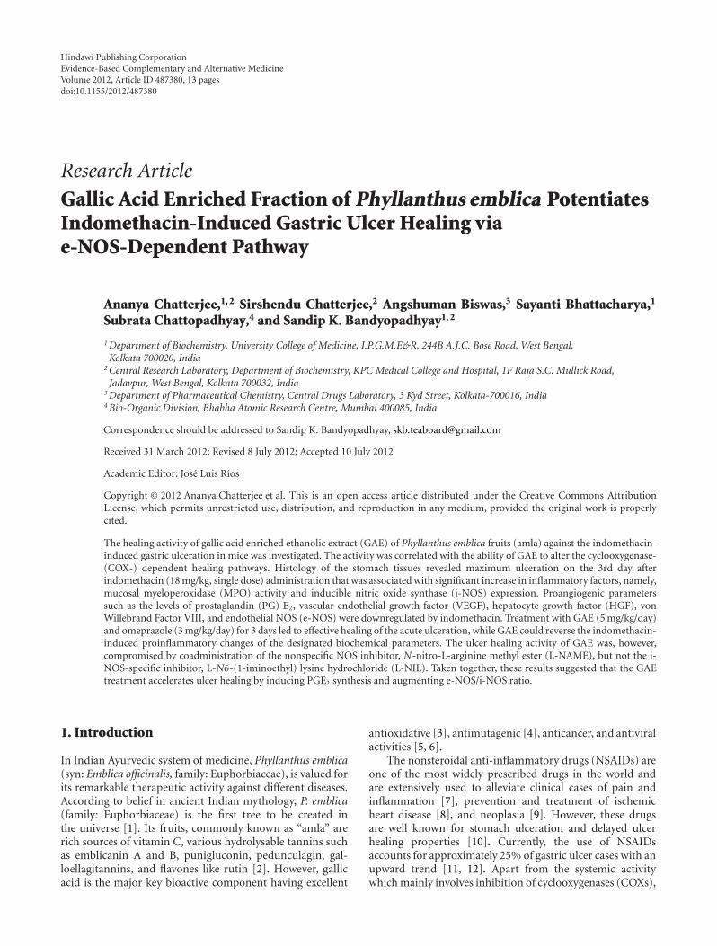
3.4. Modulation of NOS Expression. The Western blots of e-NOS and i-NOS expressions in the gastric mucosa of thecontrol, ulcerated and drug (GAE or omeprazole)-treatedmice are shown in Figure 5. The e-NOS expression wasdetected in both normal and ulcerated gastric tissues. Incontrast, the i-NOS expression was very high in the ulceratedtissues, but much less in normal gastric tissues. Our westernblot data revealed that three-day treatment with GAE sig-nificantly induced e-NOS expression, while reducing i-NOSexpression, compared to that in the untreated group. Al-though omeprazole also made similar changes in the expres-sions of the enzymes, however, the effect was much less.
3.5. Modulation of Tissue NO Level. Compared to the normalcontrol group, the tissue NO level in the ulcerated untreatedmice was suppressed by 69.4% (Figure 6). Compared to theuntreated mice, the tissue NO level was markedly increased(2.2 fold) in the GAE-treated group, while omeprazole didnot significantly alter this.
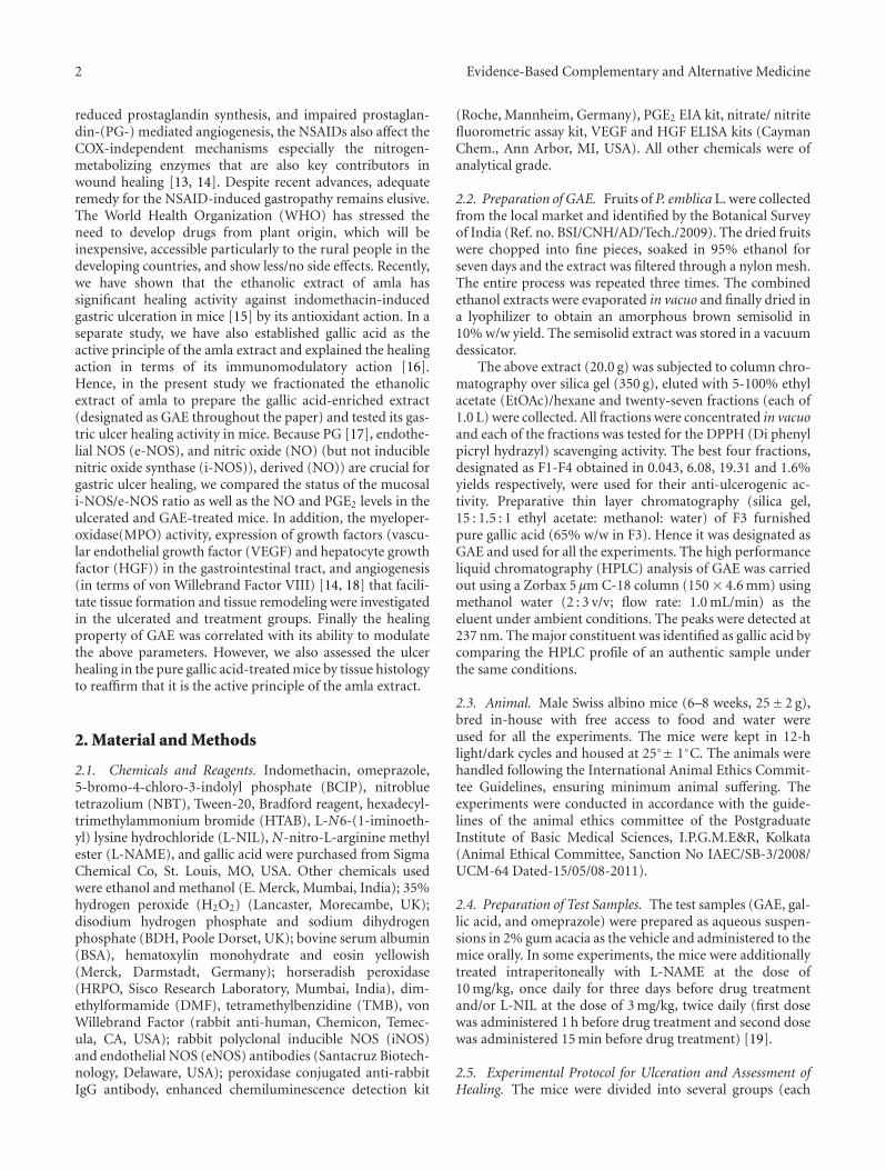
3.6. Quantification of von Willebrand Factor (vWF) VIII.The microscopic results using immunostaining of the vonWillebrand Factor VIII revealed the presence of 19.6 ± 1.15microvessels/mm2 in submucosa of control mice. This in-creased to 24.9± 1.42 in the ulcerated untreated mice Figure7(a). Treatment with GAE and omeprazole-enhanced themicrovessel number by 51.6% and 27.3% respectively com-pared to that in the untreated mice.
3.7. Regulation of Growth Factors. Indomethacin administra-tion downregulated the VEGF and HGF levels by 36.8% and34.2%, respectively, compared to sham-treated mice. TheVEGF and HGF levels in the GAE-treated group wereincreased 2.2-fold and 2.4-fold, respectively, compared to theuntreated mice (Figures 7(b) and 7(c)). Omeprazoleincreased the VEGF and HGF levels by 45.1% and 39.6%,respectively, compared to the untreated mice.
3.8. Effect of NOS Inhibitors on the Healing Property of GAE.Treatment with L-NAME in the GAE-treated group signif-icantly increased the damage score (50.7%) (Figure 8(a))and MPO activity (78.9%) (Figure 8(b)), but reduced themucosal NO level by 1.1-fold (Figure 8(c)) and microvesselsnumbers by 1.2-fold (Figure 8(d)) compared to the soleGAE-treated group. But none of these parameters changedsignificantly in the GAE + L-NIL group, compared to theGAE-treated group.
4. Discussion
Several factors such as oxidative stress, neutrophils activa-tion, as well as modulation of various enzymes, cytokines andsoluble mediators play crucial roles in the indomethacin-mediated gastric ulceration and delayed ulcer healing [26].Controlling these factors provides an opportunity to developimproved antiulcer medications. The gastrotoxicity ofindomethacin is generally explained in terms of COX inhi-bition, reduced PG synthesis and the impaired PG-mediatedangiogenesis. However, the process also involves alternateCOX-independent mechanisms, wherein other contributorssuch as the nitrogen-metabolizing enzymes [13, 14] andneutrophil infiltration [27] determine the healing process.
The impressive healing capacity of the ethanolic extractof amla against the indomethacin-induced gastric ulcer [15]encouraged us to investigate the probable modulatory effectof the extract on the COX-dependent [28] and independentpathways [29] of wound healing. For this purpose, we usedthe gallic acid-enriched fraction (GAE) of the amla extract.In the present study, indomethacin administration led tomucosal damage and augmented the MPO activity in theulcerated area of the gastric wall. Because MPO activity isincreased by the activated neutrophils, the above results sug-gested the involvement of neutrophils infiltration in gastriculceration. The MPO activity is known to increase under theulcerated conditions, and reduced during the healing process[30]. It is often used as a risk marker and diagnostic tool forassessing severity of gastric ulcer [31]. Treatment with GAEcould sufficiently restore the normal gastric mucosalintegrity, while reducing the MPO activity. Earlier, the crudeethanolic extract of amla (60 mg/kg) showed similar healingactivity as that of GAE (5 mg/kg). However, the GAE contentof the crude extract (60 mg/kg) would be ∼11 mg. Further,the extract was marginally more potent than pure gallic acidat the concentrations present in the effective dose of GAE.Taken together, these results established that gallic acid is theactive constituent of GAE, but some other constituents ofGAE may play synergistic roles in healing activity of GAE.
Evidence-Based Complementary and Alternative Medicine 7
1
1
2.62 ± 0.16 1.52 ± 0.09 1.91 ± 0.11
0.62 ± 0.06 3.27 ± 0.19 0.87 ± 0.13
i-NOS
e-NOS
β-actin
−−−
−− − −
+
+++
+
Ulcerated untreated
Ulcerated + GAE treated
Ulcerated + Omeprazole treated
(a)
0
0.5
1
1.5
2
2.5
3
NormalUlcerated untreated
Ulcerated + GAE treatedUlcerated + omeprazole treated
Rat
io o
f i-
NO
S to
β-a
ctin
Groups
(b)
4
3.5
3
2.5
2
1.5
1
0.5
0Groups
Rat
io o
f e-
NO
S to
β-a
ctin
NormalUlcerated untreated
Ulcerated + GAE treatedUlcerated + Omeprazole treated
(c)
Figure 5: The e-NOS and i-NOS expressions in normal, ulcerated and GAE (5 mg/kg, single dose daily × 3 days, p.o.) or omeprazole(3 mg/kg, p.o.) treated gastric tissues of mice, and their quantifications. Western blots of the expressions of the enzymes (a). Ratios of theintensities of i-NOS (b) and e-NOS (c) bands to that of the respective β-actin bands as quantified from the western blot images, usingKodak Gelquant software. The values (arbitrary unit, mean ± S.E.M.) are the density scanning results of three independent experiments,considering that of normal mice as 1.
These results also suggested a close relationship between thestate of the gastric inflammation and MPO activity. Earlierpretreatment with an antibody against neutrophils wasfound to prevent the indomethacin-induced gastric ulcer-ation [32]. Based on these, it is tempting to propose thatindomethacin first stimulates the neutrophils to releasesubstances which are related to inflammation. However,further studies are needed to clarify the sequence of events.
Besides indicating ulcer initiation and progression, neu-trophils infiltration is also reported to delay gastric ulcer
healing [33] and its reduction accelerates ulcer healing [34].Oxygen-free radicals derived from the activated neutrophilsdelay gastric ulcer healing in rats [35]. Furthermore, neu-trophils infiltration induces microcirculatory abnormalities[36] and its suppression promotes healing [37]. Hence, weused it as an oxidative marker in the present study.
The NSAIDs exert both therapeutic and toxic effects,mainly through reduction of the levels of circulating PGE2 atthe gastric mucosa. Besides stimulating mucus and bicarbon-ate secretion and mucosal blood flow, PGs also contribute

8 Evidence-Based Complementary and Alternative Medicine
0
50
100
150
200
250
Groups
DA
N fl
uor
esce
nce
(a.
u./
mg
tiss
ue)
∗
∗
∗
NormalUlcerated untreated
Ulcerated + GAE treatedUlcerated + Omeprazole
treated
Figure 6: Effect of GAE on mucosal NO level in indomethacin-induced ulcerated mice. Ulceration in the mice was induced byindomethacin (18 mg/kg, single dose, p.o.). Treatment was carriedout for 3 days with GAE (5 mg/kg, single dose daily× 3 days, p.o.) oromeprazole (3 mg/kg, p.o.) after ulcer induction. The mucosal NOlevels of the ulcerated untreated and treated mice were measured.The values are mean ± S.E. of four independent experiments, eachwith 8 mice/group. ∗P < 0.001, compared to normal mice; ∗∗P <0.001, compared to untreated mice.
to ulcer healing by inducing angiogenesis [17]. The reducedPGE2 causes gastric ulceration and also exacerbates preex-isting gastric ulcers in rodents and human [25]. Our datashowed that indomethacin treatment depleted the tissuePGE2 level that was increased significantly in the drug-treated groups (Figure 4). The effect of GAE was better thanthat of omeprazole. Enhanced PG synthesis is known toinhibit neutrophils-mediated free radical generation [38].Therefore, stimulation of PGE2 level by GAE might con-tribute to its antioxidative property, observed in the previousstudy.
The physiologically important NO, produced duringarginine catabolism by the NOSs plays dual roles in gastricmucosal defense and injury. The low concentration of NO,produced by e-NOS, one of the constitutive NOS isoformshelps in wound healing by increasing blood flow [39] andangiogenesis [19, 40] in the damaged gastric mucosa. How-ever, its enhanced generation by i-NOS may contribute tothe pathogenesis of various gastroduodenal disorders includ-ing peptic ulcer [30]. An increase in i-NOS activity and adecrease in e-NOS activity in the gastric mucosa are closelyrelated to the development of gastric mucosal lesions. Cur-rently we confirmed that the indomethacin-induced gastriculceration increased the mucosal i-NOS expression, butreduced the e-NOS expression in mice. Piotrowski et al. [41]showed a 12-fold increase in gastric epithelial expression ofiNOS activity in the indomethacin-administered animals,compared to controls and the increase correlated positively
with the epithelium damage. Our results showed only 1.3-fold increased i-NOS expression after ulceration. This maypossibly be due to the fact that we assayed it on the3rd day of ulceration. Despite the increased i-NOS expres-sion, the tissue NO level was significantly reduced in theindomethacin group. The apparent discrepancy may be duethe fact that i-NOS expression itself may not match with itsactivity. Also, the generated NO may be scavenged throughNADPH oxidase or MPO catalyzed reactions [42]. Thereduction of the beneficial vasodilatory NO would delay theulcer healing. Treatment with GAE raised the e-NOS/i-NOSratio to a level favourable for efficient ulcer healing. Theassociated increase in the tissue NO level must bederived through the e-NOS-catalyzed reaction, because GAEincreased e-NOS, but not i-NOS expressions. Earlier, usinge-NOS deficient mice, the importance of e-NOS and e-NOS-derived NO in regulating microvascular structure duringacute inflammation has been demonstrated [43].
Gastric ulcer healing entails several distinct repair mech-anisms. The epithelial cell proliferation and migration fromthe ulcer edge across the ulcer bed is accompanied bymaturation of granulation tissue beneath the ulcer base.Within this tissue vascular endothelial cells form newcapillaries to restore the microvasculature, while fibroblastsrestore the lamina propria. The degree of neovascularisation(angiogenesis), assessed by specific endothelial markersincluding von Willebrand Factor VIII, CD31, and CD34 inexperimental ulcer models correlates well with the extent andspeed of ulcer healing. Among these markers, von Wille-brand Factor VIII acts as a cofactor for platelet binding toexpose extracellular matrix in injured vessel walls. A largenumber of factors including several growth factors regulateangiogenic wound healing at its various stages [18, 44, 45].Amongst these, VEGF triggers endothelial proliferation andmigration and accelerate ulcer healing by promoting angio-genesis [14, 46]. Likewise HGF, expressed at the ulcer marginto act as trophic factors for the gastric mucosa helps angio-genesis by multiple mechanisms including COX activation[47]. Hence, we focused on these growth factors for thepresent studies.
Our result on the increased number of mucosal vonWillebrand Factor VIII in the ulcerated mice over that of nor-mal control mice is consistent with the requirement of moremicrovessels for ulcer healing. The increased number ofmicrovessels would assist better blood flow and transport ofoxygen and nutrients to the site of inflammation for quickerhealing. Treatment with GAE increased the von WillebrandFactor VIII further. This explains the accelerated ulcerhealing by GAE, compared to natural healing. The resultsare consistent with our previous finding with a resveratrol-analogue that also increased the e-NOS/i-NOS ratio toprovide better angiogenesis [25]. Indomethacin inhibitsADP-induced platelet aggregation and release of the α-granule, which stores VEGF. Consequently, indomethacintreatment would reduce VEGF release. We also found thatindomethacin administration suppressed the levels of VEGFand HGF. Both these parameters were increased significantlybeyond the respective normal values by GAE treatment(Figures 7(b) and 7(c)).
Evidence-Based Complementary and Alternative Medicine 9
0
10
20
30
40
50
Groups
∗
∗∗∗
#
Nu
mbe
r of
mic
rove
ssel
s/m
m2
NormalUlcerated untreated
Ulcerated + GAE treatedUlcerated + Omeprazole
treated
(a)
NormalUlcerated untreated
Ulcerated + GAE treatedUlcerated + Omeprazole
treated
0
10
20
30
40
Groups
∗∗
∗∗∗
#
Tis
sue
VE
GF
leve
l (n
g/m
L)
(b)
Normal
0
10
20
30
40
Groups
Tis
sue
HG
F le
vel (
ng/
mL)
∗∗
∗∗∗
#
Ulcerated untreatedUlcerated + GAE treatedUlcerated + Omeprazole
treated
(c)
Figure 7: Effect of GAE on different angiogenic parameters in indomethacin-induced ulcerated mice. (a) von Willebrand Factor VIII; (b)mucosal VEGF level; (c) mucosal HGF level. Ulceration in the mice was induced by indomethacin (18 mg /kg, single dose, p.o.). Treatmentwas carried out for 3 days with GAE (5 mg/kg, single dose daily × 3 days, p.o.) or omeprazole (3 mg/kg, p.o.) after ulcer induction. Themucosal von Willebrand Factor VIII (expressed as number of microvessels/mm2) and growth factors (expressed as ng/ml tissue extract)were measured by immunohistochemistry and colorimetry, respectively. ∗P < 0.05, ∗∗P < 0.01, compared to normal mice; #P < 0.01,∗∗∗P < 0.001, compared to untreated mice.
Enhanced synthesis of mucosal PGE2 and e-NOS-derivedNO by GAE might be instrumental in their ulcer-healingaction. On the other hand, omeprazole did not show anysignificant effect on NO synthesis (data not shown).
To substantiate our hypothesis that modulation of e-NOSmay primarily account for the excellent ulcer healing capacityof GAE, we studied the effects of L-NIL, a specific i-NOSinhibitor and L-NAME, a nonspecific NOS inhibitor on thehealing capacities of GAE. For this, we assessed four differentparameters, namely, (i) ulcer index, (ii) MPO activity, (iii)von Willebrand Factor VIII, and (iv) tissue NO level of the
GAE-treated mice in the absence and presence of the aboveinhibitors. Since i-NOS expression was effectively inhibitedby GAE alone (Figure 5), addition of L-NIL did not alterany of these parameters significantly. However, addition of L-NAME would suppress both e-NOS and i-NOS expressions,negating the augmented e-NOS expression, caused by GAE.Consistent with this, treatment with GAE in conjunctionwith L-NAME led to increased ulcer index and MPO activitywith associated decrease in von Willebrand Factor VIII andtissue NO level, compared to that with the only GAE-treatedmice (Figure 8). Taken together our results established that

10 Evidence-Based Complementary and Alternative Medicine
0
0.5
1
1.5
2
2.5
3
3.5
4
Groups
Ulc
er in
dex
(dam
age
scor
e)
∗
∗
∗
Untreated ulcerated
Ulcerated + GAE treated
Ulcerated + GAE +L-NAME treated
Ulcerated + GAE +L-NIL treated
(a)
0
0.5
1
1.5
2
2.5
3
3.5
Groups
MP
O a
ctiv
ity
(U/m
g pr
otei
n)
∗
∗∗
Untreated ulcerated
Ulcerated + GAE treated
Ulcerated + GAE +L-NAME treated
Ulcerated + GAE +L-NIL treated
(b)
Untreated ulcerated
0
20
40
60
80
100
120
140
160
180
200
Groups
Nu
mbe
r of
mic
rove
ssel
s/m
m2 ∗
∗∗
Ulcerated + GAE treated
Ulcerated + GAE +L-NAME treated
Ulcerated + GAE +L-NIL treated
(c)
0
10
20
30
40
50
Groups
DA
N fl
uor
escc
ence
(a.
u./
mg
tiss
ue)
∗
∗∗
Untreated ulcerated
Ulcerated + GAE treated
Ulcerated + GAE +L-NAME treated
Ulcerated + GAE +L-NIL treated
(d)
Figure 8: Effect of NOS inhibitors on the healing activity of GAE in indomethacin-induced ulcerated mice. (a) damage score, (b) MPOactivity, (c) von Willebrand Factor VIII, (d) NO level. Ulceration in the mice was induced by indomethacin (18 mg /kg, single dose, p.o.).After ulcer induction, treatment was carried out with GAE (5 mg/kg, single dose daily, p.o.) alone or in conjunction with L-NAME (15 mg/kg,once daily) or L-NIL (3 mg/kg, twice daily) for 3 days. The parameters of the ulcerated untreated and treated mice were measured. ∗P < 0.001compared to normal mice; ∗∗P < 0.01, compared to GAE-treatment.
the e-NOS-derived NO contributed maximum to the ulcerhealing property of GAE, although a role for neuronal NOS-derived NO cannot be excluded.
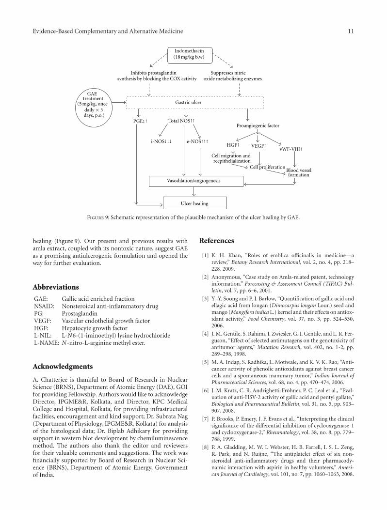
5. Conclusion
Overall, gallic acid was the active principle of the gallicacid enriched ethanolic amla extract (GAE) that promoted
healing of indomethacin-induced gastric ulcers in mice. Thebeneficial effect of GAE was due to its ability to reduceneutrophils infiltration and increase mucosal PGE2 as wellas NO levels that were downregulated by indomethacin.GAE increased the mucosal NO by augmenting the e-NOS/i-NOS ratio. All these factors, especially the modulation ofthe NOS-pathway helped in upregulating mucosal VEGF andHGF levels to promote angiogenesis and accelerate ulcer
Evidence-Based Complementary and Alternative Medicine 11
Indomethacin(18 mg/kg b.w)
Inhibits prostaglandinsynthesis by blocking the COX activity
Suppresses nitric
Gastric ulcer
GAE
daily × 3days, p.o.)
Proangiogenic factor
Vasodilation/angiogenesis
Ulcer healing
Cell migration andreepithelialization
Cell proliferationBlood vesselformation
oxide metabolizing enzymes
PGE2↑ Total NOS↑↑
i-NOS↓↓↓ e-NOS↑↑↑HGF↑ VEGF↑
vWF-VIII↑
treatment(5 mg/kg, once
Figure 9: Schematic representation of the plausible mechanism of the ulcer healing by GAE.
healing (Figure 9). Our present and previous results withamla extract, coupled with its nontoxic nature, suggest GAEas a promising antiulcerogenic formulation and opened theway for further evaluation.
Abbreviations
GAE: Gallic acid enriched fractionNSAID: Nonsteroidal anti-inflammatory drugPG: ProstaglandinVEGF: Vascular endothelial growth factorHGF: Hepatocyte growth factorL-NIL: L-N6-(1-iminoethyl) lysine hydrochlorideL-NAME: N-nitro-L-arginine methyl ester.
Acknowledgments
A. Chatterjee is thankful to Board of Research in NuclearScience (BRNS), Department of Atomic Energy (DAE), GOIfor providing Fellowship. Authors would like to acknowledgeDirector, IPGME&R, Kolkata, and Director, KPC MedicalCollege and Hospital, Kolkata, for providing infrastructuralfacilities, encouragement and kind support; Dr. Subrata Nag(Department of Physiology, IPGME&R, Kolkata) for analysisof the histological data; Dr. Biplab Adhikary for providingsupport in western blot development by chemiluminescencemethod. The authors also thank the editor and reviewersfor their valuable comments and suggestions. The work wasfinancially supported by Board of Research in Nuclear Sci-ence (BRNS), Department of Atomic Energy, Governmentof India.
References
[1] K. H. Khan, “Roles of emblica officinalis in medicine—areview,” Botany Research International, vol. 2, no. 4, pp. 218–228, 2009.
[2] Anonymous, “Case study on Amla-related patent, technologyinformation,” Forecasting & Assessment Council (TIFAC) Bul-letin, vol. 7, pp. 6–6, 2001.
[3] Y.-Y. Soong and P. J. Barlow, “Quantification of gallic acid andellagic acid from longan (Dimocarpus longan Lour.) seed andmango (Mangifera indica L.) kernel and their effects on antiox-idant activity,” Food Chemistry, vol. 97, no. 3, pp. 524–530,2006.
[4] J. M. Gentile, S. Rahimi, J. Zwiesler, G. J. Gentile, and L. R. Fer-guson, “Effect of selected antimutagens on the genotoxicity ofantitumor agents,” Mutation Research, vol. 402, no. 1-2, pp.289–298, 1998.
[5] M. A. Indap, S. Radhika, L. Motiwale, and K. V. K. Rao, “Anti-cancer activity of phenolic antioxidants against breast cancercells and a spontaneous mammary tumor,” Indian Journal ofPharmaceutical Sciences, vol. 68, no. 4, pp. 470–474, 2006.
[6] J. M. Kratz, C. R. Andrighetti-Frohner, P. C. Leal et al., “Eval-uation of anti-HSV-2 activity of gallic acid and pentyl gallate,”Biological and Pharmaceutical Bulletin, vol. 31, no. 5, pp. 903–907, 2008.
[7] P. Brooks, P. Emery, J. F. Evans et al., “Interpreting the clinicalsignificance of the differential inhibition of cyclooxygenase-1and cyclooxygenase-2,” Rheumatology, vol. 38, no. 8, pp. 779–788, 1999.
[8] P. A. Gladding, M. W. I. Webster, H. B. Farrell, I. S. L. Zeng,R. Park, and N. Ruijne, “The antiplatelet effect of six non-steroidal anti-inflammatory drugs and their pharmacody-namic interaction with aspirin in healthy volunteers,” Ameri-can Journal of Cardiology, vol. 101, no. 7, pp. 1060–1063, 2008.

12 Evidence-Based Complementary and Alternative Medicine
[9] G. A. Piazza, A. B. Keeton, H. N. Tinsley et al., “NSAIDs: olddrugs reveal new anticancer targets,” Pharmaceuticals, vol. 3,no. 5, pp. 1652–1667, 2010.
[10] A. Lanas, M. A. Perez-Aisa, F. Feu et al., “A nationwidestudy of mortality associated with hospital admission dueto severe gastrointestinal events and those associated withnonsteroidal antiinflammatory drug use,” American Journal ofGastroenterology, vol. 100, no. 8, pp. 1685–1693, 2005.
[11] A. S. Tarnawski and M. K. Jones, “Inhibition of angiogenesisby NSAIDs: molecular mechanisms and clinical implications,”Journal of Molecular Medicine, vol. 81, no. 10, pp. 627–636,2003.
[12] F. K. L. Chan, “Primer: managing NSAID-induced ulcercomplications—balancing gastrointestinal and cardiovascularrisks,” Nature Clinical Practice Gastroenterology and Hepatol-ogy, vol. 3, no. 10, pp. 563–573, 2006.
[13] J. I. Isenberg, K. R. McQuaid, L. Laine, and W. Rubin, “Acid-peptic disorders,” in Textbook of Gastroenterology, T. Yamada,D. H. Alpers, C. Owyng, D. W. Powell, and F. E. Silverstein,Eds., vol. 1, p. 1253, 1991.
[14] A. Tarnawski, I. L. Szabo, S. S. Husain, and B. Soreghan,“Regeneration of gastric mucosa during ulcer healing istriggered by growth factors and signal transduction pathways,”Journal of Physiology Paris, vol. 95, no. 1-6, pp. 337–344, 2001.
[15] A. Chatterjee, S. Chattopadhyay, and S. K. Bandyopad-hyay, “Biphasic effect of Phyllanthus emblica L. extract onNSAID-induced ulcer: an antioxidative trail weaved withimmunomodulatory effect,” Evidence-Based Complementaryand Alternative Medicine, vol. 2011, Article ID 146808, 13pages, 2011.
[16] S. Bhattacharya, S. Chatterjee, A. Bauri et al., “Immunophar-macological basis of the healing of indomethacin-induced gas-tric mucosal damage in rats by the constituents of Phyllanthusemblica,” Current Science, vol. 93, no. 1, pp. 47–53, 2007.
[17] M. K. Jones, H. Wang, B. M. Peskar et al., “Inhibition ofangiogenesis by nonsteroidal anti-inflammatory drugs: insightinto mechanisms and implications for cancer growth and ulcerhealing,” Nature Medicine, vol. 5, no. 12, pp. 1418–1423, 1999.
[18] J. Folkman and P. A. D’Amore, “Blood vessel formation: whatis its molecular basis?” Cell, vol. 87, no. 7, pp. 1153–1155, 1996.
[19] L. Ma and J. L. Wallace, “Endothelial nitric oxide synthasemodulates gastric ulcer healing in rats,” American Journal ofPhysiology, vol. 279, no. 2, pp. G341–G346, 2000.
[20] D. Banerjee, B. Maity, A. K. Bauri, S. K. Bandyopadhyay, andS. Chattopadhyay, “Gastroprotective properties of Myristicamalabarica against indometacin-induced stomach ulceration:a mechanistic exploration,” Journal of Pharmacy and Pharma-cology, vol. 59, no. 11, pp. 1555–1565, 2007.
[21] D. Banerjee, A. K. Bauri, R. K. Guha, S. K. Bandyopadhyay,and S. Chattopadhyay, “Healing properties of malabariconeB and malabaricone C, against indomethacin-induced gastriculceration and mechanism of action,” European Journal ofPharmacology, vol. 578, no. 2-3, pp. 300–312, 2008.
[22] D. Dokmeci, M. Akpolat, N. Aydogu, L. Doganay, and F. N.Turan, “L-Carnitine inhibits ethanol-induced gastric mucosalinjury in rats,” Pharmacological Reports, vol. 57, pp. 481–488,2005.
[23] K. Suzuki, H. Ota, S. Sasagawa, T. Sakatani, and T. Fujikura,“Assay method for myeloperoxidase in human polymor-phonuclear leukocytes,” Analytical Biochemistry, vol. 132, pp.345–352, 1983.
[24] M. M. Bradford, “A rapid and sensitive method for thequantitation of microgram quantities of protein utilizing theprinciple of protein-dye binding,” Analytical Biochemistry, vol.72, pp. 248–254, 1976.
[25] P. Guha, A. Dey, A. Chatterjee, S. Chattopadhyay, and S.K. Bandyopadhyay, “Pro-ulcer effects of resveratrol in micewith indomethacin-induced gastric ulcers are reversed by L-arginine,” British Journal of Pharmacology, vol. 159, pp. 726–734, 2010.
[26] S. Fiorucci, E. Antonelli, and A. Morelli, “Mechanism of non-steroidal anti-inflammatory drug-gastropathy,” Digestive andLiver Disease, vol. 33, no. 2, pp. S35–S43, 2001.
[27] L. Osborn, “Leukocyte adhesion to endothelium in inflamma-tion,” Cell, vol. 62, no. 1, pp. 3–6, 1990.
[28] R. Langenbach, S. G. Morham, H. F. Tiano et al.,“Prostaglandin synthase 1 gene disruption in mice reducesarachidonic acid- induced inflammation and indomethacin-induced gastric ulceration,” Cell, vol. 83, no. 3, pp. 483–492,1995.
[29] I. Tegeder, J. Pfeilschifter, and G. Geisslinger, “Cyclooxygenase-independent actions of cyclooxygenase inhibitors,” FASEBJournal, vol. 15, no. 12, pp. 2057–2072, 2001.
[30] M. H. L. P. Souza, H. Paula Lemos, R. B. Oliveira, and F. Q.Cunha, “Gastric damage and granulocyte infiltration inducedby indomethacin in tumour factor 1 (TNF-R1) or induciblenitric oxide synthase (iNOS) deficient mice,” Gut, vol. 53, no.6, pp. 791–796, 2004.
[31] J. E. Krawisz, P. Sharon, and W. F. Stenson, “Quantitative assayfor acute intestinal inflammation based on myeloperoxidaseactivity. Assessment of inflammation in rat and hamstermodels,” Gastroenterology, vol. 87, no. 6, pp. 1344–1350, 1984.
[32] J. L. Wallace, C. M. Keenan, and D. N. Granger, “Gastriculceration induced by nonsteroidal anti-inflammatory drugsis a neutrophil-dependent process,” American Journal ofPhysiology, vol. 259, no. 3, pp. G462–G467, 1990.
[33] H. Fujita, S. Takahashi, and S. Okabe, “Mechanism by whichindomethacin delays the healing of acetic acid- induced ulcersin rats. Role of neutrophil antichemotactic and chemotacticactivities,” Journal of Physiology and Pharmacology, vol. 49, no.1, pp. 71–82, 1998.
[34] N. Shimizu, T. Watanabe, T. Arakawa, Y. Fujiwara, K. Higuchi,and T. Kuroki, “Pentoxifylline accelerates gastric ulcer healingin rats: roles of tumor necrosis factor alpha and neutrophilsduring the early phase of ulcer healing,” Digestion, vol. 61, no.3, pp. 157–164, 2000.
[35] Y. Suzuki, M. Ishihara, T. Segami, and M. Ito, “Anti-ulcereffects of antioxidants, quercetin, α-tocopherol, nifedipine andtetracycline in rats,” Japanese Journal of Pharmacology, vol. 78,no. 4, pp. 435–441, 1998.
[36] C. F. Bou-Abboud, H. Wayland, G. Paulsen, and P. H. Guth,“Microcirculatory stasis precedes tissue necrosis in ethanol-induced gastric mucosal injury in the rat,” Digestive Diseasesand Sciences, vol. 33, no. 7, pp. 872–877, 1988.
[37] Y. Tsukimi, C. Nozue, and S. Okabe, “Effects of leminoprazole,omeprazole and sucralfate on indomethacin-induced delayedhealing of kissing gastric ulcers in rats,” Journal of Gastroen-terology and Hepatology, vol. 11, no. 4, pp. 335–340, 1996.
[38] R. J. Gryglewski, A. Szczeklik, and M. Wandzilak, “The effectof six prostaglandins, prostacyclin and iloprost on generationof superoxide anions by human polymorphonuclear leuko-cytes stimulated by zymosan or formyl-methionyl-leucyl

Evidence-Based Complementary and Alternative Medicine 13
-phenylalanine,” Biochemistry Pharmacology, vol. 36, pp.4209–4213, 1987.
[39] J. R. Whittle, “Nitric oxide in gastrointestinal physiology andpathology,” in The Physiology of the Gastrointestinal Tract, L. R.Johnson, Ed., pp. 267–294, Raven, New York, NY, USA, 1994.
[40] M. Ziche, L. Morbidelli, E. Masini et al., “Nitric oxidemediates angiogenesis in vivo and endothelial cell growthand migration in vitro promoted by substance P,” Journal ofClinical Investigation, vol. 94, no. 5, pp. 2036–2044, 1994.
[41] J. Piotrowski, A. Slomiany, and B. L. Slomiany, “Activationof apoptotic caspase-3 and nitric oxide synthase-2 in gastricmucosal injury induced by indomethacin,” ScandinavianJournal of Gastroenterology, vol. 34, no. 2, pp. 129–134, 1999.
[42] J. Morton, B. Coles, K. Wright et al., “Circulating neutrophilsmaintain physiological blood pressure by suppressing bacteriaand IFNγ-dependent iNOS expression in the vasculature ofhealthy mice,” Blood, vol. 111, no. 10, pp. 5187–5194, 2008.
[43] J. C. Luo, V. Y. Shin, E. S. L. Liu et al., “Non-ulcerogenic doseof dexamethasone delays gastric ulcer healing in rats,” Journalof Pharmacology and Experimental Therapeutics, vol. 307, no.2, pp. 692–698, 2003.
[44] J. Folkman and Y. Shin, “Angiogenesis,” Journal of BiologicalChemistry, vol. 267, pp. 10931–10934, 1992.
[45] W. Risau, “Mechanisms of angiogenesis,” Nature, vol. 386, pp.671–673, 1997.
[46] M. Takahashia, K. Oguraa, S. Maedaa et al., “Promotersof epithelialization induce expression of vascular endothelialgrowth factor in human gastric epithelial cells in primaryculture,” FEBS Letter, vol. 418, pp. 115–118, 1997.
[47] T. Brzozowski, P. C. Konturek, S. J. Konturek et al., “Involve-ment of cyclooxygenase (COX)-2 products in acceleration ofulcer healing by gastrin and hepatocyte growth factor,” Journalof Physiology and Pharmacology, vol. 51, no. 4, pp. 751–773,2000.

Submit your manuscripts athttp://www.hindawi.com
Stem CellsInternational
Hindawi Publishing Corporationhttp://www.hindawi.com Volume 2014
Hindawi Publishing Corporationhttp://www.hindawi.com Volume 2014
MEDIATORSINFLAMMATION
of
Hindawi Publishing Corporationhttp://www.hindawi.com Volume 2014
Behavioural Neurology
EndocrinologyInternational Journal of
Hindawi Publishing Corporationhttp://www.hindawi.com Volume 2014
Hindawi Publishing Corporationhttp://www.hindawi.com Volume 2014
Disease Markers
Hindawi Publishing Corporationhttp://www.hindawi.com Volume 2014
BioMed Research International
OncologyJournal of
Hindawi Publishing Corporationhttp://www.hindawi.com Volume 2014
Hindawi Publishing Corporationhttp://www.hindawi.com Volume 2014
Oxidative Medicine and Cellular Longevity
Hindawi Publishing Corporationhttp://www.hindawi.com Volume 2014
PPAR Research
The Scientific World JournalHindawi Publishing Corporation http://www.hindawi.com Volume 2014
Immunology ResearchHindawi Publishing Corporationhttp://www.hindawi.com Volume 2014
Journal of
ObesityJournal of
Hindawi Publishing Corporationhttp://www.hindawi.com Volume 2014
Hindawi Publishing Corporationhttp://www.hindawi.com Volume 2014
Computational and Mathematical Methods in Medicine
OphthalmologyJournal of
Hindawi Publishing Corporationhttp://www.hindawi.com Volume 2014
Diabetes ResearchJournal of
Hindawi Publishing Corporationhttp://www.hindawi.com Volume 2014
Hindawi Publishing Corporationhttp://www.hindawi.com Volume 2014
Research and TreatmentAIDS
Hindawi Publishing Corporationhttp://www.hindawi.com Volume 2014
Gastroenterology Research and Practice
Hindawi Publishing Corporationhttp://www.hindawi.com Volume 2014
Parkinson’s Disease
Evidence-Based Complementary and Alternative Medicine
Volume 2014Hindawi Publishing Corporationhttp://www.hindawi.com