ganodermataceae (polyporales): diversity in greater mekong
TRANSCRIPT

Submitted 16 January 2019, Accepted 25 March 2019, Published 29 April 2019
Corresponding Author: Author: Ting-Chi Wen – e-mail – [email protected] 221
Ganodermataceae (Polyporales): Diversity in Greater Mekong
Subregion countries (China, Laos, Myanmar, Thailand and Vietnam)
Hapuarachchi KK2,3,4, Karunarathna SC5, Phengsintham P6, Yang HD2,7,
Kakumyan P4, Hyde KD3,4,5, Wen TC1,2
1State Key Laboratory Breeding Base of Green Pesticide and Agricultural Bioengineering, Key Laboratory of Green
Pesticide and Agricultural Bioengineering, Ministry of Education, Guizhou University, Guiyang 550025, China 2The Engineering Research Center of Southwest Bio–Pharmaceutical Resource Ministry of Education, Guizhou
University, Guiyang 550025, Guizhou Province, China 3Center of Excellence in Fungal Research, Mae Fah Luang University, Chiang Rai 57100, Thailand 4School of Science, Mae Fah Luang University, Chiang Rai 57100, Thailand 5Key Laboratory for Plant Diversity and Biogeography of East Asia, Kunming Institute of Botany, Chinese Academy of
Sciences, 132 Lanhei Road, Kunming 650201, China 6National University of Laos, Dongdok, Vientiane, Vientiane, Lao PDR 7College of life science, Southwest Forestry University, Kunming 650224, China
Hapuarachchi KK, Karunarathna SC, Phengsintham P, Yang HD, Kakumyan P, Hyde KD, Wen TC
2019 – Ganodermataceae (Polyporales): Diversity in Greater Mekong Subregion countries (China,
Laos, Myanmar, Thailand and Vietnam). Mycosphere 10(1), 221–309, Doi
10.5943/mycosphere/10/1/6
Abstract
Taxa of Ganodermataceae have been widely used as traditional medicines for centuries in
Asia. Despite several taxonomic investigations, relationships and classification of many species are
still unresolved. Species in this family are either pathogenic, wood decaying and/or wood
inhabiting. In this paper, we introduce, a collection of Ganodermataceae species based on fresh and
dried specimens found within the Greater Mekong Subregion countries; China, Laos, Myanmar,
Thailand and Vietnam. Amauroderma schomburgkii, A. rude, Haddowia longipes, Ganoderma
lingzhi, G. luteomarginatum, G. subresinosum and G. tropicum from Laos, G. australe and G.
multiplicatum from Myanmar, G. donkii from Thailand, G. adspersum from Thailand and
Myanmar, G. flexipes, G. gibbosum, G. orbiforme, and G. neojaponicum from both Laos and
Myanmar, are newly recorded species for these countries. We also identified A. schomburgkii and
A. rude, based on morphology and the other species based on both morphology and DNA sequence
data. Two species; G. nasalanense Hapuar., Pheng., & K.D. Hyde, sp. nov., and G. sandunense
Hapuar., T.C. Wen & K.D. Hyde, sp. nov., are new to science and established with morphological
and DNA sequence based evidence. All taxa collected are described and illustrated with coloured
photographs. We present an updated phylogeny for Ganodermataceae based on nrLSU, ITS,
nrSSU, TEF1 and RPB2 DNA sequence data and species relationships and classification are
discussed.
Key words – new taxa – new records – morphology – pathogenic species – phylogeny
Introduction
Ganodermataceae is a large family of polypores with seven accepted genera: Amauroderma
Murril, Foraminispora Robledo et al., Furtadoa Costa-Rezende et al., Ganoderma P. Karst,
Mycosphere 10(1): 221–309 (2019) www.mycosphere.org ISSN 2077 7019
Article
Doi 10.5943/mycosphere/10/1/6

222
Haddowia Steyaert, Humphreya Steyaert and Polyporopsis Audet (Richter et al. 2015, Costa-
Rezende et al. 2017) including 596 epithets, of which most are known as Ganoderma species
(www.indexfungorum.org, accessed 22 March 2019). This family has received great attention from
mycologists for over many decades. Recently the phylogeny of Ganodermataceae and its allied
genera has been reconstructed using multigene DNA sequences including ITS, IGS, nrLSU, nrSSU,
RPB2, TEF1, β-tubulin, mtSSU, mtLSU, and ATP6 genes (Wang & Yao 2005, Wang 2012, Zhou
et al. 2015). These species have a worldwide distribution in green ecosystems, both in tropical and
temperate geographical regions, such as East Asia (China, Japan and South Korea), East Africa
(Ghana, Kenya and Tanzania) as well as Europe (almost all the European countries), North
America (Canada and U.S.A.), Oceania (Australia), South America (Argentina, Brazil and
Uruguay), South and Southeast Asia (India, Indonesia, Philippines, Thailand and Vietnam) (Wang
et al. 2012, Pilotti et al. 2003). The majority of taxa are facultative parasites that live as saprobes on
rotting stumps and roots (Pilotti et al. 2004, Dai et al. 2007).
Ganodermataceae is distinct from other families of polypores, in having a peculiar type of
double-walled basidiospores (Adaskaveg & Gilbertson 1988). The inner walls of the
Ganodermataceae spores are quite often colored, and usually the surface is ornamented (Donk
1964). Many polypores have bigger basidia than Ganoderma species. The hyphal system is usually
trimitic and occasionally dimitic or monomitic, which contains hyaline, thin-walled, branched,
clamped generative hyphae, pigmented, thick walled, arboriform or aciculiform, branched skeletal
hyphae and colorless, terminal branched binding hyphae (Seo & Kirk 2000). Ganoderma represents
the largest genus of Ganodermataceae (Wu & Zhang 2003). The genus is characterized by double-
walled basidiospores with a truncate apex and ornamented endospores (Moncalvo & Ryvarden
1997). Most of the species of Ganoderma belong to the Ganoderma lucidum species complex. The
second largest genus is Amauroderma with tropical stipitate to sessile species with non-truncate,
globose to subglobose, verrucose to asperulate (smooth in only one species) basidiospores
(Ryvarden 2004b). These characteristics distinguish Amauroderma from the other genera, as
Ganoderma and Humphreya which have truncated basidiospores and Haddowia which has crested
basidiospores while Polyporopsis has ellipsoidal basidiospores with oily drops (Ryvarden 2004b,
Le et al. 2012, Audet 2010, Hapuarachchi et al. 2018c).
Taxa of Ganodermataceae have been widely used as traditional medicines for millennia in
Asia (Dai et al. 2007, Zhou et al. 2015). Its species are widely researched, because of their highly
prized medicinal value, since they contain many chemical constituents with potential nutritional
and therapeutic values (Hapuarachchi et al. 2016a, b, 2017). Ganoderma and Amauroderma include
several species which possess great economic and ecological importance (Correia de Lima et al.
2014). Ganoderma species have gained wide popular use as dietary supplements in China, Japan,
North America and the other regions of the world (Paterson 2006, Hapuarachchi et al. 2018a).
Ganoderma has been used as a functional food to prevent and treat immunological diseases (Wang
et al. 2012). Several hundreds of metabolites have been obtained from the species of this family
including G. lucidum, which is the most sought after species of the genus (Dong & Han 2015).
Amauroderma rugosum is traditionally used by the Chinese to reduce inflammation, to treat
diuretic and upset stomach, and to prevent cancer (Dai & Yang 2008).
The traditional taxonomy of Ganodermataceae is based on its morphological traits, however
species identification and circumscriptions have been unclear and taxonomic segregation of the
genera has been controversial because of different viewpoints among mycologists (Moncalvo et al.
1995a, Moncalvo & Ryvarden 1997). Most of the taxonomists judge the current nomenclatural
situation of Ganodermataceae as chaotic and poorly studied (Ryvarden 1991, Smith &
Sivasithamparam 2003, Hapuarachchi et al. 2015). Earlier it was believed that this family
represented an old lineage from which other groups of polypores have been derived (Corner 1983).
Phenotypic plasticity observed in the genus is an indication that species are not evolving rapidly
resulting in low speciation rate (Ryvarden 1994), an observation supported by Moncalvo &
Buchanan (2008). Use of DNA sequence data especially from ribosomal DNA sequences have
clarified to a certain extent classification and species relationships. Some researchers suggested

223
using a combination of morphological, chemotaxonomic and molecular methods to develop a more
stable taxonomy for Ganodermataceae and resolve taxonomic ambuiguities (Richter et al. 2015,
Welti et al. 2015). Table 1 lists the recent taxonomic classification system for Ganodermataceae.
The Greater Mekong Subregion covers an area of approximately 2.5 million km2 including parts of
China’s Yunnan Province, Cambodia, Laos, Myanmar, Thailand and Vietnam. The Mekong River
is one of the largest watercourses flowing through the region. The Mekong is well known for its
high levels of biodiversity and famous for its mushroom diversity with at least 650 edible and
medicinal species (Mortimer et al. 2014). There is potentially a high number of novel species in the
region (Hyde et al. 2018). These mushrooms not only provide a source of food, but a very
important source of income and play a vital role in supporting people’s livelihoods. Furthermore,
the ecological contributions of mushrooms in this region are immense and incredibly important
(Mortimer et al. 2014). Furthermore, some species are pathogens for various economic plants and
these diseases are major problems can result in consequent economic loss in these regional
countries. Hence, it is important to study the diversity of this family in the Greater Mekong
Subregion.
Table 1 The recent taxonomic classification system for Ganodermataceae
Taxonomic rank Taxon Introduced by (Reference) Other main references
Family Ganodermataceae Donk, M.A. 1948. Notes
on Malesian fungi. I.
Bulletin du Jardin
Botanique de Buitenzorg.
17:473-482
Genus Ganoderma Karsten 1881 Steyaert 1972
Amauroderma
(Magoderna)
Murrill 1905a Steyaert 1972
Magoderna Steyaert 1972 Moncalvo &
Ryvarden 1997,
Gomes-Silva et al.
2015
Haddowia Steyaert 1972
Humphreya Steyaert 1972
Polyporopsis Audet 2010 Richter et al. 2015
Foraminispora Costa-Rezende et al 2017
Furtadoa Costa-Rezende et al 2017
The objectives of the present study are to document species of Ganodermataceae based on
morphological characteristics and compared with similar taxa. Furthermore, we provide a
phylogeny for the Ganodermataceae based on combined nrLSU, ITS, nrSSU, EF and RPB2
analyses. A checklist of species of Ganodermataceae reported from Greater Mekong Subregion
countries is also given in Table 2.
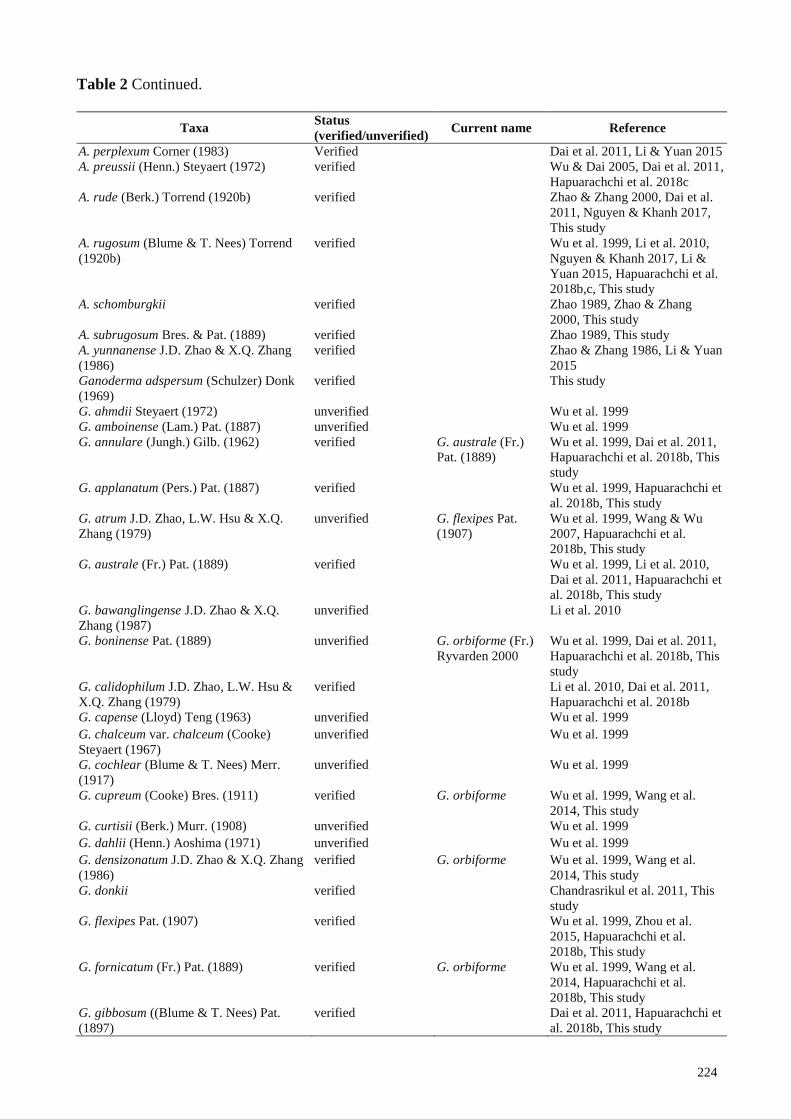
Table 2 List of Ganodermataceae species known for Greater Mekong Subregion countries.
Taxa Status
(verified/unverified) Current name Reference
Amauroderma austrosinense J.D. Zhao &
L.W. Hsu (1984)
verified Wu et al. 1999, Li & Yuan
2015, Hapuarachchi et al. 2018b
A. concentricum J. Song, Xiao L. He &
B.K. Cui (2016)
verified Song et al. 2016
A. conjunctum (Lloyd) Torrend (1920b) verified Nguyen & Khanh 2017
A. exile (Berk.) Torrend (1920b) verified Nguyen & Khanh 2017
224
Table 2 Continued.
Taxa Status
(verified/unverified) Current name Reference
A. perplexum Corner (1983) Verified Dai et al. 2011, Li & Yuan 2015
A. preussii (Henn.) Steyaert (1972) verified Wu & Dai 2005, Dai et al. 2011,
Hapuarachchi et al. 2018c
A. rude (Berk.) Torrend (1920b) verified Zhao & Zhang 2000, Dai et al.
2011, Nguyen & Khanh 2017,
This study
A. rugosum (Blume & T. Nees) Torrend
(1920b)
verified Wu et al. 1999, Li et al. 2010,
Nguyen & Khanh 2017, Li &
Yuan 2015, Hapuarachchi et al.
2018b,c, This study
A. schomburgkii verified Zhao 1989, Zhao & Zhang
2000, This study
A. subrugosum Bres. & Pat. (1889) verified Zhao 1989, This study
A. yunnanense J.D. Zhao & X.Q. Zhang
(1986)
verified Zhao & Zhang 1986, Li & Yuan
2015
Ganoderma adspersum (Schulzer) Donk
(1969)
verified This study
G. ahmdii Steyaert (1972) unverified Wu et al. 1999
G. amboinense (Lam.) Pat. (1887) unverified Wu et al. 1999
G. annulare (Jungh.) Gilb. (1962) verified G. australe (Fr.)
Pat. (1889)
Wu et al. 1999, Dai et al. 2011,
Hapuarachchi et al. 2018b, This
study
G. applanatum (Pers.) Pat. (1887) verified Wu et al. 1999, Hapuarachchi et
al. 2018b, This study
G. atrum J.D. Zhao, L.W. Hsu & X.Q.
Zhang (1979)
unverified G. flexipes Pat.
(1907)
Wu et al. 1999, Wang & Wu
2007, Hapuarachchi et al.
2018b, This study
G. australe (Fr.) Pat. (1889) verified Wu et al. 1999, Li et al. 2010,
Dai et al. 2011, Hapuarachchi et
al. 2018b, This study
G. bawanglingense J.D. Zhao & X.Q.
Zhang (1987)
unverified Li et al. 2010
G. boninense Pat. (1889) unverified G. orbiforme (Fr.)
Ryvarden 2000
Wu et al. 1999, Dai et al. 2011,
Hapuarachchi et al. 2018b, This
study
G. calidophilum J.D. Zhao, L.W. Hsu &
X.Q. Zhang (1979)
verified Li et al. 2010, Dai et al. 2011,
Hapuarachchi et al. 2018b
G. capense (Lloyd) Teng (1963) unverified Wu et al. 1999
G. chalceum var. chalceum (Cooke)
Steyaert (1967)
unverified Wu et al. 1999
G. cochlear (Blume & T. Nees) Merr.
(1917)
unverified Wu et al. 1999
G. cupreum (Cooke) Bres. (1911) verified G. orbiforme Wu et al. 1999, Wang et al.
2014, This study
G. curtisii (Berk.) Murr. (1908) unverified Wu et al. 1999
G. dahlii (Henn.) Aoshima (1971) unverified Wu et al. 1999
G. densizonatum J.D. Zhao & X.Q. Zhang
(1986)
verified G. orbiforme Wu et al. 1999, Wang et al.
2014, This study
G. donkii verified Chandrasrikul et al. 2011, This
study
G. flexipes Pat. (1907) verified Wu et al. 1999, Zhou et al.
2015, Hapuarachchi et al.
2018b, This study
G. fornicatum (Fr.) Pat. (1889) verified G. orbiforme Wu et al. 1999, Wang et al.
2014, Hapuarachchi et al.
2018b, This study
G. gibbosum ((Blume & T. Nees) Pat.
(1897)
verified Dai et al. 2011, Hapuarachchi et
al. 2018b, This study

225
Table 2 Continued.
Taxa Status
(verified/unverified) Current name Reference
G. hainanense J.D. Zhao, L.W. Hsu &
X.Q. Zhang (1979)
unverified G. flexipes Wu et al. 1999, Wang & Wu
2007, Hapuarachchi et al.
2018b, This study
G. hoehnelianum Bres. (1912) verified Wu et al. 1999, Wang & Wu
2010, Hapuarachchi et al. 2018b
G. jiangfenglingense X.L. Wu. (1996) unverified Wu et al. 1999
G. 1eytense Steyaert (1972) unverified Wu et al. 1999
G. limushanense J.D. Zhao & X.Q. Zhang
(1986)
verified G. orbiforme Wu et al. 1999, Wang et al.
2014, Hapuarachchi et al.
2018b, This study
G. lobatum (Schwein.) G.F. Atk. (1908) unverified Wu et al. 1999
G. luteomarginatum J.D. Zhao, L.W. Hsu
& X.Q. Zhang (1979)
verified Wu et al. 1999, Zhao et al. 1979,
This study
G. mastoporum (Lev.) Pat. (1889) verified G. orbiforme Wu et al. 1999, Wang et al.
2014, Hapuarachchi et al.
2018b, This study
G. multipileum Ding Hou. (1950) verified Dai et al. 2011, Wang et al.
2005, Zhou et al. 2015
G. multiplicatum (Mont.) Pat. (1889) verified Zhao & Zhang 2000, Wang &
Wu 2007, Hapuarachchi et al.
2018b, This study
G. neojaponicum Imazeki (1939) verified Tan et al. 2015, This study
G. nigrolucidum (Lloyd) D.A. Reid (1975) unverified Dai et al. 2004, Li et al. 2010
G. parviungulatum J.D. Zhao & X.Q.
Zhang (1986)
unverified G. flexipes Wu et al. 1999, Cao et al. 2012,
This study
G. ramosissimum J.D. Zhao (1989) unverified Wu et al. 1999
G. resinaceum Boud (1890) verified Wu et al. 1999
Hapuarachchi et al. 2018b, This
study
G. shangsiense J.D. Zhao. (1988) verified G. hoehnelianum Li et al. 2010, Wang & Wu
2010, Hapuarachchi et al. 2018b
G. sinense J.D. Zhao. L.W. Hsu & X.Q.
Zhang (1979)
verified Dai et al. 2004, Wang et al.
2005, Hapuarachchi et al.
2018b, This study
G. subresinosum (Murrill) C.J. Humphrey
(1938)
verified Wu et al. 1999, Li et al. 2010,
Hapuarachchi et al. 2018b, This
study
G. theaecola J.D. Zhao (1984) verified Zhao et al. 1984, This study
G. tornatum (Pers.) Bres. (1912) verified G. australe Li et al. 2010, This study
G. tropicum (Jungh.) Bres. (1910) verified Li et al. 2010, Zhou et al. 2015
Hapuarachchi et al. 2018b, This
study
G. tsugae Murrill (1902) verified Dai et al. 2007, Zhou et al. 2015
G. valesiacum Boud. (1895) unverified Wu et al. 1999
G. weberianum (Bres. & Henn. ex Sacc.)
Steyaert (1972)
verified Wu et al. 1999, Pan & Dai 2001,
Wang et al. 2012, This study
G. williamsianum Murrill (1907) verified Wang & Wu 2010, Xing et al.
2018, This study
Haddowia longipes (Lév.) Steyaert (1972) verified Zhao & Zhang 2000, Zhang et
al. 2015, This study
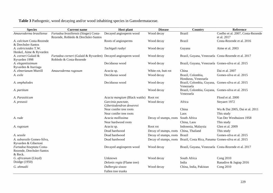
Ecological aspects
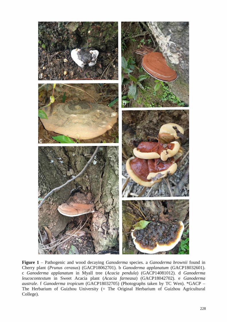
Ganoderma species have a global distribution in green ecosystems both in tropical and
temperate geographical regions of Asia, Africa, America and Europe (Wang et al. 2012). They are
226
usually found in subtropical and tropical regions since they live in hot and humid conditions (Pilotti
2004). These species are important wood decaying fungi. Most species of Ganoderma are
pathogenic (Fig. 1) causing root and stem rot on a variety of monocotyledons, dicotyledons and
gymnosperms including wide range of economically important trees and perennial crops which
results in the death of affected trees (Lee & Chang 2016). Some species are saprobic and cause
white rot of wood (Muthelo 2009). Hence, they have ecological importance in the breakdown of
woody plants for nutrient mobilization. They possess effective machineries of lignocellulose-
decomposing enzymes useful for bioenergy production and bioremediation (Hepting 1971,
Adaskaveg et al. 1991, Coetzee et al. 2015, Kües et al. 2015). Plant pathogenic species in this
genus can cause severe diseases (stem, butt, and root rot) in economically important trees and
perennial crops, especially in tropical countries (Coetzee et al. 2015). Ganoderma disease
development can be triggered by environmental factors and plant death could be either slow or
rapid depending on water availability and temperature. Furthermore, Amauroderma species are also
considered as parasitic on the roots of living trees (Glen et al. 2009). Ganoderma boninense is the
most aggressive pathogen to cause the basal stem rot in oil palm (Turner 1981, Wong et al. 2012).
Different species have different features and pathogenicity. Accuarate identification of these
pathogenic species is problematic and hence results in problems for proper disease mangament
(Wong et al. 2012). Members of Ganodermataceae can be of significant importance in horticulture,
infecting landscape plants (Acacia sp., Cassia sp., Pinus sp.) and fruit trees (Avocado) (Kinge &
Mih (2015). Pathogenic, wood decaying and/or wood inhabiting members of Ganodermataceae,
diseases caused and corresponding host plants are listed in Table 3.
Materials and methods
Sample collections
Samples of Ganodermataceae were collected during 2014 to 2018 from China, Laos,
Thailand, Myanmar and Vietnam and dealt with as in Cao et al. (2012). The materials were
deposited at Guizhou University (GACP) and Mae Fah Luang University (MFLU) herbaria.
Macroscopic and microscopic characterization
Macro-morphological characteristics were described based on fresh materials, and the
photographs provided here. Colour codes (e.g. 3A3) are from Kornerup & Wanscher (1978).
Specimens were dried and placed separately in plastic ziplock bags. For micro-morphological
observations, basidiomes were examined under a stereo dissecting microscope (Motic SMZ 168
series) and sections were cut with a razor blade, mounted in 5% KOH, and then observed,
measured and illustrated under a compound microscope (Nikon ECLIPSE 80i) equipped with a
camera (Canon 600D). Measurements were made using Tarosoft (R) Image Frame Work v. 0.9.7.
At least 20 basidiospores were measured from each mature specimen except for very scanty
materials. The basidiospore size was measured both with and without the myxosporium, but only
spore sizes with myxosporium were used for comparisons. Basidiospore dimensions are given as
(a–) b–c–d (–e), where a represents the minimum, b (mean average-standard deviation), c the
average, d (mean average+standard deviation) and e the maximum. Q, the length/width ratio (L/W)
of a spore in side view and Qm is the average, smallest and largest Q values given as Q. Pellis
sections were taken from the mature pileus portion and mounted in Melzer’s reagent for
observation. The Facesoffungi number is provided as explained in Jayasiri et al. (2015).
DNA Extraction, PCR and Sequencing
Dried samples of basidiomes were used to extract genomic DNA. Genomic DNA was
extracted using an EZgene TM Fungal gDNA Kit (Biomiga, CA, USA) according to the
manufacturer instructions. DNA concentrations were estimated visually in agarose gel by
comparing band intensity with a DNA ladder 1Kb (Invitrogen Biotech). Reaction mixtures (50 µl)
contained 2 µl template DNA (ca. 10 ng), 19 µl distilled water, and 2 µl (10 µM) of each primer
227
and 25 µl 2x BenchTopTM Taq Master Mix (Biomigas). Amplification conditions were 40 cycles
of 95 °C for 30 s, 59 °C for 30 s and 72 °C for 1 min, followed by a final extension at 72 °C for 10
min for all DNA fragments. The ITS rDNA regions were amplified using the universal primer pair
ITS4/ITS5 and the 18S and 28S rDNA genes were amplified using the universal primer pair
NS1/NS4 and primer pair LROR/LR5 respectively (Vilgalys & Hester 1990, White et al. 1990,
Rehner & Samuels 1994). Two protein coding genes: translation elongation factor-1α (TEF1) and
RNA polymerase II gene (RPB2) were amplified using corresponding primer pair 983F/2218R
(Rehner & Buckley 2005) and fRPB26f/7CR (Liu et al. 1999). Amplified PCR products were
verified by 1% agarose gel electrophoresis stained with ethidium bromide in 1x TBE. The PCR
products were sequenced with primers mentioned above by SinoGenoMax Co., Ltd (Beijing).
Sequence Alignment and Phylogenetic Analysis
All the other sequences except which were obtained from this study (Table 3) was retrieved
from GenBank based on ITS BLAST searches in GenBank (Benson et al. 2017) and recently
published data. Sequences that had possibly been contaminated by micro fungi or other unnamed
species (such as those with aff. in the species name) were discarded, ambiguous regions were
excluded and gaps were treated as missing data in the analysis (Nilsson et al. 2012). One hundred
sixty two nucleotide sequences representing 70 species of Ganodermataceae from Asia, America
and Europe were retrieved from GenBank and those retrieved sequences and the newly generated
sequences were aligned with MAFFT v. 7 (http://mafft.cbrc.jp/alignment/server/index.html; Katoh
& Standley 2013). The resulting alignment was improved manually when necessary using BioEdit
v. 7.0.5.2 (Hall 1999). The Maximum Likelihood (ML) analyses were performed using RAxML-
HPC2 (Stamatakis 2014) on the CIPRES Science Gateway V. 3.3 (Miller & Blair 2009), with
default settings except that the number of bootstrap replicates was set to 1,000. For Bayesian
analysis (BY), the GTR+I+G model of nucleotide evolution was selected with the help of
MrModeltest 2.2 (Nylander 2004) as the best-fit model and posterior probabilities (PP) (Rannala &
Yang 1996) were determined by Markov Chain Monte Carlo sampling (BMCMC) using MrBayes
v3.1.2 (Ronquist et al. 2012). BY analyses were conducted with six simultaneous Markov chains
and trees were summarized every 100th generation. The analyses were stopped after 5,000,000
generations when the average standard deviation of split frequencies was below 0.01. The
convergence of the runs was checked using TRACER v1.6 (Rambaut et al. 2013). The first 25% of
the resulting trees were discarded as burn-in, and PP were calculated from the remaining sampled
trees. In both ML and BY analyses, Tomophagus colossus was selected as the outgroup. ML
bootstrap values and BY posterior probabilities greater than or equal to 70% and 0.95, respectively,
were considered as significant support. The phylogenetic tree was visualized with FigTree version
1.4.0 (Rambaut 2012) available at http://tree.bio.ed.ac.uk/software/figtree/.
Results
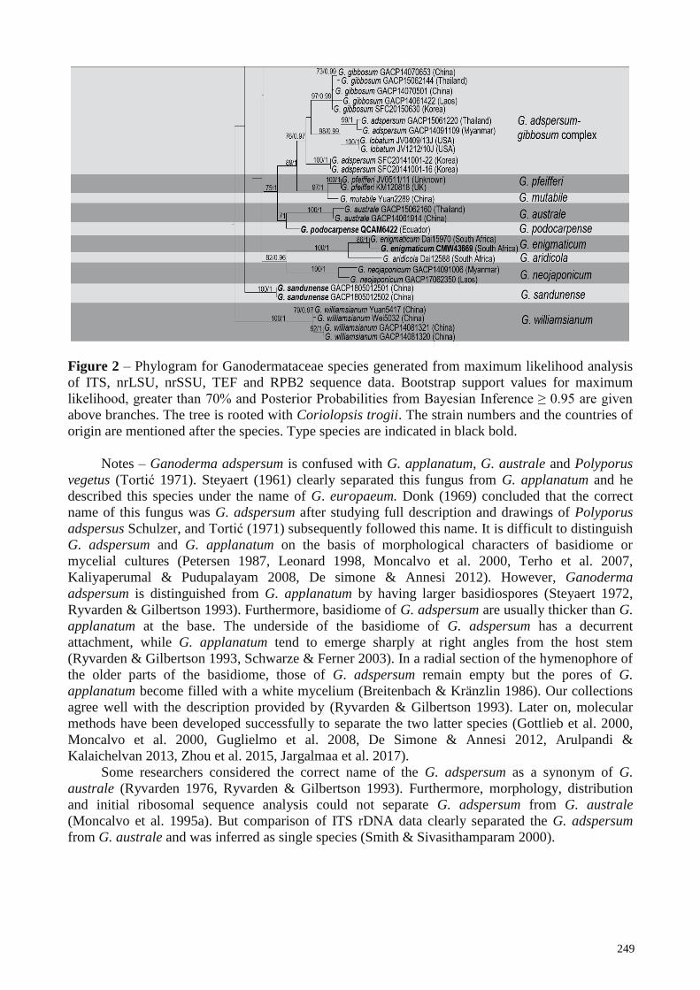
Phylogeny
The tree topologies obtained from ML and BY were identical. Therefore, only the ML tree is
shown (Fig. 2). The 162 sequences of Amauroderma, Foraminispora, Furtadoa, Haddowia,
Humphreya and Ganoderma clustered in 45 clades in Ganodermataceae (Fig. 2).
Our collections from China, Laos, Myanmar, Thailand and Vietnam clustered with all other
Amauroderma, Haddowia and Ganoderma species, including the holotypes (Amauroderma
calcitum, A. concentricum, A. floriformum, A. subsessile, Furtadoa biseptata G. aridicola, G.
austroafricanum, G. carocalcareus, G. destructans, G. enigmaticum G. ecuadoriense, G.
leucocontextum, G. lingzhi, G. sichuanense, G. ryvardenii, G. mebrekobenum, G. mizoramense, G.
podocarpense and G. wiiroense,), paratypes (G. wiiroense and G. mebrekobenum), and isotype (A.
laccatostipitatum) in well-
228
Figure 1 – Pathogenic and wood decaying Ganoderma species. a Ganoderma brownii found in
Cherry plant (Prunus cerasus) (GACP18062701). b Ganoderma applanatum (GACP18032601).
c Ganoderma applanatum in Myall tree (Acacia pendula) (GACP14081012). d Ganoderma
leucocontextum in Sweet Acacia plant (Acacia farneasa) (GACP18042702). e Ganoderma
australe. f Ganoderma tropicum (GACP18032705) (Photographs taken by TC Wen). *GACP –
The Herbarium of Guizhou University (= The Original Herbarium of Guizhou Agricultural
College).
229
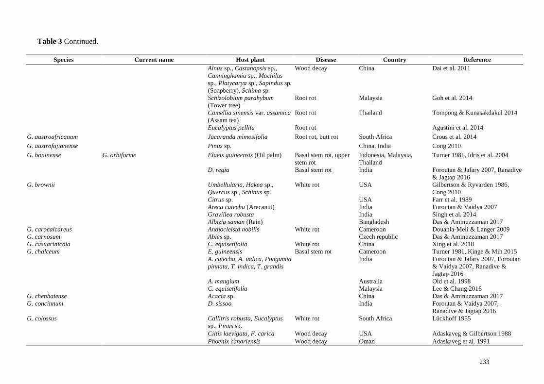
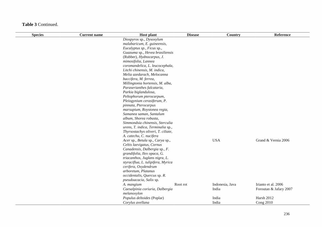
Table 3 Pathogenic, wood decaying and/or wood inhabiting species in Ganodermataceae.
Species Current name Host plant Disease Country Reference
Amauroderma brasiliense Furtadoa brasiliensis (Singer) Costa-
Rezende, Robledo & Drechsler-Santos
Decayed angiosperm wood Wood decay Brazil Coelho et al. 2007, Costa-Rezende
et al. 2017
A. calcitum Costa-Rezende
& Drechsler-Santos
Roots of angiosperms Wood decay Brazil Costa-Rezende et al. 2016
A. coltricioides T.W.
Henkel, Aime & Ryvarden
Tachigali rusbyi Wood decay Guyana Aime et al. 2003
A. corneri Gulaid &
Ryvarden 1998
Furtadoa corneri (Gulaid & Ryvarden)
Robledo & Costa-Rezende
Decayed angiosperm wood Wood decay Brazil, Guyana, Venezuela Costa-Rezende et al. 2017
A. elegantissimum
Ryvarden & Iturriaga
Deciduous wood Wood decay Brazil, Guyana, Venezuela Gomes-silva et al. 2015
A. elmerianum Murrill Amauroderma rugosum Acacia sp. White rot, butt rot China Dai et al. 2007
A. exile Deciduous wood Wood decay Brazil, Colombia,
Honduras, Venezuela
Gomes-silva et al. 2015
A. omphalodes Deciduous wood Wood decay Brazil, Colombia, Guyana,
Venezuela
Gomes-silva et al. 2015
A. partitum Wood decay Brazil, Colombia, Guyana,
Venezuela
Gomes-silva et al. 2015
A. Parasiticum Acacia mangium (Black wattle) Root rot Flood et al. 2000
A. preussii Garcinia punctate,
Gibertiodendron dewevrei
Wood decay Africa Steyaert 1972
Near conifer tree roots China Wu & Dai 2005, Dai et al. 2011
Near conifer tree roots Laos This study
A. rude Acacia mollissima Decay of stumps, roots South Africa Van Der Westhuizen 1958
Near hardwood roots China, Laos This study
A. rugosum Acacia sp. Root rot Indonesia, Malaysia Glen et al. 2009
Dead hardwood Decay of stumps, roots China, Thailand This study
A. sessile Dead hardwood Decay of stumps, roots Brazil Gomes-silva et al. 2015
A. subsessile Gomes-Silva,
Ryvarden & Gibertoni
Dead hardwood Decay of stumps, roots Brazil, Costa Rica, Panama Gomes-silva et al. 2015
Furtadoa biseptata Costa-
Rezende, Drechsler-Santos
& Reck.
Decayed angiosperm wood Wood decay Brazil, Guyana, Venezuela Costa-Rezende et al. 2017
G. africanum (Lloyd)
Doidge (1950)
Unknown Wood decay South Africa Cong 2010
Delonix regia (Flame tree) India Ranadive & Jagtap 2016
G. ahmadii Dalbergia sissoo Wood decay China, India, Pakistan Cong 2010
Fallen tree trunks

230
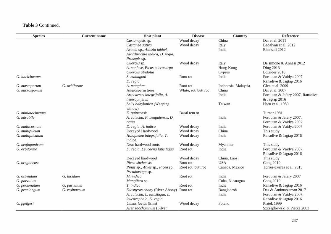
Table 3 Continued.
Species Current name Host plant Disease Country Reference
G. adspersum Ficus carica, Prunus dulcis
(Almonds)
Root rot, butt rot Cyprus Viney 2005
Salix sp. (Willow) Belgium Guglielmo et al. 2008
Juglans regia, Aesculus
hippocastanum (Horse
chestnut), Cercis siliquastrum,
Pterocarya fraxinifolia, Fagus
sylvatica, Morus sp.
Italy
Carpinus betulus, Tilia sp.
(Lime), Quercus sp. (Oak),
Fagus, Aesculus
Germany, USA, Czech
Republic
Cong 2010
Melia azadirachta, Prunus
armeniaca, Tamarindus indica
(Tamarind), Terminalia
bellerica, Grevillea parallela
Indonesia Flood et al. 2010
Salix sp., Ceratonia siliqua
(Carob tree)
Cong 2010
Fraxinus sp., Acacia sp. Armenia Badalyan et al. 2012
Abies sp. (fir), C. siliquastrum,
F. sylvatica L., Pinus pinea,
Prunus avium, Quercus sp.,
Robinia sp., L. nobilis
Italy De simone & Annesi 2012
Alnus orientalis, Quercus sp., F.
carica, On buried tree trunk
India Arulpandi & Kalaichelvan 2013,
Ranadive & Jagtap 2016
P. dulcis USA Johnson 2017
Quercus infectoria, C. siliqua,
Salix alba
Cyprus Loizides 2018
G. applanatum Fagus sp. (Beech), Ulmus sp.
(Elms), Quercus sp. (Oak),
Platanus sp., (Sycamore), A.
hippocastanum, Salix (Willow),
Juglans (Walnut)
Root rot, butt rot UK Cartwright & Findlay 1958
Citrus sp. USA Farr et al. 1989
Rhizophora apiculata
(Mangrove)
Butt rot Thailand Chalermpongse 1991
Gleditsia triacanthos (Honey
locust)
USA Report in Plant diseases 1999
Acacia auriculiformis Australia Old et al. 1998

231
Table 3 Continued.
Species Current name Host plant Disease Country Reference
Acer sp. (Maple), Alnus rubra
(Red alder), Amelanchier
arborea, Betula sp., Carya
tomentosa, Cercis Canadensis,
Fagus grandifolia, Juglans
cinerera, Liriodendron
tulipifera, Magnolia fraseri,
Malus sylvestris, Populus sp.,
Prunus pensylvanica,
Pseudotsuga menziesii
(Douglas-fir), Quercus sp.,
Robinia pseudoacacia, Salix sp.,
Tilia heterophylla, Thuja plicata
(Western Redcedar), Tsuga
canadensis (Hemlock), Ulmus
rubra
White mottled rot USA, Canada Allen et al. 1996, Grand & Vernia
2006
Tectona grandis (Teak), Xylia
xylocarpa, Bombax ceiba,
Terminalia sp.
India Florence & Yesodharan 2000
Acer sp., Abies sp., Betula sp.,
Carpinus (Hornbeam ), Pinus
sibirica (Siberian pine), Tilia,
Salix sp.
Russia Kuz'michev & Kulikova 2001
A. mangium Indonesia Glen et al. 2009
Abies pindrow, Acacia sp.,
Albizia sp., Artocarpus sp.,
Azadirachta indica, Camellia
sinensis (Tea), Caryota urens,
Cassia sp., Celtis tetrandra,
Cinnamomum cecidodaphne,
Cocos nucifera (Coconut), D.
sissoo, Dipterocarpus
macrocarpus, Leucaena
leucocephala, Mallotus
philippensis, Morus alba, Picea
smithiana, Pieris ovalifolia,
Pinus roxburghii, Santalum sp.,
Shorea sp., Sterculia, T.
India Sankaran et al. 2005

232
Table 3 Continued.
Species Current name Host plant Disease Country Reference
grandis, T. bellirica, Toona
ciliata
Phoenix sylvestris India Bhosle et al. 2010, Ranadive &
Jagtap 2016
Betuleceae alnus (Alder),
Maleace malus (Apple),
Ulmaceae Ulmus (Elm),
Aesculus (Horse-chestnut), Acer
sp., Quercus, Juglans (Walnut)
Denmark, Norway,
Germany, China, Vietnam,
Czech republic
Cong 2010
Laurus nobilis (Bay laurel) Wood decay USA Glaeser & Smith 2010
F. sylvatica, L. nobilis Wood decay Italy De simone & Annesi 2012
Mangifera indica (Mango), Wood decay India Singh et al. 2014
Swietenia mahagoni (Mahagoni) Wood decay Bangladesh Das & Aminuzzaman 2017
A. mangium Wood decay Australia Tchotet Tchoumi et al. 2017
Hardwoods Wood decay China, Thailand,
Myanmar, Laos
This study
G. amazonense Spondiae lutae Wood decay Brazil Cong 2010
T. grandis India Foroutan & Vaidya 2007,
Ranadive & Jagtap 2016
G. amboinense Acacia confusa, Dead wood Wood decay Indonesia, India Cong 2010, Ranadive & Jagtap
2016
G. angustisporum Casuarina equisetifolia White rot China Xing et al. 2018
G. aridicola Ficus sp. White rot South Africa Xing et al. 2016
G. argillaceum G. resinaceum Mangifera sp. Cuba Cong 2010
G. atrum Fagus sp., Coniferous sp. China Cong 2010
G. atkinsonii G. carnosum Abies alba Bohemia Cong 2010
G. australe Angiosperm trees,
Cinnamomum sp.
White rot, butt rot China Turner 1981, Dai et al. 2004, 2007
M. indica Wood decay Malaysia Abdullah et al. 1997
Hardwoods Laos, Thailand, Myanmar This study
X. xylocarpa, Cassia sp., Mesua
ferrea
White rot India
Florence & Yesodharan 2000
Coffea arabica (Coffee),
Grevillea robusta, M. ferrea
India
Sankaran et al. 2005, Ranadive &
Jagtap 2016
A. mangium Root rot Indonesia, Malaysia Glen et al. 2009
233
Table 3 Continued.
Species Current name Host plant Disease Country Reference
Alnus sp., Castanopsis sp.,
Cunninghamia sp., Machilus
sp., Platycarya sp., Sapindus sp.
(Soapberry), Schima sp.
Wood decay China Dai et al. 2011
Schizolobium parahybum
(Tower tree)
Root rot Malaysia Goh et al. 2014
Camellia sinensis var. assamica
(Assam tea)
Root rot Thailand Tompong & Kunasakdakul 2014
Eucalyptus pellita Root rot Agustini et al. 2014
G. austroafricanum Jacaranda mimosifolia Root rot, butt rot South Africa Crous et al. 2014
G. austrofujianense Pinus sp. China, India Cong 2010
G. boninense G. orbiforme Elaeis guineensis (Oil palm) Basal stem rot, upper
stem rot
Indonesia, Malaysia,
Thailand
Turner 1981, Idris et al. 2004
D. regia Basal stem rot India Foroutan & Jafary 2007, Ranadive
& Jagtap 2016
G. brownii Umbellularia, Hakea sp.,
Quercus sp., Schinus sp.
White rot USA Gilbertson & Ryvarden 1986,
Cong 2010
Citrus sp. USA Farr et al. 1989
Areca catechu (Arecanut) India Foroutan & Vaidya 2007
Gravillea robusta India Singh et al. 2014
Albizia saman (Rain) Bangladesh Das & Aminuzzaman 2017
G. carocalcareus Anthocleista nobilis White rot Cameroon Douanla-Meli & Langer 2009
G. carnosum Abies sp. Czech republic Das & Aminuzzaman 2017
G. casuarinicola C. equisetifolia White rot China Xing et al. 2018
G. chalceum E. guineensis Basal stem rot Cameroon Turner 1981, Kinge & Mih 2015
A. catechu, A. indica, Pongamia
pinnata, T. indica, T. grandis
India Foroutan & Jafary 2007, Foroutan
& Vaidya 2007, Ranadive &
Jagtap 2016
A. mangium Australia Old et al. 1998
C. equisetifolia Malaysia Lee & Chang 2016
G. chenhaiense Acacia sp. China Das & Aminuzzaman 2017
G. concinnum D. sissoo India Foroutan & Vaidya 2007,
Ranadive & Jagtap 2016
G. colossus Callitris robusta, Eucalyptus
sp., Pinus sp.
White rot South Africa Lückhoff 1955
Ciltis laevigata, F. carica Wood decay USA Adaskaveg & Gilbertson 1988
Phoenix canariensis Wood decay Oman Adaskaveg et al. 1991
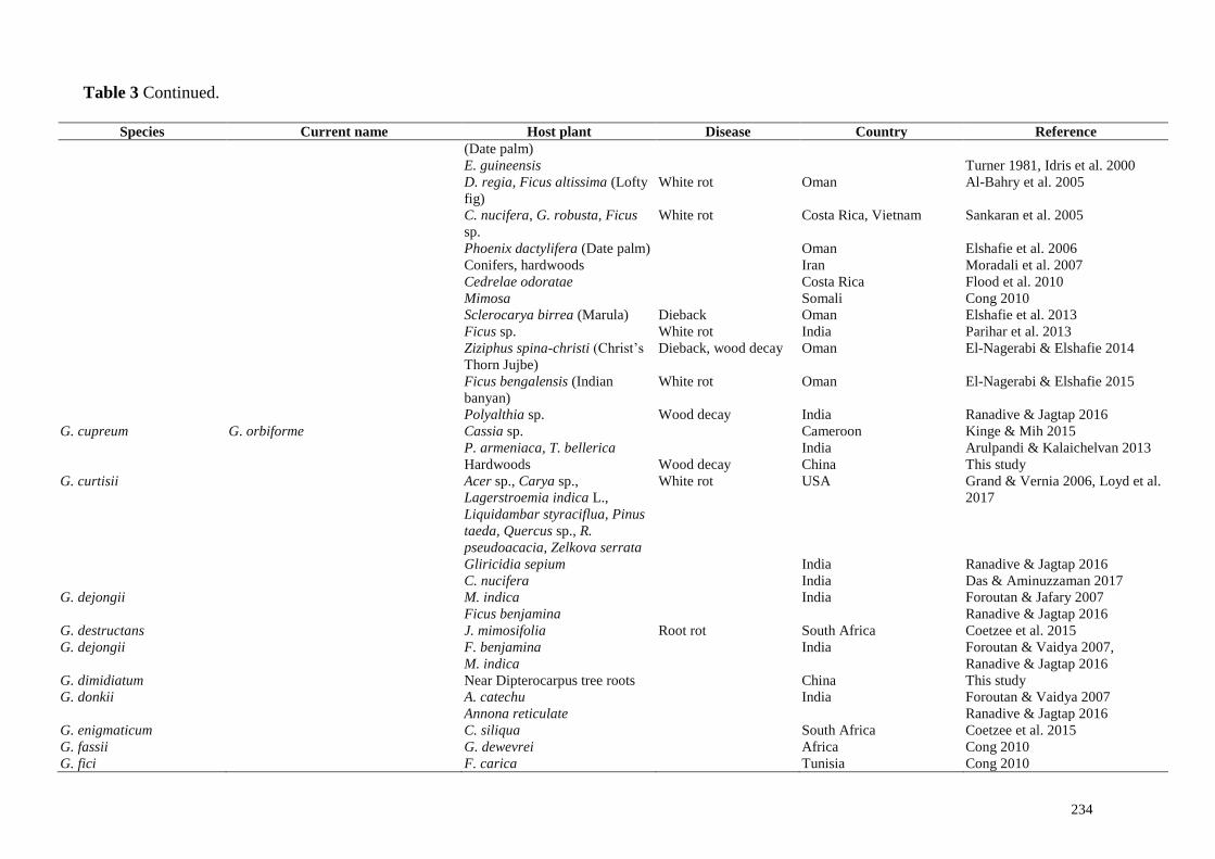
234
Table 3 Continued.
Species Current name Host plant Disease Country Reference
(Date palm)
E. guineensis Turner 1981, Idris et al. 2000
D. regia, Ficus altissima (Lofty
fig)
White rot Oman Al-Bahry et al. 2005
C. nucifera, G. robusta, Ficus
sp.
White rot Costa Rica, Vietnam Sankaran et al. 2005
Phoenix dactylifera (Date palm) Oman Elshafie et al. 2006
Conifers, hardwoods Iran Moradali et al. 2007
Cedrelae odoratae Costa Rica Flood et al. 2010
Mimosa Somali Cong 2010
Sclerocarya birrea (Marula) Dieback Oman Elshafie et al. 2013
Ficus sp. White rot India Parihar et al. 2013
Ziziphus spina-christi (Christ’s
Thorn Jujbe)
Dieback, wood decay Oman El-Nagerabi & Elshafie 2014
Ficus bengalensis (Indian
banyan)
White rot Oman El-Nagerabi & Elshafie 2015
Polyalthia sp. Wood decay India Ranadive & Jagtap 2016
G. cupreum G. orbiforme Cassia sp. Cameroon Kinge & Mih 2015
P. armeniaca, T. bellerica India Arulpandi & Kalaichelvan 2013
Hardwoods Wood decay China This study
G. curtisii Acer sp., Carya sp.,
Lagerstroemia indica L.,
Liquidambar styraciflua, Pinus
taeda, Quercus sp., R.
pseudoacacia, Zelkova serrata
White rot USA Grand & Vernia 2006, Loyd et al.
2017
Gliricidia sepium India Ranadive & Jagtap 2016
C. nucifera India Das & Aminuzzaman 2017
G. dejongii M. indica India Foroutan & Jafary 2007
Ficus benjamina Ranadive & Jagtap 2016
G. destructans J. mimosifolia Root rot South Africa Coetzee et al. 2015
G. dejongii F. benjamina India Foroutan & Vaidya 2007,
Ranadive & Jagtap 2016 M. indica
G. dimidiatum Near Dipterocarpus tree roots China This study
G. donkii A. catechu India Foroutan & Vaidya 2007
Annona reticulate Ranadive & Jagtap 2016
G. enigmaticum C. siliqua South Africa Coetzee et al. 2015
G. fassii G. dewevrei Africa Cong 2010
G. fici F. carica Tunisia Cong 2010
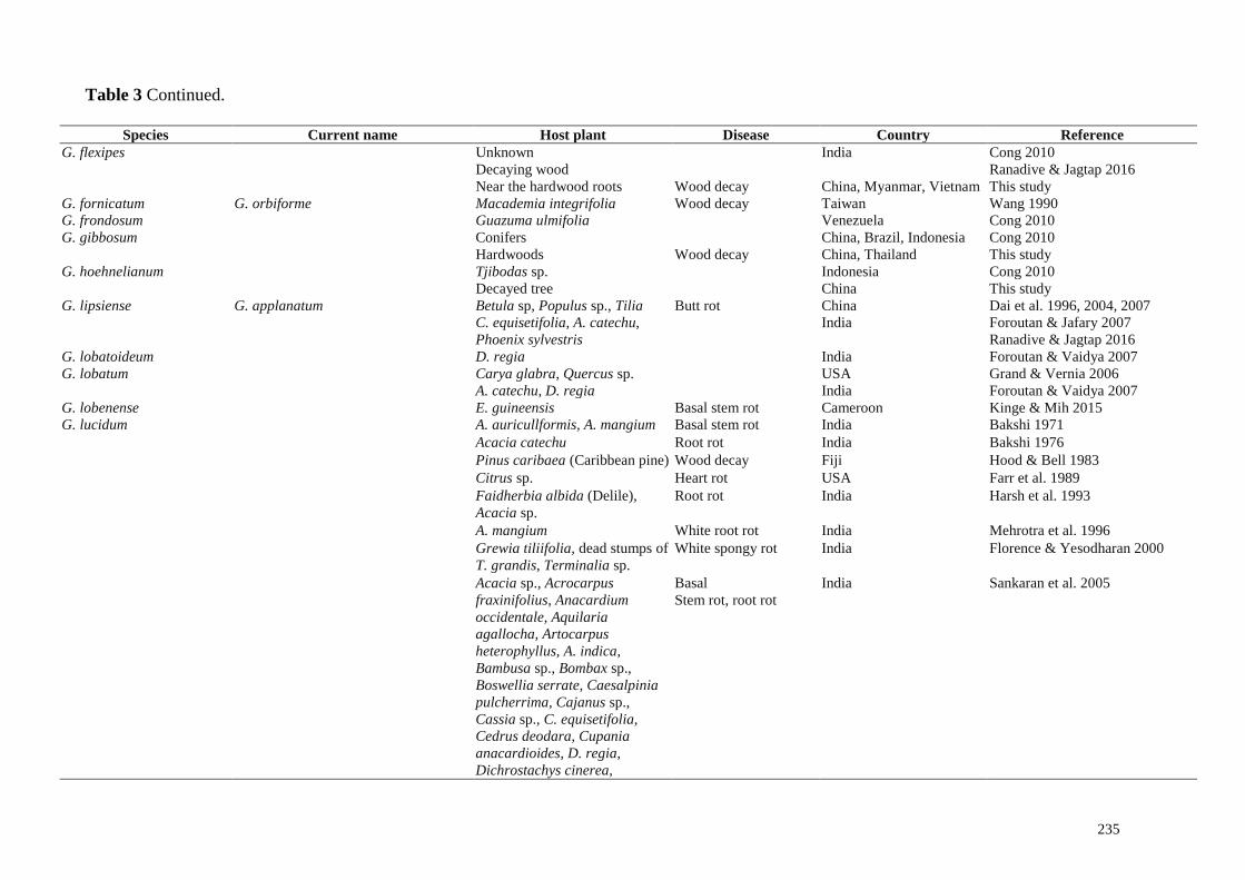
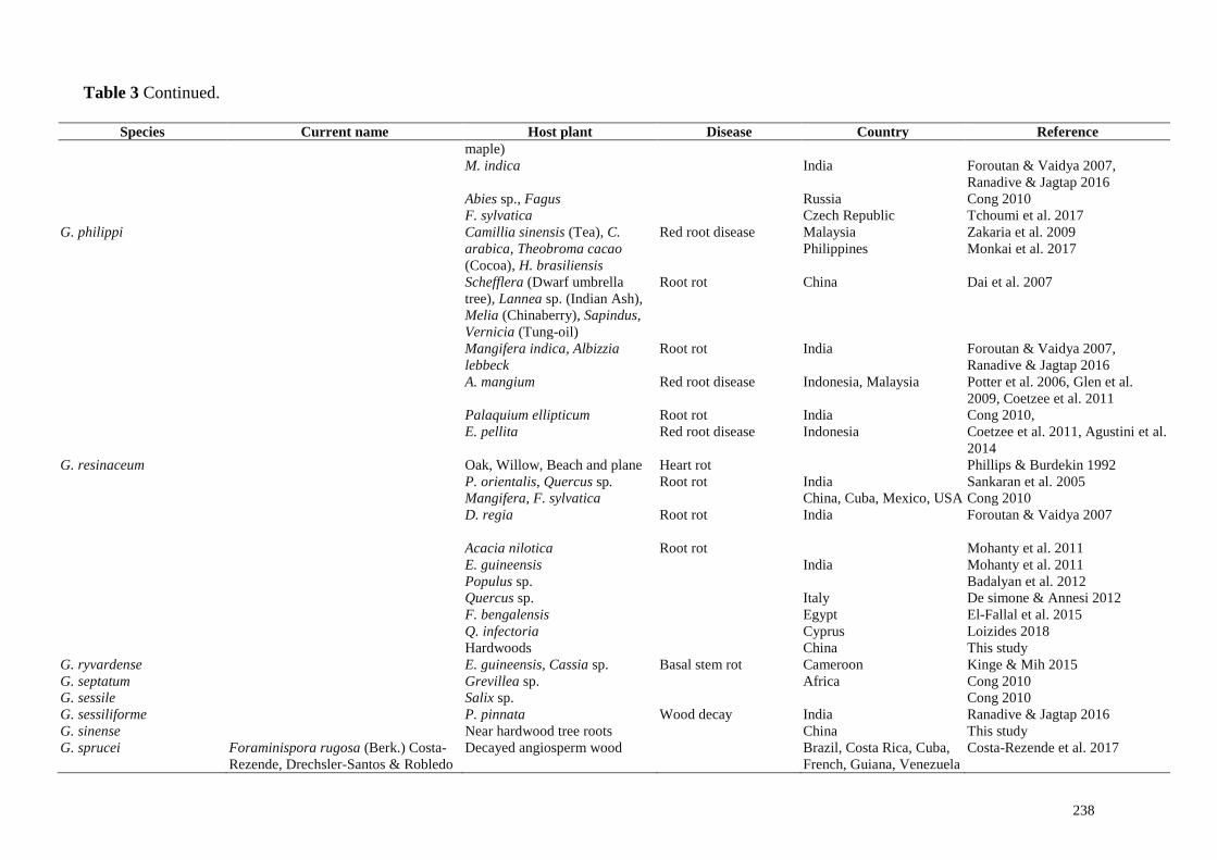
235
Table 3 Continued.
Species Current name Host plant Disease Country Reference
G. flexipes Unknown India Cong 2010
Decaying wood Ranadive & Jagtap 2016
Near the hardwood roots Wood decay China, Myanmar, Vietnam This study
G. fornicatum G. orbiforme Macademia integrifolia Wood decay Taiwan Wang 1990
G. frondosum Guazuma ulmifolia Venezuela Cong 2010
G. gibbosum Conifers China, Brazil, Indonesia Cong 2010
Hardwoods Wood decay China, Thailand This study
G. hoehnelianum Tjibodas sp. Indonesia Cong 2010
Decayed tree China This study
G. lipsiense G. applanatum Betula sp, Populus sp., Tilia Butt rot China Dai et al. 1996, 2004, 2007
C. equisetifolia, A. catechu, India Foroutan & Jafary 2007
Phoenix sylvestris Ranadive & Jagtap 2016
G. lobatoideum D. regia India Foroutan & Vaidya 2007
G. lobatum Carya glabra, Quercus sp. USA Grand & Vernia 2006
A. catechu, D. regia India Foroutan & Vaidya 2007
G. lobenense E. guineensis Basal stem rot Cameroon Kinge & Mih 2015
G. lucidum A. auricullformis, A. mangium Basal stem rot India Bakshi 1971
Acacia catechu Root rot India Bakshi 1976
Pinus caribaea (Caribbean pine) Wood decay Fiji Hood & Bell 1983
Citrus sp. Heart rot USA Farr et al. 1989
Faidherbia albida (Delile),
Acacia sp.
Root rot India Harsh et al. 1993
A. mangium White root rot India Mehrotra et al. 1996
Grewia tiliifolia, dead stumps of
T. grandis, Terminalia sp.
White spongy rot India Florence & Yesodharan 2000
Acacia sp., Acrocarpus
fraxinifolius, Anacardium
occidentale, Aquilaria
agallocha, Artocarpus
heterophyllus, A. indica,
Bambusa sp., Bombax sp.,
Boswellia serrate, Caesalpinia
pulcherrima, Cajanus sp.,
Cassia sp., C. equisetifolia,
Cedrus deodara, Cupania
anacardioides, D. regia,
Dichrostachys cinerea,
Basal
Stem rot, root rot
India Sankaran et al. 2005
236
Table 3 Continued.
Species Current name Host plant Disease Country Reference
Diospyros sp., Dysoxylum
malabaricum, E. guineensis,
Eucalyptus sp., Ficus sp.,
Guazuma sp., Hevea brasiliensis
(Rubber), Hydnocarpus, J.
mimosifolia, Lannea
coromandelica, L. leucocephala,
Litchi chinensis, M. indica,
Melia azedarach, Melocanna
baccifera, M. ferrea,
Millingtonia hortensis, M. alba,
Paraserianthes falcataria,
Parkia biglandulosa,
Peltophorum pterocarpum,
Pleiogynium cerasiferum, P.
pinnata, Pterocarpus
marsupium, Roystonea regia,
Samanea saman, Santalum
album, Shorea robusta,
Simmondsia chinensis, Sterculia
urens, T. indica, Terminalia sp.,
Thyrsostachys oliveri, T. ciliate,
A. catechu, C. nucifera
Acer sp., Betula sp., Carya sp.,
Celtis laevigatus, Cornus
Canadensis, Dalbergia sp., F.
grandifolia, Ilex opaca, G.
triacanthos, Juglans nigra, L.
styraciflua, L. tulipifera, Myrica
cerifera, Oxydendrum
arboretum, Platanus
occidentalis, Quercus sp. R.
pseudoacacia, Salix sp.
USA Grand & Vernia 2006
A. mangium Root rot Indonesia, Java Irianto et al. 2006
Caesalpinia coriuria, Dalbergia
melanoxylon
India Foroutan & Jafary 2007
Populus deltoides (Poplar) India Harsh 2012
Corylus avellana India Cong 2010
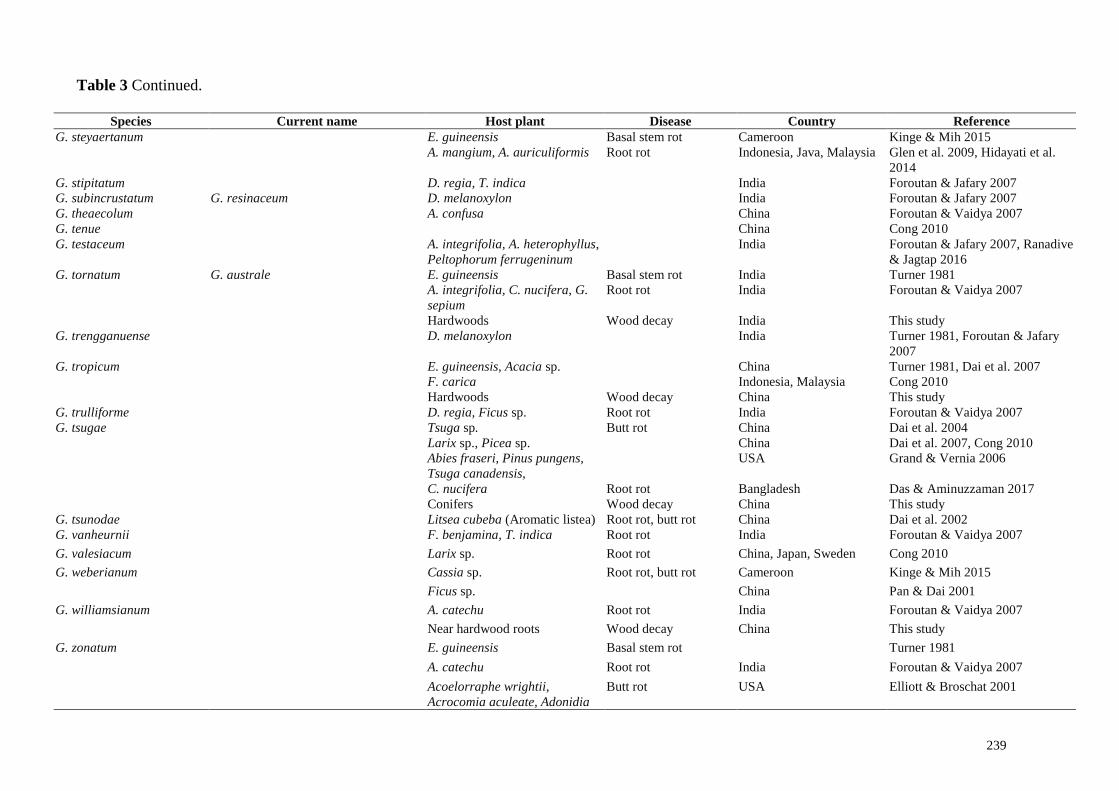
237
Table 3 Continued.
Species Current name Host plant Disease Country Reference
Castanopsis sp. Wood decay China Dai et al. 2011
Castanea sativa Wood decay Italy Badalyan et al. 2012
Acacia sp., Albizia labbek,
Azardirachta indica, D. regia,
Prosopis sp.
India Bhansali 2012
Quercus sp. Wood decay Italy De simone & Annesi 2012
A. confuse, Ficus microcarpa Hong Kong Ding 2013
Quercus alnifolia Cyprus Loizides 2018
G. luteicinctum S. mahagoni Root rot India Foroutan & Vaidya 2007
D. regia Ranadive & Jagtap 2016
G. mastoporum G. orbiforme A. mangium Root rot Indonesia, Malaysia Glen et al. 2009
G. microsporum Angiosperm trees White, rot, butt rot China Dai et al. 2007
Artocarpus integrifolia, A.
heterophyllus
India Foroutan & Jafary 2007, Ranadive
& Jagtap 2016
Salix babylonica (Weeping
willow)
Taiwan Hseu et al. 1989
G. miniatocinctum E. guineensis Basal tem ot Turner 1981
G. mirabile A. catechu, F. bengalensis, D.
regia
India Foroutan & Jafary 2007,
Foroutan & Vaidya 2007
G. multicornum D. regia, A. indica Wood decay India Foroutan & Vaidya 2007
G. multipileum Decayed Hardwood Wood decay China This study
G. multiplicatum Holoptelea integrifolia, T.
indica
Wood decay India Ranadive & Jagtap 2016
G. neojaponicum Near hardwood roots Wood decay Myanmar This study
G. orbiforme D. regia, Leucaena latisiliqua Root rot India Foroutan & Vaidya 2007,
Ranadive & Jagtap 2016
Decayed hardwood Wood decay China, Laos This study
G. oregonense Picea sitchensis Root rot USA Cong 2010
Pinus sp., Abies sp., Picea sp.,
Pseudotsuga sp.
Root rot, butt rot Canada, Mexico Torres-Torres et al. 2015
G. ostreatum G. lucidum M. indica Root rot India Foroutan & Jafary 2007
G. parvulum Mangifera sp. Cuba, Nicaragua Cong 2010
G. perzonatum G. parvulum T. indica Root rot India Ranadive & Jagtap 2016
G. praelongum G. resinaceum Diospyros ebony (River Abony) Root rot Bangladesh Das & Aminuzzaman 2017
A. catechu, L. latisiliqua, L.
leucocephala, D. regia
India Foroutan & Vaidya 2007,
Ranadive & Jagtap 2016
G. pfeifferi Ulmus laevis (Elm) Wood decay Poland Piatek 1999
Acer saccharinum (Silver Szczepkowski & Pietka 2003
238
Table 3 Continued.
Species Current name Host plant Disease Country Reference
maple)
M. indica India Foroutan & Vaidya 2007,
Ranadive & Jagtap 2016
Abies sp., Fagus Russia Cong 2010
F. sylvatica Czech Republic Tchoumi et al. 2017
G. philippi Camillia sinensis (Tea), C.
arabica, Theobroma cacao
(Cocoa), H. brasiliensis
Red root disease
Malaysia Zakaria et al. 2009
Philippines Monkai et al. 2017
Schefflera (Dwarf umbrella
tree), Lannea sp. (Indian Ash),
Melia (Chinaberry), Sapindus,
Vernicia (Tung-oil)
Root rot China
Dai et al. 2007
Mangifera indica, Albizzia
lebbeck
Root rot India Foroutan & Vaidya 2007,
Ranadive & Jagtap 2016
A. mangium Red root disease Indonesia, Malaysia Potter et al. 2006, Glen et al.
2009, Coetzee et al. 2011
Palaquium ellipticum Root rot India Cong 2010,
E. pellita Red root disease Indonesia Coetzee et al. 2011, Agustini et al.
2014
G. resinaceum Oak, Willow, Beach and plane Heart rot Phillips & Burdekin 1992
P. orientalis, Quercus sp. Root rot India Sankaran et al. 2005
Mangifera, F. sylvatica China, Cuba, Mexico, USA Cong 2010
D. regia Root rot
India
Foroutan & Vaidya 2007
Acacia nilotica Root rot Mohanty et al. 2011
E. guineensis India Mohanty et al. 2011
Populus sp. Badalyan et al. 2012
Quercus sp. Italy De simone & Annesi 2012
F. bengalensis Egypt El-Fallal et al. 2015
Q. infectoria Cyprus Loizides 2018
Hardwoods China This study
G. ryvardense E. guineensis, Cassia sp. Basal stem rot Cameroon Kinge & Mih 2015
G. septatum Grevillea sp. Africa Cong 2010
G. sessile Salix sp. Cong 2010
G. sessiliforme P. pinnata Wood decay India Ranadive & Jagtap 2016
G. sinense Near hardwood tree roots China This study
G. sprucei Foraminispora rugosa (Berk.) Costa-
Rezende, Drechsler-Santos & Robledo
Decayed angiosperm wood Brazil, Costa Rica, Cuba,
French, Guiana, Venezuela
Costa-Rezende et al. 2017
239
Table 3 Continued.
Species Current name Host plant Disease Country Reference
G. steyaertanum E. guineensis Basal stem rot Cameroon Kinge & Mih 2015
A. mangium, A. auriculiformis Root rot Indonesia, Java, Malaysia Glen et al. 2009, Hidayati et al.
2014
G. stipitatum D. regia, T. indica India Foroutan & Jafary 2007
G. subincrustatum G. resinaceum D. melanoxylon India Foroutan & Jafary 2007
G. theaecolum A. confusa China Foroutan & Vaidya 2007
G. tenue China Cong 2010
G. testaceum A. integrifolia, A. heterophyllus,
Peltophorum ferrugeninum
India Foroutan & Jafary 2007, Ranadive
& Jagtap 2016
G. tornatum G. australe E. guineensis Basal stem rot India Turner 1981
A. integrifolia, C. nucifera, G.
sepium
Root rot India Foroutan & Vaidya 2007
Hardwoods Wood decay India This study
G. trengganuense D. melanoxylon India Turner 1981, Foroutan & Jafary
2007
G. tropicum E. guineensis, Acacia sp. China Turner 1981, Dai et al. 2007
F. carica Indonesia, Malaysia Cong 2010
Hardwoods Wood decay China This study
G. trulliforme D. regia, Ficus sp. Root rot India Foroutan & Vaidya 2007
G. tsugae Tsuga sp. Butt rot China Dai et al. 2004
Larix sp., Picea sp. China Dai et al. 2007, Cong 2010
Abies fraseri, Pinus pungens,
Tsuga canadensis,
USA Grand & Vernia 2006
C. nucifera Root rot Bangladesh Das & Aminuzzaman 2017
Conifers Wood decay China This study
G. tsunodae Litsea cubeba (Aromatic listea) Root rot, butt rot China Dai et al. 2002
G. vanheurnii F. benjamina, T. indica Root rot India Foroutan & Vaidya 2007
G. valesiacum Larix sp. Root rot China, Japan, Sweden Cong 2010
G. weberianum Cassia sp. Root rot, butt rot Cameroon Kinge & Mih 2015
Ficus sp. China Pan & Dai 2001
G. williamsianum A. catechu Root rot India Foroutan & Vaidya 2007
Near hardwood roots Wood decay China This study
G. zonatum E. guineensis Basal stem rot Turner 1981
A. catechu Root rot India Foroutan & Vaidya 2007
Acoelorraphe wrightii,
Acrocomia aculeate, Adonidia
Butt rot USA Elliott & Broschat 2001

240
Table 3 Continued.
Species Current name Host plant Disease Country Reference
merrillii, Aiphanes sp., Arenga
sp., Attalea sp., Bactris major,
Brahea sp., Carpentaria
acuminate, Caryota mitis,
Chamaerops humilis,
Coccothrinax sp., Cocos
nucifera, Copernicia curtisii,
Dictyosperma album, Dypsis
cabadae, Dypsis lutescens
(Areca palm), Euterpe edulis,
Gastrococos crispa, Hyophorbe
indica, Livistona sp.,
Nannorrhops ritchiana, Phoenix
sp., Ptychosperma sp.,
Roystonea sp., Sabal palmetto
(Sabal palm), Satakentia
liukiuensis, Scheelea sp.,
Serenoa repens, Syagrus
romanzoffiana (Queen palm),
Washingtonia robusta
Cocos nucifera Basal stem rot Brazil, USA Cong 2010
Butia odorata (Jelly palm) Butt rot USA Loyd et al. 2017
Haddowia longipes Near hardwood roots Wood decay Laos This study
Humphreya coffeata Elaeocarpus lancifolius Root rot India Lyngdoh & Dhakar 2014
Sequences, A. rugosum from, China (GACP14081009, GACP14080929), Laos (GACP16072714, GACP14061012), and Thailand (GACP14062120);
Haddowia longipes (GACP17072708, GACP17072709) from Thailand; G. adspersum (GACP15061220) from Thailand and Myanmar
(GACP14091108); G. applanatum from China (GACP14080601, GACP14080603); G. australe from, China (GACP14061914) and Thailand
(GACP15062160); G. flexipes from, Vietnam (GACP17102301) and Laos (GACP17073037); G. gibbosum from, China (GACP14070501,
GACP14070653), Laos (GACP14061422) and Thailand (GACP15062144); G. lingzhi from Laos (GACP18011910, GACP18011911); G.
multiplicatum from Myanmar (GACP14091107, GACP14091108); G. neojaponicum, from Laos (GACP17062350) and Myanmar (GACP14091006);
G. orbiforme from, Laos (GACP14061420, GACP14061414), Thailand (GACP15061260) and Myanmar (GACP140910138); G. sinense from China
(GACP17092559, GACP16072729), G. subresinosum from Laos (GACP18011907); G. tropicum from Thailand (GACP15081610) and G.
williamsianum from China (GACP14081320, GACP14081321) obtained from our collections, clustered in well-supported clades forming
241
monophyletic groups with, A. rugosum (BS = 100%, BPP = 0.99), H. longipes (BS = 100%, BPP =
1.0), G. applanatum (BS = 100%, BPP = 1.0), G. australe (BS = 100%, BPP = 1.0), G. flexipes (BS
= 100%, BPP = 1.00), G. gibbosum (BS = 94%, BPP = 0.97), G. lingzhi (BS = 100%, BPP = 1.0),
G. multiplicatum (BS = 100%, BPP = 1), G. neojaponicum (BS = 100%, BPP = 1.00), G. orbiforme
(BS = 100%, BPP = 1.00), G. sinense (BS = 100%, BPP = 1.00), G. subresinosum (BS = 100%,
BPP = 1.0) and G. tropicum (BS = 100%, BPP=1.0), G. williamsianum (BS = 100%, BPP=1.0),
respectively (Fig. 2).
Taxonomy
Ganoderma P. Karst., 1881, Rev. Mycol. (Toulouse) 3, p. 17.
= Dendrophagus Murrill, Bull. Torrey bot. Club 32(9): 473 (1905)
= Elfvingia P. Karst., Bidr. Känn. Finl. Nat. Folk 48: 333 (1889)
= Friesia Lázaro Ibiza, Revista Real Acad. Ci. Madrid 14: 587 (1916)
= Ganoderma subgen. Trachyderma Imazeki, Bull. Tokyo Sci. Mus.1: 49 (1939)
= Tomophagus Murrill, Torreya 5: 197 (1905)
= Trachyderma (Imazeki) Imazeki, Bull. Gov. Forest Exp. Stn Tokyo 57: 97 (1952)
See the description at Ryvarden (2004b)
Basidiomes annual or perennial, stipitate to sessile; pileal surface with a thick, dull cuticle or
shiny and laccate with a thin cuticle or cuticle of clavate end cells. Context cream coloured to dark
purplish brown, soft and spongy to firm-fibrous; pore surface cream coloured, bruising brown, the
pores regular, 4–7 per mm; tube layers single or stratified, pale to purplish brown; stipe when
present central or lateral; hyphal system dimitic; generative hyphae with clamps; skeletal hyphae
hyaline to brown, non-septate, often with long, tapering branches; basidia broadly ellipsoid,
tapering abruptly at the base; cystidia absent. Basidiospores broadly to narrowly ellipsoid with a
truncate apex and apical germ pore, wall two-layered, endosporium brown and separated from the
hyaline exosporium by inter-wall pillars, negative in Melzer's reagent, 7–30 μm long.
Type species – Ganoderma lucidum (Curtis) P. Karst.
Notes – Ganoderma was established by Karsten (1881) with Ganoderma lucidum (Curtis) P.
Karst. as the type species. Traditional Chinese books classified Ganoderma into six species with
reference to the colour of the basidiome (Szedlay 2002). Patouillard (1889) listed 48 species of
Ganoderma worldwide. Species of Ganoderma have been studied primarily by many researchers
(Ryvarden & Johansen 1980, Furtado 1981, Corner 1983, Moncalvo & Ryvarden 1997) who have
made major contributions to the nomenclature and taxonomy of the genus. The traditional
taxonomy of Ganoderma is based on morphological traits and this genus was divided into two
distinct groups, the laccate (G. lucidum complex) and the non–laccate (G. applanatum complex)
species, which refer to the subgenera Ganoderma and Elfvingia respectively (Zheng et al. 2007).
Donk (1933) reunite all the taxa under subfamily Ganodermatoideae which previously belong to
Polyporaceae. In 1948, he raised up this taxon to family level and established Ganodermataceae on
the basis of spore peculiarities with the laccate and stipitate white rot fungus Polyporus lucidus
Curtis as its type species (Moncalvo & Ryvarden 1997) and placed the family in Polyporales,
Basidiomycetes (Schwarze & Ferner 2003).
This classification has subsequently been accepted by most recent workers, however Jülich (1981)
introduced the ordinal name Ganodermatales and this was accepted by Pegler in the eighth edition
of the Dictionary of the fungi, though other workers have continued to use the traditional
Aphyllophorales in a broad sense. The genus Ganoderma was initially classified on the basis of
morphological characteristics, however, environmental factors, variability, interhybridization, and
morphological propensity can lead to the inaccurate identification of Ganoderma species (Zheng et
al. 2007). There are 449 epithets listed in Index Fungorum (2019) for Ganoderma, while Kirk et al.
(2008) estimates there are 80 species. The taxonomic circumscription within Ganoderma is unclear
as species and generic concepts are confused because similar fungi are found in Fomes (Fr.),
Polyporus P. Micheli and Tomophagus Murril (Paterson 2006). Ganoderma species identification

242
and circumscriptions have often been problematic and taxonomic segregation of the genus has been debatable from long time (Moncalvo et al. 1995c).
Table 4 Sequences used in the phylogenetic analysis.
Species Voucher/strain Origin ITS nrLSU RPB2 nrSSU TEF Reference
Amauroderma aurantiacum (Torrend) Gibertoni &
Bernicchia
URM 78847 Brazil JX310840 - - - - Gomes-Silva et al. 2015
A. aurantiacum FLOR:52205 Brazil KR816510 KU315205 - - - Costa-Rezende et al. 2016
A. austrosinense Cui 13618 China KU219973 KU219996 - - - Song et al. 2016
A. calcigenum Berk.) Torrend URM 83864 Brazil JX982565 - - - - Gomes-Silva et al. 2015
A. calcigenum URM 86847 Brazil KT006601 - - - - Gomes-Silva et al. 2015
A. calcitum D.H. Costa & Drechsler-Santos FLOR:52230 (holotype) Brazil KR816529 - - - - Costa-Rezende et al. 2016
A. calcitum FLOR:50931 Brazil KR816528 - - - - Costa-Rezende et al. 2016
A. camerarium (Berk.) J.S. Furtado FLOR:52169 Brazil KR816523 - - - - GenBank
A. concentricum J. Song, Xiao L. He & B.K. Cui Cui 12644 (holotype) Sichuan,
China
KU219974 KU219997 - - - Song et al. 2016
A. concentricum Cui 12648 Sichuan,
China
KU219975 KU219998 - - - Song et al. 2016
A. elegantissimum Ryvarden & Iturr. URM 82787 Brazil JX310843 KT006616 - - - Gomes-Silva et al. 2015
A. elegantissimum URM 82789 Brazil JX310844 KT006617 - - - Gomes-Silva et al. 2015
A. exile (Berk.) Torrend URM 82794 Brazil JX310845 - - - - Gomes-Silva et al. 2015
A. floriformum Gomes-Silva, Ryvarden & Gibertoni URM83250 (holotype) Brazil JX310846 - - - - Gomes-Silva et al. 2015
A. laccatostipitatum Gomes-Silva, Ryvarden &
Gibertoni
URM83238 (isotype) Brazil JX310847 - - - - Gomes-Silva et al. 2015
A. laccatostipitatum HFSL ACGS7 Brazil KT006602 - - - - Gomes-Silva et al. 2015
A. omphalodes (Berk.) Torrend HUEFS:DHCR499 Brazil MF409956 - - - - Costa-Rezende et al. 2017
A. omphalodes HUEFS:DHCR500 Brazil MF409957 - - - MF421239 Costa-Rezende et al. 2017
A. perplexum Corner Cui 6496 Brazil KJ531650 KU220001 - - - Li & Yuan 2015
A. perplexum Dai 10811 Brazil KJ531651 KU220002 - - - Li & Yuan 2015
A. praetervisum (Pat.) Torrend URM87611 Brazil KT006606 - - - - Gomes-Silva et al. 2015
A. praetervisum URM84223 Brazil KC348460 - - - - Gomes-Silva et al. 2015
A. rugosum (Blume & T. Nees) Torrend GACP1408929 China MK345420 - - - - This study
A. rugosum GACP14081009 China MK345421 - - - - This study
A. rugosum GACP16072714 Laos MK077647 - - - - Hapuarachchi et al. 2018c
A. rugosum GACP14061012 Laos MK345422 - - - - This study
A. rugosum GACP14062120 Thailand MK077648 - - - Hapuarachchi et al. 2018c
A. rugosum Cui9012 China KJ531665 KJ531665 - - KU572503 Li & Yuan 2015
A. sessile Gomes-Silva, Ryvarden & Gibertoni URM83905 Brazil JX982570 - - - - Gomes-Silva et al. 2015

243
Table 4 Continued.
Species Voucher/strain Origin ITS nrLSU RPB2 nrSSU TEF Reference
A. subsessile Gomes-Silva, Ryvarden & Gibertoni URM83239 (holotype) Brazil JX310860 - - - - Gomes-Silva et al. 2015
A. schomburgkii (Mont. & Berk.) Torrend URM 84228 Brazil KT006608 - - - - Gomes-Silva et al. 2015
A. schomburgkii URM 84254 Brazil KT006611 - - - - Gomes-Silva et al. 2015
A. yunnanense J.D. Zhao & X.Q. Zhang Cui 7974 Yunnan, China KJ531653 KU220013 - KJ531653 - Li & Yuan 2015
A. yunnanense Yuan 2253 Yunnan, China KJ531655 - - - - Li & Yuan 2015
Furtadoa brasiliensis (Singer) Costa-Rezende,
Robledo & Drechsler-Santos
URM83578 Brazil JX310841 - - - - Gomes-Silva et al. 2015
F. brasiliensis TBG58 Brazil JX982569 - - - - Gomes-Silva et al. 2015
F. biseptata (Singer) Costa-Rezende, Robledo &
Drechsler-Santos
FLOR50932 (holotype) Brazil KU315196 KU315206 - - - Gomes-Silva et al. 2015
Foraminisporus rugosa (Berk.) Costa-Rezende,
Drechsler-Santos & Robledo
DHCR554 (HUEFS) Brazil MF409962 MF409954 - - - Costa-Rezende et al. 2017
F. rugosa DHCR560 Brazil MF409963 MF409955 - - - Costa-Rezende et al. 2017
Haddowia longipes (Lév.) Steyaert 2012BZ01 China KP226862 - - - Zhang et al. 2015
H. longipes DN128 Vietnam MG663597 - - - - GenBank
H. longipes GACP17072708 Laos MK345423 MK346828 - MK346836 - This study
H. longipes GACP17072709 Laos MK345424 MK346829 - MK346837 - This study
Humphreya coffeata QCAM2955 Ecuador MH124633 - - - - GenBank
Ganoderma sp. FRIM138 Malaysia AJ698114 - - - - Glen et al. 2009
Ganoderma sp. FMD13 Vietnam KT965501 - - - - GenBank
Ganoderma sp. G31 Malaysia KR093030 - - - - Goh et al. 2016
Ganoderma adspersum (Schulzer) Donk SFC20141001-16 Korea KY364251 - KY393270 - KY393284 Jargalmaa et al. 2017
G. adspersum SFC20141001-22 Korea KY364252 - KY393271 - KY393285 Jargalmaa et al. 2017
G. adspersum GACP15061220 Thailand MK345425 - MK371437 - MK371431 This study
G. adspersum GACP14091109 Myanmar MK345435 This study
G. applanatum (Pers.) Pat. SFC20150930-02 Korea KY364258 - KY393274 - KY393288 Jargalmaa et al. 2017
G. applanatum SFC20141001-24 Korea KY364255 - KY393273 - KY393287 Jargalmaa et al. 2017
G. applanatum GACP XC14080601 China MK345426 - - MK346838 - This study
G. applanatum GACP XC14080603 China MK345427 - - - - This study
G. applanatum Dai8924 China KU219987 - - - - Song et al. 2016
G. aridicola J.H. Xing & B.K. Cui Dai12588(holotype) Durban, South
Africa
KU572491 - - - KU572502 Xing et al. 2016
G. australe K(M)120828 UK AY884183 - - AY884183 - Arulpandi & Kalaichelvan
2013
G. australe GDGM25344 China JX195198 JX195198 JX195198 GenBank
G. australe GACP14061914 China MK345428 - - - MK371432 This study
244
Table 4 Continued.
Species Voucher/strain Origin ITS nrLSU RPB2 nrSSU TEF Reference
G. australe GACP15062160 Thailand MK345429 - - - - This study
G. austroafricanum M.P.A. Coetzee, M.J. Wingf.,
Marinc. & Blanchette
CBS138724 (ex-type) South Africa KM507324 KM507325
- - - Crouse et al. 2014
G. boninense Pat. WD 2028 (FFPRI) Japan KJ143905 - KJ143964 - KJ143924 Zhou et al. 2015
G. boninense WD 2085 (FFPRI) Japan KJ143906 - KJ143965 - KJ143925 Zhou et al. 2015
G. carocalcareus DMC 322 (holotype) Cameroon EU089969 - - - - Douanla-Meli & Langer 2009
G. carocalcareus DMC 513 Cameroon EU089970 - - - - Douanla-Meli & Langer 2009
G. carnosum MQN001 Nepal AB763348 GenBank
G. curtisii CBS 100132 NC, USA JQ781849 - KJ143967 - KJ143927 Zhou et al. 2015
G. destructans M.P.A. Coetzee, Marinc. & M.J.
Wingf.
CMW43670 (ex-type) South Africa KR183856 - - KR183856 - Coetzee et al. 2015
G. destructans CMW43671 South Africa KR183857 - - - - Coetzee et al. 2015
G. enigmaticum M.P.A. Coetzee, Marinc. & M.J.
Wingf.
CMW43669 (ex-type) South Africa KR183855 -
- - - Coetzee et al. 2015
G. enigmaticum Dai 15970 South Africa KU572486 - - - - Xing et al. 2016
G. ecuadoriense A. Salazar, C.W. Barnes & Ordoñez ASL799 (holotype) Ecuador KU128524 - - - - Crous et al. 2016
G. ecuadoriense PMC126 Ecuador KU128525 - - - - Crous et al. 2016
G. flexipes GACP17102301 Vietnam MK345430 MK346830 MK346839 This study
G. flexipes GACP17073037 Laos MK345431 This study
G. gibbosum (Cooke) Pat. SFC20150630-23 Korea KY364264 - - - - Jargalmaa et al. 2017
G. gibbosum GACP 14070501 China MK345432 - MK371436 - - This study
G. gibbosum GACP14070653 China MK345433 - - - - This study
G. gibbosum GACP15062144 Thailand MK345434 - - - - This study
G. gibbosum GACP14061422 Laos MK345436 - - - - This study
G. leucocontextum T.H. Li, W.Q. Deng, Dong M.
Wang & H.P. Hu
Dai15601 China KU572485 - - - KU572495
Xing et al. 2016
G. leucocontextum GDGM40200 (holotype) China KF011548 - - - - Li et al. 2015
G. lingzhi Sheng H. Wu, Y. Cao & Y.C. Dai Wu 1006-38 (holotype) China JQ781858 - - - -
G. lingzhi Cui6982 China JQ781862 - - - - Cao et al. 2012
G. lingzhi Dai12573 China JQ781855 - - - - Cao et al. 2012
G. lingzhi Li245 China JQ781863 - - - - Cao et al. 2012
G. lingzhi GACP18011910 Laos MK345437 - - MK346840 - This study
G. lingzhi GACP18011911 Laos MK345438 - - MK346841 - This study
G. lobatum (Cooke) G.F. Atk. JV1212/10J USA KF605676 - - - KU572501 GenBank
G. lobatum JV0409/13J USA KF605675 - - - - GenBank
G. lucidum (Curtis) P. Karst. K 175217 UK KJ143911 - KJ143971 - KJ143929 Zhou et al. 2015
245
Table 4 Continued.
Species Voucher/strain Origin ITS nrLSU RPB2 nrSSU TEF Reference
G. lucidum MT 26/10 (BRNM) Czech
Republic
KJ143912 - - - KJ143930 Zhou et al. 2015
G. martinicense Welti & Courtec LIP SW-Mart08-44 France KF963257 - - - - GenBank
G. martinicense LIP SW-Mart08-55 France KF963256 - - - - GenBank
G. mebrekobenum E.C. Otto, Blanchette, Held, C.W.
Barnes & Obodai
UMN7-3 GHA (holotype) Ghana KX000896 - - - - Crous et al. 2016
G. mebrekobenum UMN7-4GHA (paratype) Ghana KX000898 - - - - Crous et al. 2016
G. mereditheae Adask. & Gilb. ATCC 64492 USA JQ520190 - - - - Park et al. 2012
G. mereditheae ASI 7140 Unknown JQ520191 - - - - Park et al. 2012
G. mizoramense Zothanz., Blanchette, Held & C.W.
Barnes
UMN-MZ4 (holotype) India KY643750 - - - - Crous et al. 2017
G. mizoramense UMN-MZ5 India KY643751 - - - - Crous et al. 2017
G. mutabile Yuan2289 China JN383977 - - - - Cao & Yuan 2013
G. multipileum Ding Hou CWN 04670 (TNM) Taiwan, China KJ143913 - KJ143972 - KJ143931 Zhou et al. 2015
G. multipileum Dai 9447 (IFP) Hainan, China KJ143914 - KJ143973 - KJ143932 Zhou et al. 2015
G. multiplicatum (Mont.) Pat. Dai 13122 China KU572488 - - - KU572498 Xing et al. 2016
G. multiplicatum Dai 13710 China KU572489 - - - KU572499 Xing et al. 2016
G. multiplicatum GACP14091107 Myanmar MK345439 - - - - This study
G. multiplicatum GACP14091108 Myanmar MK345440 - - - - This study
G. nasalanense Hapuar., Pheng., & K.D. Hyde. GACP17060211 (holotype) Laos MK345441 MK346831 - MK346842 - This study
G. nasalanense GACP17060212 (paratype) Laos MK345442 MK346832 - MK346843 - This study
G. neojaponicum Imazeki ASI 7032 Korea JQ520193 - - - - Park et al. 2012
G. neojaponicum GACP14091006 Myanmar MK345443 - - - - This study
G. neojaponicum GACP17062350 Laos MK345444 - - - - This study
G. orbiforme (Fr.) Ryvarden GACP14081202 Hainan, China MK345445 - - - - This study
G. orbiforme GACP14061420 Laos MK345447 MK346833 - MK346844 - This study
G. orbiforme GACP14061414 Laos MK345446 - - - - This study
G. orbiforme GACP15061260 Thailand MK345448 - - - - This study
G. orbiforme GACP140910138 Myanmar MK345449 - - - - This study
G. oregonense Murrill CBS 265.88 USA JQ781875 - KJ143974 - KJ143933 Zhou et al. 2015
G. oregonense CBS 266.88 USA JQ781876 - KJ143975 - - Zhou et al. 2015
G. oregonense JLF1614 USA MH277958 GenBank
G. oregonense JLF1625 USA MH277959 GenBank
G. philippi (Bres. & Henn. ex Sacc.) Bres. E7098 Malaysia AJ536662 - - Glen et al. 2009
G. philippi E7425 Malaysia AJ608713 AJ608713 - AJ608713 - GenBank
G. pfeifferi Bres. JV 0511/11 Unknown KF605660 - - - GenBank

246
Table 4 Continued.
Species Voucher/strain Origin ITS nrLSU RPB2 nrSSU TEF Reference
G. pfeifferi K(M)120818 UK AY884185 - - - - GenBank
G. podocarpense J.A. Flores, C.W. Barnes &
Ordoñez
QCAM6422 (holotype)
Ecuador MF796661 MF796660 - - - Crous et al. 2017
G. resinaceum Boud. BCRC 36147 Netherlands KJ143916 - - - - Zhou et al. 2015
G. resinaceum BR4150 France KJ143915 - - - Zhou et al. 2015
G. ryvardense Tonjock & Mih 2010 HKAS58053 (holotype) Cameroon HM138671 - - - - Kinge & Mih 2011
G. ryvardense GanoTK32 Cameroon JN105698 - - - - Kinge & Mih 2011
G. sandunense Hapuar., T.C. Wen & K.D. Hyde. GACP18012501 (holotype) China MK345450 - - - - This study
G. sandunense GACP18012502 China MK345451 - - - - This study
G. sessile Murrill JV 1209/27 AZ, USA KF605630 KF605630 KJ143976 - KJ143937 Zhou et al. 2015
G. sessile JV1209/9 AZ, USA KF605629 - - - KJ143936 Zhou et al. 2015
G. sessile 165MO USA MG654312 Loyd et al. 2018
G. sessile 298NJ MG654215 Loyd et al. 2018
G. sichuanense J.D. Zhao & X.Q. Zhang HMAS42798 (holotype) China JQ781877 - - - - Cao et al. 2012
G. sichuanense Cui7691 China JQ781878 - - - - Cao et al. 2012
G. sinense J.D. Zhao, L.W. Hsu & X.Q. Zhang GACP17092559 China MK345452 MK346834 MK371435 MK346845 - This study
G. sinense GACP17092539 China MK345453 MK346835 - - - This study
G. sinense GACP17092587 China MK345454 This study
G. sinense Wei5327 China KF494998 KF495008 - - KF494976 GenBank
G. steyaertanum B.J. Sm. & Sivasith. MEL:2382783 Australia: NT KP012964 - - - - GenBank
G. steyaertanum 6-WN-20(BL)-B Indonesia KJ654462 - - - - Glen et al. 2014
G. stipitatum (Murrill) Murrill THC 16 Unknown KC884264 - - - - GenBank
G. subresinosum (Murrill) C.J. Humphrey 5-D-3-D-26 Indonesia KJ654467 - - - - Glen et al. 2014
G. subresinosum GACP18011907 Laos MK345455 - - - - This study
G. tropicum (Jungh.) Bres. Dai9724 China JQ781879 - - - - Cao et al. 2012
G. tropicum Yuan3490 China JQ781880 - - - - Cao et al. 2012
G. tropicum GACP14081518 China MH106884 - - - - Hapuarachchi et al. 2018b
G. tropicum GACP 5081610 Thailand MK345456 - - - - This study
G. tsugae Murrill Dai 12751b (BJFC) CT, USA KJ143919 - KJ143977 - KJ143939 Zhou et al. 2015
G. tsugae Dai12760 CT, USA KJ143920 - - - KJ143940 Zhou et al. 2015
G. weberianum (Bres. & Henn. ex Sacc.) Steyaert CBS 219.36 Philippines JQ520219 - - - - Park et al. 2012
G. weberianum GanoTK17 Cameroon JN105705 - - JN105726 - GenBank
G. williamsianum Murrill Wei 5032 China KU219994 KU220024 - - Song et al. 2016
G. williamsianum Yuan 5417 China KU219995 KU220025 - - - Song et al. 2016
G. williamsianum GACP14081320 China MK345457 - - - - This study
247
Table 4 Continued.
Species Voucher/strain Origin ITS nrLSU RPB2 nrSSU TEF Reference
G. williamsianum GACP14081321 China MK345458 - - - - This study
G. wiiroense E.C. Otto, Blanchette, C.W. Barnes &
Held
UMN-20-GHA (para type) Ghana KT952361 - - - - Crous et al. 2015
G. wiiroense UMN-21-GHA (holotype) Ghana KT952363 - - - - Crous et al. 2015
G. zonatum Murrill FL-02 (TNM) FL, USA KJ143921 - KJ143979 - KJ143941 Zhou et al. 2015
G. zonatum FL-03 FL, USA KJ143922 - KJ143980 - - Zhou et al. 2015
Coriolopsis trogii (Berk.) Domański RLG4286sp USA JN164993 - JN164867 - JN164898 Jargalmaa et al. 2017
Annotated list of species of Ganodermataceae in Mekong Subregion Countries
1. Ganoderma adspersum (Schulzer) Donk Proc. K. Ned. Akad. Wet., Ser. C, Biol. Med. Sci.
72(3): 273 (1969) Fig. 3
≡ Polyporus adspersus Schulzer (1878)
Facesoffungi number: FoF05600
Basidiome annual, with a distinctly contracted base, non-laccate weakly laccate, woody. Pileus 10–15 × 4–7 cm, up to 4 cm thick at the base,
flabelliform, plano convex, applanate, upper surface; hard, several layers thick, brownish orange (6C8) to light brown (6D4), crust overlies the pellis,
concentrically sulcate zones with turberculate bumps and rivulose depressions, differentiated zone at the point of attachment; margin soft, 5 mm thick,
rounded, concolourous with the pileus, lower surface greyish yellow (4B3) to light brown (6D5). Hymenophore up to 10 mm long, indistinctly
stratose, orange grey (6B2), pores circular or sub-circular. Context up to 2.5 cm thick, dry, triplex; upper layer dark brown (7F8), pithy, composed of
coarse loose fibrils, soft; lower layer light brown (5D4), woody. Basidiospores (n = 25) (7.2)8.5–9.6–10.5(10.9) × (4.8)5.4–6.1–7.3(7.8) µm, (Qm =
1.1, Q = 0.9–1.8, with myxosporium). (n = 25) (5.9)6.4–8.2–9.1(9.7) × (3.2)4.1–5.2–5.8(6.1) µm (Qm = 1.1, Q = 1.0–1.8, without myxosporium),
yellowish brown (5D8), ovoid to subglobose, eusporium bearing fine, short, and distinct echinulae, overlaid by a hyaline myxosporium. Pileipellis a
hymeniderm, dark brown (7F8), composed of apically clavate like branched cells. Context trimitic; generative hyphae (n = 25) (0.8–2.0–2.5) μm in
width, thin-walled, hyaline; skeletal hyphae (n = 25) (1.8–3.4–4.2) μm in width, light brown (5D6), thick-walled; binding hyphae (n = 25) (1.4–3.1–
3.9) μm in width, light brown (5D6), thick-walled, branched, intertwined the skeletal hyphae.
Habitat – On a living Dipterocarpus tree, accompanied in humus rich soil with over heavily rotted litter on the ground.
Specimens examined – THAILAND, Chiang Mai Province, Mushroom Research Center, mixed deciduous forest, 19°20′N–98°44′E, elev. 770
m, 12 June 2015, K.K Hapuarachchi (GACP15061220, GACP15061225, GACP15061226).

248
249
Figure 2 – Phylogram for Ganodermataceae species generated from maximum likelihood analysis
of ITS, nrLSU, nrSSU, TEF and RPB2 sequence data. Bootstrap support values for maximum
likelihood, greater than 70% and Posterior Probabilities from Bayesian Inference ≥ 0.95 are given
above branches. The tree is rooted with Coriolopsis trogii. The strain numbers and the countries of
origin are mentioned after the species. Type species are indicated in black bold.
Notes – Ganoderma adspersum is confused with G. applanatum, G. australe and Polyporus
vegetus (Tortić 1971). Steyaert (1961) clearly separated this fungus from G. applanatum and he
described this species under the name of G. europaeum. Donk (1969) concluded that the correct
name of this fungus was G. adspersum after studying full description and drawings of Polyporus
adspersus Schulzer, and Tortić (1971) subsequently followed this name. It is difficult to distinguish
G. adspersum and G. applanatum on the basis of morphological characters of basidiome or
mycelial cultures (Petersen 1987, Leonard 1998, Moncalvo et al. 2000, Terho et al. 2007,
Kaliyaperumal & Pudupalayam 2008, De simone & Annesi 2012). However, Ganoderma
adspersum is distinguished from G. applanatum by having larger basidiospores (Steyaert 1972,
Ryvarden & Gilbertson 1993). Furthermore, basidiome of G. adspersum are usually thicker than G.
applanatum at the base. The underside of the basidiome of G. adspersum has a decurrent
attachment, while G. applanatum tend to emerge sharply at right angles from the host stem
(Ryvarden & Gilbertson 1993, Schwarze & Ferner 2003). In a radial section of the hymenophore of
the older parts of the basidiome, those of G. adspersum remain empty but the pores of G.
applanatum become filled with a white mycelium (Breitenbach & Kränzlin 1986). Our collections
agree well with the description provided by (Ryvarden & Gilbertson 1993). Later on, molecular
methods have been developed successfully to separate the two latter species (Gottlieb et al. 2000,
Moncalvo et al. 2000, Guglielmo et al. 2008, De Simone & Annesi 2012, Arulpandi &
Kalaichelvan 2013, Zhou et al. 2015, Jargalmaa et al. 2017).
Some researchers considered the correct name of the G. adspersum as a synonym of G.
australe (Ryvarden 1976, Ryvarden & Gilbertson 1993). Furthermore, morphology, distribution
and initial ribosomal sequence analysis could not separate G. adspersum from G. australe
(Moncalvo et al. 1995a). But comparison of ITS rDNA data clearly separated the G. adspersum
from G. australe and was inferred as single species (Smith & Sivasithamparam 2000).

250
Figure 3 – Ganoderma adspersum (GACP15061220). a Upper surface. b Lower surface. c Section
of pileus. d Pores in the lower surface (5×). e–g Spores (100×). h Generative hyphae (100×).
i Skeletal hyphae (100×). j Binding hyphae (100×). Scale bars: e–g = 10 μm, g–i = 5 μm.
251
Ganoderma adspersum has been recorded almost exclusively on hardwoods causing butt rot
or root rot (De Simone & Annesi 2012) (Table 3). Ganoderma adspersum and G. applanatum
differ in their ability to break through the reaction zones formed in infected trees. Ganoderma
adspersum can penetrate intact reaction zones of infected wood blocks, while G. applanatum
cannot. In the absence of reaction zones, however, G. applanatum causes more extensive and
intense decay (Schwarze & Ferner 2003). Hence, the correct identification of the causal agent is
important for a reliable assessment of the potential risks caused by infected trees.
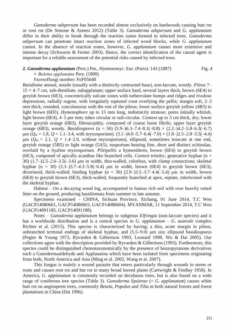
2. Ganoderma applanatum (Pers.) Pat., Hymenomyc. Eur. (Paris): 143 (1887) Fig. 4
≡ Boletus applanatus Pers. (1800)
Facesoffungi number: FoF05648
Basidiome annual, sessile (usually with a distinctly contracted base), non-laccate, woody. Pileus 7–
15 × 4–7 cm, sub-dimidiate, subapplanate; upper surface hard, several layers thick, brown (6E4) to
greyish brown (6E3), concentrically sulcate zones with turberculate bumps and ridges and rivulose
depressions, radially rugose, with irregularly ruptured crust overlying the pellis; margin soft, 2–3
mm thick, rounded, concolourous with the rest of the pileus; lower surface greyish yellow (4B3) to
light brown (6D5). Hymenophore up to 15 mm long, indistinctly stratose; pores initially whitish,
light brown (6E4), 4–5 per mm; tubes circular or sub-circular. Context up to 3 cm thick, dry; lower
layer greyish orange (6B3), fibrous/pithy, composed of coarse loose fibrils; upper layer greyish
orange (6B3), woody. Basidiospores (n = 50) (5.9–)6.3–7.4–8.1(–8.8) × (2.2–)4.2–5.8–6.3(–6.7)
μm (Qm = 1.8, Q = 1.1–3.4, with myxosporium). (3.1–)4.0–5.7–6.4(–7.0) × (1.8–)2.5–2.8–3.5(–4.4)
μm (Qm = 2.1, Q = 1.4–2.9, without myxosporium), ellipsoid, sometimes truncate at one end,
greyish orange (5B5) to light orange (5A5), eusporium bearing fine, short and distinct echinulae,
overlaid by a hyaline myxosporium. Pileipellis a hymeniderm, brown (6E4) to greyish brown
(6E3), composed of apically acanthus like branched cells. Context trimitic; generative hyphae (n =
30) (1.7–)2.5–2.6–3.5(–3.6) μm in width, thin-walled, colorless, with clamp connections; skeletal
hyphae (n = 30) (3.3–)3.7–4.3–5.9(–6.4) μm in width, brown (6E4) to greyish brown (6E3),
dextrinoid, thick-walled; binding hyphae (n = 30) (2.9–)3.5–3.7–4.4(–5.4) μm in width, brown
(6E4) to greyish brown (6E3), thick-walled, frequently branched at apex, septate, intertwined with
the skeletal hyphae.
Habitat – On a decaying wood log, accompanied in humus rich soil with over heavily rotted
litter on the ground, producing basidiomata from summer to late autumn.
Specimens examined – CHINA, Sichuan Province, Xichang, 01 June 2014, T.C Wen
(GACP14080601, GACP14080603, GACP14080604). MYANMAR, 11 September 2014, T.C Wen
(GACP14091185, GACP14091188).
Notes – Ganoderma applanatum belongs to subgenus Elfvingia (non-laccate species) and it
has a worldwide distribution and is a central species in G. applanatum – G. australe complex
Richter et al. (2015). This species is characterized by having; a thin, acute margin in pileus,
unbranched terminal endings of skeletal hyphae, and (5.5–9.0) μm size ellipsoid basidiospores
(Pegler & Young 1973, Ryvarden & Gilbertson 1993, Leonard 1998, Wu & Dai 2005). Our
collections agree with the description provided by Ryvarden & Gilbertson (1993). Furthermore, this
species could be distinguished chemotaxonomically by the presence of benzopyranone derivatives
such a Ganodermaaldehyde and Applanatins which have been isolated from specimens originating
from both, North America and Asia (Ming et al. 2002, Wang et al. 2007).
This fungus is mainly a wound parasite that enters particularly through wounds in stems or
roots and causes root rot and but rot in many broad leaved plants (Cartwright & Findlay 1958). In
America, G. applanatum is commonly recorded on deciduous trees, but is also found on a wide
range of coniferous tree species (Table 3). Ganoderma lipsiense (= G. applanatum) causes white
butt rot on angiosperm trees, commonly Betula, Populus and Tilia in both natural forests and forest
plantations in China (Dai 1996).
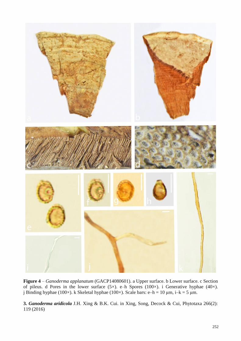
252
Figure 4 – Ganoderma applanatum (GACP14080601). a Upper surface. b Lower surface. c Section
of pileus. d Pores in the lower surface (5×). e–h Spores (100×). i Generative hyphae (40×).
j Binding hyphae (100×). k Skeletal hyphae (100×). Scale bars: e–h = 10 μm, i–k = 5 μm.
3. Ganoderma aridicola J.H. Xing & B.K. Cui. in Xing, Song, Decock & Cui, Phytotaxa 266(2):
119 (2016)
253
See the description and illustration at Xing et al. 2016)
Notes – Ganoderma aridicola, a species belong to G. lucidum species complex, was
introduced by Xing et al. (2016) from South Africa with the aid of molecular data. This species is a
white rot fungus found in Ficus species (Xing et al. 2016) (Table 3). Ganoderma aridicola is
characterized by its fuscous to black pileal surface with distinct concentric zones and small pores,
furthermore, it was found in an environment with contrasted dry and humid seasons (Xing et al.
2016).
4. Ganoderma angustisporum J.H. Xing, B.K. Cui & Y.C. Dai. in Xing, Sun, Han, Cui & Dai,
MycoKeys 34: 93–108 (2018)
See the description and illustration at Xing et al. (2018)
Notes – Ganoderma angustisporum was introduced from China recently, based on molecular
and morphological data (Xing et al. 2016). This species causes white rot in Casuarina equisetifolia
plants (Xing et al. 2016). Ganoderma angustisporum is characterized by its sessile basidiomata,
white pore surface, almond-shaped, slightly truncate and narrow basidiospores (Xing et al. 2016).
5. Ganoderma australe (Fr.: Fr.) Pat., 1889, Bull. Soc. mycol. Fr. 5, p. 71. Fig. 5
≡ Polyporus australis Fr.: Fr., 1828, Elench. Fung. 1, p. 108
Facesoffungi number: FoF05649
Basidiome annual, sessile, non-laccate, woody. Pileus 6–13 × 5.5–6 cm, sub orbicular, plano-
convex, applante, at center slightly swollen: upper surface brownish orange (6C4) to brown (6D4),
distinctly concentrically sulcate, with irregularly ruptured crust overlying the pellis; margin soft,
slightly lobate and concolorous with the pileus; lower surface yellowish white (4A2).
Hymenophore up to 9 mm long, indistinctly stratose; pores initially yellowish white (4A2), later
brown (6D6), 4–5 per mm; pores circular or sub circular. Context up to 1.5 cm thick, dry, duplex;
lower layer, brown (6D6), fibrous/pithy, composed of coarse loose fibrils; upper layer dark brown
(7F8), woody. Basidiospores (n = 40) (6.1–)7.6–9.2–10.8(–11.5) × (4.7–)5.3–7.6–7.9(–8.5) μm (Qm
= 1.5, Q = 0.9–2.6, with myxosporium). (4.9–)5.3–6.6–7.9(–8.5) × (3.1–)3.4–4.2–5.1(–5.8) μm (Qm
= 1.5, Q = 0.9–2.9, without myxosporium), ellipsoid, brownish orange (6C8) to brown (6D8), with
a brown eusporium bearing fine, short, and distinct echinulae, overlaid by a hyaline myxosporium.
Pileipellis a hymeniderm, (6–28) μm, brownish orange (6C8) to brown (6D8), composed of
apically acanthus like branched cells, dextrinoid. Context trimitic; generative hyphae not observed;
skeletal hyphae (n = 30) (4.5–3.1–3.9–4.1(–4.8) μm in width, thick-walled, sometimes branched,
brownish orange (6C8); binding hyphae (n = 30) (3.5–)4.2–5.6–5.8(–6.1) μm in width, thick-
walled, branched, brownish orange (6C8).
Habitat – On a decaying wood log, accompanied in humus rich soil with over heavily rotted
litter on the ground.
Specimens examined – CHINA, Jiangxi Province, 19 June 2014, T.C Wen (GACP14061912,
GACP14061914, GACP14061915). THAILAND, Chiang Mai Province, Mushroom Research
Center, Coniferous rainforest, 19°20′N–98°44′E, elev. 770 m, 12 June 2015, K.K Hapuarachchi
(GACP15061230, GACP15061231). MYANMAR, 11 September 2014, T.C Wen
(GACP14091187, GACP14091195).
Notes – Ganoderma australe was initially described from Pacific Islands but the type
specimen was lost or destroyed and currently only the neotype available is from Europe (Ryvarden
& Gilbertson 1993). This species has worldwide distribution (Martinez et al. 1991). Ganoderma
applanatum and G. australe from Europe have been confused longtime with each other (Leonard
1998). Ganoderma australe belongs to G. applanatum – australe complex and can be clearly
distinguished from G. applanatum by having larger basidiospores (Martinez et al. 1991, Moncalvo
& Ryvarden 1997). Furthermore, basidiospore dimensions, thickness of the cuticle and color of the
context layer were considered to be reliable discriminating characters in delimiting G. applanatum
and G. australe (Ryvarden & Gilbertson 1993, Leonard 1998). Our collections agree with the
description provided by Ryvarden & Gilbertson (1993). Ganoderma australe is common in the
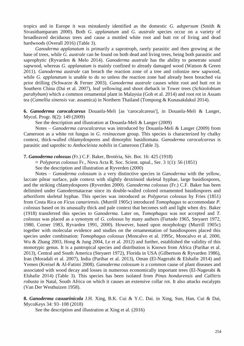
254
tropics and in Europe it was mistakenly identified as the domestic G. adspersum (Smith &
Sivasithamparam 2000). Both G. applanatum and G. australe species occur on a variety of
broadleaved deciduous trees and cause a mottled white root and butt rot of living and dead
hardwoods (Overall 2016) (Table 3).
Ganoderma applanatum is primarily a saprotroph, rarely parasitic and then growing at the
base of trees, while G. australe can be found on both dead and living trees, being both parasitic and
saprophytic (Ryvarden & Melo 2014). Ganoderma australe has the ability to penetrate sound
sapwood, whereas G. applanatum is mainly confined to already damaged wood (Watson & Green
2011). Ganoderma australe can breach the reaction zone of a tree and colonize new sapwood,
while G. applanatum is unable to do so unless the reaction zone had already been breached via
prior drilling (Schwarze & Ferner 2003). Ganoderma australe causes white root and butt rot in
Southern China (Dai et al. 2007), leaf yellowing and shoot dieback in Tower trees (Schizolobium
parahybum) which a common ornamental plant in Malaysia (Goh et al. 2014) and root rot in Assam
tea (Camellia sinensis var. assamica) in Northern Thailand (Tompong & Kunasakdakul 2014).
6. Ganoderma carocalcareus Douanla-Meli [as 'carocalcareus'], in Douanla-Meli & Langer,
Mycol. Progr. 8(2): 149 (2009)
See the description and illustration at Douanla-Meli & Langer (2009)
Notes – Ganoderma carocalcareus was introduced by Douanla-Meli & Langer (2009) from
Cameroon as a white rot fungus in G. resinaceum group. This species is characterized by chalky
context, thick-walled chlamydospores and dimorphic basidiomata. Ganoderma carocalcareus is
parasitic and saprobic to Anthocleista nobilis in Cameroon (Table 3).
7. Ganoderma colossus (Fr.) C.F. Baker, Brotéria, Sér. Bot. 16: 425 (1918)
≡ Polyporus colossus Fr., Nova Acta R. Soc. Scient. upsal., Ser. 3 1(1): 56 (1851)
See the description and illustration at Ryverden (2000)
Notes – Ganoderma colossum is a very distinctive species in Ganoderma with the yellow,
laccate pilear surface, pale context with slightly dextrinoid skeletal hyphae, large basidiospores,
and the striking chlamydospores (Ryverden 2000). Ganoderma colossus (Fr.) C.F. Baker has been
delimited under Ganodermataceae since its double-walled colored ornamented basidiospores and
arboriform skeletal hyphae. This species was introduced as Polyporus colossus by Fries (1851)
from Costa Rica on Ficus canariensis. (Murrill 1905c) introduced Tomophagus to accommodate P.
colossus based on its unusually thick and pale context that becomes soft and light when dry. Baker
(1918) transferred this species to Ganoderma. Later on, Tomophagus was not accepted and T.
colossus was placed as a synonym of G. colossus by many authors (Furtado 1965, Steyaert 1972,
1980, Corner 1983, Ryvarden 1991, 2000). However, based upon morphology (Murrill 1905c)
together with molecular evidence and studies on the ornamentation of basidiospores placed this
species under combination: Tomophagus colossus (Moncalvo et al. 1995c, Moncalvo et al. 2000,
Wu & Zhang 2003, Hong & Jung 2004, Le et al. 2012) and further, established the validity of this
monotypic genus. It is a pantropical species and distribution is Known from Africa (Parihar et al.
2013), Central and South America (Steyaert 1972), Florida in USA (Gilbertson & Ryvarden 1986),
Iran (Moradali et al. 2007), India (Parihar et al. 2013), Oman (El-Nagerabi & Elshafie 2014) and
Yemen (Kreisel & Al-Fatimi 2008). Ganoderma colossum is a common cause of plant diseases and
associated with wood decay and losses in numerous economically important trees (El-Nagerabi &
Elshafie 2014) (Table 3). This species has been isolated from Pinus hondurensis and Callitris
robusta in Natal, South Africa on which it causes an extensive collar rot. It also attacks eucalypts
(Van Der Westhuizen 1958).
8. Ganoderma casuarinicola J.H. Xing, B.K. Cui & Y.C. Dai. in Xing, Sun, Han, Cui & Dai,
MycoKeys 34: 93–108 (2018)
See the description and illustration at Xing et al. (2016)
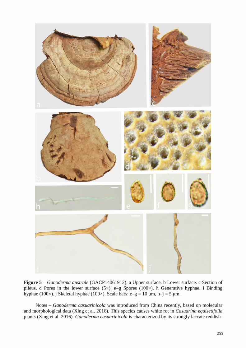
255
Figure 5 – Ganoderma australe (GACP14061912). a Upper surface. b Lower surface. c Section of
pileus. d Pores in the lower surface (5×). e–g Spores (100×). h Generative hyphae. i Binding
hyphae (100×). j Skeletal hyphae (100×). Scale bars: e–g = 10 μm, h–j = 5 μm.
Notes – Ganoderma casuarinicola was introduced from China recently, based on molecular
and morphological data (Xing et al. 2016). This species causes white rot in Casuarina equisetifolia
plants (Xing et al. 2016). Ganoderma casuarinicola is characterized by its strongly laccate reddish-
256
brown pileal surface, white pore surface, and luminous yellow to yellowish-brown cutis (Xing et al.
2016).
9. Ganoderma donkii Steyaert, Persoonia 7(1): 75 (1972) Fig. 6
Facesoffungi number: FoF05650
Basidiome annual to perennial, corky, with distinctly contracted base to host, becoming hard
corky to woody hard when dry. Pileus 3–5.5 cm, up to 1 cm thick at the base, suborbicular, plano
convex, applanate; upper surface hard, light brown (6D6) to dark brown (6F6), non laccate,
distinctly concentrically sulcate zones, crust overlies the pellis, differentiated zone at the point of
attachment; margin 3mm thick, concolorous with the pileus, rounded, inflexed; lower surface
yellowish white (4A2) to light brown (6D6). Hymenophore up to 8 mm long, indistinctly stratose,
orange grey (6B2), pores circular. Context duplex, not completely homogeneous in color; upper
layer dark brown (7F8), pithy, composed of coarse loose fibrils, soft; lower layer light brown
(5D4), woody. Basidiospores (n = 25) (6.6)6.7–7.7–8.7(11.3) × (4.2)4.6–5.4–6.2(7.8) µm (Qm =
1.4, Q = 1.1–1.7, with myxosporium). (n = 25) (4.7)4.9–5.8–6.7(7.8) × (2.5)3.2–3.9–4.7(6.2) µm
(Qm = 1.4, Q = 1.0–1.8.without myxosporium), ellipsoid, with a yellowish brown (5D8) to brown
(6D6) eusporium bearing fine and short echinulae, overlaid by a hyaline myxosporium. Pileipellis a
hymeniderm, dark brown (6F8), composed of apically acanthus like branched cells. Context
trimitic; generative hyphae (n = 20) (0.5–1.1–2.0) µm in width, colorless, thin-walled; skeletal
hyphae (n = 25) (0.9–1.4–1.9) µm in width, light brown (6D6), thick walled, sometimes branched;
binding hyphae (n = 25) (0.6–1.0–1.6) µm in width, light brown (6D6), branched, with clamp-
connections, intertwined the skeletal hyphae.
Habitat – On a living Dipterocarpus tree, accompanied in humus rich soil with over heavily
rotted litter on the ground.
Specimens examined – THAILAND, Chiang Mai Province, Mushroom research Center,
19°20′N–98°44′E, elev. 770 m, 21 December 2015, K.K Hapuarachchi (GACP15122107,
GACP15122108, GACP151221010, GACP151221012).
Notes – Ganoderma donkii was introduced by Steyaert (1972) from Indonesia and
characterized by radially corrugated pileus, hard context and small, ovoid (truncate when mature)
basidiospores (7.5–9 × 5.5–7) µm. This species belongs to G. applanatum – australe complex and
can be clearly distinguished from G. applanatum and G. australe by having smaller basidiospores
(Steyaert 1972, Martinez et al. 1991, Moncalvo & Ryvarden 1997). Ganoderma donkii is
distributed in China (Wu & Dai 2005), West Java (Indonesia) (Steyaert (1972) and Thailand
(Chandrasrikul et al. 2011). Our collections agree well with the description provided by (Wu & Dai
2005).
10. Ganoderma destructans M.P.A. Coetzee, Marinc. & M.J. Wingf., in Coetzee, Marincowitz,
Muthelo & Wingfield, Mycosphere 6(1): 253 (2015)
See the description and illustration at Coetzee et al. (2015)
Notes – Ganoderma destructans was introduced by Coetzee et al. (2015) from South Africa
with the aid of morphological and molecular data. This species is characterized by the pileus which
contains creamy soft non-poroid tissue showing obvious continuity to hymenophore, ovoid
basidiospores (11–14 × 7–9) μm and unique nucleotide polymorphisms at ITS and nrLSU (Coetzee
et al. 2015). Ganoderma destructans is the most important pathogen resulting in the death of
Jacaranda mimosifolia trees by root rot in Pretoria, South Africa (Coetzee et al. 2015).
11. Ganoderma flexipes Pat., Bull. Soc. mycol. Fr. 23(2): 75 (1907) Fig. 7
≡ Polyporus flexipes (Pat.) Lloyd, Mycol. Writ. 3 (Syn. Stip. Polyporoids): 104 (1912)
Facesoffungi number: FoF05651
Basidiome annual, stipitate, laccate, corky, becoming hard corky to woody hard when dry.
Pileus 2.5–3.5 × 1.0–2.0 cm, up to 0.5 cm thick at the base, flabelliform, rotund, upper surface;
reddish brown (8E8), concentrically sulcate zones with turberculate bumps and ridges and rivulose
257
depressions, with irregularly ruptured crust overlying the pellis, margin; soft, yellowish brown
(5E8), lower surface; light brown (7D5). Hymenophore up to 10 mm long, indistinctly stratose;
pores initially greyish brown (7D3), bruising brown (8E8), pores circular or sub-circular or
isodiametric, 3–4 per mm. Context up to 8 mm thick, triplex, dry; lower layer reddish brown (8E8),
fibrous, composed of coarse loose fibrils; upper layer dark brown (8F8), corky to woody. Stipe
eccentric, dorsally lateral to nearly dorsal, sub-cylindrical, dark brown (8F8), 4.0 × 7.0 cm, 0.4 cm
thick at the base. Basidiospores (n = 25) (8.3–)9.1–9.8–10.5(11.1–) × (–6.5)7.3–6.1–9.2(–10.5) μm
(Qm = 1.4, Q = 0.9–2.1 with myxosporium). (6.5–)7.3–8.3–9.2(–10.5) × (4.1–)4.4–4.9–5.4(–5.9)
μm (Qm = 1.4, Q = 0.9–2.9, without myxosporium), ellipsoid, light orange (5A5), brownish orange
(6C8) to light brown (5A5), with a brown eusporium bearing fine, short and distinct echinulae,
overlaid by a hyaline myxosporium. Pileipellis a hymeniderm, brownish orange (6C8), clavate like
cells, dextrinoid. Context trimitic; generative hyphae (n = 30) (1.5–)1.2–1.8–2.6(–3.9) μm in width,
hyaline, thin-walled without clamp connections; skeletal hyphae (n = 30) (3.1–)1.5–4.4–5.5(–6.8)
μm in width, thick-walled, nearly solid, sometimes branched, greyish brown (5B5) to brownish
orange (6C8); binding hyphae (n = 30) (2.4–)3.2–4.1–4.9(–5.4) μm in width, thick-walled,
branched, nearly solid, light orange (5A5) to brownish orange (6C8).
Habitat – Growing near hardwood roots or rotten woods, in dry dipterocarp forest, coniferous
rain forest and in upper mixed deciduous forest, growing up from soil.
Specimens examined – LAOS, Savvanakhet Province, Phin District, Dong Phou Vieng
Protected Area, 16°23′N–105°59′E, elev. 283 m, mixed deciduous forest, 27 June 2017, P.
Phengsintham (GACP17062724), Huaphanh Province, Samneua District, Nasala Village, 20°22′N-
103°98′E, elev. 1346 m, Coniferous rain forest, 30 July 2017, P. Phengsintham (GACP17073037).
VIETNAM, Lam Dong Province, Bidoup Nui Ba National Park, 12°26′N–108°30′E, elev. 2050 m,
coniferous rain forest, 19 October 2017, T.C Wen (GACP17101903, GACP17101910,
GACP17102101, GACP17102301).
Notes – Ganoderma flexipes was introduced by Patouillard (1907) from Vietnam and has
since been recorded from China, India, Nepal, Pakistan (Steyaert 1972, Zhao & Zhang 2000, Wang
& Wu 2007). This species can easily be recognized by its small reddish brown pileus, long and thin
stipe, reddish brown to dark brown context, and ellipsoid or ovoid basidiospores. Our collections
fall within the range (8–13 × 5.5–8) μm, measured by Steyaert (1972) and fit that of the lectotype
(9–11 × 6–7.5) μm, measured by Ryvarden (1983) for G. flexipes. We report G. flexipes from Laos
as a new record.
12. Ganoderma gibbosum (Cooke) Pat., Ann. Jard. Bot. Buitenzorg, suppl. 1: 114 (1897) Fig. 8
≡ Fomes amboinensis var. gibbosus Cooke (1885)
Facesoffungi number: FoF05652
Basidiome annual, sessile (usually with a distinctly contracted base), non laccate, woody.
Pileus 6–12 × 3.0–6.0 cm, up to 3 cm thick at the base, spathulate, upper surface; hard, several
layers thick, light brown (6D5) to light brown (6D6), crust overlies the pellis, concentric zones with
turberculate bumps and ridges and rivulose depressions; margin with numerous undulations and
irregularities, wavy, 2 mm thick, concolorous with the pileus; lower surface light brown (5D4).
Hymenophore up to 6 mm long, indistinctly stratose, light brown (5D4), pores circular or sub
circular, 4–5 pores per mm. Context up to 3 cm thick, dry, duplex, lower layer; brown (6E8), pithy,
composed of coarse loose fibrils, upper layer; dark brown (6F8), woody. Basidiospores (n = 50)
(4.3–)6.9–7.6–9.2(–10.5) × (3.8–)4.6–5.6–5.7(–6.1) μm (Qm = 1.7, Q = 0.8–2.5, with
myxosporium). (–3.6)4.8–6.0–7.6(–8.7) × (–2.0)2.7–3.5–4.5(–6.7) μm (Qm = 1.7, Q = 0.8–3.2,
without myxosporium), ellipsoid to elongate, brown (6D8) to light brown (6D6), eusporium
bearing fine, short, and distinct echinulae, overlaid by a hyaline myxosporium, bitunicate.
Pileipellis a hymeniderm, brown (6D8) to light brown (6D6), composed of apically acanthus like
branched cells, dextrinoid. Context trimitic; generative hyphae (n = 30) (1.3–)1.6–2.1–2.7(–3.6) μm
in width, thin-walled, hyaline; skeletal hyphae (n = 30) (4.0–)4.8–4.7–5.2(–5.8) μm in width, brown
(6F8), dextrinoid, thick-walled; binding hyphae (n = 30) (2.6–)3.6–4.7–5.8(–6.1) μm in width,
brown (6F8), thick-walled, branched, intertwined the skeletal hyphae.
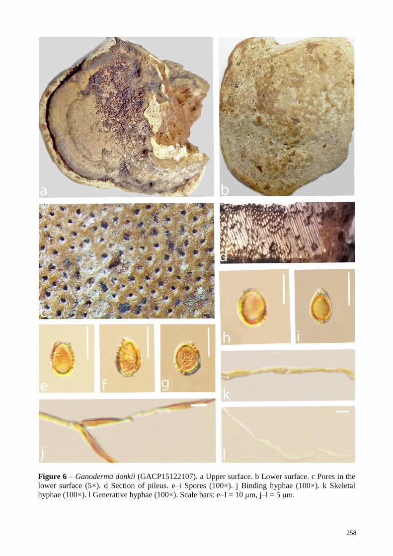
258
Figure 6 – Ganoderma donkii (GACP15122107). a Upper surface. b Lower surface. c Pores in the
lower surface (5×). d Section of pileus. e–i Spores (100×). j Binding hyphae (100×). k Skeletal
hyphae (100×). l Generative hyphae (100×). Scale bars: e–I = 10 μm, j–l = 5 μm.
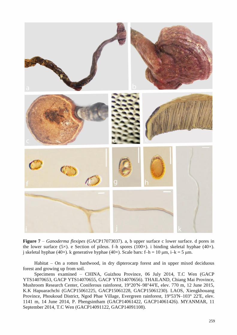
259
Figure 7 – Ganoderma flexipes (GACP17073037). a, b upper surface c lower surface. d pores in
the lower surface (5×). e Section of pileus. f–h spores (100×). i binding skeletal hyphae (40×).
j skeletal hyphae (40×). k generative hyphae (40×). Scale bars: f–h = 10 μm, i–k = 5 μm.
Habitat – On a rotten hardwood, in dry dipterocarp forest and in upper mixed deciduous
forest and growing up from soil.
Specimens examined – CHINA, Guizhou Province, 06 July 2014, T.C Wen (GACP
YTS14070653, GACP YTS14070655, GACP YTS14070656). THAILAND, Chiang Mai Province,
Mushroom Research Center, Coniferous rainforest, 19°20′N–98°44′E, elev. 770 m, 12 June 2015,
K.K Hapuarachchi (GACP15061225, GACP15061228, GACP15061230). LAOS, Xiengkhouang
Province, Phoukoud District, Ngod Phae Village, Evergreen rainforest, 19°53′N–103° 22′E, elev.
1141 m, 14 June 2014, P. Phengsintham (GACP14061422, GACP14061426). MYANMAR, 11
September 2014, T.C Wen (GACP14091122, GACP14091108).
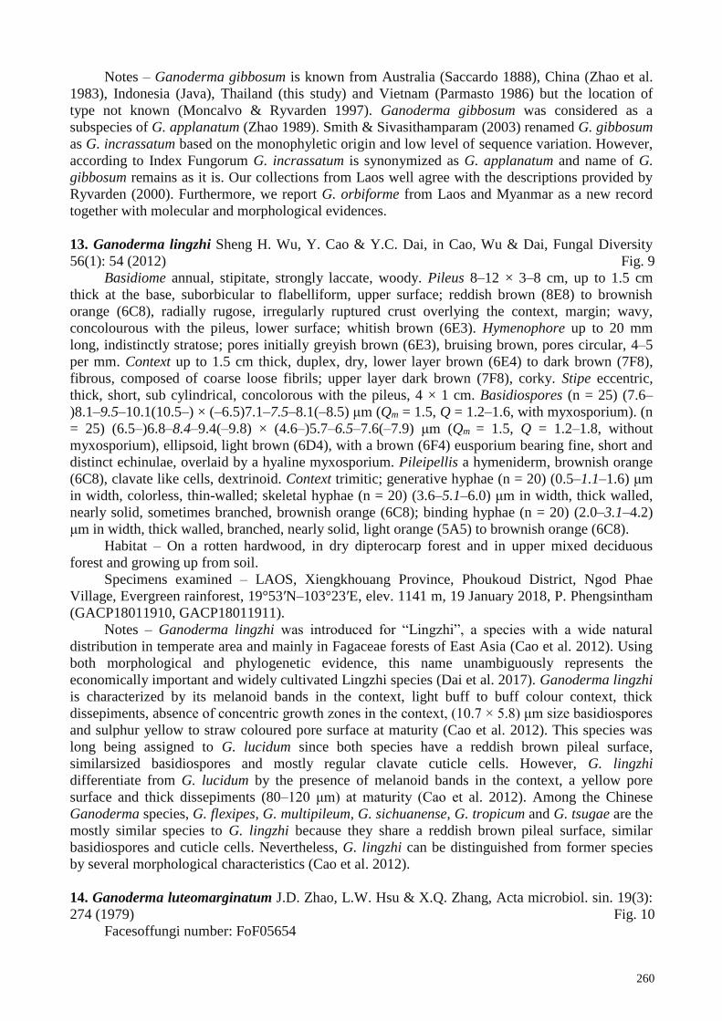
260
Notes – Ganoderma gibbosum is known from Australia (Saccardo 1888), China (Zhao et al.
1983), Indonesia (Java), Thailand (this study) and Vietnam (Parmasto 1986) but the location of
type not known (Moncalvo & Ryvarden 1997). Ganoderma gibbosum was considered as a
subspecies of G. applanatum (Zhao 1989). Smith & Sivasithamparam (2003) renamed G. gibbosum
as G. incrassatum based on the monophyletic origin and low level of sequence variation. However,
according to Index Fungorum G. incrassatum is synonymized as G. applanatum and name of G.
gibbosum remains as it is. Our collections from Laos well agree with the descriptions provided by
Ryvarden (2000). Furthermore, we report G. orbiforme from Laos and Myanmar as a new record
together with molecular and morphological evidences.
13. Ganoderma lingzhi Sheng H. Wu, Y. Cao & Y.C. Dai, in Cao, Wu & Dai, Fungal Diversity
56(1): 54 (2012) Fig. 9
Basidiome annual, stipitate, strongly laccate, woody. Pileus 8–12 × 3–8 cm, up to 1.5 cm
thick at the base, suborbicular to flabelliform, upper surface; reddish brown (8E8) to brownish
orange (6C8), radially rugose, irregularly ruptured crust overlying the context, margin; wavy,
concolourous with the pileus, lower surface; whitish brown (6E3). Hymenophore up to 20 mm
long, indistinctly stratose; pores initially greyish brown (6E3), bruising brown, pores circular, 4–5
per mm. Context up to 1.5 cm thick, duplex, dry, lower layer brown (6E4) to dark brown (7F8),
fibrous, composed of coarse loose fibrils; upper layer dark brown (7F8), corky. Stipe eccentric,
thick, short, sub cylindrical, concolorous with the pileus, 4 × 1 cm. Basidiospores (n = 25) (7.6–
)8.1–9.5–10.1(10.5–) × (–6.5)7.1–7.5–8.1(–8.5) μm (Qm = 1.5, Q = 1.2–1.6, with myxosporium). (n
= 25) (6.5–)6.8–8.4–9.4(–9.8) × (4.6–)5.7–6.5–7.6(–7.9) μm (Qm = 1.5, Q = 1.2–1.8, without
myxosporium), ellipsoid, light brown (6D4), with a brown (6F4) eusporium bearing fine, short and
distinct echinulae, overlaid by a hyaline myxosporium. Pileipellis a hymeniderm, brownish orange
(6C8), clavate like cells, dextrinoid. Context trimitic; generative hyphae (n = 20) (0.5–1.1–1.6) μm
in width, colorless, thin-walled; skeletal hyphae (n = 20) (3.6–5.1–6.0) μm in width, thick walled,
nearly solid, sometimes branched, brownish orange (6C8); binding hyphae (n = 20) (2.0–3.1–4.2)
μm in width, thick walled, branched, nearly solid, light orange (5A5) to brownish orange (6C8).
Habitat – On a rotten hardwood, in dry dipterocarp forest and in upper mixed deciduous
forest and growing up from soil.
Specimens examined – LAOS, Xiengkhouang Province, Phoukoud District, Ngod Phae
Village, Evergreen rainforest, 19°53′N–103°23′E, elev. 1141 m, 19 January 2018, P. Phengsintham
(GACP18011910, GACP18011911).
Notes – Ganoderma lingzhi was introduced for “Lingzhi”, a species with a wide natural
distribution in temperate area and mainly in Fagaceae forests of East Asia (Cao et al. 2012). Using
both morphological and phylogenetic evidence, this name unambiguously represents the
economically important and widely cultivated Lingzhi species (Dai et al. 2017). Ganoderma lingzhi
is characterized by its melanoid bands in the context, light buff to buff colour context, thick
dissepiments, absence of concentric growth zones in the context, (10.7 × 5.8) μm size basidiospores
and sulphur yellow to straw coloured pore surface at maturity (Cao et al. 2012). This species was
long being assigned to G. lucidum since both species have a reddish brown pileal surface,
similarsized basidiospores and mostly regular clavate cuticle cells. However, G. lingzhi
differentiate from G. lucidum by the presence of melanoid bands in the context, a yellow pore
surface and thick dissepiments (80–120 μm) at maturity (Cao et al. 2012). Among the Chinese
Ganoderma species, G. flexipes, G. multipileum, G. sichuanense, G. tropicum and G. tsugae are the
mostly similar species to G. lingzhi because they share a reddish brown pileal surface, similar
basidiospores and cuticle cells. Nevertheless, G. lingzhi can be distinguished from former species
by several morphological characteristics (Cao et al. 2012).
14. Ganoderma luteomarginatum J.D. Zhao, L.W. Hsu & X.Q. Zhang, Acta microbiol. sin. 19(3):
274 (1979) Fig. 10
Facesoffungi number: FoF05654

261
Basidiome annual, stipitate, strongly laccate, corky. Pileus 1.5–5 × 1–3 cm, up to 0.4 cm thick at
the base, flabelliform to suborbicular to spathulate, upper surface; reddish black (8E8) to brownish
black (6C8), undulate, ferruginous, irregularly ruptured crust overlying the context, margin; 2mm
thick, blunt, yellow (4A8); lower surface greyish brown (6E3). Hymenophore up to 10 mm long,
indistinctly stratose; pores initially brown (6E3), bruising dark brown (6F6), pores circular, 3–5 per
mm. Context up to 0.8 cm thick, duplex, dry, upper layer dark brown (7F8), fibrous, composed of
coarse loose fibrils, corky; lower layer dark brown (7F8), woody. Stipe eccentric, sub cylindrical,
concolorous with the pileus, 4 × 8 cm, 0.5 cm at the base. Basidiospores (n = 20) (8.6–)9.6–10.7–
11.8(12.1–) × (–6.1)6.3–7.2–8.1(–8.6) μm (Qm = 1.5, Q = 1.3–1.6, with myxosporium). (n = 20)
(6.6–)7.9–9.2–10.4(–11.7) × (3.7–)4.6–5.7–6.9(–7.7) μm (Qm = 1.6, Q = 1.3–2.3, without
myxosporium), ellipsoid to truncate, truncate at the apex, brown (6E6), with a light brown (6D6)
eusporium bearing fine, short and distinct echinulae, overlaid by a hyaline myxosporium.
Pileipellis a hymeniderm, brownish orange (6C8), clavate like cells. Context trimitic; generative
hyphae (n = 20) (0.4–0.9–1.4) μm in width, thin-walled, hyaline; skeletal hyphae (n = 20) (1.6–2.5–
3.2) μm in width, thick walled, nearly solid, sometimes branched, brownish orange (6C8); binding
hyphae (n = 20) (2.5–3.9–4.9) μm in width, thick walled, branched, nearly solid, light orange (5A5)
to brownish orange (6C8).
Habitat – On a rotten hardwood, in dry dipterocarp forest and in upper mixed deciduous
forest and growing up from soil.
Specimens examined – LAOS, Sayabouli Province, Paklay District, Nakhan Village, mixed
deciduous forest, 18°41′N–101°46′E, elev. 354 m, 25 August 2016, P. Phengsintham
(GACP16082545, GACP6082548, GACP16082549) (from Herbarium of Guizhou Agricultural
sciences; GAS3356). MYANMAR, 11 September 2014, T.C Wen (GACP140911224).
Notes – Ganoderma luteomarginatum was introduced by Zhao et al. (1979) from China and
characterized by strongly laccate, thin, reddish black pileus with yellow margin, small
basidiospores (10.7 × 7.2) μm and trimitic hyphal system. The morphology of G. luteomarginatum
collections from Laos and Myanmar agree well with that of the holotype as described by Zhao et al.
(1979). This species morphologically resembles G. sinense and G. austrofujianense. However, G.
sinense can be distinguished by having thick, black pileus with a margin concolourous to the pileus
and larger basidiospores, and G. austrofujianense differed by having white and brown color zones
in the pileus. Furthermore, this species has been recorded from Thailand and Vietnam (Kha 2014)
with morphological evidence. There is no phylogenetic data available for this species in GenBank.
Ganoderma luteomarginatum is newly recorded taxa from Laos and Myanmar.
15. Ganoderma mbrekobenum E.C. Otto, Blanchette, Held, C.W. Barnes & Obodai (2016)
See the description ad illustration at Crouse et al. (2016)
Notes – Ganoderma mbrekobenum was introduced by Crous et al. 2016 from Ghana based on
morphology and molecular data. This species is characterized by ovoid to broadly ellipsoid with a
truncate base, bitunicate, verruculose basidiospores, dimitic hyphal system and a homogenous
context. Ganoderma mbrekobenum causes decay in the roots and trunks of angiosperm trees in the
southern regions of Ghana (Crous et al. 2016) (Table 3).
16. Ganoderma microsporum R.S. Hseu, in Hseu, Chen & Wang, Mycotaxon 35(1): 36 (1989)
See the description at Hseu et al. (1989)_
Notes – Ganoderma microsporum was introduced by Hseu et al. (1989) based on
morphological data from Taiwan. This species is characterized by small size, (4–5) × (6–8) μm, and
ovoid to subspherical basidiospores. However, G. weberianum reported from Asia could not be
separated from those representing G. microsporum based on molecular data (Moncalvo et al.
1995a, 1995b). Later, Smith & Sivasithamparam (2003) showed that the two species could be
separated by the presence of gasterospores in G. weberianum. This fungus causes white butt rot on
angiosperm trees and reported from China and India (Table 3).
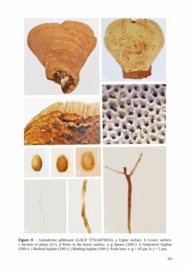
262
Figure 8 – Ganoderma gibbosum (GACP YTS14070653). a Upper surface. b Lower surface.
c Section of pileus (5×). d Pores in the lower surface. e–g Spores (100×). h Generative hyphae
(100×). i Skeletal hyphae (100×). j Binding hyphae (100×). Scale bars: e–g = 10 μm, h–j = 5 μm.

263
Figure 9 – Ganoderma lingzhi (GACP18011910). a Upper surface. b Lower surface. c Pores in the
lower surface (5×). d Section of pileus. e–h Spores (100×). i Skeletal hyphae (100×). j Binding
hyphae (100×). k Generative hyphae (100×). Scale bars: e–h = 10 μm, i–k = 5 μm.

264
Figure 10 – Ganoderma luteomarginatum (GACP16082545). a,b Upper surface. c Lower surface.
d Section of pileus . e Pores in the lower surface (5×). f–i Spores (100×). j Generative hyphae
(100×). k Skeletal hyphae (100×). l Binding hyphae (100×). Scale bars: f–i = 10 μm, j–l = 5 μm.
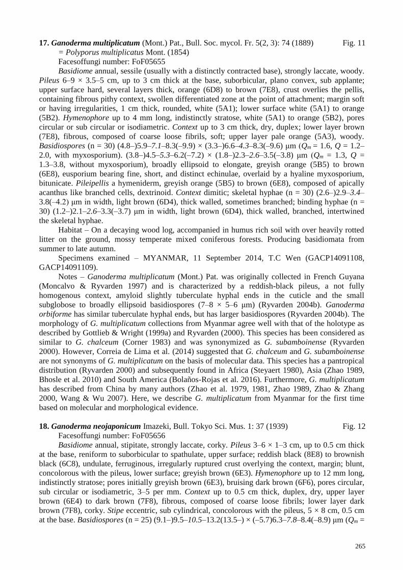
265
17. Ganoderma multiplicatum (Mont.) Pat., Bull. Soc. mycol. Fr. 5(2, 3): 74 (1889) Fig. 11
= Polyporus multiplicatus Mont. (1854)
Facesoffungi number: FoF05655
Basidiome annual, sessile (usually with a distinctly contracted base), strongly laccate, woody.
Pileus 6–9 × 3.5–5 cm, up to 3 cm thick at the base, suborbicular, plano convex, sub applante;
upper surface hard, several layers thick, orange (6D8) to brown (7E8), crust overlies the pellis,
containing fibrous pithy context, swollen differentiated zone at the point of attachment; margin soft
or having irregularities, 1 cm thick, rounded, white (5A1); lower surface white (5A1) to orange
(5B2). Hymenophore up to 4 mm long, indistinctly stratose, white (5A1) to orange (5B2), pores
circular or sub circular or isodiametric. Context up to 3 cm thick, dry, duplex; lower layer brown
(7E8), fibrous, composed of coarse loose fibrils, soft; upper layer pale orange (5A3), woody.
Basidiospores (n = 30) (4.8–)5.9–7.1–8.3(–9.9) × (3.3–)6.6–4.3–8.3(–9.6) μm (Qm = 1.6, Q = 1.2–
2.0, with myxosporium). (3.8–)4.5–5.3–6.2(–7.2) × (1.8–)2.3–2.6–3.5(–3.8) μm (Qm = 1.3, Q =
1.3–3.8, without myxosporium), broadly ellipsoid to elongate, greyish orange (5B5) to brown
(6E8), eusporium bearing fine, short, and distinct echinulae, overlaid by a hyaline myxosporium,
bitunicate. Pileipellis a hymeniderm, greyish orange (5B5) to brown (6E8), composed of apically
acanthus like branched cells, dextrinoid. Context dimitic; skeletal hyphae (n = 30) (2.6–)2.9–3.4–
3.8(–4.2) μm in width, light brown (6D4), thick walled, sometimes branched; binding hyphae (n =
30) (1.2–)2.1–2.6–3.3(–3.7) μm in width, light brown (6D4), thick walled, branched, intertwined
the skeletal hyphae.
Habitat – On a decaying wood log, accompanied in humus rich soil with over heavily rotted
litter on the ground, mossy temperate mixed coniferous forests. Producing basidiomata from
summer to late autumn.
Specimens examined – MYANMAR, 11 September 2014, T.C Wen (GACP14091108,
GACP14091109).
Notes – Ganoderma multiplicatum (Mont.) Pat. was originally collected in French Guyana
(Moncalvo & Ryvarden 1997) and is characterized by a reddish-black pileus, a not fully
homogenous context, amyloid slightly tuberculate hyphal ends in the cuticle and the small
subglobose to broadly ellipsoid basidiospores (7–8 × 5–6 μm) (Ryvarden 2004b). Ganoderma
orbiforme has similar tuberculate hyphal ends, but has larger basidiospores (Ryvarden 2004b). The
morphology of G. multiplicatum collections from Myanmar agree well with that of the holotype as
described by Gottlieb & Wright (1999a) and Ryvarden (2000). This species has been considered as
similar to G. chalceum (Corner 1983) and was synonymized as G. subamboinense (Ryvarden
2000). However, Correia de Lima et al. (2014) suggested that G. chalceum and G. subamboinense
are not synonyms of G. multiplicatum on the basis of molecular data. This species has a pantropical
distribution (Ryvarden 2000) and subsequently found in Africa (Steyaert 1980), Asia (Zhao 1989,
Bhosle et al. 2010) and South America (Bolaños-Rojas et al. 2016). Furthermore, G. multiplicatum
has described from China by many authors (Zhao et al. 1979, 1981, Zhao 1989, Zhao & Zhang
2000, Wang & Wu 2007). Here, we describe G. multiplicatum from Myanmar for the first time
based on molecular and morphological evidence.
18. Ganoderma neojaponicum Imazeki, Bull. Tokyo Sci. Mus. 1: 37 (1939) Fig. 12
Facesoffungi number: FoF05656
Basidiome annual, stipitate, strongly laccate, corky. Pileus 3–6 × 1–3 cm, up to 0.5 cm thick
at the base, reniform to suborbicular to spathulate, upper surface; reddish black (8E8) to brownish
black (6C8), undulate, ferruginous, irregularly ruptured crust overlying the context, margin; blunt,
concolorous with the pileus, lower surface; greyish brown (6E3). Hymenophore up to 12 mm long,
indistinctly stratose; pores initially greyish brown (6E3), bruising dark brown (6F6), pores circular,
sub circular or isodiametric, 3–5 per mm. Context up to 0.5 cm thick, duplex, dry, upper layer
brown (6E4) to dark brown (7F8), fibrous, composed of coarse loose fibrils; lower layer dark
brown (7F8), corky. Stipe eccentric, sub cylindrical, concolorous with the pileus, 5 × 8 cm, 0.5 cm
at the base. Basidiospores (n = 25) (9.1–)9.5–10.5–13.2(13.5–) × (–5.7)6.3–7.8–8.4(–8.9) μm (Qm =
266
1.5, Q = 1.1–1.7, with myxosporium). (n = 25) (7.8–)8.1–8.9–9.2(–10.5) × (4.7–)5.1–5.8–6.2(–6.8)
μm (Qm = 1.5, Q = 1.2–1.8, without myxosporium), ellipsoid, truncate at the apex, brown (6E6),
with a light brown (6D6) eusporium bearing fine, short and distinct echinulae, overlaid by a hyaline
myxosporium. Pileipellis a hymeniderm, brownish orange (6C8), clavate like cells. Context
dimitic; generative hyphae not observed; skeletal hyphae (n = 30) (3.6–5.0–6.2) μm in width, thick
walled, nearly solid, sometimes branched, brownish orange (6C8); binding hyphae (n = 30) (2.8–
4.5–5.4) μm in width, thick walled, branched, nearly solid, light orange (5A5) to brownish orange
(6C8).
Habitat – On a rotten wood, in dry dipterocarp forest and in upper mixed deciduous forest and
growing up from soil.
Specimens examined – LAOS, Savannakhet province, Phin district, Nathom Xe village, Phou
Xang hae, mixed deciduous forest, 16 58′N–105 89′E, 23 June 2017, P. Phengsintham
(GACP17062350, GACP17062355). MYANMAR, 10 September 2014, TC Wen
(GACP14091009, GACP14091014).
Notes – Ganoderma neojaponicum was introduced by Imazeki (1939) from Japan and it’s a
member of the Ganoderma lucidum species complex (Hattori & Ryvarden 1994). This species is
rarely saprotrophic and unique with its slender long spores, a black shiny basidiocarp, slender stipe
and host range (dead coniferous trees or bamboos) (Imazeki et al 1988, Hsieh & Yeh 2004). This
mushroom is distributed in Asian countries including China, Korea, Japan, Taiwan, Malaysia (Tan
et al. 2015), Laos and Myanmar (this study).
19. Ganoderma orbiforme (Fr.) Ryvarden (as 'orbiformum') Mycologia 92(1): 187 (2000) Fig. 13
≡ Polyporus orbiformis Fr. (1838)
= Ganoderma mastoporum (Lév.) Pat. [as 'malosporum'], Bull. Soc. mycol. Fr. 5(2, 3): 75
(1889)
= Ganoderma fornicatum (Fr.) Pat. sensu Imazeki, Bull. Tokyo Sci. Mus. 1: 47 (1939)
= Ganoderma cupreum (Cooke) Bres, Annls mycol. 9(3): 268 (1911)
= Ganoderma subtornatum Murrill, Bull. Torrey bot. Club 34: 477 (1907)
= Ganoderma densizonatum J.D. Zhao & X.Q. Zhang, in Zhao, Zhang & Xu, Acta Mycol.
Sin. 5(2): 86 (1986)
= Ganoderma limushanense J.D. Zhao & X.Q. Zhang, Acta Mycol. Sin. 5(4): 219 (1986)
See Index Fungorum for other synonyms
Facesoffungi number: FoF05657
Basidiome annual to perennial, with a distinctly contracted base, weakly laccate, woody.
Pileus 8–21 × 5–12 cm, up to 4 cm thick at the base, flabelliform, plano convex, applanate, upper
surface; hard, several layers thick, alternating brownish orange (6C8) to light brown (6D4)
concentrically zones, crust overlies the pellis, concentrically sulcate zones with turberculate bumps
and rivulose depressions, differentiated zone at the point of attachment; margin with numerous
undulations and irregularities, 4 mm thick, rounded and white (4A1) to yellowish white (4A2).
Hymenophore up to 3 mm long, indistinctly stratose, orange grey (6B2), pores circular or sub-
circular. Context up to 2 cm thick, dry, triplex; lower layer dark brown (7F8), pithy, composed of
coarse loose fibrils, soft; upper layer light brown (5D4), woody. Basidiospores (n = 25) (8.2)10.1–
10.7–11.4(11.6) × (4.8)5.7–6.5–7.2(7.4) µm, (Qm = 1.6, Q = 1.3–2.1, with myxosporium). (n = 25)
(6.8)7.8–8.7–9.6(10.3) × (3.7)4.7–5.4–6.1(6.7) µm (Qm = 1.6, Q = 1.2–2.1, without myxosporium),
yellowish brown (5D8), elongate, eusporium bearing fine, short, and distinct echinulae, overlaid by
a hyaline myxosporium, bitunicate. Pileipellis a hymeniderm, dark brown (7F8), composed of
apically acanthus like branched cells, dextrinoid. Context trimitic; generative hyphae (n = 25) (0.8–
2.2–2.5) μm in width, thin-walled, hyaline; skeletal hyphae (n = 28) (1.9–3.7–4.9) μm in width,
light brown (5D6), thick-walled; binding hyphae (n = 28) (1.6–3.4–7.2) μm in width, light brown
(5D6), thick-walled, branched, intertwined the skeletal hyphae.
Habitat – On a decaying hardwood log, accompanied in humus rich soil with over heavily
rotted litter on the ground, producing basidiomata from summer to late autumn.
267
Figure 11 – Ganoderma multiplicatum (GACP14091107). a Upper surface. b Lower surface.
c Pores in the lower surface (4.5×). d Section of pileus, e–g Spores (100×). h Generative hyphae
(100×). i Skeletal hyphae (100×). j Binding hyphae (100×). Scale bars: e–g = 10 μm, h–j = 5 μm.

268
Figure 12 – Ganoderma neojaponicum (GACP17062350). a Upper surface. b Lower surface.
c Section of pileus (5×). d Pores in the lower surface. e–h Spores (100×). i Cuticle cells (100×).
j Skeletal hyphae (100×). k Binding hyphae (100×). Scale bars: e–i = 10 μm, j–k = 5 μm.
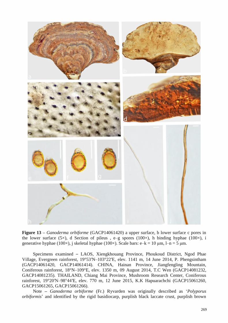
269
Figure 13 – Ganoderma orbiforme (GACP14061420) a upper surface, b lower surface c pores in
the lower surface (5×), d Section of pileus , e–g spores (100×), h binding hyphae (100×), i
generative hyphae (100×), j skeletal hyphae (100×). Scale bars: e–k = 10 μm, l–n = 5 μm.
Specimens examined – LAOS, Xiengkhouang Province, Phoukoud District, Ngod Phae
Village, Evergreen rainforest, 19°53′N–103°22′E, elev. 1141 m, 14 June 2014, P. Phengsintham
(GACP14061420, GACP14061414). CHINA, Hainan Province, Jiangfengling Mountain,
Coniferous rainforest, 18°N–109°E, elev. 1350 m, 09 August 2014, T.C Wen (GACP14081232,
GACP14081235). THAILAND, Chiang Mai Province, Mushroom Research Center, Coniferous
rainforest, 19°20′N–98°44′E, elev. 770 m, 12 June 2015, K.K Hapuarachchi (GACP15061260,
GACP15061265, GACP15061266).
Note – Ganoderma orbiforme (Fr.) Ryvarden was originally described as ‘Polyporus
orbiformis’ and identified by the rigid basidiocarp, purplish black laccate crust, purplish brown
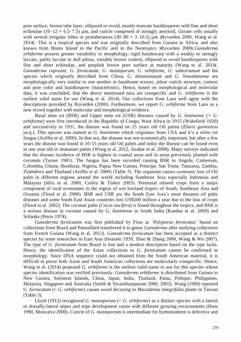
270
pore surface, brown tube layer, ellipsoid or ovoid, mostly truncate basidiospores with fine and short
echinulae (10–12 × 6.5–7.5) µm, and cuticle composed of strongly amyloid, clavate cells usually
with several irregular lobes or protuberances (30–80 × 3–10.5) µm (Ryvarden 2000, Wang et al.
2014). This is a tropical species and was originally described from Guinea in Africa, and also
known from Bonin Island in the Pacific and in the Neotropics (Ryvarden 2000).Ganoderma
orbiforme possess greater variability in morphology; rigid basidiocarp with a weakly to strongly
laccate, partly laccate or dull pileus, variably brown context, ellipsoid to ovoid basidiospores with
fine and short echinulae, and purplish brown pore surface at maturity (Wang et al. 2014).
Ganoderma cupreum, G. fornicatum, G. mastoporum, G. orbiforme, G. subtornatum and the
species which originally described from China; G. densizonatum and G. limushanense are
morphologically very similar to one another in basidiome texture, pilear cuticle structure, context
and pore color and basidiospore characteristics. Hence, based on morphological and molecular
data, it was concluded, that the above mentioned taxa are conspecific and G. orbiforme is the
earliest valid name for use (Wang et al. 2014). Our collections from Laos well agree with the
descriptions provided by Ryvarden (2000). Furthermore, we report G. orbiforme from Laos as a
new record together with molecular and morphological evidence.
Basal stem rot (BSR) and Upper stem rot (USR) diseases caused by G. boninense (= G.
orbiforme) were first introduced in the Republic of Congo, West Africa in 1915 (Wakefield 1920)
and successively in 1931, the disease was found on 25 years old Oil palms (Elaeis guineensis
jacq.). This species was named as G. boninense which originates from USA and it’s a white rot
fungus (Ariffin et al. 2000). In that era, the disease was not economically important, but after a few
years the disease was found in 10-15 years old Oil palms and today the disease can be found even
in one year old or immature palms (Wong et al. 2012, Azahar et al. 2008). Many surveys indicated
that the disease incidence of BSR is highest in coastal areas and in areas previously planted with
coconuts (Turner 1981). The fungus has been recorded causing BSR in Angola, Cameroon,
Colombia, Ghana, Honduras, Nigeria, Papua New Guinea, Principe, San Tome, Tanzania, Zambia,
Zimbabwe and Thailand (Ariffin et al. 2000) (Table 3). The organism causes economic loss of Oil
palm in different regions around the world including Southeast Asia especially Indonesia and
Malaysia (Idris et al. 2000, Corley & Tinker 2003). Perennial oilseed crops form a major
component of rural economies in the region of wet lowland tropics of South, Southeast Asia and
Oceania (Flood et al. 2000). BSR and USR are the South East Asia’s most threaten oil palm
diseases and some South East Asian countries loss US$500 million a year due to the loss of crops
(Flood et al. 2002). The coconut palm (Cocos nucifera) is found throughout the tropics, and BSR is
a serious disease in coconut caused by G. boninense in South India (Kandan et al. 2009) and
Srilanka (Peiris 1974).
Ganoderma fornicatum was first published by Fries as ‘Polyporus fornicatus’ based on
collections from Brazil and Patouillard transferred it to genus Ganoderma after studying collections
from French Guiana (Wang et al. 2012). Ganoderma fornicatum has been accepted as a distinct
species by some researches in East Asia (Imazeki 1939, Zhao & Zhang 2000, Wang & Wu 2007).
The type of G. fornicatum from Brazil is lost and a modern description based on the type lacks.
Hence, the identification of the Asian collections to G. fornicatum cannot be confirmed in
morphology. Since DNA sequence could not obtained from the South American material, it is
difficult to prove both Asian and South American collections are molecularly conspecific. Hence,
Wang et al. (2014) proposed G. orbiforme is the earliest valid name to use for this species whose
species identification was verified previously. Ganoderma orbiforme is distributed from Guinea to
New Guinea, Solomon Islands, China, Japan, India, Thailand, Palau, Pohnpei, Philippines,
Malaysia, Singapore and Australia (Smith & Sivasithamparam 2000, 2003). Wang (1990) reported
G. fornicatum (= G. orbiforme) causes wood decaying in Macademia integrifolia plants in Taiwan
(Table 3).
Lloyd (1912) recognized G. mastoporum (= G. orbiforme) as a distinct species with a lateral
or dorsally-lateral stipes and stipe development varies with different growing environments (Hseu
1990, Moncalvo 2000). Cuticle of G. mastoporum is intermediate for hymenioderm is defective and

271
clavate cells disappear in the mature crust (Corner 1983). However, Wang et al. (2014) concluded,
based on morphological and molecular data, G. mastoporum is conspecific with G. orbiforme and
the latter is the earliest valid name for use. Ganoderma mastoporum occurs widely in Asia and
Africa (Corner 1983). This species has been recorded with the association of diseased A. mangium,
and sometimes co-occurred with G. philippii (Table 3). Furthermore, G. mastoporum on A.
mangium in Malaysia conspecific with South Sumatran collections (Glen et al. 2009).
20. Ganoderma oregonense Murrill, N. Amer. Fl. (New York) 9(2): 119 (1908)
= Fomes oregonensis (Murrill) Sacc. & Traverso, Syll. fung. (Abellini) 19: 715 (1910)
= Ganoderma nevadense Murrill, N. Amer. Fl. (New York) 9(2): 119 (1908)
= Ganoderma sequoiae Murrill, N. Amer. Fl. (New York) 9(2): 119 (1908)
See the description and illustration at Torres-Torres et al. (2015)
Notes – Ganoderma oregonense was introduced by Murril (1908) from Oregon, USA on
Picea sitchensis tree and characterized by dark colored pileus, large size basidiomata, large pores,
wider basidiospores (13–17 × 8–10) μm and homogeneous context (Gilbertson & Ryvarden 1986).
This species is morphologically very similar to G. tsugae (Torres-Torres et al. 2015), however, the
thickness and length of the tubes are different and G. tsugae has smaller basidiospores (9–11 × 6–8)
μm (Overholts 1953). Furthermore, G. oregonense is closely related to G. lucidum for which it may
be mistaken (Gilbertson & Ryvarden 1986). This species causes root and butt rot of living and dead
trees (Table 3). Distribution is known from Canada, Central and South America, and Mexico
(Torres-Torres et al. 2015).
21. Ganoderma philippii (Bres. & Henn. ex Sacc.) Bres., Iconogr. Mycol. 21: tab. 1014 (1932)
≡ Fomes philippii Bres. & Henn. ex Sacc. (1891)
= Scindalma philippii (Bres. & Henn. ex Sacc.) Kuntze, Revis. gen. pl. (Leipzig) 3(2): 519
(1898)
= Fomes pseudoferreus Wakef., Bull. Misc. Inf., Kew: 208 (1918)
= Ganoderma pseudoferreum (Wakef.) Overeem & B.A. Steinm., Bull. Jard. bot. Buitenz, 3
Sér. 7: 437 (1925)
See the description at Hood (2006)
Notes – Ganoderma philippii was introduced as Fomes philippii by Bresadola & Hennings in
1891 (Saccardo 1891). Later on, Bresadola (1932) transferred this species to Ganoderma. This
species is characterized by a non-laccate abhymenial surface and sessile basidiome, plecodermis
type of pilear crust consisting of densely entwined sub hyaline hyphae impregnated with melanoid
substances forming a layer distinct from the context, dimitic or trimitic hyphal system with
clamped generative hyphae and especially by a characteristic spore with tiny spines or echinulae
positioned between two walls (Hood 2006, Singh et al. 2013). Ganoderma philippii causes red root
rot disease in a wide range of commercial perennial woody crops such as rubber and tropical
acacias (Agustini et al. 2014) (Table 3). This species can survive longer in the soil and then active
after two years, furthermore the fungus is especially active in heavy soils with high water content
(Steyaert 1975).This species is distributed through South East Asia, from Myanmar through
Malaysia and Vietnam in the north; Sri Lanka, Southern India through Indonesia to Papua New
Guinea in the south (Steyaert 1975).
22. Ganoderma pfeifferi Bres. in Patouillard, Bull. Soc. mycol. Fr. 5(2, 3): 70 (1889)
See Index Fungorum for synonyms
See the description at Corner (1983)
Notes – Ganoderma pfeifferi was introduced by Bresadola in 1889 (Patouillard 1889) from
Germany. This species is easily identified by its cracked and wrinkled resinous layer on the pileus
and its sweet aroma in winter and the production of chlamydospores in culture Hong & Jung 2004).
Furthermore, it is distinguished from old specimens of G. lucidum and G. resinaceum by its dark
brown context (Lindequist et al. 2015). Phylogenetically G. pfeifferi grouped together with G.

272
resinaceum, G. subamboinense and G. lucidum from the United States and Taiwan into one
monophyletic group (Hong & Jung 2004). Ganoderma pfeifferi is a unique species that grows quite
exclusively in Europe (Ryvarden & Gilbertson 1993). Mycelia and fruiting bodies of this
mushroom species can also be obtained by cultivation in liquid media or on solid substrates
(Lindequist et al. 2015). Ganoderma pfeifferi is a weak parasitic and inhabits many species of
deciduous trees such as Abies, Acer and Ulmus and causes severe root, stem and branch decay
(Ryverden & Gilbertson 1993, Szczepkowski & Peitka 2003) (Table 3).
23. Ganoderma ryvardenii Tonjock & Mih [as 'ryvardense'], Mycosphere 2(2 (no. 8)): 181 (2010)
See the description and illustration at Kinge & Mih (2011)
Notes – Ganoderma ryvardenii, a species causes Basal Stem Rot (BSR) in oil palms in south
western Cameroon (Table 3) was introduced by Kinge & Mih (2011) and characterized by having
ellipsoid basidiospores with a slightly truncated apex and a truncated base. This species is
morphologically similar to G. boninense by having clavate pellipelis and similar spore shape Kinge
& Mih (2011). However, G. ryvardense differs from G. boninense that the latter has ellipsoid
spores which are slightly truncated at the sides and the apices with slightly different basidiospore
measurements. Furthermore, G. ryvardense is similar to G. hildebrandii in having ellipsoid
basidiospores with slightly truncated at the apex. Despite in all other aspects, such as length and
width of basidiospores, pileipellis and macro structure, G. hildebrandii has drop-shaped
basidiospores (Moncalvo & Ryvarden 1995a). Ganoderma ryvardense formed a sister group with
G. steyaertanum but morphologically they are different species Kinge & Mih (2011).
24. Ganoderma nasalanense Hapuar., Pheng., & K.D. Hyde, sp. nov. Fig. 14
Index Fungorum number: IF555783, Facesoffungi number: FoF05658
Etymology – Refers to the collecting site‘Nasala’, Laos
Holotype – GACP17060211
Basidiome annual, laccate, sessile, usually with a distinctly contracted base, corky, becoming
hard corky to woody hard when dry. Pileus 4.5–10 × 2–5 cm, several layers thick, applanate,
subreniform; upper surface dark brown (8F8) to grey (8B1), distinctly concentrically sulcate,
irregularly ruptured crust overlying the context; margin 3 mm thick, soft, concolorous with the
pileus; lower surface dark brown (7F8). Hymenophore up to 15 mm long, indistinctly stratose;
pores initially greyish brown (6E3), bruising dark brown (8F6), pores circular or sub circular, 3–5
per mm. Context 2 cm thick, duplex; upper layer brown (6E4) to dark brown (7F8), fibrous,
composed of coarse loose fibrils; lower layer dark brown (7F8), woody. Basidiospores (n = 21)
(6.6)8.6–9.5–10.4(10.7) × (4.4)4.9–5.5–6.1(6.7) µm (with myxosporium, Qm = 1.7, Q = 1.2–2.9).
(5.0)6.9–7.8–8.8(10.0) × (3.4)4.0–4.4–4.9(5.2) µm (without myxosporium, Qm = 1.7, Q = 1.1–2.6),
ellongate, with a light brown (6D6) eusporium bearing fine, short and distinct echinulae, overlaid
by a hyaline myxosporium. Pileipellis a hymeniderm, brownish orange (5C5), clavate like cells.
Context dimitic; generative hyphae not observed; binding hyphae (n = 25), (1.8–3.2–4.8) µm in
width, brownish orange (6C8), branched, with clamp-connections, skeletal hyphae (n = 25) (4.2–
6.1–7.3) µm in width, brownish orange (6C8), thick walled, sometimes branched (Fig. 14).
Habitat – On a decaying hardwood tree trunk, accompanied in humus rich soil with over
heavily rotted litter on the ground.
Specimens examined – LAOS, Huaphanh Province, Samneua District, Nasala Village,
20°22′N–103°98′E, elev. 1346 m, 25 August 2016, P Phengsintham (GACP17060211,
GACP17060212).
Notes – Ganoderma nasalanense is a new member of Ganoderma (Fig. 15) and it is
distinguished by dark brown to grey pileus, duplex context, trimitic hyphal system and relatively
large, broadly ellipsoid to ellipsoid, light brown spores (12.1–13.8 × 9.2–10.5) μm. It clusters with
G. orbiforme (Fr.) Ryvarden and morphologically similar in having rigid basidiocarp, purplish
black laccate crust, purplish brown pore surface, brown tube layer, mostly truncate basidiospores
with fine and short echinulae (10–12 × 6.5–7.5) µm, and cuticle composed of strongly amyloid,
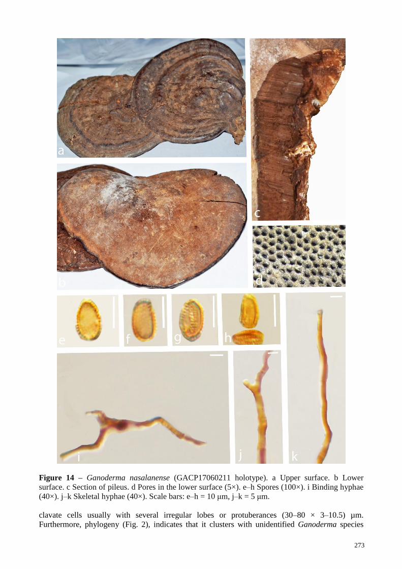
273
Figure 14 – Ganoderma nasalanense (GACP17060211 holotype). a Upper surface. b Lower
surface. c Section of pileus. d Pores in the lower surface (5×). e–h Spores (100×). i Binding hyphae
(40×). j–k Skeletal hyphae (40×). Scale bars: e–h = 10 μm, j–k = 5 μm.
clavate cells usually with several irregular lobes or protuberances (30–80 × 3–10.5) µm.
Furthermore, phylogeny (Fig. 2), indicates that it clusters with unidentified Ganoderma species
274
recorded from Malaysia (FRIM138 and G31) and Vietnam (FMD13) (descriptions unavailable).
The species is currently only known from the type locality, Nasala village, Huaphanh Province,
Laos.
25. Ganoderma sandunense Hapuar., T.C. Wen & K.D. Hyde, sp. nov. Fig. 15
Index Fungorum number: IF555784, Facesoffungi number: FoF05659
Etymology – Refers to the collecting site‘Sandu’, China
Holotype – GACP18012501
Basidiome annual, stipitate, strongly laccate, corky, becoming woody hard when drying.
Pileus 2–4 × 1–2.5 cm, several layers thick, orbicular, upper surface; reddish black (8E8) to
brownish black (6C8), distinctly concentrically sulcate, undulate, ferruginous, irregularly ruptured
crust overlying the context, margin; wavy, concolorous with the pileus, lower surface;
whitish yellow (4A2) to light brown (6D4). Hymenophore up to 10 mm long, indistinctly stratose;
pores initially greyish brown (6E3), bruising dark brown (8F6), pores circular or isodiametric, 3–5
per mm. Context up to 1 cm thick, duplex, dry, upper layer brown (6E4) to dark brown (7F8),
fibrous, composed of coarse loose fibrils; lower layer dark brown (7F8), woody. Stipe central, sub
cylindrical, concolorous with the pileus, 4 × 8 cm, 0.5 cm at the base. Basidiospores (n = 25) (10.8–
)12.1–12.9–13.8(14.3–) × (–8.6)9.2–9.8–10.5(–11.1) μm (Qm = 1.3, Q = 1.1–1.5, with
myxosporium). (n = 25) (9.6–)10.4–11.3–12.3(–12.7) × (6.8–)7.5–8.3–9.0(–9.8) μm (Qm = 1.3, Q =
1.1–1.7, without myxosporium), broadly ellipsoid to ellipsoid, brown (7E5), with a light brown
(6D6) eusporium bearing thick, short and distinct echinulae, overlaid by a hyaline myxosporium.
Pileipellis a hymeniderm, brownish orange (5C5), clavate like cells. Context trimitic; generative
hyphae (n = 20) (1.2–3.1–2.0) μm in width, hyaline, thin-walled, with clamp connections, rarely
seen; skeletal hyphae (n = 20) (3.0–3.6–4.6) μm in width, thick walled, nearly solid, brownish
orange (6C8); binding hyphae (n = 20) (1.2–3.4–2.1) μm in width, thick walled, branched, nearly
solid, light brown (6D4).
Habitat – Rotten wood, in dry dipterocarp forest and in upper mixed deciduous forest and
growing up from soil.
Specimens examined – CHINA, Guizhou Province, Sandu Shui Autonomous Country,
24°54′N–107°53′E, elev. 590 m, 25 September 2017, T.C Wen (GACP18012501,
GACP18012502).
Notes – Ganoderma sandunense is a new to science and it is distinguished by several layers
thick, orbicular, ferruginous laccate pileus, relatively large broadly ellipsoid to ellipsoid spores
(12.1–13.8 × 9.2–10.5) μm, with a light brown eusporium bearing fine, short and distinct echinulae.
Initial blast results of this species in GenBank showed 96% similarity with G. applanatum, G.
oregonense, G. lucidum and G. carnosum. However, there are 1.7%, 1.6%, 1.5%, 1.8% base pair
differences in ITS region between G. sandunense and other species; G. applanatum, G. oregonense,
G. lucidum and G. carnosum, respectively. The new species is established based on
recommendations provided by Jeewon & Hyde (2016). In addition, none of these species cluster
with G. sandunense (Fig. 2) and also the morphology is very different from former species.
Ganoderma sandunense is currently only known from the type locality, Sandu Shui Autonomous
Country, Guizhou Province, China.
26. Ganoderma sinense J.D. Zhao, L.W. Hsu & X.Q. Zhang, Acta Microbiol. Sin. 19(3): 272
(1979) Fig. 16
Ganoderma formosanum T.T. Chang & T. Chen in Trans. Or. Mycol. Soc. 82: 731 (1984)
(Nom. Invalid)
= Ganoderma japonicum (Fr. Lloyd in Teng, Fungi of China; 447 (1963): Tai, Syll. Fung.
Sin.: 469 (1979), Teng, Fungi of China: 326 (1996), non Polyporus japonicus Fr.,Epicrisis:
442 (1838)
= Ganoderma japonicum (Fr.) Lloyd, Mycol. Writ. 3: Syn. Stip. Polyp.: 102(1912)

275
= Ganoderma lucidum (Leyss.) P. Karst. var. japonicum (Fr.) Bres. in Teng in Sinensia 5:199
(1934). non Polyporus japonicus Fr., Epicrisis: 442 (1838).
Facesoffungi number: FoF05660
Figure 15 – Ganoderma sandunense (GACP18012501). a upper surface. b lower surface. c Section
of pileus (5×). d pores in the lower surface. e–g spores (100×). h generative hyphae (100×).
i skeletal hyphae (100×), j binding hyphae (100×). Scale bars: e–g = 10 μm, h–j = 5 μm.
276
Basidiome annual, stipitate, strongly laccate, branched, corky. Pileus 3–7 × 2–4 cm, up to 1.0
cm thick at the base, orbicular; upper surface brown (6E4) to brownish black (6C8), yellowish
brown (5D8) at the margin, irregularly ruptured crust overlying the context; margin blunt, yellow
brown (5D8); lower surface light brown (6D6). Hymenophore up to 20 mm long, indistinctly
stratose; pores brownish grey (6D2), circular, 2–4 per mm. Context up to 1 cm thick, duplex, dry,
upper layer dark brown (7F8), corky; lower layer pale brown (5A5), fibrous, composed of coarse
loose fibrils; woody. Stipe central, sub cylindrical, concolorous with the pileus, 5 × 8 cm, 1.5 cm at
the base. Basidiospores (n = 20) (8.0–)9.7–11.6–11.2(13.4–) × (–6.3) 7.3–8.1–8.8(–9.6) μm (Qm =
1.4, Q = 1.1–1.6, with myxosporium). (6.6–)8.2–9.6–10.9(–11.4) × (5.8–)7.3–6.7–7.5(–8.5) μm
(Qm = 1.4, Q = 1.2–1.7, without myxosporium), ellipsoid, brown (6E4), with a pale brown (5A5)
eusporium bearing fine, short and distinct echinulae, overlaid by a hyaline myxosporium.
Pileipellis a hymeniderm, brown (6E4), clavate like cells, dextrinoid. Context trimitic; generative
hyphae (n = 20) (0.3–1.3–2.2) μm in width, colorless, thin-walled; skeletal hyphae (n = 20) (2.6–
3.6–5.1) μm in width, thick walled, nearly solid, sometimes branched, brown (6E4); binding
hyphae (n = 20 (0.9–2.6–4.5) μm in width, thick walled, branched, nearly solid, pale brown (5A5).
Habitat – Rotten wood, in dry dipterocarp forest and in upper mixed deciduous forest and
growing up from soil.
Specimens examined – CHINA, Guizhou Province, Sandu Shui Autonomous Country,
24°54′N–107°53′E, elev. 590 m, 25 September 2017, T.C Wen (GACP17092530,
GACP17092532).
Notes – See Hapuarachchi et al. (2018b, 2019)
27. Ganoderma steyaertianum B.J. Sm. & Sivasith. [as 'steyaertanum'], Aust. Syst. Bot. 16(4): 495
(2003)
See the description and illustration at Smith & Sivasithamparam (2003)
Notes – Ganoderma steyaertanum was introduced by Smith & Sivasithamparam (2003) from
Bali, Indonesia and has previously been mistaken for Ganoderma lucidum (Curtis) P. Karst. in
Australia and Indonesia. Recent studies have attempted to determine the correct name for non-
European collections of ‘G. lucidum’ (Wang et al. 2009, 2012, Cao et al. 2012, Yao et al. 2013).
This laccate species can be readily distinguished in its large, 50–500 mm diameter, sessile or
broadly stipitate basidiome, concentric zones and dark brown (Chestnut) to almost black pileus,
thick walled, brown, ovoid and ellipsoid basidiospores Smith & Sivasithamparam (2003). Analysis
of ITS rDNA sequence (Smith & Sivasithamparam 2000) indicating allopatric speciation of G.
steyaertanum from a species from India and the Philippines, suggests the distribution of may not
extend much further north of Indonesia. Ganoderma steyaertanum has been confirmed as a primary
pathogen of A. mangium root rot (Hidayati et al. 2014) (Table 3).
28. Ganoderma subresinosum (Murrill) C.J. Humphrey, Mycologia 30(3): 332 (1938) Fig. 17
= Fomes subresinosus Murrill (1908)
= Polyporus mamelliporus Beeli, Bull. Soc. R. Bot. Belg. 62: 62 (1929)
= Trachyderma subresinosum (Murrill) Imazeki, Bull. Gov. Forest Exp. Stn Tokyo 57: 119
(1952)
= Magoderna subresinosum (Murrill) Steyaert, Persoonia 7(1): 112 (1972)
= Amauroderma subresinosum (Murrill) Corner, Beih. Nova Hedwigia 75: 93 (1983)
Facesoffungi number: FoF03808
Basidiome annual, sessile (with distinctly contracted base), weakly laccate, woody. Pileus
16–20 × 11–13 cm, up to 4 cm thick at the base, sub-orbicular; upper surface dark brown (8F4),
radially rugose, concentrically sulcate with irregularly ruptured crust overlying the context; margin
blunt or wavy, yellow brown; lower surface brownish orange (6C4). Hymenophore up to 20 mm
long, indistinctly stratose; pores initially greyish orange (5B3), bruising brownish orange (6C4),
pores circular, 3–5 per mm. Context up to 2 cm thick, duplex, dry; lower layer, light orange (5A4),
fibrous, composed of coarse loose fibrils; upper layer brownish orange (6C5), corky. Basidiospores

277
Figure 16 – Ganoderma sinense (GACP17092559). a, b Upper surface. c Lower surface. d Section
of pileus. e Pores in the lower surface (5×). f–h Spores (100×). i Skeletal hyphae (100×).
j Generative hyphae (100×). k Binding hyphae (100×). Scale bars: f – i =10 μm, j – l =5 μm.
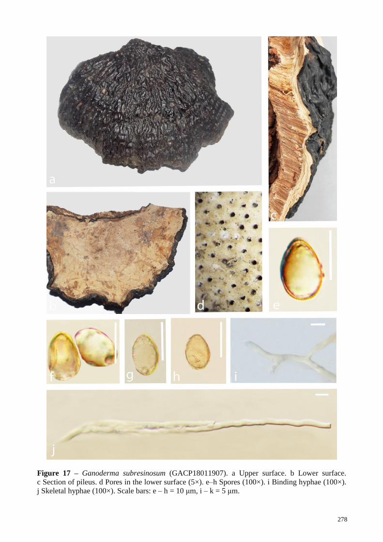
278
Figure 17 – Ganoderma subresinosum (GACP18011907). a Upper surface. b Lower surface.
c Section of pileus. d Pores in the lower surface (5×). e–h Spores (100×). i Binding hyphae (100×).
j Skeletal hyphae (100×). Scale bars: e – h = 10 μm, i – k = 5 μm.
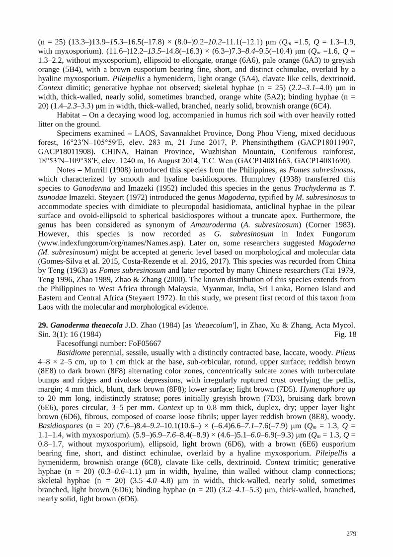
279
(n = 25) (13.3–)13.9–15.3–16.5(–17.8) × (8.0–)9.2–10.2–11.1(–12.1) μm (Qm =1.5, Q = 1.3–1.9,
with myxosporium). (11.6–)12.2–13.5–14.8(–16.3) × (6.3–)7.3–8.4–9.5(–10.4) μm (Qm =1.6, Q =
1.3–2.2, without myxosporium), ellipsoid to ellongate, orange (6A6), pale orange (6A3) to greyish
orange (5B4), with a brown eusporium bearing fine, short, and distinct echinulae, overlaid by a
hyaline myxosporium. Pileipellis a hymeniderm, light orange (5A4), clavate like cells, dextrinoid.
Context dimitic; generative hyphae not observed; skeletal hyphae (n = 25) (2.2–3.1–4.0) μm in
width, thick-walled, nearly solid, sometimes branched, orange white (5A2); binding hyphae (n =
20) (1.4–2.3–3.3) μm in width, thick-walled, branched, nearly solid, brownish orange (6C4).
Habitat – On a decaying wood log, accompanied in humus rich soil with over heavily rotted
litter on the ground.
Specimens examined – LAOS, Savannakhet Province, Dong Phou Vieng, mixed deciduous
forest, 16°23′N–105°59′E, elev. 283 m, 21 June 2017, P. Phensinthgthem (GACP18011907,
GACP18011908). CHINA, Hainan Province, Wuzhishan Mountain, Coniferous rainforest,
18°53′N–109°38′E, elev. 1240 m, 16 August 2014, T.C. Wen (GACP14081663, GACP14081690).
Notes – Murrill (1908) introduced this species from the Philippines, as Fomes subresinosus,
which characterized by smooth and hyaline basidiospores. Humphrey (1938) transferred this
species to Ganoderma and Imazeki (1952) included this species in the genus Trachyderma as T.
tsunodae Imazeki. Steyaert (1972) introduced the genus Magoderna, typified by M. subresinosus to
accommodate species with dimidiate to pleuropodal basidiomata, anticlinal hyphae in the pilear
surface and ovoid-ellipsoid to spherical basidiospores without a truncate apex. Furthermore, the
genus has been considered as synonym of Amauroderma (A. subresinosum) (Corner 1983).
However, this species is now recorded as G. subresinosum in Index Fungorum
(www.indexfungorum/org/names/Names.asp). Later on, some researchers suggested Magoderna
(M. subresinosum) might be accepted at generic level based on morphological and molecular data
(Gomes-Silva et al. 2015, Costa-Rezende et al. 2016, 2017). This species was recorded from China
by Teng (1963) as Fomes subresinosum and later reported by many Chinese researchers (Tai 1979,
Teng 1996, Zhao 1989, Zhao & Zhang (2000). The known distribution of this species extends from
the Philippines to West Africa through Malaysia, Myanmar, India, Sri Lanka, Borneo Island and
Eastern and Central Africa (Steyaert 1972). In this study, we present first record of this taxon from
Laos with the molecular and morphological evidence.
29. Ganoderma theaecola J.D. Zhao (1984) [as 'theaecolum'], in Zhao, Xu & Zhang, Acta Mycol.
Sin. 3(1): 16 (1984) Fig. 18
Facesoffungi number: FoF05667
Basidiome perennial, sessile, usually with a distinctly contracted base, laccate, woody. Pileus
4–8 × 2–5 cm, up to 1 cm thick at the base, sub-orbicular, rotund, upper surface; reddish brown
(8E8) to dark brown (8F8) alternating color zones, concentrically sulcate zones with turberculate
bumps and ridges and rivulose depressions, with irregularly ruptured crust overlying the pellis,
margin; 4 mm thick, blunt, dark brown (8F8); lower surface; light brown (7D5). Hymenophore up
to 20 mm long, indistinctly stratose; pores initially greyish brown (7D3), bruising dark brown
(6E6), pores circular, 3–5 per mm. Context up to 0.8 mm thick, duplex, dry; upper layer light
brown (6D6), fibrous, composed of coarse loose fibrils; upper layer reddish brown (8E8), woody.
Basidiospores (n = 20) (7.6–)8.4–9.2–10.1(10.6–) × (–6.4)6.6–7.1–7.6(–7.9) μm (Qm = 1.3, Q =
1.1–1.4, with myxosporium). (5.9–)6.9–7.6–8.4(–8.9) × (4.6–)5.1–6.0–6.9(–9.3) μm (Qm = 1.3, Q =
0.8–1.7, without myxosporium), ellipsoid, light brown (6D6), with a brown (6E6) eusporium
bearing fine, short, and distinct echinulae, overlaid by a hyaline myxosporium. Pileipellis a
hymeniderm, brownish orange (6C8), clavate like cells, dextrinoid. Context trimitic; generative
hyphae (n = 20) (0.3–0.6–1.1) μm in width, hyaline, thin walled without clamp connections;
skeletal hyphae (n = 20) (3.5–4.0–4.8) μm in width, thick-walled, nearly solid, sometimes
branched, light brown (6D6); binding hyphae (n = 20) (3.2–4.1–5.3) μm, thick-walled, branched,
nearly solid, light brown (6D6).

280
Habitat – On a decaying hardwood tree trunk, accompanied in humus rich soil with over
heavily rotted litter on the ground.
Specimens examined – CHINA, Hainan Province, Diaoluoshan National Nature Reserve,
18°68′N-109°95′E, elev. 1058 m, collection date unknown, X.L Wu (GACP HNU02, GACP
HNU58) (from Herbarium of Guizhou Agricultural sciences; GAS3359).
Notes – Ganoderma theaecola was introduced from Guangxi, China as a laccate Ganoderma
species (Zhao et al. 1984). In Index Fungorum, as an editorial comment, it is mentioned that the
orthography of this specific epithet is uncertain (www.indexfungorum.org, accessed 22 December
2018). Since the type specimens located in herbarium of Chinese Academy of Sciences, is scanty
and difficult to observe, we have loaned a reference specimen from the herbarium of Guizhou
Agricultural Sciences. Basidiospore size is mentioned as (7–9 × 5.2–6.2) μm (Zhao et al. 1984), (6–
9 × 5–6) μm (Wu & Dai 2005) and (8.4–10.1 × 6.6–7.6) μm (This study). Furthermore, there is no
molecular data available for this species in GenBank.
30. Ganoderma tropicum (Jungh.) Bres. Annls mycol. 8(6): 586 (1910) Fig. 19
≡Polyporus tropicus Jungh. (1838)
= Polyporus tropicus Jungh. (1838)
= Fomes tropicus (Jungh.) Cooke, Grevillea 14(no. 69): 19 (1885)
= Scindalma tropicum (Jungh.) Kuntze, Revis. gen. pl. (Leipzig) 3(2): 519 (1898)
Facesoffungi number: FoF05661
Basidiome annual, sessile (usually with a distinctly contracted base), strongly laccate, woody.
Pileus 4–8 × 2.5–6 cm, up to 1 cm thick at the base, spathulate, plano convex; upper surface hard,
several layers thick, yellowish brown (5F8), orange (6D8) to brown (7E8), crust overlies the pellis,
containing fibrous pithy context, swollen differentiated zone at the point of attachment; margin
soft, 1 cm thick, rounded, white (5A1); lower surface white (5A1) to orange (5B2). Hymenophore
up to 4 mm long, indistinctly stratose, white (5A1) to orange (5B2), pores sub circular or
isodiametric, 3–5 per mm. Context up to 3 cm thick, dry, duplex; lower layer brown (7E8), fibrous,
composed of coarse loose fibrils, soft; upper layer light brown (5A5), woody. Basidiospores (n =
25) (10.8–)11.2–12.1–12.8(–13.1) × (8.3–)9.6–10.1–10.8(–11.1) μm (Qm = 1.3, Q = 0.9–1.5, with
myxosporium). (7.9–)8.8–9.1–10.2(–10.8) × (5.8–)6.4–7.3–7.8(–9.8) μm (Qm = 1.3, Q = 1.1–1.7,
without myxosporium), broadly ellipsoid, brownish orange (6C8) to light brown (5A5) eusporium
bearing fine, short and distinct echinulae, overlaid by a hyaline myxosporium, bitunicate. Pileipellis
a hymeniderm, greyish orange (5B5) to brown (6E8), composed of apically acanthus like branched
cells, dextrinoid. Context trimitic; generative hyphae (n = 30) (0.4–)0.6–1.4–1.8(–2.6) μm in width,
thin-walled, hyaline skeletal hyphae (n = 30) (2.8–)3.1–3.4–3.8(–4.3) μm in width, light brown
(6D4), thick walled, sometimes branched; binding hyphae (n = 30) (1.1–)2.2–2.5–3.4(–3.8) μm in
width, light brown (6D4), thick walled, branched, intertwined the skeletal hyphae.
Habitat – On a decaying Dipterocarpus tree, wood log, accompanied in humus rich soil with
over heavily rotted litter in forest, mossy coniferous forests.
Specimens examined – CHINA, Hainan Province, Jiangfengling Mountain, Coniferous
rainforest, 18°44′N–108°51′E, elev. 550 m, collection date unknown, X.L Wu (GACP HNU21,
GACP HNU25), Wuzshishan Mountain, Coniferous rainforest, 18°53′N–109°38′E, elev. 1240 m,
15 August 2014, T.C Wen (GACP14081511, GACP14081518). LAOS, Savannakhet Province,
Phin District, Dong Phou Vieng Protected Area, 16 58′N–105 89′E, elev. 173 m, 07 June 2017, P.
Phengsintham (GACP17060701).
Notes – Ganoderma tropicum was introduced as Polyporus tropicus by Junghuhn (1838)
from Java, Indonesia and later, Bresadola (1910) transferred this species to Ganoderma. This
species widely distributed in lowland of tropical Asia and its taxonomy has been well resolved with
the aid of molecular data (Wang et al. 2012, Yang & Feng 2013). Ganoderma tropicum consider as
a member of G. lucidum species complex (Zhou et al. 2015). Among the Chinese Ganoderma
species, G. flexipes, G. multipileum, G. sichuanense, and G. tsugae are the mostly similar species to
G. tropicum since they share a reddish brown pileal surface, similar basidiospores and cuticle cells,
281
Figure 18 – Ganoderma theaecola (GAS3359). a Upper surface. b Lower surface. c Section of
pileus. d Pores in the lower surface (5×). e–h Spores (100×). i Generative hyphae (100×). j Skeletal
hyphae (100×). k Binding hyphae (100×). Scale bars: e – h =10 μm, i – k = 5 μm.
282
Figure 19 – Ganoderma tropicum (GACP14081511). a Upper surface. b Lower surface. c Section
of pileus. d Pores in the lower surface (5×). e–g Spores (100×). h Binding hyphae (100×).
i Generative hyphae (100×). j Skeletal hyphae (100×). Scale bars: e–g = 10 μm, h–j = 5 μm.
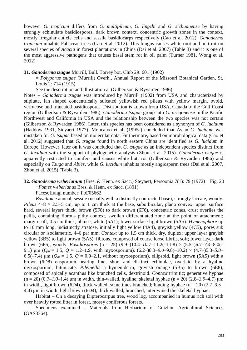
283
however G. tropicum differs from G. multipileum, G. lingzhi and G. sichuanense by having
strongly echinulate basidiospores, dark brown context, concentric growth zones in the context,
mostly irregular cuticle cells and sessile basidiocarps respectively (Cao et al. 2012). Ganoderma
tropicum inhabits Fabaceae trees (Cao et al. 2012). This fungus causes white root and butt rot on
several species of Acacia in forest plantations in China (Dai et al. 2007) (Table 3) and it is one of
the most aggressive pathogens that causes basal stem rot in oil palm (Turner 1981, Wong et al.
2012).
31. Ganoderma tsugae Murrill, Bull. Torrey bot. Club 29: 601 (1902)
= Polyporus tsugae (Murrill) Overh., Annual Report of the Missouri Botanical Garden, St.
Louis 2: 714 (1915)
See the description and illustration at (Gilbertson & Ryvarden 1986)
Notes – Ganoderma tsugae was introduced by Murrill (1902) from USA and characterized by
stipitate, fan shaped concentrically sulcated yellowish red pileus with yellow margin, ovoid,
verrucose and truncated basidiospores. Distribution is known from USA, Canada to the Gulf Coast
region (Gilbertson & Ryvarden 1986). Ganoderma tsugae group into G. oregonense in the Pacific
Northwest and California in USA and the relationship between the two species was not certain
(Gilbertson & Ryvarden 1986). Later, this species has been considered as a synonym of G. lucidum
(Haddow 1931, Steyaert 1977). Moncalvo et al. (1995a) concluded that Asian G. lucidum was
mistaken for G. tsugae based on molecular data. Furthermore, based on morphological data (Cao et
al. 2012) suggested that G. tsugae found in north eastern China are identified as G. lucidum in
Europe. However, later on it was concluded that G. tsugae as an independent species distinct from
G. lucidum with the support of phylogenetic analysis (Zhou et al. 2015). Ganoderma tsugae is
apparently restricted to conifers and causes white butt rot (Gilbertson & Ryvarden 1986) and
especially on Tsuga and Abies, while G. lucidum inhabits mostly angiosperm trees (Dai et al. 2007,
Zhou et al. 2015) (Table 3).
32. Ganoderma weberianum (Bres. & Henn. ex Sacc.) Steyaert, Persoonia 7(1): 79 (1972) Fig. 20
≡Fomes weberianus Bres. & Henn. ex Sacc. (1891)
Facesoffungi number: FoF05662
Basidiome annual, sessile (usually with a distinctly contracted base), strongly laccate, woody.
Pileus 4–8 × 2.5–5 cm, up to 1 cm thick at the base, suborbicular, plano convex; upper surface
hard, several layers thick, brown (5F8) to dark brown (6F6), concentric zones, crust overlies the
pellis, containing fibrous pithy context, swollen differentiated zone at the point of attachment;
margin soft, 0.5 cm thick, obtuse, white (5A1); lower surface light brown (5A5). Hymenophore up
to 10 mm long, indistinctly stratose, initially light yellow (4A4), greyish yellow (4C5), pores sub
circular or isodiametric, 4–6 per mm. Context up to 1.5 cm thick, dry, duplex; upper layer grayish
yellow (3B5) to light brown (5A5), fibrous, composed of coarse loose fibrils, soft; lower layer dark
brown (6F6), woody. Basidiospores (n = 25) (9.9–)10.4–10.7–11.2(–11.8) × (5.5–)6.7–7.4–8.0(–
9.1) μm (Qm = 1.5, Q = 1.2–1.9, with myxosporium). (6.2–)8.3–9.0–9.8(–10.2) × (4.7–)5.3–5.8–
6.5(–7.4) μm (Qm = 1.5, Q = 0.9–2.1, without myxosporium), ellipsoid, light brown (5A5) with a
brown (6D8) eusporium bearing fine, short and distinct echinulae, overlaid by a hyaline
myxosporium, bitunicate. Pileipellis a hymeniderm, greyish orange (5B5) to brown (6E8),
composed of apically acanthus like branched cells, dextrinoid. Context trimitic; generative hyphae
(n = 20) (0.7–1.0–1.4) μm in width, thin-walled, hyaline; skeletal hyphae (n = 20) (2.8–3.9–4.7) μm
in width, light brown (6D4), thick walled, sometimes branched; binding hyphae (n = 20) (2.7–3.5–
4.4) μm in width, light brown (6D4), thick walled, branched, intertwined the skeletal hyphae.
Habitat – On a decaying Dipterocarpus tree, wood log, accompanied in humus rich soil with
over heavily rotted litter in forest, mossy coniferous forests.
Specimens examined – Materials from Herbarium of Guizhou Agricultural Sciences
(GAS3364).

284
Figure 20 – Ganoderma weberianum (GAS3364). a Upper surface. b Lower surface. c Pores in the
lower surface (5×). d Section of pileus. e–g Spores (100×). h Generative hyphae (100×). i Binding
hyphae (100×). j Skeletal hyphae (100×). Scale bars: e–g = 10 μm, h–j = 5 μm.

285
Notes – Ganoderma weberianum is characterized by hard pileus crust, pale context that
changes to yellow when cut with resinous incrustations, cylindrical pileipellis cells with
granulations, and small basidiospores (Torres-Torres & Guzmán-Dávalos 2005) with abundant
gasterospores in basidiome (Quanten 1997). This species is distributed Africa, Asia and Samoa
Island (Steyaert 1972), Southeast Asia and Papua New Guinea (Corner 1983, Quanten 1997),
Australia (Smith & Sivasithamparam 2000), China (Pan & Dai 2001, Wang et al. 2005), Singapore
(Corner 1983), Brazil (Torres-Torres & Guzmán-Dávalos 2005) and India (Mohanty et al. 2011).
Ganoderma weberianum is a pathogen of Ficus, and causes a white root and butt rot (Pan & Dai
2001).
33. Ganoderma wiiroense E.C. Otto, Blanchette, C.W. Barnes & Held. (2015)
See the description and illustration at (Crous et al. 2015)
Notes – Ganoderma wiiroense was introduced as a wood decaying Ganoderma species from Ghana
recently, with the support of molecular data (Crous et al. 2015). This species is characterized by
dimidiate, zonate and rounded margin pileus, trimitic hyphal system, ellipsoid to cylindrical-
ellipsoid, truncate base, bitunicate and verrucose basidiospores (Crous et al. 2015). Ganoderma
wiiroense causes decay in the roots and trunks of angiosperm trees in the upper west region of
Ghana (Table 3) (Crous et al. 2015).
34. Ganoderma williamsianum Murrill, Bull. Torrey bot. Club 34: 478 (1907) Fig. 21
= Elfvingia williamsiana (Murrill) Imazeki, Bull. Gov. Forest Exp. Stn Tokyo 57: 106 (1952)
Facesoffungi number: FoF05663
Basidiome annual, sessile (usually with a distinctly contracted base), weakly laccate, woody. Pileus
3–7 × 2–6 cm, up to 2 cm thick at the base, suborbicular, plano convex, applanate; upper surface
hard, several layers thick, brown (5F8), dark brown (6F6) to brownish red (8C8), orange yellow
(4A8) near to edge, concentric zones, crust overlies the pellis, containing fibrous pithy context,
swollen differentiated zone at the point of attachment; margin soft, 0.5 cm thick, obtuse, white
(5A1); lower surface white (8A1). Hymenophore up to 8 mm long, not stratose, initially light
brown (6D8), bruising greyish brown (6D3), pores circular, 4–6 per mm. Context up to 2 cm thick,
dry, duplex; upper layer grayish brown (6D3) to light brown (5A5), fibrous, composed of coarse
loose fibrils, soft; lower layer dark brown (6F6), woody. Basidiospores (n = 50) (8.7–)9.9–10.8–
11.7(–12.5) × (5.1–)6.6–7.5–8.3(–9.6) μm (Qm = 1.5, Q = 1.3–1.8, with myxosporium). (7.4–)8.1–
8.9–9.8(–11.7) × (3.2–)5.0–5.7–6.6(–8.4) μm (Qm = 1.5, Q = 1.3–1.9, without myxosporium),
ellipsoid, light brown (5A5) with a brown (6D8) eusporium bearing fine, short and distinct
echinulae, overlaid by a hyaline myxosporium. Pileipellis a hymeniderm, greyish orange (5B5) to
brown (6E8), composed of apically acanthus like branched cells, dextrinoid. Context trimitic;
generative hyphae (n = 30) (0.3–0.8–1.2) μm in width, thin-walled, hyaline; skeletal hyphae (n =
20) (0.4–2.6–4.4) μm in width, light brown (6D4), thick walled, sometimes branched; binding
hyphae (n = 20) (1.2–2.1–3.7) μm in width, light brown (6D4), thick walled, branched, intertwined
the skeletal hyphae.
Habitat – On a decaying Dipterocarpus tree, wood log, accompanied in humus rich soil with
over heavily rotted litter in forest, mossy coniferous forests.
Specimens examined – CHINA, Hainan Province, Jiangfengling Mountain, Coniferous
rainforest, 18°44′N–108°51′E, elev. 550 m, 13 August 2014, T.C Wen (GACP14081320,
GACP14081321).
Notes – This common species is introduced from Philippines and easily recognized by small,
dense, ungulate pileus with anamixodermic cuticle and pale yellow pores, rather large spores and
very short skeletals (Corner 1983, Moncalvo & Ryvarden 1997). Ganoderma williamsianum
superficially resembles members of the laccate Ganoderma group (Wang & Wu 2010).
Furthermore, Ganoderma brownii is very similar to G. williamsianum in color of pore surface
(Lowe & Gilbertson 1961, Gilbertson & Ryvarden 1986). However, G. brownii can be easily
distinguished from G. williamsianum by having a dull pileus with a hard crust (Lowe & Gilbertson

286
1961) formed by hyphae arranged in a trichoderm (Steyaert 1972, Gottlieb & Wright 1999a),
skeletal hyphae with occasional branching (Gilbertson & Ryvarden 1986), and smaller
basidiospores (Lowe & Gilbertson 1961, Steyaert 1972, Gilbertson & Ryvarden 1986, Gottlieb &
Wright 1999a). Ganoderma williamsianum is similar to G. meijiangense but distinguished from the
latter by having dark brown context without any black crustose layer and a distinct cuticular
composition (Zhao 1988). Later, based on type specimens it was suggested that G. williamsianum
is the earliest valid name for G. meijiangense (Wang & Wu 2010). Our collections from Hainan
Province, China are similar with the descriptions provided by Wang & Wu (2010). This species is
distributed in China (Wang & Wu 2010, Xing et al. 2018), Indonesia (Imazeki 1952, Steyaert
1972), Philippines (Murrill 1907, Steyaert 1972) and Malaysia (Steyaert 1972).
35. Ganoderma zonatum Murrill, Bull. Torrey bot. Club 29: 606 (1902)
See the description and illustration at Ryvarden (2000)
Notes – Ganoderma zonatum is recognized by the distinct strongly amyloid irregular cuticle
cells and the oblong basidiospores (Ryvarden 2000). This is a white rot fungus which cause butt rot
in Palms (Schubert et al. 1997) and Cycads (Elliot & Broschat 2001) (Table 3) and often referred to
as a member of the G. lucidum species complex (Adaskaveg & Gilbertson 1989). (Ryvarden 2000)
mentioned that this species distribution is only known from USA, however, Foroutan & Vaidya
(2007) and Cong (2010) reported this species from India and Brazil respectively.
Amauroderma P. Karst., 1881, Rev. Mycol. (Toulouse) 3, p. 17.
= Amauroderma (Pat.) Torrend, Brotéria, sér. Bot. 18: 121 (1920)
= Ganoderma sect. Amauroderma Pat., Bull. Soc. mycol. Fr. 5(2, 3): 75 (1889)
= Lazulinospora Burds. & M.J. Larsen, Mycologia 66(1): 97 (1974)
= Magoderna Steyaert, Persoonia 7(1): 111 (1972)
= Whitfordia Murrill, Bull. Torrey bot. Club 35: 407 (1908)
See the description at Ryvarden (2004b)
Basidiocarps annual or reviving for a second season, centrally-laterally stipitate, solitary or in
small groups with several fused pilei, consistency coriaceous, corky to woody hard, seldom brittle.
Pileus round, reniform to fan-shaped, concave, umbilicate to strongly infundibuliform, upper
surface in varying colours from white, ochraceous, brown to almost black, finely tomentose to
glabrous, dull to glossy with a distinct cortex or cuticle, often concentrically zoned and radially
wrinkled, stipe rather thin and long, finely tomentose to glabrous, pore surface whitish to
ochraceous when fresh, darkens when dry to brownish colours, pores round to angular and entire,
large to small, tubes seldom stratified, context white, ochraceous to dark brown, cystidia absent,
hyphal system dimitic, generative hyphae with clamps hyaline and thin-walled, skeletal hyphae
arboriform to more rarely unbranched, hyaline to brown, those being hyaline often dextrinoid or
without reaction in Melzer´s reagent, basidia bladder-like with 4 large curved sterigmata,
basidiospores hyaline to pale yellow, sub-globose to cylindrical, dextrinoid to non-dextrinoid,
bitunicate with the inner wall finely asperulate or very rarely smooth.
Type species – Amauroderma regulicolor (Berk. ex Cooke) Murrill (= Amauroderma
schomburgkii)
Notes – Amauroderma is a widespread tropical genus that usually occurs on roots of living or
dead trees or, is more rarely, wood inhabiting and cause white rots (Furtado 1981, Ryvarden
2004a). After Amauroderma was described, Torrend (1920a,b) worked extensively on this genus in
South America and published a crucial work based mainly on spore shape (globose or oblong,
never truncate) and the presence of a stipe (usually dull, like the pilear surface) and recorded 28
species of Amauroderma placed within three sections. Furtado (1981) carefully revised this genus
and recognized 27 species and defined Amauroderma by the globose to subglobose basidiospores,
with double walls, stipitate basidiomes and a tropical distribution pattern. Twenty one species of
Amauroderma were described by Ryvarden (2004a) from the Neotropics, using the same genus
circumscription. However, according to Index Fungorum (www.indexfungorum.org, accessed 22
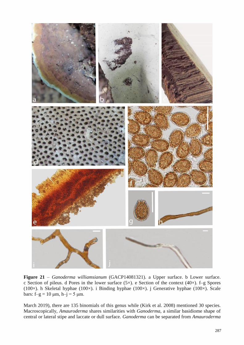
287
Figure 21 – Ganoderma williamsianum (GACP14081321). a Upper surface. b Lower surface.
c Section of pileus. d Pores in the lower surface (5×). e Section of the context (40×). f–g Spores
(100×). h Skeletal hyphae (100×). i Binding hyphae (100×). j Generative hyphae (100×). Scale
bars: f–g = 10 μm, h–j = 5 μm.
March 2019), there are 135 binomials of this genus while (Kirk et al. 2008) mentioned 30 species.
Macroscopically, Amauroderma shares similarities with Ganoderma, a similar basidiome shape of
central or lateral stipe and laccate or dull surface. Ganoderma can be separated from Amauroderma
288
by its distinctly truncate basidiospores, and most Ganoderma species grow on dead wood, while
most Amauroderma species grow in the ground from buried roots/woods (Ryvarden 2004b).
Amauroderma species are regarded as economically valuable because of their important medicinal
properties and pathogenicity (Dai et al. 2007, 2009, Jiao et al. 2013, Chan et al. 2013). Taxonomic
studies of Amauroderma in Asia have been carried out over many years by various researchers
(Teng 1936, Zhao et al. 1979, Zhao & Zhang 1987, Li & Yuan 2015, Song et al. 2016). Twenty
species have been recorded in China (Zhao & Zhang 2000), but among these, only six have been
confirmed as Amauroderma based on both morphological characters and phylogenetic analyses: A.
austrosinense J.D. Zhao & L.W. Hsu, A. concentricum Song, Xiao L. He & B.K. Cui, A. perplexum
Corner, A. rugosum (Blume & T. Nees) Torrend, A. subresinosum (Murrill) Corner and A.
yunnanense J.D. Zhao & X.Q. Zhang (Li & Yuan 2015). The other Amauroderma species recorded
from China, have not been fully studied yet. Furthermore, members of this genus have been
subsequently recorded from Thailand (Chandrasrikul et al. 2011), Myanmar (Thaung 2007) and
Vietnam (Quang et al. 2011). Here, we record five Amauroderma species found within Sub
Mekong region countries based on micro- and macro morphological characteristics together with
molecular data.
36. Amauroderma rude (Berk.) Torrend [as ‘rudis’], Brotéria, sér. bot. 18: 127 (1920) Fig. 22
≡Fomes rudis Berk. (1885)
See Index Fungorum for synonyms
Facesoffungi number: FoF05664
Basidiome annual, stipitate, laccate, corky. Pileus 12–6.5 × 5.5–2.5 cm, up to 0.5 cm thick at
the base, orbicular to subreniform, mesopodal; upper surface reddish brown (8E5, 8E6, 8D5) to
grayish brown (8F1) alternating colour zones, slightly concentrically sulcate with irregularly
ruptured crust; margin soft, grayish brown (8F1); lower surface white (8A1), turns reddish white
(8A2) when drying. Hymenophore up to 15 mm long, indistinctly stratose; pores initially brownish
orange (5C4), bruising brown (6E8), pores circular, 4–6 per mm. Context up to 6 mm thick, duplex,
dry; upper layer light brown (5D6), fibrous, composed of coarse loose fibrils, brown (6E8), corky;
lower layer light brown (5A5), woody. Stipe eccentric or central, sub cylindrical, concolorous with
the pileus, 4 × 9 cm. Basidiospores (n = 20) (11.2–)11.8–12.5–13.3(14.5–) × (–8.2)8.8–9.8–10.7(–
11.3) μm (Qm = 1.3, Q = 1.1–1.4, with myxosporium). (11.2–)8.3–9.3–10.4(–10.6) × (5.6–)5.8–6.9–
8.0(–8.4) μm (Qm = 1.3, Q = 1.1-1.6)), subglobose to broadly ellipsoid, brownish yellow (5B3),
with a brown eusporium, overlaid by a hyaline myxosporium. Pileipellis a hymeniderm, brownish
orange (5C4), clavate like cells, dextrinoid. Context trimitic; generative hyphae (n = 25) (0.3–1.2–
2.6) μm, hyaline, thin-walled with clamp connections; skeletal hyphae (n = 25) (1.1–2.8–3.6) μm,
thick-walled, nearly solid, sometimes branched, light brown (7D5); binding hyphae (n = 20) (0.7–
1.9–2.5) μm, thick-walled, branched, nearly solid, light brown (7D5).
Habitat – Rotten wood, in dry dipterocarp forest and in upper mixed deciduous forest and
growing up from soil.
Specimens examined – LAOS, Savvanakhet province, Phin district Phouxang Hae Protected
Area, mixed deciduous forest, 16 58′N–105 89′E, elev. 173 m, 23 June 2017, P. Phengsintham
(GACP17062326, GACP17062328). CHINA, Guizhou Province, Kaili, Coniferous rainforest,
18°44′N–108°51′E, elev. 550 m, 12 October 2017, T.C Wen (GACP17101201, GACP17101202).
THAILAND, Chiang Mai Province, Mushroom Research Center, Coniferous rainforest, 19°20′N–
98°44′E, elev. 770 m, 21 June 2015, K.K Hapuarachchi (GACP15062130, GACP 15062134).
Notes – Amauroderma rude was introduced as Fomes rudis by Berkeley in 1885 (Cooke
1885). Later, it was transferred to Ganoderma and Amauroderma (as A. rude) by Patouillard (1889)
and Torrend (1920b) respectively. This species occurs in Africa, Asia and Oceania, and is mainly
characterized by a soft, corky to spongy basidiomata, trichodermal pilear surface and globose to
subglobose (9–11 × 7.5–9) μm, double-walled basidiospores with an ornamented inner layer
(Furtado 1981). It was considered that A. intermedium was a synonym of A. rude due to a similar
microstructures in pilear surface and spores (Furtado 1981). Later, Gomes-Silva et al. (2015)

289
suggested to keep these two taxa as distinct species based on the analysis of the type species, on the
characteristics described by Furtado (1981) and on geographic distribution. Furthermore, A.
pseudoboletus is treated as a synonym of A. rude (www.indexfungorum.org). However, Furtado
(1981) and Ryvarden (2004b) differentiate A. pseudoboletus with a cortex-like pileipellis and larger
(12–13 × 9–11) μm basidiospores from A. rude with a derm-like pileipellis and smaller (9–11 ×
7.5–9) μm basidiospores. This concept was subsequently followed by other authors (Campacci &
Gugliotta 2009). Amauroderma rude is distributed through Tasmania, Australia, Hong Kong, South
Africa, New Caledonia (Furtado 1981) and China (Zhao & Zhang 2000). This species is very
common in wattle plantations in South Africa and has been reported as the cause of decay of
stumps and roots of Acacia mollissima (Van Der Westhuizen 1958). Even we were unable to obtain
DNA, our collections agree well with the description provided by (Furtado 1981). In this study, we
report A. rude as the first record from Laos based on morphological evidence.
37. Amauroderma schomburgkii (Mont. & Berk.) Torrend, Brotéria, sér. bot. 18(no. 2): 140
(1920) Fig. 23
≡ Polyporus schomburgkii Mont. & Berk. (1844)
See Index Fungorum for synonyms
Facesoffungi number: FoF05665
Basidiome annual to perennial, stipitate, corky, becoming hard corky to woody hard when
dry. Pileus 3.0–4.5 cm, up to 1 cm thick at the base, suborbicular; upper surface when young pale
yellow (4A3) to light brown (6D6), becoming brownish orange (6C8), alternating light brown
(6D6) to brownish orange (6C8) concentric zones, weakly laccate to non laccate; lower surface
usually pale orange; margin 2 mm thick, concolorous with pileus, slightly wavy. Hymenophore up
to 12 mm long, indistinctly stratose; pores initially brownish orange (5C4), bruising brown (6E8),
pores sub circular or isodiametric, 4-6 per mm. Context up to 6 mm thick, duplex, dry; upper layer
light brown (5D6), fibrous, composed of coarse loose fibrils, brown (6E8), corky; lower layer light
brown (5A5), corky. Stipe sub cylindrical to cylindrical, lateral to horizontally lateral or eccentric,
dark brown (6F6), 2.5 ×4, 0.5 cm at the base. Basidiospores (n = 30) (7.5)8.6–9.8–11.0(12.3) ×
(5.4)6.6–7.9–9.3(10.8) µm (Qm = 1.3, Q = 1.1–1.7, with myxosporium) (6.1)6.9–7.9–9.0(10.0) ×
(4.4)5.3–6.4–7.5(8.5) µm (Qm = 1.3, Q = 1.1–1.7, without myxosporium), light brown (6D6), with
a dark brown (6F4) eusporium bearing thick echinulae, overlaid by a hyaline myxosporium, sub
globose to broadly. Pileipellis a hymeniderm, brownish orange (5C4), clavate like cells, dextrinoid.
Context dimitic; generative hyphae not observed; binding hyphae (n = 25) (1.2–2.3–3.1) µm in
width, branched, with clamp-connections, light brown (6D6) to brown (6E6); skeletal hyphae (n =
25) (1.3–2.6–3.7) µm in width, thick walled, sometimes branched, brown (6E6).
Habitat – Rotten wood, on the soil near in humus rich soil with over heavily rotted litter on
the ground, growing up from soil.
Specimens examined – LAOS, Xiengkhouang Province, Phoukoud District, Yai village,
Evergreen forest, 19 58′N–103 00′E, elev. 1120 m, 27 July 2016, P. Phengsintham
(GACP16072703, GACP16072833). CHINA, Hainan Province, Jiangfengling Mountain,
Coniferous rainforest, 18°44′N–108°51′E, elev. 550 m, 25 September 2017, T.C Wen
(GACP14080901, GACP14080905).
Notes – Amauroderma schomburgkii is the type species of Amauroderma. The reddish brown
pileus, brown context, small pores and the globose basidiospores characterize this species and A.
sprucei is separated only by its white to pale cream context (Ryvarden 2004a). Our collections
agree well with the descriptions provided by Furtadoa (1981). Basidiomes of Amauroderma show a
wide variation in dimension, texture, shape and color, which has caused the description of many
taxa having same microstructure as A. schomburgkii (Furtadoa 1981). This is the most common
taxon of Amauroderma in Neotropical collections and known from southern Brazil to Cuba, Puerto
Rico, Jamaica (Ryvarden 2004a) and China (Zhao 1989, Zhao & Zhang 2000). We report A.
schomburgkii from Laos as a new record based on morphological evidence.
290
Haddowia Steyaert, Persoonia 7(1): 108 (1972)
See the description at Ryvarden & Johansen (1980)
Basidiome stipitate, pileate with shiny laccate pileus, reddish to blackish-brown, stipe laccate
and shiny, pore surface light-colored, context white to pale straw-colored, pores 2–3 per mm,
hyphal system trimitic with clamped generative hyphae, binding hyphae and skeletal hyphae,
spores with longitudinal double crests connected by small transverse membranes.
Type species: Haddowia longipes (Lév.) Steyaert.
Notes – Haddowia was introduced by Steyaert (1972) to accommodate Polyporus longipes
and Haddowia aetii. This genus is characterized by pale context and longitudinally crested
'amaurodermatoid' basidiospores (Costa-Rezende et al. 2017).
38. Haddowia longipes (Lév.) Steyaert, Persoonia 7(1): 109 (1972) Fig. 24
≡Polyporus longipes Lév. (1846)
Facesoffungi number: FoF05666
Basidiome annual to perennial, stipitate, mesopodal, corky, becoming woody when dry.
Pileus 3–8 cm, up to 0.3 cm thick at the base, reniform; upper surface light yellow (3A5) to
yellowish white (3A2) when young, becoming brown (6E4) when old, strongly laccate, slightly
concentrically sulcate, slightly depressed in the mid; margin soft, obtuse, 1 mm thick, white (4A1);
lower surface white (4A1). Hymenophore up to 5 mm long, indistinctly stratose, pores circular, sub
circular or iso diametric, 2–3 pores per mm. Context up to 3 mm thick, dry, duplex, not completely
homogeneous in color, upper layer light brown (5D6), fibrous, composed of coarse loose fibrils,
brown (6E8), corky; lower layer pale brown (5A5), woody. Stipe stout cylindric, concolorous with
the pileus, lateral to horizontally lateral or eccentric, 12 × 7 cm. Basidiospores (n = 25) (10.8)11.6–
12.7–13.8(14.6) × (9.3)10.3–10.9–11.6(12.0) µm (Qm = 1.2, Q = 0.9–1.3, with myxosporium)
(8.8)10.0–10.9–11.8(12.8) × (7.7)8.7–9.5–10.3(10.5) µm (Qm = 1.2, Q = 1.0–1.4, without
myxosporium), sub globose to broadly ellipsoid, brown (6E8) to dark brown (6F8), with a
yellowish orange (4B7) eusporium bearing thick echinulae, overlaid by a hyaline myxosporium,
with longitudinal double crests partly connected with short transverse walls. Pileipellis a
hymeniderm, pale yellow (4A3) streaks the cutis, a closely-packed palisade, yellowish white (4A2),
club shaped terminal elements. Context trimitic; generative hyphae (n = 25) (1.1–1.3–1.6) µm in
width, colorless, thin-walled; skeletal hyphae (n = 25) (2.4–3.1–4.1), thick walled, sometimes
branched, grey (4B1), dextrinoid; binding hyphae (n = 25) (2.6–3.5–4.4) µm in width, grey (4B1),
branched.
Habitat – Rotten wood, dry root area, in dry dipterocarp forest and in upper mixed deciduous
forest and growing up from soil.
Specimens examined – LAOS, Xiengkhouang Province, Phoukoud District, Yai village,
Evergreen forest, 19o58′N–103o00′Eo, elev. 1120 m, 27 July 2017, P. Phengsintham
(GACP17072708, GACP17072710).
Notes – Haddowia longipes was introduced as Polyporus longipes (Steyaert 1972). Torrend
(1920b) transferred this species into Amauroderma based on morphology: spherical stalk, distinct
thorns and longitudinally parallel strips with crested basidiospores with the size of (12.0–14.0) μm.
Microscopically the species should be easy to recognize because of the crested spores and the
basidiome reminds of a Ganoderma species (Ryvarden & Johansen (1980).Steyaert (1972)
combined this species into the genus Haddowia describing the ellipsoid basidiospores: (12.0–15.3–
19.0 × 10.0–12.0–14.5) μm. Ryvarden & Johansen (1980) mentioned the basidiospore size as
(12.0–17.0–19.0 × 10.0–14.0–15.0) μm. Furtado (1981) observed oval shape basidiospores with the
size of (12.0–14.0 × 10.0–12.0) μm. In our study, we observed crested basidiospores with the size
of (12.0–14.0 × 10.0–12.0) μm. Combined molecular data of ITS, nrLSU and nrSSU clearly make
Haddowia as an independent clade from Ganoderma and Amauroderma. This species is reported
from Africa and Neotropical countries (Steyaert 1972, Ryvarden & Johansen 1980, Corner 1983),
Australia's overseas territory, Christmas Island, Angola, French Guiana, Costa Rica, Kenya
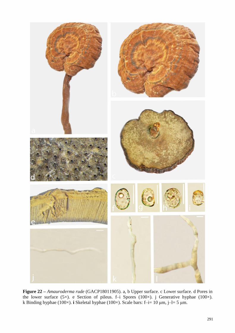
291
Figure 22 – Amauroderma rude (GACP18011905). a, b Upper surface. c Lower surface. d Pores in
the lower surface (5×). e Section of pileus. f–i Spores (100×). j Generative hyphae (100×).
k Binding hyphae (100×). i Skeletal hyphae (100×). Scale bars: f–i= 10 μm, j–l= 5 μm.
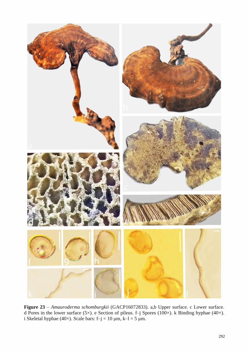
292
Figure 23 – Amauroderma schomburgkii (GACP16072833). a,b Upper surface. c Lower surface.
d Pores in the lower surface (5×). e Section of pileus. f–j Spores (100×). k Binding hyphae (40×).
i Skeletal hyphae (40×). Scale bars: f–j = 10 μm, k–l = 5 μm.
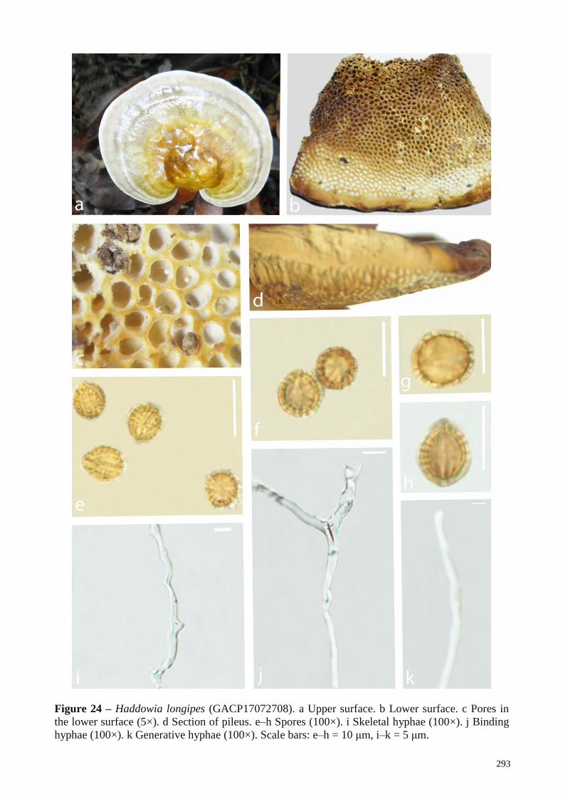
293
Figure 24 – Haddowia longipes (GACP17072708). a Upper surface. b Lower surface. c Pores in
the lower surface (5×). d Section of pileus. e–h Spores (100×). i Skeletal hyphae (100×). j Binding
hyphae (100×). k Generative hyphae (100×). Scale bars: e–h = 10 μm, i–k = 5 μm.
294
(Furtado 1981), China (Teng 1963, Tai 1979, Zhao 1989, Zhao & Zhang 2000, Zhang et al. 2015),
India, Philippines (Ryvarden & Johansen 1980) and Laos (This study).concept of Humphreya may
represent a genus independent of Ganoderma (Costa-Rezende et al. 2017). However, Humphreya’s
position at genus level is still uncertain given the lack of sufficient DNA sequence data analyses
(Costa-Rezende et al. 2017). Currently there are four species, i.e. H. coffeata, H. eminii, H. endertii
and H. lloydii described under this genus according to Index Fungorum (www.indexfungorum.org,
accessed 22 March 2019).
Humphreya Steyaert, Persoonia 7(1): 98 (1972)
See the description at Steyaert (1972)
Basidiome stipitate, pileus dull to glossy, yellow to dark brown, shiny to dull, pores small to
medium, context ochraceous. Hyphal system di-trimitic, generative hyphae with clamps, hyaline,
skeletal hyphae arboriform with long unbranched lower segments, moderately to richly-branched,
in the top, hyaline to pale-yellowish, cuticle a palisade of clavaroid elements arising from
generative hyphae, with age becoming thickened and brown at the apex. Cystidia none. Spores
large, 10–35 µm long, truncate and ornamented with a reticulate pattern of ridges, covered with a
thin hyaline episporium, usually collapsed in dry specimens.
Type species – Humphreya lloydii (Pat. & Har.) Steyaer
Notes – Steyaert (1972) proposed the genus Humphreya to accommodate Amauroderma lloydii,
Polyporous coffeatus and Humphreya endertii due to their hyphal disposition and basidiospore
ornamentation. Humphreya species has pale context and basidiospores with truncate apex and the
endosporium ornamented by typical longitudinal ridges (Costa-Rezende et al. 2017). However,
Ganoderma coffeatum has typical basidiospores with endosporic ornamentation as predominantly
longitudinal ridges and with a known distribution in South and Central America (Decock &
Herrera-Figueroa 2007). Hence, latter authors declined the combination of Steyaert (1972) since the
vicinity of G. coffeatum and H. lloydii is uncertain. Later, it was concluded that the G. coffeatum
clustered in an independent clade from the typical Ganoderma species and the
Foraminispora Robledo, Costa-Rezende & Drechsler-Santos, Persoonia 39, 254–269 (2017)
See the description at Costa-Rezende et al. (2017)
Basidiomata annual, stipe pleuropodal to pseudomesopodal, pileus circular to spathulate.
Pilear surface glabrous, greyish brown to dark brown, concentrically zonate with thin blackish
bands, radially rugose. Context white, homogenous, in section with a shiny black cuticle. Tubes
slightly darker than context. Pore surface whitish to vivid orange. Pores regular, circular to angular.
Dissepiments thick, entire. Stipe cylindrical, pale to dark brown, finely tomentose, solid to hollow,
context homogeneous, whitish, in section with a shiny dark cuticle. Hyphal system dimitic,
generative hyphae clamped, arboriform and skeleto-binding hyphae almost hyaline, dextrinoid.
Cystidia and cystidioles absent. Basidia clavate, with four sterigmata. Basidiospores subglobose,
hyaline to pale brown, double walled, with conspicuous ornamentation as endosporic projections
column-like, some of them with a hole, that persists up to the exospore, IKI-.
Type species – Foraminispora rugosa (Berk.) Costa-Rezende, Drechsler-Santos & Robledo
Notes – Foraminispora was introduced to accommodate Porothelium rugosum Berk. (=
Amauroderma sprucei) and is characterized by stipitate basidiomata, dull pilear surface, whitish
context, a dimitic hyphal system, skeleto-binding hyphae with lateral and apical branches and
arboriform skeletal hyphae, both dextrinoid, and globose to subglobose, hyaline to pale brown
spores, with conspicuous endosporic projections. These columnar endosporic projections are
hollow and these holes persist until the exospore wall. This feature is unique for this genus (Costa-
Rezende et al. 2017). Foraminispora belongs to Ganodermataceae because it has clamped
generative and arboriform skeletal hyphae, the double-walled basidiospores, with the inner layer
ornamented (Costa-Rezende et al. 2017). Macro- and microscopic features of Foraminispora are
similar with Amauroderma, however, perforated columns in basidiospores are absent in this genus
(Furtado 1962, 1981, Ryvarden & Johansen 1980, Corner 1983, Ryvarden 2004a). Ganoderma also
presents species with pale context and double-walled spores with endosporic ornamentation
295
(Ryvarden & Johansen 1980, Corner 1983, Ryvarden 2004b, Torres-Torres & Guzmán-Dávalos
2012), but absence of hollow columns (some species) and the truncate apex of basidiospores clearly
distinguish Ganoderma from Foraminispora (Costa-Rezende et al. 2017). Haddowia and
Humphreya differ from this genus by having double-walled spores with endosporic ornamentation
formed by ridges (Costa-Rezende et al. 2017). Tomophagus mainly differs from Foraminispora by
its laccate and soft pileus and truncate basidiospores (Murrill 1905c, Steyaert 1972, Ryvarden
2004b, Le et al. 2012).
39. Foraminispora rugosa (Berk.) Costa-Rezende, Drechsler-Santos & Robledo (2017)
= Porothelium rugosum Berk., Hooker’s J. Bot. Kew Gard. Misc. 8: 237. (1856)
See the description and illustration at Decock & Herrera-Figueroa (2005) as Amauroderma
sprucei.
Notes – This species is characterized by the predominantly subglobose basidiospores with
conspicuous hollow columnar ornamentation, di-trimitic hyphal system and the strongly dextrinoid
skeletal hyphae (Decock & Herrera-Figueroa 2005, Costa-Rezende et al. 2017). When Porothelium
rugosum was combined in Ganoderma the epithet ‘rugosum’ was already occupied by Ganoderma
rugosum, hence, the name Ganoderma sprucei was proposed. Torrend (1920b) continued to use
‘sprucei’, the earliest epithet available in Amauroderma (as A. sprucei). Considering the
combination of Porothelium rugosum in Foraminispora the epithet is available (Costa-Rezende et
al. 2017). Specimens growing on the ground or on decayed angiosperm wood in Brazil, Venezuela,
French Guiana, Costa Rica and Cuba (Decock & Herrera-Figueroa 2005).
Furtadoa Costa-Rezende, Robledo & Drechsler-Santos, in Costa-Rezende, Robledo, Góes-Neto,
Reck, Crespo & Drechsler-Santos, Persoonia 39: 263 (2017)
See the description at Costa-Rezende et al. (2017)
Basidiomata annual, stipe pleuropodal to pseudomesopodal, soft when fresh, light and fragile
when dried, pileus circular to almost flabelliform or funnel-shaped. Pilear surface dull, glabrous,
greyish brown, azonate. Context white to pale brown, homogenous. Tubes slightly darker than
context. Pore surface pale brown. Pores angular, sometimes radially elongated. Dissepiments thin,
entire to lacerate. Stipe yellowish brown, finely tomentose, solid to hollow, context homogeneous,
pale brown. Hyphal system dimitic. Context composed of clamped to simple septate generative
hyphae, thin to slightly thick-walled, some distinctly wider, with a swollen apex. Trama of tubes
composed of clamped generative and arboriform skeletal hyphae. Cystidia and cystidioles not seen.
Basidia clavate, with four sterigmata. Basidiospores subglobose to ellipsoid, hyaline, double
walled, with ornamentation as endosporic projections column-like, IKI-.
Type species: Furtadoa biseptata Costa-Rezende, Drechsler-Santos & Reck
Notes – Furtadoa was introduced to accommodate Furtadoa biseptata, F. brasiliensis (=
Amauroderma brasiliensis), F. corneri (= A. corneri) and is characterized by a stipitate
basidiomata, soft when fresh, dull pilear surface, pale context, a dimitic hyphal system, with a
monomitic context, composed of both clamped and simple-septate generative hyphae, thin to
slightly thick-walled and dimitic trama of tubes, composed of clamped generative hyphae and
arboriform skeletal hyphae and double-walled, ornamented basidiospores (Costa-Rezende et al.
2017). Furtadoa is considered as member of Ganodermataceae due to its double-walled
basidiospores with ornamented inner layer. Macro- and microscopic features of Furtadoa are
similar to Amauroderma, however the monomitic context with simple-septate generative hyphae is
exclusive of this new genus (Costa-Rezende et al. 2017).
40. Furtadoa biseptata Costa-Rezende, Drechsler-Santos & Reck, in Costa-Rezende, Robledo,
Góes-Neto, Reck, Crespo & Drechsler-Santos, Persoonia 39: 265 (2017).
See the description and illustration at Costa-Rezende et al. (2017)
Notes — Furtadoa was typified by F. biseptata and this species differs from Furtadoa
brasiliensis by its thinner basidiomata, darker context, and the presence of simple septate
generative hyphae in the context. F. corneri differs from F. biseptata by the funnel-shaped
296
basidiomata and the thinner pileus, and slightly larger basidiospores. This species grows on the
ground or on decayed angiosperm wood and found rarely in the field (Costa-Rezende et al. 2017).
41. Furtadoa brasiliensis (Singer) Costa-Rezende, Drechsler- Santos & Robledo, in Costa-
Rezende, Robledo, Góes-Neto, Reck, Crespo & Drechsler-Santos, Persoonia 39: 265 (2017).
= Scutiger brasiliensis Singer, Nova Hedwigia, Beih. 77: 22, 1983.
= Amauroderma brasiliense (Singer) Ryvarden, Syn. Fungorum 19: 44, 2004 ‘as A.
brasilensis’.
See the description and illustration at Singer et al. (1983) 22, ‘as Scutiger brasiliensis’
Notes – This species was introduced as Scutiger brasiliense from Brazil with stipitate
basidiomata with a white and soft-flesh context, monomitic hyphal system and inamyloid and
ellipsoid to almost subglobose spores (Singer et al. 1983). Then, it was transferred to Amauroderma
(as A. corneri) based on Amauroderma-like basidiospores by observing a specimen from Atlantic
Rain Forest in Brazil (Gulaid & Ryvarden 1998). The species was later considered as a synonymy
of A. brasiliense (Ryvarden 2004b, Coelho et al. 2007, Gomes-Silva et al. 2015). However, based
on morphological differences and high genetic divergence between the taxa Costa-Rezende et al.
(2017) decided to maintain both A. brasiliense and A. corneri as independent species. Furthermore,
the latter authors suggested a separate genus; Furtadoa to accommodate A. brasiliense and A.
corneri based on monomitic context with simple-septate generative hyphae. Furtadoa brasiliensis
is growing on the ground or on decayed angiosperm wood (Costa-Rezende et al. 2017).
42. Furtadoa corneri (Gulaid & Ryvarden) Robledo & Costa- Rezende in Costa-Rezende,
Robledo, Góes-Neto, Reck, Crespo & Drechsler-Santos, Persoonia 39: 265 (2017).
= Amauroderma corneri Gulaid & Ryvarden, Mycol. Helv. 10 (1): 28. 1998.
See the description and illustration at Gulaid & Ryvarden (1998) 28, as ‘A. corneri ’
Notes – Furtadoa corneri is characterized by a thin, funnel-to fan-shaped pileus, monomitic
context and subglobose to ellipsoid basidiospores. This species is growing on the ground or on
decayed angiosperm wood (Costa-Rezende et al. 2017).
Polyporopsis Mycotaxon 111: 447 (2010)
Type species: Polyporopsis mexicanus (Laferr. & Gilb.) Audet
43. Polyporopsis mexicanus (Laferr. & Gilb.) Audet, Mycotaxon 111: 447 (2010)
See the description Audet (2010)
Notes – Polyporopsis was introduced to accommodate Albatrellus mexicanus (Laferr. &
Gilb.) and this genus is characterized by circular pileus, dimitic hyphal system and ellipsoidal
basidiospores with oily drops. Laferrière & Gilbertson (1990) have classified this species as
Albatrellus without considering the double wall of the spores or the hyphal system. Microscopic
characters of P. mexicanus are close to Amauroderma but some microscopic features do not
correspond to this genus and the morphology of the basidiome is very distinct according to
Ryvarden (1991). Furthermore, Zheng & Liu (2006) synonymized this species with Polyporoletus
sublividus without examining the dimitic hyphal system or other characters of Polyporoletus
(Audet 2010). However, P. sublividus is a species with gray pores, with a trichodermic coating, and
larger spores, while Polyporopsis mexicanus is a yellow-brown species with resinous brown pores
in the lower surface and non-aborted tubes on the stipe, with an indeterminate coating, and a
dimitic hyphal system and with smaller spores (Audet 2010).
Acknowledgements
This work was financed by the Science and Technology Foundation of Guizhou Province
(No. [2017]2511-1), and the Science Research Foundation of Guizhou University (No. 201309).
Samantha C. Karunarathna thanks CAS President’s International Fellowship Initiative (PIFI) for
funding his postdoctoral research (number 2018PC0006). Kalani K. Hapuarachchi is grateful to
297
Prof. Xingliang Wu, Dr. Olivier Raspé, Hansika Perera and Milan Samarakoon for their valuable
comments and suggestions.
References
Abdullah F, Vijaya SK, Zurina H. 1997 – First report of Ganoderma australe on mango (Mangifera
indica) stumps. In: Abstracts of Research Seminar 1997, Faculty of Science and
Environmental Studies, Budin H, Daud I, Basri M, Yaziz MI. (Eds.). Universiti Putra
Malaysia, Serdang, Selangor 67–68.
Al-Bahry S, Elshafie AE, Deadman M, AlSa’di A et al. 2005 – First report of Ganoderma colossum
on Ficus altissima and Delonix regia in Oman. Plant Pathology 54, 245.
Allen E, Morrison D, Wallis G. 1996 – Common tree disease of British Columbia. Victoria, BC:
Canadian Forest Service, Pacific Forestry Centre 178.
Adaskaveg JE, Gilbertson RL. 1988 – Basidiospores, pilocystidia, and other basidiocarp characters
in several species of the Ganoderma lucidum complex. Mycology 80, 493–507.
Adaskaveg JE, Gilbertson RL. 1989 – Cultural studies of four North American species in the
Ganoderma lucidum complex with comparisons to G. lucidum and G. tsugae. Mycological
Research 92, 182–191.
Adaskaveg JE, Blanchette RA, Gilbertson RL. 1991 – Decay of date palm wood by white–rot and
brown– rot fungi. Canadian Journal of Botany 69, 615–629.
Agustini L, Francis A, Glen M, Indrayadi H et al. 2014 – Signs and identification of fungal root‐rot
pathogens in tropical Eucalyptus pellita plantations. Forest Pathology 44, 486–495.
Aime MC, Henkel TW, Ryvarden L. 2003 – Studies in Neotropical polypores 15: new and
interesting species from Guyana. Mycologia 95, 614–619.
Ariffin D, Idris AS, Singh G. 2000 – Status of Ganoderma in oil palm. In Flood J, Bridge PD,
Holderness M (Eds.) Ganoderma diseases of perennial crops. CABI Publishing: Wallingford.
Arulpandi I, Kalaichelvan PT. 2013 – Ganoderma adspersum and Ganoderma cupreum from South
India, First report based on molecular phylogeny. International Journal of Current
Microbiology and Applied Sciences 2, 693–702.
Audet SA. 2010 – Essai de découpage systématique du genre Scutiger (Basidiomycota):
Albatrellopsis, Albatrellus, Polyporoletus, Scutiger et description de six nouveaux genres.
Mycotaxon. 111, 431–464 (In French).
Azahar TM, Boursier P, Seman IA. 2008 – Spatial analysis of basal stem rot disease using
geographical information system. GIS Development 1–6.
Baker CF. 1918 – Ganoderma colossus (Fr.) C.F. Baker, Brotéria 425.
Badalyan SM, Gharibyan NG, Iotti M, Zambonelli A. 2012 – Morphological and genetic
characteristics of different collections of Ganoderma P. Karst. species. In: Mushroom
Science XVIII, Proceedings of the 18th ISMS Congress (Zhang J, Wang H, Chen M, eds),
26-30 August 2012, Beijing, China Agriculture press, 247-254.
Bakshi BK. 1971 – Indian Polyporaceae (on trees and timber). New Delhi: Indian Council of
Agricultural Research.
Bakshi BK. 1976 – Forest pathology: Principles and practice in forestry. Controller of Publication,
F.R.I. & Colleges, Dehradun, India. 400
Benson DA, Cavanaugh M, Clark K, Karsch-Mizrachi I et al. 2017 – GenBank. Nucleic Acids
Research 45 (D1), D37–D42.
Bhosle S, Ranadive K, Bapat G, Garad S et al. 2010 – Taxonomy and diversity of Ganoderma from
the Western parts of Maharashtra (India). Mycosphere 1, 249–262.
Bhansali RR. 2012 – Ganoderma diseases of woody plants of indian arid zone and their biological
control. J.M. Mérillon and K.G. Ramawat (Eds.), Plant Defence: Biological Control, Progress
in Biological Control 12.

298
Bolaños-Rojas AC, Bononi VL, de Mello Gugliotta A. 2016 – New records of Ganoderma
multiplicatum (Mont.) Pat.(Polyporales, Basidiomycota) from Colombia and its geographic
distribution in South America. Check List 12, 1948.
Bresadola G. 1910 – Adnotanda in fungos aliquot exoticos regii Musei lugdunensis. Annals
Mycologici 8, 585–589.
Bresadola G. 1932 – Iconographia Mycologica 21, 1001–1050.
Breitenbach J, Kränzlin F. 1986 – Fungi of Switzerland, 2. Verlag Mykologia, Luzern. 412.
Cao Y, Wu SH, Dai YC. 2012 – Species clarification of the prize medicinal Ganoderma mushroom
‘‘Lingzhi’’. Fungal Diversity 56, 49–62.
Cao Y, Yuan HS. 2013 – Ganoderma mutabile sp. nov. from southwestern China based on
morphological and molecular data. Mycological Progress 12, 121–126.
Campacci TV, Gugliotta AD. 2009 – A review of Amauroderma in Brazil, with A. oblongisporum
newly recorded from the neotropics. Mycotaxon 110, 423–436.
Cartwright KSTG, Findlay WPK. 1958 – Decay of Timber and its Prevention. 2nd (Edn.). London:
HMSO.
Coelho G, Cortez VG, Guerrero RT. 2007 – New morphological data on Amauroderma brasiliense
(Polyporales, Basidiomycota). Mycotaxon 100, 177–183.
Costa-Rezende DH, Gugliotta AM, Góes-Neto A, Reck MA et al. 2016 – Amauroderma calcitum
sp. nov. and notes on taxonomy and distribution of Amauroderma species
(Ganodermataceae). Phytotaxa 244 101–124.
Costa-Rezende DH, Robledo GL, Goes-Neto A, Reck MA et al. 2017 – Morphological
reassessment and molecular phylogenetic analyses of Amauroderma s.lat. raised new
perspectives in the generic classification of the Ganodermataceae family. Persoonia 39, 254–
269.
Cooke MC. 1885 –Fungi of Malayan Peninsula. Grevillea 14, 43–44.
Chan PM, Kanagasabapathy G, Tan YS, Sabaratnam V et al. 2013 – Amauroderma rugosum
(Blume & T. Nees) Torrend: Nutritional composition and antioxidant and potential anti-
inflammatory properties. Evidence-Based Complementary and Alternative Medicine 2013, 1–
10.
Chandrasrikul A, Suwanarit P, Sangwanit U, Lumyong S et al. 2011 – Checklist of mushrooms
(Basidiomycetes) in Thailand. Office of Natural Resources and Environmental Policy and
Planning, Bangkok, Thailand 448.
Chalermpongse A. 1991 – Fungal diseases in mangrove ecosystem. In: Proceeding, the 5th
Silviculture Seminar in Thailand, Division of Silviculture, Royal Forest Department,
Bangkok, Thailand, 307–338.
Correia de Lima Júnior N, Baptista Gibertoni T, Malosso E. 2014 – Delimitation of some
neotropical laccate Ganoderma (Ganodermataceae): Molecular phylogeny and morphology.
Revista de Biología Tropical 62, 1197–1208.
Cong VT. 2010 – Ganoderma spp-biology, species and culture in Vietnam and in the Czech
Republic. PhD thesis. Mendel University in Brno, Czech Republic.
Coetzee MPA, Wingfield BD, Golani GD, Tjahjono B et al. 2011 – A single dominant Ganoderma
species is responsible for root rot of Acacia mangium and Eucalyptus in Sumatra. Southern
Forests: A Journal of Forest Science 1, 73, 175–180.
Coetzee MPA, Marincowitz S, Vuledzani VG, Wingfield MJ. 2015 –Ganoderma species, including
new taxa associated with root rot of the iconic Jacaranda mimosifolia in Pretoria, South
Africa. IMA Fungus 6, 249–256.
Corley R, Tinker P. 2003 – Vegetative propagation and biotechnology. The Oil Palm 4, 201–215.
Corner EJH. 1983 – Ad Ployporaceas I. Amauroderma and Ganoderma. Nova Hedwigia, 75, 1182.
Crous PW, Shivas RG, Quaedvlieg W, Van der Bank M et al. 2014 – Fungal planet description
sheets: 214–280. Persoonia 32, 184–306.
Crous PW, Wingfield MJ, Guarro J, Hernández-Restrepo M et al. 2015 – Fungal planet description
sheets: 320–370. Persoonia 34, 167–266.
299
Crous PW, Wingfield MJ, Burgess TI, Hardy GS et al. 2016 – Fungal planet description sheets:
469–557. Persoonia 37, 218–403.
Crous PW, Wingfield MJ, Burgess TI, Hardy GS et al. 2017 – Fungal planet description sheets:
558–624. Persoonia 38, 240–384.
Dai YC. 1996 – Changbai wood-rotting fungi 7. A checklist of the polypores. Fungal Science 11,
79–105.
Dai YC, Vainio E, Hantula J, Niemela T, Korhonen K. 2002 – Sexuality and intersterility within
the Heterobasidion insulare complex. Mycological Research 106, 1435–1448.
Dai YC, Wei YL, Wu XL. 2004 – Polypores from Hainan Province 1. Journal of Fungal Research
2, 53–57.
Dai YC, Cui BK, Yuan HS, Li BD. 2007 – Pathogenic wood–decaying fungi in China. Forest
Pathology 37, 105–120.
Dai YC, Yang ZL. 2008 – A revised checklist of medicinal fungi in China. Mycosystema 27, 801–
824.
Dai YC, Yang ZL, Cui BK, Yu CJ et al. 2009 – Species diversity and utilization of medicinal
mushrooms and fungi in China. International Journal of Medicinal Mushrooms 11, 287–302.
Dai YC, Cui BK, Yuan HS, He SH et al. 2011 – Wood-inhabiting fungi in southern China. 4.
Polypores from Hainan Province. Annales Botanici Fennici 48, 219–231.
Dai YC, Zhou LW, Hattori T, Cao Y et al. 2017 – Ganoderma lingzhi (Polyporales,
Basidiomycota): the scientific binomial for the widely cultivated medicinal fungus Lingzhi.
Mycological Progress 16, 1051–1055.
Das K, Aminuzzaman FM. 2017 – Morphological and ecological characterization of xylotrophic
fungi in mangrove forest regions of Bangladesh. Journal of Advances in Biology &
Biotechnology 11, 1–15.
De Simone D, Annesi T. 2012 – Occurrence of Ganoderma adspersum on Pinus pinea.
Phytopathologia Mediterranea 374–382.
Decock C, Herrera Figueroa S. 2005 – Neotropical Ganodermataceae (Basidiomycetes):
Amauroderma sprucei and A. dubiopansum. Cryptogamie, Mycologie 26.
Decock C, Herrera-Figueroa S. 2007 – Studies in Ganodermataceae (Basidiomycota): the concept
of Ganoderma coffeatum in the Neotropics and East Asia. Cryptogamie Mycologie 28, 77–
89.
Ding S. 2013 – A survey of fungi associated with trees in subtropical Hong Kong. HKU Theses
Online (HKUTO).
Doidge EM. 1950 – The South African fungi and lichens to the end of 1945. Bothalia 5, 1–1094.
Donk MA. 1933 – Revision der Niederländischen Homobasidiomycetae – Aphyllophoraceae II.
Mededelingen van het botanisch Museum en Herbarium van de Rijksuniversiteit Utrecht. 9,
1–278.
Donk MA. 1948 – Notes on Malesian fungi I. Bulletin du Jardin Botanique de Buitenzorg 3, 473–
482.
Donk MA. 1964 – A conspectus of families of Aphyllophorales. Persoonia 3, 199–324.
Donk MA. 1969 – Notes on European polypores. IV. on some species of Ganoderma. Kon Neth
Akad Wetensch Proc Ser C Biol Med Sci. 72, 273–282.
Dong C, Han Q. 2015 – Ganoderma lucidum (Lingzhi, Ganoderma): Fungi, algae, and other
materials In: Liu Y, Wang Z, Zhang J. (Eds.) Dietary Chinese Herbs Chemistry:
Pharmacology and Clinical Evidence. Springer, London 759–765.
Douanla-Meli C, Langer E. 2009 – Ganoderma carocalcareus sp. nov. with crumbly-friable
context parasite to saprobe on Anthocleista nobilis and its phylogenetic relationship in G.
resinaceum group. Mycological Progress 8, 145–155.
El-Nagerabi SAF, Elshafie AE. 2014 – First record of Ganoderma colossum dieback and wood
decay of Ziziphus spina-christi. Journal on New Biological Reports 3, 75–79.
El-Nagerabi SAF, Elshafie AE. 2015 – New Record of Ganoderma colossum White Rot on Ficus
bengalensis. Journal of New Biological Reports 4, 228–232.
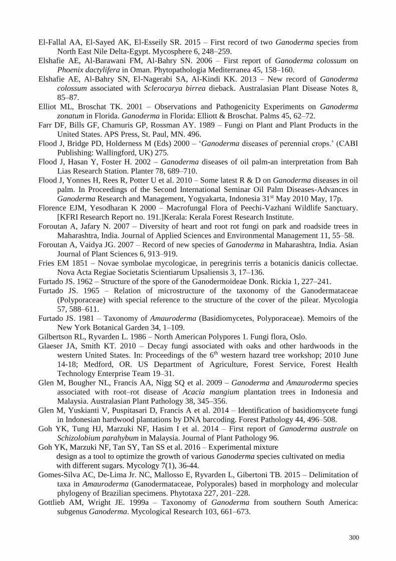
300
El-Fallal AA, El-Sayed AK, El-Esseily SR. 2015 – First record of two Ganoderma species from
North East Nile Delta-Egypt. Mycosphere 6, 248–259.
Elshafie AE, Al-Barawani FM, Al-Bahry SN. 2006 – First report of Ganoderma colossum on
Phoenix dactylifera in Oman. Phytopathologia Mediterranea 45, 158–160.
Elshafie AE, Al-Bahry SN, El-Nagerabi SA, Al-Kindi KK. 2013 – New record of Ganoderma
colossum associated with Sclerocarya birrea dieback. Australasian Plant Disease Notes 8,
85–87.
Elliot ML, Broschat TK. 2001 – Observations and Pathogenicity Experiments on Ganoderma
zonatum in Florida. Ganoderma in Florida: Elliott & Broschat. Palms 45, 62–72.
Farr DF, Bills GF, Chamuris GP, Rossman AY. 1989 – Fungi on Plant and Plant Products in the
United States. APS Press, St. Paul, MN. 496.
Flood J, Bridge PD, Holderness M (Eds) 2000 – ‘Ganoderma diseases of perennial crops.’ (CABI
Publishing: Wallingford, UK) 275.
Flood J, Hasan Y, Foster H. 2002 – Ganoderma diseases of oil palm-an interpretation from Bah
Lias Research Station. Planter 78, 689–710.
Flood J, Yonnes H, Rees R, Potter U et al. 2010 – Some latest R & D on Ganoderma diseases in oil
palm. In Proceedings of the Second International Seminar Oil Palm Diseases-Advances in
Ganoderma Research and Management, Yogyakarta, Indonesia 31st May 2010 May, 17p.
Florence EJM, Yesodharan K 2000 – Macrofungal Flora of Peechi-Vazhani Wildlife Sanctuary.
[KFRI Research Report no. 191.]Kerala: Kerala Forest Research Institute.
Foroutan A, Jafary N. 2007 – Diversity of heart and root rot fungi on park and roadside trees in
Maharashtra, India. Journal of Applied Sciences and Environmental Management 11, 55–58.
Foroutan A, Vaidya JG. 2007 – Record of new species of Ganoderma in Maharashtra, India. Asian
Journal of Plant Sciences 6, 913–919.
Fries EM 1851 – Novae symbolae mycologicae, in peregrinis terris a botanicis danicis collectae.
Nova Acta Regiae Societatis Scientiarum Upsaliensis 3, 17–136.
Furtado JS. 1962 – Structure of the spore of the Ganodermoideae Donk. Rickia 1, 227–241.
Furtado JS. 1965 – Relation of microstructure of the taxonomy of the Ganodermataceae
(Polyporaceae) with special reference to the structure of the cover of the pilear. Mycologia
57, 588–611.
Furtado JS. 1981 – Taxonomy of Amauroderma (Basidiomycetes, Polyporaceae). Memoirs of the
New York Botanical Garden 34, 1–109.
Gilbertson RL, Ryvarden L. 1986 – North American Polypores 1. Fungi flora, Oslo.
Glaeser JA, Smith KT. 2010 – Decay fungi associated with oaks and other hardwoods in the
western United States. In: Proceedings of the 6th western hazard tree workshop; 2010 June
14-18; Medford, OR. US Department of Agriculture, Forest Service, Forest Health
Technology Enterprise Team 19–31.
Glen M, Bougher NL, Francis AA, Nigg SQ et al. 2009 – Ganoderma and Amauroderma species
associated with root–rot disease of Acacia mangium plantation trees in Indonesia and
Malaysia. Australasian Plant Pathology 38, 345–356.
Glen M, Yuskianti V, Puspitasari D, Francis A et al. 2014 – Identification of basidiomycete fungi
in Indonesian hardwood plantations by DNA barcoding. Forest Pathology 44, 496–508.
Goh YK, Tung HJ, Marzuki NF, Hasim I et al. 2014 – First report of Ganoderma australe on
Schizolobium parahybum in Malaysia. Journal of Plant Pathology 96.
Goh YK, Marzuki NF, Tan SY, Tan SS et al. 2016 – Experimental mixture
design as a tool to optimize the growth of various Ganoderma species cultivated on media
with different sugars. Mycology 7(1), 36-44.
Gomes-Silva AC, De-Lima Jr. NC, Mallosso E, Ryvarden L, Gibertoni TB. 2015 – Delimitation of
taxa in Amauroderma (Ganodermataceae, Polyporales) based in morphology and molecular
phylogeny of Brazilian specimens. Phytotaxa 227, 201–228.
Gottlieb AM, Wright JE. 1999a – Taxonomy of Ganoderma from southern South America:
subgenus Ganoderma. Mycological Research 103, 661–673.
301
Gottlieb AM, Ferrer E, Wright JE. 2000 – rDNA analyses as an aid to the taxonomy of species of
Ganoderma. Mycological Research 104, 1033–1045.
Grand LF, Vernia CS. 2006 – Biogeography and hosts of poroid wood decay fungi in North
Carolina: species of Fomes, Fomitopsis, Fomitella and Ganoderma. Mycotaxon 94, 231–234.
Gulaid H, Ryvarden L. 1998 – Two new species of Amauroderma (Ganodermataceae,
Basidiomycetes). Mycologia Helvetica 10, 25–30.
Guglielmo F, Gonthier P, Garbelotto M, Nicolott G. 2008 – A PCR-based method for the
identification of important wood rotting fungal taxa within Ganoderma, Inonotus s.l. and
Phellinus s.l. FEMS Microbiology Letters 282, 228–237.
Haddow WR. 1931 – Studies in Ganoderma. Journal of the Arnold Arboretum 12, 25–46.
Hall TA. 1999 – BioEdit: a user–friendly biological sequence alignment editor and analysis
program for Windows 95/98/NT. In: Nucleic Acids Symposium Series. 41, 95–98.
Hapuarachchi KK, Wen TC, Deng CY, Kang JC et al. 2015 – Mycosphere Essays 1: Taxonomic
confusion in the Ganoderma lucidum species complex. Mycosphere 6, 542–559.
Hapuarachchi KK, Wen TC, Jeewon R, Wu XL et al. 2016a – Mycosphere Essays 7: Ganoderma
lucidum - are the beneficial anti-cancer properties substantiated? Mycosphere 7, 305–332.
Hapuarachchi KK, Wen TC, Jeewon R, Wu XL et al. 2016b – Mycosphere Essays 15: Ganoderma
lucidum - are the beneficial medical properties substantiated? Mycosphere 7, 687–715.
Hapuarachchi KK, Cheng CR, Wen TC, Jeewon R et al. 2017 – Mycosphere Essays 20:
Therapeutic potential of Ganoderma species: Insights into its use as traditional medicine.
Mycosphere 8, 1653–1694.
Hapuarachchi KK, Elkhateeb WA, Karunarathna SC, Cheng CR et al. 2018a – Current status of
global Ganoderma cultivation, products, industry and market. Mycosphere 9, 1025–1052.
Hapuarachchi KK, Karunarathna SC, Raspé O, De Silva KHWL et al. 2018b – High diversity of
Ganoderma and Amauroderma (Ganodermataceae, Polyporales) in Hainan Island, China.
Mycosphere 9, 931–982.
Hapuarachchi KK, Karunarathna SC, Phengsintham P, Kakumyan P et al. 2018c – Amauroderma
(Ganodermataceae, Polyporales) – bioactive compounds, beneficial properties and two new
records from Laos. Asian Journal of Mycology 1, 121–136.
Hapuarachchi KK, Karunarathna SC, McKenzie EHC, Wu XL et al. 2019 – High phenotypic
plasticity of Ganoderma sinense (Ganodermataceae, Polyporales) in China. Asian Journal of
Mycology 2(1), 1–47.
Harsh NSK, Rai BK, Tiwari DP. 1993 – Use of Ganoderma lucidum in folk medicines. Indian
Journal Tropical Biodiversity 1, 324–326.
Harsh NSK. 2012 – Fungal Decay in Poplar Trees and Wood. Forestry Bulletin, Forest Pathology
Division, Forest Research Institute, Dehradun, India 12, 100–104.
Hattori T, Ryvarden L. 1994 – Type studies in the Polyporaceae 25. Species described from Japan
by R. Imazeki & A. Yasuda. Mycotaxon 50, 27–46.
Hepting GH. 1971 – Diseases of forest and shade trees of the United States. US Department of
Agriculture, Agricultural Handbook 386, 1–658.
Hidayati N, Glen M, Nurrohmah SH, Rimbawanto A, Mohammed CL. 2014 – Ganoderma
steyaertanum as a root‐rot pathogen of forest trees. Forest Pathology 44, 460–471.
Hong SG, Jung HS. 2004 – Phylogenetic analysis of Ganoderma based on nearly complete
mitochondrial small–subunit ribosomal DNA sequences. Mycologia 96, 742–755.
Hood IA, Bell TIW. 1983 – Inoculation of Pinus caribaea var. hondurensis seedlings with
Ganoderma lucidum in Fiji. New Zealand Journal of Forestry Science 13, 53–57.
Hood IA. 2006 – The mycology of the Basidiomycetes. In ACIAR Proceedings February, ACIAR;
1998, 124, 34.
Hseu RS, Chen CY, Ueng YC, Wang HH. 1989 – Application of laccase isozyme electrophoretic
patterns in the identification of Ganoderma species. Journal of the Chinese Agricultural
Chemical Society 27, 209–217.
302
Hseu RS. 1990 – An identification system for cultures of Ganoderma species. Ph. D. Dissertation,
National Taiwan University, Taipei, Taiwan.
Hsieh FG, Yeh ZY. 2004 – Cultural and physiological studies of Ganoderma neo-japonicum and
G. zonatum. BioFormosa 39, 23–32.
Humphrey CJ. 1938 – Notes on some Basidiomycetes from the Orient. Mycologia 30, 327–335.
Hyde KD, Norphanphoun C, Chen J, Dissanayake AJ et al. 2018 – Thailand’s amazing diversity –
up to 96% of fungi in northern Thailand are novel. Fungal Diversity 93, 215–239.
Idris AS, Arifin D, Swinburne TR, Watt TA. 2000 – The identity of Ganoderma species
responsible for BSR disease of oil palm in Malaysia-Morphological Characteristics.
Malaysian Palm Oil Board 102, 77a.
Idris A, Kushairi A, Ismail S, Ariffin D. 2004 – Selection for partial resistance in oil palm
progenies to Ganoderma basal stem rot. Journal of Oil Palm Research 16, 12–18.
Irianto RS, Barry K, Hidayati N, Ito S et al. 2006 – Incidence and spatial analysis of root rot of
Acacia mangium in Indonesia. Journal of Tropical Forest Science 1, 157–165.
Imazeki R. 1939 – Studies on Ganoderma of Nippon. Bulletin of the National Science Museum,
Tokyo 1, 29–52.
Imazeki R. 1952 – A contribution to the fungus flora of Dutch New Guinea. Bulletin of the
Government Forest Experiment Station Tokyo 57, 87–128.
Imazeki R, Otani Y, Hongo T. 1988 – Coloured illustration of fungi of Japan. YAMAKEI
Publishers Co. Ltd., Tokyo, Japan.
Index Fungorum. 2019 – http://www.indexfungorum.org (accessed 22 March 2019).
Jayasiri SC, Hyde KD, Ariyawansa HA, Bhat J et al. 2015 – The Faces of Fungi database: fungal
names linked with morphology, phylogeny and human impacts. Fungal Diversity 74, 3–18.
Jargalmaa S, Eimes JA, Park MS, Park JY et al. 2017 – Taxonomic evaluation of selected
Ganoderma species and database sequence validation. Peer J 5, p.e3596.
Jeewon R, Hyde KD. 2016 –Establishing species boundaries and new taxa among fungi:
recommendations to resolve taxonomic ambiguities. Mycosphere 7(11), 1669–1669.
Jiao C, Xie YZ, Yang X, Li H et al. 2013 – Anticancer Activity of Amauroderma rude. PLoS ONE
8, e66504.
Johnson B. 2017 – Ganoderma root and butt rot: an emerging threat to California almonds. NPDN
News.
Jülich W. 1981 – Higher taxa of Basidiomycetes. In: J Cramer (Eds.) Bibliotheca Mycologia,
Vaduz 85, 1–485.
Junghuhn FW. 1838 – Praemissae in floram cryptogamicam Java insulae (Batavia). In Verh. Batav.
Genootsch 17, 1–86 (preprint).
Kaliyaperumal M, Kalaichelvan PT. 2008 – Ganoderma australe from southern India.
Microbiological Research 163, 286–292.
Katoh K, Standley DM. 2013 – MAFFT multiple sequence alignment software version 7:
improvements in performance and usability. Molecular Biology and Evolution 30, 772–780.
Kandan A, Radjacommare R, Ramanathan A, Raguchander T et al. 2009 – Molecular biology of
Ganoderma pathogenicity and diagnosis in coconut seedlings. Folia Microbiologica 54, 147–
152.
Karsten PA. 1881 – Enumeralio boletinearum et polypore arum fennicarum, systemate novo
dispositarum. Reviews of Mycology 3, 16–19.
Kha TT. 2014 – Study on characteristics of medicinal fungi on wood in Bavi national park, Hanoi,
Vietnam. 44–51 (In Vietnamese).
Kinge TR, Mih AM. 2011 – Ganoderma ryvardense sp. nov. associated with basal stem rot (BSR)
disease of oil palm in Cameroon. Mycosphere 2, 179–188.
Kinge TR, Mih AM. 2015 – Diversity and distribution of species of Ganoderma in South Western
Cameroon. Journal of Yeast and Fungal Research 6, 17–24.
Kirk PM, Cannon PF, Minter DW, Stalpers JA. 2008 – Dictionary of the fungi. 10th (Edn.). 272.
Kornerup A, Wanscher JH. 1978 – Methuen handbook of colour, 3rd (Edn.). Methuen, London.

303
Kreisel H, Al‐Fatimi M. 2008 – Further basidiomycetes from Yemen. Feddes Repertorium. 119,
463–483.
Kües U, Nelson DR, Liu C, Yu GJ et al. 2015 – Genome analysis of medicinal Ganoderma spp.
with plant-pathogenic and saprotrophic life-styles. Phytochemistry 114, 18–37.
Kuntze O. 1898 – Revisio generum plantarum 3, 1– 576.
Kuz'michev EP, Kulikova EG. 2001 – Common fungal diseases of Russian forests. Gen. Tech. Rep.
NE-279. Newtown Square, PA; US Department of Agriculture, Forest Service, Northeastern
Research Station. 129.
Laferrière JE, Gilbertson RL. 1990 – A new species of Albatrellus (Aphyllophorales:
Albatrellaceae) from Mexico. Mycotaxon 37, 183–186.
Le XT, Le QHN, Pham ND, Duong VH et al. 2012 – Tomophagus cattienensis sp. nov., a new
Ganodermataceae species from Vietnam: Evidence from morphology and ITS DNA
barcodes. Mycological Progress 11, 775–780.
Lee SS, Chang YS. 2016 – Ganoderma - Jekyll and Hyde mushrooms. Utar Agriculture Science
Journal 2, 21–31.
Leonard AC. 1998 – Two Ganoderma Species Compared. Mycologist 12, 65–68.
Li MJ, Yuan HS. 2015 – Type studies on Amauroderma species described by J.D. Zhao et al. and
the phylogeny of species in China. Mycotaxon 130, 79–89.
Li TH, Hu HP, Deng WQ, Wu SH et al. 2015 – Ganoderma leucocontextum, a new member of the
G. lucidum complex from southwestern China. Mycoscience 56, 81–85.
Lindequist U, Jülich WD, Witt S. 2015 – Ganoderma pfeifferi–A European relative of Ganoderma
lucidum. Phytochemistry 114, 102–108.
Liu YL, Whelen S, Hall BD. 1999 – Phylogenetic relationships among ascomycetes: evidence from
an RNA polymerase II subunit. Molecular Biology and Evolution 16, 1799–1808.
Lloyd CG. 1912 – Synopsis of the stipitate polyporoids. Mycological Writters 3, 95–208.
Loyd AL, Smith JA, Richter BS, Blanchette RA et al. 2017 – The Laccate Ganoderma of the
Southeastern United States: A Cosmopolitan and important genus of wood decay fungi. Plant
Pathology Department. UF/IFAS Extension, 333.
Loyd AL, Barnes CW, Held BW, Schink MJ et al. 2018 – Elucidating “lucidum”: Distinguishing
the diverse laccate Ganoderma species of the United States. PloS one. 13(7):e0199738.
Loizides M. 2018 – Diversity of wood-inhabiting aphyllophoraceous basidiomycetes on the island
of Cyprus. Mycotaxon-Ithaca Ny- 132, 985–986.
Lowe JL, Gilbertson RL. 1961 – Synopsis of the Polyporaceae of the Western United States and
Canada. Mycologia 53, 474–511.
Lückhoff HA. 1955 – Two hitherto unrecorded fungal diseases attacking pines and eucalypts in
South Africa. Journal of the South African Forestry Association 26, 47–61.
Lyngdoh A, Dkhar MS. 2014 – First report of two wood-rotting fungi, Cyclomyces fuscus and
Humphreya coffeatum, from India. Journal on New Biological Reports 3, 25–48.
Martinez AT, Barrasa JM, Prieto A, Blanco MN. 1991 – Fatty acid composition and taxonomic
status of Ganoderma australe from Southern Chile. Mycological Research 95, 782–784.
Mehrotra MD, Pandey PC, Chakrabarti K, Suresh S et al. 1996 – Root and heart rots in Acacia
mangium plantations in India. Indian Forester 122, 155–160.
Miller RE, Blair PD. 2009 – Input-output analysis: foundations and extensions. Cambridge
University Press.
Ming D, Chilton J, Fogarty F, Towers GHN. 2002 – Chemical constituents of Ganoderma
applanatum of British Columbia forests. Fitoterapia 73, 147–152.
Mohanty PS, Harsh NSK, Pandey A. 2011 – First report of Ganoderma resinaceum and G.
weberianum from north India based on ITS sequence analysis and micromorphology.
Mycosphere 2, 469–474.
Moncalvo JM. 2000 – Systematics of Ganoderma. In Ganoderma Diseases of Perennial Crops
(Edn. J. Flood, P. D. Bridge and M. Holderness), 23–45. CABI Bioscience, Egham, UK.
304
Moncalvo JM, Buchanan PK. 2008 – Molecular evidence for long distance dispersal across the
Southern Hemisphere in the Ganoderma applanatum-australe species complex
(Basidiomycota). Mycological Research 112, 425–436.
Moncalvo JM, Lutzoni FM, Rehner SA, Johnson J et al. 2000 – Phylogenetic relationships of
agaric fungi based on nuclear large subunit ribosomal DNA sequences. Systems Biology 49,
278–305.
Moncalvo JM, Ryvarden L. 1997 – A nomenclatural study of the Ganodermataceae Donk. Fungi
Flora 10, 1–114.
Moncalvo JM, Wang HF, Hseu RS. 1995a – Gene phylogeny of the Ganoderma lucidum complex
based on ribosomal DNA sequences. Comparison with traditional taxonomic characters.
Mycological Research 99, 1489–1499.
Moncalvo JM, Wang HF, Wang HH, Hseu RS. 1995b – The use of ribosomal DNA sequence data
for species identification and phylogeny in the Ganodermataceae. In Ganoderma:
Systematics, Phytopathology and Pharmacology. Proceedings of Contributed Symposium
59AB. 5th International Mycological Conference (Edn. P. K. Buchanan, R. S. Hseu and J. M.
Monocalvo), 31-44. National Taiwan University, Taipei, Vancouver.
Moncalvo JM, Wang HH, Hseu RS. 1995c – Phylogenetic relationships in Ganoderma inferred
from the internal transcribed spacers and 25S ribosomal DNA sequences. Mycologia 87,
223–238.
Monkai JM, Hyde KD, Xu JC, Mortimer PE. 2017 – Diversity and Ecology of soil fungal
communities in rubber plantation. Fungal Biology Reviews 31, 1–11.
Moradali MF, Hedjaroude GA, Mostafavi H, Abbasi M et al. 2007 – The genus Ganoderma
(Basidiomycota) in Iran. Mycotaxon 99, 251–269.
Mortimer PE, Xu J, Karunarathna SC, Hyde KD. 2014 – Mushrooms for trees and people: a field
guide to useful mushrooms of the Mekong region 125.
Murrill WA. 1902 – The Polyporaceae of North America I. The genus Ganoderma. Bulletin of the
Torrey Botanical Club 29, 599–608.
Murrill WA. 1905a – The Polyporaceae of North America XI. A synopsis of the brown pileate
species. Bulletin of the Torreya Botanical Club 32, 353–371.
Murrill WA. 1905b – The Polyporaceae of North America XII. A synopsis of the white and bright-
colored pileate species. Bulletin of the Torreya Botanical Club 32, 469–493.
Murrill WA. 1905c – Tomophagus for Dendrophagus. Torreya 5, 197.
Murrill WA. 1907 – Some Philippine Polyporaceae. Bulletin of the Torreya Botanical Club 34,
465–481.
Murrill WA. 1908 – Polyporaceae, Part 2. North American Flora 9, 73–131.
Muthelo VG. 2009 – Molecular Characterization of Ganoderma Species. (Doctoral Desseration,
University of Pretoria).
Nguyen NPD, Khanh TD. 2017 – Impacts of ecological factors on the distribution of Amauroderma
murrill genus in central highlands of Vietnam. Journal of Scientific and Engineering
Research 4, 238–243.
Nylander JAA. 2004 – MrModeltest v2.2. Program distributed by the author: 2. Evolutionary
Biology Centre, Uppsala University, 1–2.
Nilsson RH, Tedersoo L, Abarenkov K. 2012 – Five simple guidelines for establishing basic
authenticity and reliability of newly generated fungal ITS sequences. MycoKeys 4, 37–63.
Old KM, Lee SS, Sharma JK. 1998 – Diseases of tropical acacias. In ACIAR Proceedings.
Australian Centre for International Agricultural Research 224–233.
Overall A. 2016 – Southern Bracket or Artist’s Conk?. Field mycology 17, 124–128.
Overholts LO. 1953 – Polyporaceae of the United States, Alaska, and Canada. Ann Arbor: The
University of Michigan Press.
Parihar A, Hembrom ME, Das K. 2013 – distributional records of Ganoderma colossus
(Ganodermataceae) from Jharkhand and Rajasthan. Indian Journal of Plant Sciences 2, 49–
53.

305
Park YJ, Kwon OC, Son ES, Yoon DE et al. 2012 – Genetic diversity analysis of Ganoderma
species and development of a specific marker for identification of medicinal mushroom
Ganoderma lucidum. African Journal of Microbiology Research 25, 5417–5425.
Parmasto E. 1986 – Preliminary list of Vietnamese Aphyllophorales and Polyporaceae s. str.
Tanlin. Scripta Mycologica 14, 88 (In Vietnamese, English and Russian; English
Introduction, 10–12).
Patouillard NT. 1889 – Le genre Ganoderma. Bulletin de la Société Mycologique de France 5, 64–
80.
Patouillard N. 1907 – Basidiomycètes nouveaux du Brésil recueillis par F. Noack. Annales
Mycologici 5, 364–366.
Pan HY, Dai YC. 2001 – Ganoderma weberianum newly recorded from mainland of China. Fungal
Science 16, 31–34.
Paterson RRM. 2006 – Ganoderma – a therapeutic fungal bio factory. Phytochemistry 67, 1985–
2001.
Pegler DN, Young TWK. 1973 – Basidiospores form in the British species of Ganoderma Karst. –
Kew Bulletin 28, 351–369.
Peiris OS. 1974 – Ganoderma basal stem rot of coconut: A new record of the disease in Sri Lanka.
Plant Disease Reporter 58, 293–295.
Petersen JE. 1987 – Ganoderma in Northern Europe. Mycologist 1, 62–67.
Phillips DH, Burdekin DA. 1992 – Diseases of forest and ornamental trees. Springer.
Piatek M. 1999 – Ganoderma pfeifferi – an interesting polyporoid fungus, as found in Tarnow.
Chrońmy Przyr.Ojczystą 55, 98–102 (in Polish).
Pilotti CA, Sanderson FR, Aitken EAB. 2003 – Genetic structure of a population of Ganoderma
boninense on oil palm. Plant Pathology 52, 455–463.
Pilotti CA, Sanderson FR, Aitken AB, Armstrong W. 2004 – Morphological variation and host
range of two Ganoderma species from Papua New Guinea. Mycopathologia. 158, 251–265.
Potter K, Rimbawanto A, Beadle C. 2006 – Heart rot and root rot in tropical Acacia plantations. In:
Proceedings of a Workshop held in Yogyakarta, Indonesia, February 7–9. Canberra. ACIAR
Proceedings No. 124, 7–10.
Quanten E. 1997 – The polypores (Polyporaceae s.l.) of Papua New Guinea. Opera Botanica
Belgica 11, 1–352.
Quang DN, Nga TT, Tham LX. 2011 – Chemical Composition of Vietnamese Black Lingzhi
Amauroderma Subresinosum Murr. Research Journal of Phytochemistry 5, 216–221.
Rambaut A, Suchard MA, Xie D, Drummond AJ. 2013 – Tracer version 1.6. University of
Edinburgh. (Online). http://tree.bio.ed.ac.uk/software/tracer (accessed on 19.11.2018).
Rambaut A. 2012 – FigTree version 1.4.0. http://tree.bio.ed.ac.uk/software/figtree/.
Rannala B, Yang Z. 1996 – Probability distribution of molecular evolutionary trees: a new method
of phylogenetic inference. Journal of molecular evolution 43: 304–311.
Ranadive KR, Jagtap NV. 2016 – Checklist of Ganoderma P. Karst (Ganodermataceae) From
India. International Conference on Plant Research and Resource Management and 25th APSI
Silver Jubilee Scientists Meet. 2016. India.
Rehner SA, Buckley E. 2005 – A Beauveria phylogeny inferred from nuclear ITS and EF1-a
sequences: evidence for cryptic diversification and links to Cordyceps teleomorphs.
Mycologia 97, 84–98.
Rehner SA, Samuels GJ. 1994 – Taxonomy and phylogeny of Gliocladium analysed from nuclear
large subunit ribosomal DNA sequences. Mycological Research 98, 625–634.
Report in Plant diseases. 1999 – Wood Rots and decays. Department of Crop Sciences University
of Illinois at urbana-champaign.
Richter C, Wittstein K, Kirk PM, Stadler M. 2015 – An assessment of the taxonomy and
chemotaxonomy of Ganoderma. Fungal Diversity 71, 1–15.
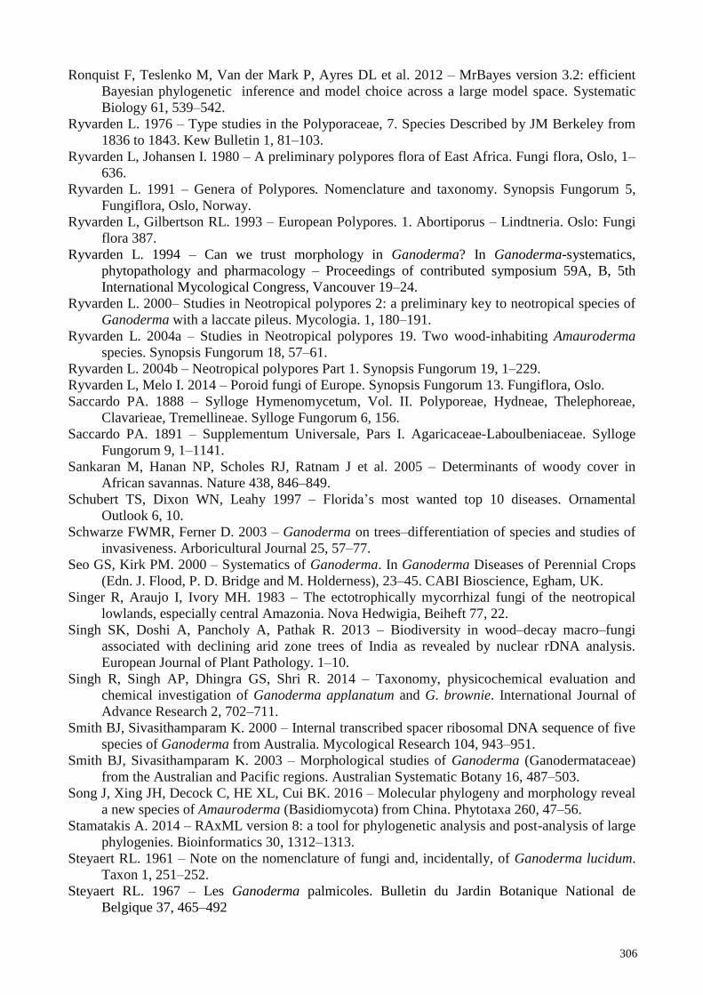
306
Ronquist F, Teslenko M, Van der Mark P, Ayres DL et al. 2012 – MrBayes version 3.2: efficient
Bayesian phylogenetic inference and model choice across a large model space. Systematic
Biology 61, 539–542.
Ryvarden L. 1976 – Type studies in the Polyporaceae, 7. Species Described by JM Berkeley from
1836 to 1843. Kew Bulletin 1, 81–103.
Ryvarden L, Johansen I. 1980 – A preliminary polypores flora of East Africa. Fungi flora, Oslo, 1–
636.
Ryvarden L. 1991 – Genera of Polypores. Nomenclature and taxonomy. Synopsis Fungorum 5,
Fungiflora, Oslo, Norway.
Ryvarden L, Gilbertson RL. 1993 – European Polypores. 1. Abortiporus – Lindtneria. Oslo: Fungi
flora 387.
Ryvarden L. 1994 – Can we trust morphology in Ganoderma? In Ganoderma-systematics,
phytopathology and pharmacology – Proceedings of contributed symposium 59A, B, 5th
International Mycological Congress, Vancouver 19–24.
Ryvarden L. 2000– Studies in Neotropical polypores 2: a preliminary key to neotropical species of
Ganoderma with a laccate pileus. Mycologia. 1, 180–191.
Ryvarden L. 2004a – Studies in Neotropical polypores 19. Two wood-inhabiting Amauroderma
species. Synopsis Fungorum 18, 57–61.
Ryvarden L. 2004b – Neotropical polypores Part 1. Synopsis Fungorum 19, 1–229.
Ryvarden L, Melo I. 2014 – Poroid fungi of Europe. Synopsis Fungorum 13. Fungiflora, Oslo.
Saccardo PA. 1888 – Sylloge Hymenomycetum, Vol. II. Polyporeae, Hydneae, Thelephoreae,
Clavarieae, Tremellineae. Sylloge Fungorum 6, 156.
Saccardo PA. 1891 – Supplementum Universale, Pars I. Agaricaceae-Laboulbeniaceae. Sylloge
Fungorum 9, 1–1141.
Sankaran M, Hanan NP, Scholes RJ, Ratnam J et al. 2005 – Determinants of woody cover in
African savannas. Nature 438, 846–849.
Schubert TS, Dixon WN, Leahy 1997 – Florida’s most wanted top 10 diseases. Ornamental
Outlook 6, 10.
Schwarze FWMR, Ferner D. 2003 – Ganoderma on trees–differentiation of species and studies of
invasiveness. Arboricultural Journal 25, 57–77.
Seo GS, Kirk PM. 2000 – Systematics of Ganoderma. In Ganoderma Diseases of Perennial Crops
(Edn. J. Flood, P. D. Bridge and M. Holderness), 23–45. CABI Bioscience, Egham, UK.
Singer R, Araujo I, Ivory MH. 1983 – The ectotrophically mycorrhizal fungi of the neotropical
lowlands, especially central Amazonia. Nova Hedwigia, Beiheft 77, 22.
Singh SK, Doshi A, Pancholy A, Pathak R. 2013 – Biodiversity in wood–decay macro–fungi
associated with declining arid zone trees of India as revealed by nuclear rDNA analysis.
European Journal of Plant Pathology. 1–10.
Singh R, Singh AP, Dhingra GS, Shri R. 2014 – Taxonomy, physicochemical evaluation and
chemical investigation of Ganoderma applanatum and G. brownie. International Journal of
Advance Research 2, 702–711.
Smith BJ, Sivasithamparam K. 2000 – Internal transcribed spacer ribosomal DNA sequence of five
species of Ganoderma from Australia. Mycological Research 104, 943–951.
Smith BJ, Sivasithamparam K. 2003 – Morphological studies of Ganoderma (Ganodermataceae)
from the Australian and Pacific regions. Australian Systematic Botany 16, 487–503.
Song J, Xing JH, Decock C, HE XL, Cui BK. 2016 – Molecular phylogeny and morphology reveal
a new species of Amauroderma (Basidiomycota) from China. Phytotaxa 260, 47–56.
Stamatakis A. 2014 – RAxML version 8: a tool for phylogenetic analysis and post-analysis of large
phylogenies. Bioinformatics 30, 1312–1313.
Steyaert RL. 1961 – Note on the nomenclature of fungi and, incidentally, of Ganoderma lucidum.
Taxon 1, 251–252.
Steyaert RL. 1967 – Les Ganoderma palmicoles. Bulletin du Jardin Botanique National de
Belgique 37, 465–492

307
Steyaert RL. 1972 – Species of Ganoderma and related genera mainly of the Bogor and Leiden
Herbaria. Persoonia 7, 55–118.
Steyaert RL. 1975 – Ganoderma applanatum. CMI Descriptions of Pathogenic Fungi and Bacteria.
443, 1–2.
Steyaert RL. 1977 – Ganoderma resinaceum Boud: should it be considered as a parasite?[on
Quercus rubra, in Belgium]. Short communication. Parasitica.
Steyaert RL. 1980 – Study of some Ganoderma species. Bulletin du Jardin Botanique National de
Belgique 50, 135–186.
Szedlay G. 2002 – is the widely used medicinal fungus the Ganoderma lucidum (fr.) karst. sensu
stricto?. Acta Microbiologica Immunologica Hungarica 49, 235–243.
Szczepkowski A, Pietka J. 2003 – New localities and new host of Ganoderma pfeifferi in Poland.
Acta Mycologica 38, 59–63.
Tan WC, Kuppusamy UR, Phan CW, Tan YS et al. 2015 – Ganoderma neo-japonicum Imazeki
revisited: Domestication study and antioxidant properties of its basidiocarps and mycelia.
Scientific Reports 5, 12515.
Tai FL. 1979 – Sylloge Fungorum Sinicorum. Beijing: Science Press, 1527 (in Chinese).
Terho M, Hantula J, Hallaaksela AM. 2007 – Occurrence and decay patterns of common wood
decay fungi in hazardous trees felled in the Helsinki City. Forest Pathology 37, 420–432.
Tchotet Tchoumi JM, Coetzee MP, Vivas M, Rajchenberg M et al. 2017 – Wood‐rotting
basidiomycetes associated with declining native trees in timber‐harvesting compartments of
the Garden Route National Park of South Africa. Austral Ecology 42, 947–963.
Teng SC.1934 – Notes on Polyporaceae from China. Sinensia 5, 198–200.
Teng SC. 1936 – Additional fungi from China III. Sinensia. 7, 529–569.
Teng SC. 1963 – Fungi of China. Beijing: Science Press, 808. (in Chinese).
Teng SC. 1996 – Fungi of China. Mycotaxon Ithaca 1–586.
Thaung MM. 2007 – A preliminary survey of macromycetes in Burma. Australasian Mycologist
26, 16–36.
Tompong S, Kunasakdakul K. 2014 – Causal agent, symptoms and environmental factors of root
rot disease of organic Assam tea in Mae Taeng district, Chiang Mai province. International
Journal of Agricultural Technology 10, 767– 77.
Torrend C. 1920a – Les polyporacées du Bresil. Broteria, série botânica 18, 23–43.
Torrend C. 1920b – Les polyporacées du Bresil. Broteria, série botânica 18, 121–142.
Torres-Torres MG, Guzmán-Dávalos L. 2005 – Notas sobre la variación morfológica de
Ganoderma curtisii en México. Revista Mexicana de Micología 21, 39–47.
Torres-Torres MG, Guzmán-Dávalos L. 2012 – The morphology of Ganoderma species with a
laccate surface. Mycotaxon 119. 201–216.
Torres-Torres MG, Ryvarden L, Guzmán-Dávalos L. 2015 – Ganoderma subgénero Ganoderma en
México. Revista Mexicana de Micología 41, 27–45.
Tortić M. 1971 – Ganoderma adspersum (S. Schulz.) Donk (= Ganoderma europaeum Steyaert)
and its distribution in Jugoslavia. Acta Botanica Croatica 30, 113–118.
Turner PD. 1981 – Oil Palm Diseases and Disorders. Oxford University Press, 88–110.
Van Der Westhuizen GCA. 1958 – Studies of Wood-Rotting Fungi: 1. Cultural Characteristics of
some Common Species. Bothalia 7, 83–99.
Vilgalys R, Hester M. 1990 – Rapid genetic identification and mapping of enzymatically amplified
ribosomal DNA from several Cryptococcus species. Journal of Bacteriology 172, 4239–4246.
Viney DE. 2005 – An illustrated introduction to the larger fungi of north Cyprus. Published by the
author. 302.
Watson G, Green T. 2011 – Fungi on Trees – An Arborists’ Field Guide. Arboricultural
Association, UK.
Wakefield EM. 1920 – Diseases of the oil palm in West Africa. In: Flood J, Bridge PD, Holderness
M (Edn.) Ganoderma diseases of perennial crops. CABI Publisher, UK.

308
Wang ZN. 1990 – Three species of Ganoderma pathogenic to Macadamia tree and their control.
Report of the Taiwan Sugar Research Institute 129, 1–10.
Wang DM, Yao YJ. 2005 – Intrastrain internal transcribed spacer heterogeneity in Ganoderma
species. Canadian Journal of Microbiology 5, 113–121.
Wang DM, Zhang XQ, Yao YJ. 2005 – Type studies of some Ganoderma species from China.
Mycotaxon 93, 61–70.
Wang DM, Wu SH. 2007 – Two species of Ganoderma new to Taiwan. Mycotaxon 102, 373–378.
Wang F, Dong ZJ, Liu JK. 2007 – Benzopyran-4-one derivatives from the fungus Ganoderma
applanatum. Zeitschrift für Naturforschung 62b, 1329–1332.
Wang DM, Wu SH, Su CH, Peng JT et al. 2009 – Ganoderma multipileum, the correct name for
‘G. lucidum’ in tropical Asia. Botanical Studies 50, 451–458.
Wang DM, Wu SH. 2010 – Ganoderma hoehnelianum has priority over G. shangsiense, and G.
williamsianum over G. meijiangense. Mycotaxon 113, 343–349.
Wang XC. 2012 – Phylogenetic study on Ganodermataceae Donk (PhD thesis). Unpublished.
Wang XC, Xi RJ, Li Y, Wang DM et al. 2012 – The species identity of the widely cultivated
Ganoderma, ‘G. lucidum’ (Ling–zhi), in China. PLoS ONE 7–40857.
Wang DM, Wu SH, Yao YJ. 2014 – Clarification of the concept of Ganoderma orbiforme with
high morphological plasticity. PLoS ONE 9: e98733.
Welti S, Moreau PA, Decock C, Danel C et al. 2015 – Oxygenated lanostane-type triterpenes
profiling in laccate Ganoderma chemotaxonomy. Mycological Progress 14, 45.
White TJ, Bruns T, Lee S, Taylor J. 1990 –Amplification and direct sequencing of fungal
ribosomal RNA genes for phylogenetics. In: Innis, MA, Gelfand DH, Sninsky JJ. & White,
T.J. (Edn.) PCR protocols: a guide to methods and applications. San Diego.
Wong CL, Bong JFC, Idris AS. 2012 – Ganoderma species associated with basal stem rot disease
of oil palm. American Journal of Applied Science 9, 879–885.
Wu SH, Zhang XQ. 2003 – The Finding of Three Ganodermataceae Species in Taiwan. Collection
and Research 16, 61–66.
Wu XL, Dai YC 2005 – Coloured illustrations of' Ganodermataceae' of China. Science Press.
Wu XL, Guo JR, Chen HQ, Liao QZ et al. 1999 – The resources and ecological distribution of the
family Ganodermataceae in Jiangfengling, Hainan Island. Acta Ecologica Sinica 19, 159–
163. (in Chinese).
Xing J, Song J, Decock C, Cui B. 2016 – Morphological characters and phylogenetic analysis
reveal a new species within the Ganoderma lucidum complex from South Africa. Phytotaxa
266, 115–124.
Xing JH, Sun YF, Han YL, Cui BK et al. 2018 – Morphological and molecular identification of
two new Ganoderma species on Casuarina equisetifolia from China. MycoKeys 7, 93–108.
Yao YJ, Wang XC, Wang B. 2013 – Epitypification of Ganoderma sichuanense J. D. Zhao and X.
Q. Zhang (Ganodermataceae). Taxon 62, 1025–1031.
Yang LZ, Feng B. 2013 – What is the Chinese “Lingzhi”? – A taxonomic mini–review, Mycologia
4, 1–4.
Zakaria L, Ali NS, Salleh B, Zakaria M. 2009 – Molecular analysis of Ganoderma species from
different hosts in Peninsula Malaysia. Journal of Biological Sciences 9, 12–20.
Zhang SS, Ma QY, Huang SZ, Dai HF et al. 2015 – Lanostanoids with Acetyl cholinesterase
inhibitory activity from the mushroom Haddowia longipes. Phytochemistry 7, 133–139.
Zheng L, Jia D, Fei X, Luo X et al. 2007 – An assessment of the genetic diversity within
Ganoderma strains with AFLP and ITSPCR–RFLP. Microbiology Research 164, 312–321.
Zheng HD, Liu PG. 2006 – Albatrellus yunnanensis, a new species from China. Mycotaxon 97,
145–151.
Zhou LW, Cao Y, Wu SH, Vlasák J et al. 2015 – Global diversity of the Ganoderma lucidum
complex (Ganodermataceae, Polyporales) inferred from morphology and multilocus
phylogeny. Phytochemistry 114, 7–15.
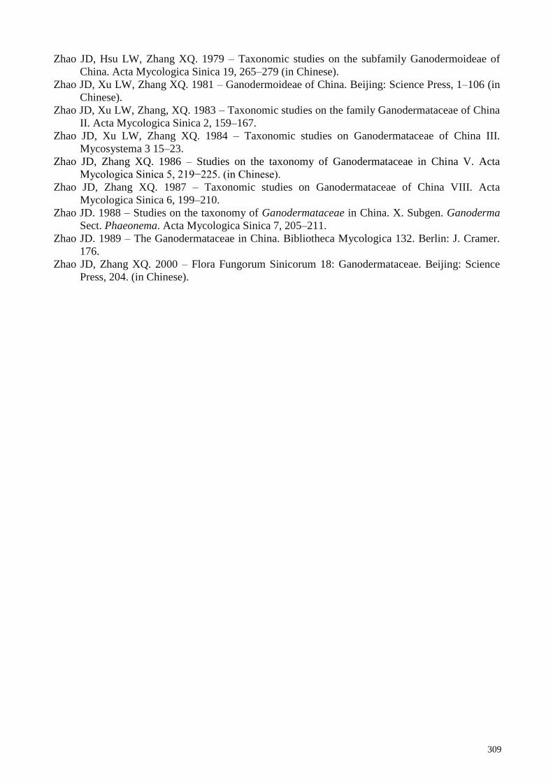
309
Zhao JD, Hsu LW, Zhang XQ. 1979 – Taxonomic studies on the subfamily Ganodermoideae of
China. Acta Mycologica Sinica 19, 265–279 (in Chinese).
Zhao JD, Xu LW, Zhang XQ. 1981 – Ganodermoideae of China. Beijing: Science Press, 1–106 (in
Chinese).
Zhao JD, Xu LW, Zhang, XQ. 1983 – Taxonomic studies on the family Ganodermataceae of China
II. Acta Mycologica Sinica 2, 159–167.
Zhao JD, Xu LW, Zhang XQ. 1984 – Taxonomic studies on Ganodermataceae of China III.
Mycosystema 3 15–23.
Zhao JD, Zhang XQ. 1986 – Studies on the taxonomy of Ganodermataceae in China V. Acta
Mycologica Sinica 5, 219−225. (in Chinese).
Zhao JD, Zhang XQ. 1987 – Taxonomic studies on Ganodermataceae of China VIII. Acta
Mycologica Sinica 6, 199–210.
Zhao JD. 1988 – Studies on the taxonomy of Ganodermataceae in China. X. Subgen. Ganoderma
Sect. Phaeonema. Acta Mycologica Sinica 7, 205–211.
Zhao JD. 1989 – The Ganodermataceae in China. Bibliotheca Mycologica 132. Berlin: J. Cramer.
176.
Zhao JD, Zhang XQ. 2000 – Flora Fungorum Sinicorum 18: Ganodermataceae. Beijing: Science
Press, 204. (in Chinese).