gas exchange unit circulation and urry • cain • …6.3.pdf · urry • cain • wasserman •...
TRANSCRIPT
CAMPBELL BIOLOGY IN FOCUS
© 2014 Pearson Education, Inc.
Urry • Cain • Wasserman • Minorsky • Jackson • Reece
Lecture Presentations by Kathleen Fitzpatrick and Nicole Tunbridge
Unit 6.3
Circulation and Gas Exchange

© 2014 Pearson Education, Inc.
Overview: Trading Places
▪ The resources that animal cells require, such as nutrients and O2, enter the cytoplasm by crossing the plasma membrane
▪ In unicellular organisms, these exchanges occur directly with the environment
▪ Most multicellular organisms rely on specialized systems that carry out exchange with the environment and transport materials through the body

© 2014 Pearson Education, Inc.
▪ Gills are an example of a specialized exchange system in animals
▪ O2 diffuses from the water into blood vessels▪ CO2 diffuses from blood into the water
▪ Internal transport and gas exchange are functionally related in most animals
© 2014 Pearson Education, Inc.
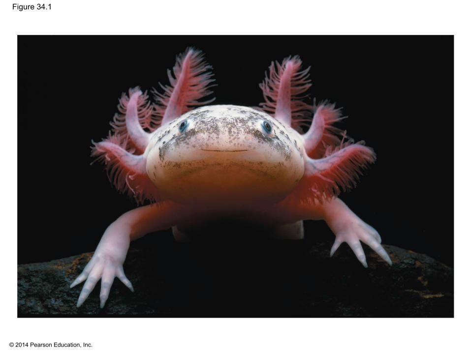
Figure 34.1
© 2014 Pearson Education, Inc.
Concept 34.1: Circulatory systems link exchange surfaces with cells throughout the body
▪ Small, nonpolar molecules such as O2 and CO2 move between cells and their immediate surroundings by diffusion
▪ Diffusion time is proportional to the square of the distance travelled
▪ Diffusion is only efficient over small distances

© 2014 Pearson Education, Inc.
▪ In small or thin animals, cells can exchange materials directly with the surrounding medium
▪ In most animals, cells exchange materials with the environment via a fluid-filled circulatory system
© 2014 Pearson Education, Inc.
Gastrovascular Cavities
▪ Some animals lack a circulatory system▪ Some cnidarians, such as jellies, have elaborate
gastrovascular cavities▪ A gastrovascular cavity functions in both digestion
and distribution of substances throughout the body▪ The body wall that encloses the gastrovascular
cavity is only two cells thick▪ Flatworms have a gastrovascular cavity and a flat
body shape to optimize diffusional exchange with the environment

© 2014 Pearson Education, Inc.
Figure 34.2
Gastrovascularcavity
Mouth
1 mm

© 2014 Pearson Education, Inc.
Open and Closed Circulatory Systems
▪ A circulatory system has a circulatory fluid, a set of interconnecting vessels, and a muscular pump, the heart
▪ Several basic types of circulatory systems have arisen during evolution, each representing adaptations to constraints of anatomy and environment

© 2014 Pearson Education, Inc.
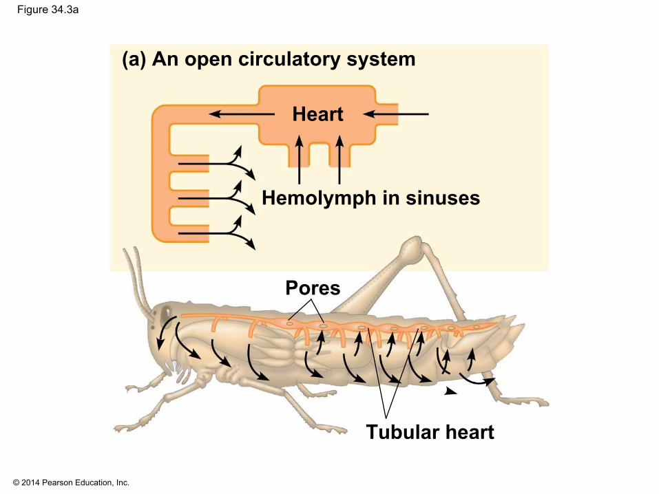
▪ All circulatory systems are either open or closed▪ In insects, other arthropods, and some molluscs,
circulatory fluid bathes the organs directly in an open circulatory system
▪ In an open circulatory system, there is no distinction between circulatory fluid and interstitial fluid, and this general body fluid is called hemolymph

© 2014 Pearson Education, Inc.
Figure 34.3
Branch vesselsin each organ
Tubular heart
Pores
Hemolymph in sinuses
(a) An open circulatory system
Heart
(b) A closed circulatory system
HeartBlood
Dorsal vessel(main heart)
Auxiliaryhearts
Ventral vessels
Interstitialfluid
© 2014 Pearson Education, Inc.
Figure 34.3a
Tubular heart
Pores
Hemolymph in sinuses
(a) An open circulatory system
Heart

© 2014 Pearson Education, Inc.
▪ In closed circulatory systems the circulatory fluid called blood is confined to vessels and is distinct from interstitial fluid
▪ These systems are found in annelids, most cephalopods, and all vertebrates
▪ One or more hearts pump blood through the vessels▪ Chemical exchange occurs between blood and
interstitial fluid and between interstitial fluid and body cells

© 2014 Pearson Education, Inc.
Figure 34.3b
Branch vesselsin each organ
(b) A closed circulatory system
HeartBlood
Dorsal vessel(main heart)
Auxiliaryhearts
Ventral vessels
Interstitialfluid

© 2014 Pearson Education, Inc.
Organization of Vertebrate Circulatory Systems
▪ Humans and other vertebrates have a closed circulatory system called the cardiovascular system
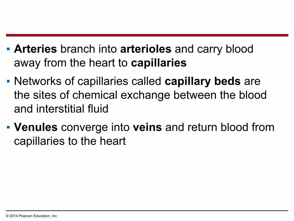
▪ The three main types of blood vessels are arteries, veins, and capillaries
▪ Blood flow is one-way in these vessels
© 2014 Pearson Education, Inc.
▪ Arteries branch into arterioles and carry blood away from the heart to capillaries
▪ Networks of capillaries called capillary beds are the sites of chemical exchange between the blood and interstitial fluid
▪ Venules converge into veins and return blood from capillaries to the heart

© 2014 Pearson Education, Inc.
▪ Arteries and veins are distinguished by the direction of blood flow, not by O2 content
▪ Vertebrate hearts contain two or more chambers▪ Blood enters through an atrium and is pumped out
through a ventricle

© 2014 Pearson Education, Inc.
Single Circulation
▪ Bony fishes, rays, and sharks have single circulation with a two-chambered heart
▪ In single circulation, blood leaving the heart passes through two capillary beds before returning

© 2014 Pearson Education, Inc.
Figure 34.4
Lungand skincapillaries
Body capillaries
Vein
Gill capillaries
(a) Single circulation: fish
Heart:
(b) Double circulation:amphibian
Key
Systemiccapillaries
Pulmocutaneous circuit
Artery
Ventricle (V)Atrium (A)
Oxygen-rich bloodOxygen-poor blood
Right Left
A A
V
Systemic circuit
Lungcapillaries
(c) Double circulation:mammal
Systemiccapillaries
Pulmonary
circuit
Right Left
A AV
Systemic circuit
V

© 2014 Pearson Education, Inc.
Figure 34.4a
Body capillaries
Vein
Gill capillaries
(a) Single circulation: fish
Heart:
Key
Artery
Ventricle (V)Atrium (A)
Oxygen-rich bloodOxygen-poor blood

© 2014 Pearson Education, Inc.
Double Circulation
▪ Amphibians, reptiles, and mammals have double circulation
▪ Oxygen-poor and oxygen-rich blood is pumped separately from the right and left sides of the heart
▪ Having both pumps within a heart simplifies coordination of the pumping cycle

© 2014 Pearson Education, Inc.
Figure 34.4b
Lungand skincapillaries
(b) Double circulation:amphibian
Key
Systemiccapillaries
Pulmocutaneous
circuit
Oxygen-rich bloodOxygen-poor blood
Right
Left
A A
V
Systemic circuit

© 2014 Pearson Education, Inc.
Figure 34.4c
Key Oxygen-rich blood
Oxygen-poor blood
Lungcapillaries
(c) Double circulation:mammal
Systemiccapillaries
Pulmonary
circuit
Right Left
A AV
Systemic circuit
V

© 2014 Pearson Education, Inc.
▪ In reptiles and mammals, oxygen-poor blood flows through the pulmonary circuit to pick up oxygen through the lungs
▪ In amphibians, oxygen-poor blood flows through a pulmocutaneous circuit to pick up oxygen through the lungs and skin
▪ Oxygen-rich blood delivers oxygen through the systemic circuit
▪ Double circulation maintains higher blood pressure in the organs than does single circulation

© 2014 Pearson Education, Inc.
Evolutionary Variation in Double Circulation
▪ Some vertebrates with double circulation are intermittent breathers
▪ These animals have adaptations that enable the circulatory system temporarily to bypass the lungs

© 2014 Pearson Education, Inc.
▪ Frogs and other amphibians have a three-chambered heart: two atria and one ventricle
▪ The ventricle pumps blood into a forked artery that splits the ventricle’s output into the pulmocutaneous circuit and the systemic circuit
▪ When underwater, blood flow to the lungs is nearly shut off

© 2014 Pearson Education, Inc.
▪ Turtles, snakes, and lizards have a three-chambered heart: two atria and one ventricle
▪ Their circulatory system allows control of relative amounts of blood flowing to the lungs and body
▪ In alligators, caimans, and other crocodilians a septum divides the ventricle
▪ A connection to atrial valves can temporarily shunt blood away from the lungs, as needed

© 2014 Pearson Education, Inc.
▪ Mammals and birds have a four-chambered heart with two atria and two ventricles
▪ The left side of the heart pumps and receives only oxygen-rich blood, while the right side receives and pumps only oxygen-poor blood
▪ There is no mechanism to vary relative blood flow to the lungs and body
▪ Mammals and birds are endotherms and require more O2 than ectotherms
© 2014 Pearson Education, Inc.
Concept 34.2: Coordinated cycles of heart contraction drive double circulation in mammals
▪ The mammalian cardiovascular system meets the body’s continuous demand for O2
© 2014 Pearson Education, Inc.
Mammalian Circulation
▪ Blood begins its flow with the right ventricle pumping blood to the lungs via the pulmonary arteries
▪ The blood loads O2 and unloads CO2 in the capillary beds of the lungs
▪ Oxygen-rich blood from the lungs enters the heart at the left atrium via the pulmonary veins and is pumped through the aorta to the body tissues by the left ventricle
© 2014 Pearson Education, Inc.
▪ The aorta provides blood to the heart through the coronary arteries
▪ Diffusion of O2 and CO2 takes place in the capillary beds throughout the body
▪ Blood returns to the heart through the superior vena cava (blood from head, neck, and forelimbs) and inferior vena cava (blood from trunk and hind limbs)
▪ The superior vena cava and inferior vena cava flow into the right atrium

© 2014 Pearson Education, Inc.
Figure 34.5
Capillaries ofabdominal organsand hind limbs
Aorta
Capillariesof right lung
Superiorvena cava
Pulmonaryartery
PulmonaryveinRight atriumRight ventricleInferior vena cava
Capillariesof left lung
Pulmonary artery
Pulmonaryvein
Left atriumLeft ventricle
Capillaries ofhead andforelimbs
Aorta
9
7
6
42
11
3
5
8
101
3
© 2014 Pearson Education, Inc.
The Mammalian Heart: A Closer Look
▪ A closer look at the mammalian heart provides a better understanding of double circulation
▪ When the heart contracts, it pumps blood; when it relaxes, its chambers fill with blood
▪ One complete sequence of pumping and filling is called the cardiac cycle
© 2014 Pearson Education, Inc.
▪ Atria have relatively thin walls and serve as collection chambers for blood returning to the heart
▪ The ventricles are more muscular and contract much more forcefully than the atria
▪ The volume of blood each ventricle pumps per minute is called the cardiac output
© 2014 Pearson Education, Inc.
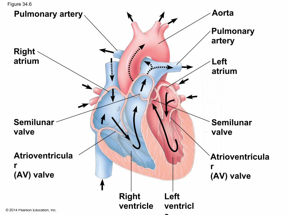
Figure 34.6
Aorta
Atrioventricular(AV) valve
Semilunarvalve
Pulmonary artery
Rightatrium
Right ventricle
Pulmonary artery
Left atrium
Left ventricle
Atrioventricular(AV) valve
Semilunarvalve
© 2014 Pearson Education, Inc.
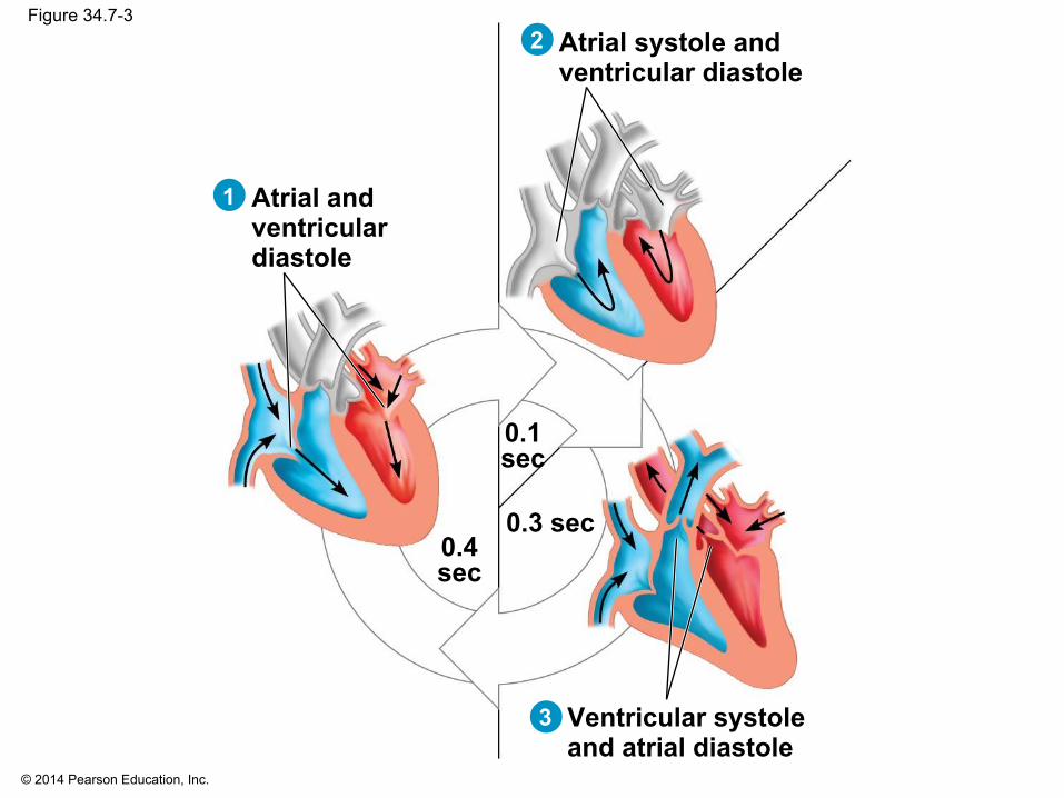
Figure 34.7-3
Atrial andventriculardiastole
Atrial systole andventricular diastole
Ventricular systole and atrial diastole
0.4sec
0.3 sec
0.1sec
1
2
3
© 2014 Pearson Education, Inc.
▪ The heart rate, also called the pulse, is the number of beats per minute
▪ The stroke volume is the amount of blood pumped in a single contraction
▪ Cardiac output depends on both the heart rate and stroke volume
© 2014 Pearson Education, Inc.
▪ Four valves prevent backflow of blood in the heart▪ The atrioventricular (AV) valves separate each
atrium and ventricle▪ The semilunar valves control blood flow to the
aorta and the pulmonary artery

© 2014 Pearson Education, Inc.
▪ The “lub-dup” sound of a heart beat is caused by the recoil of blood against the AV valves (lub) then against the semilunar (dup) valves
▪ Backflow of blood through a defective valve causes a heart murmur

© 2014 Pearson Education, Inc.
Maintaining the Heart’s Rhythmic Beat
▪ Some cardiac muscle cells are autorhythmic, meaning they contract without any signal from the nervous system
▪ The sinoatrial (SA) node, or pacemaker, sets the rate and timing at which all other cardiac muscle cells contract
▪ The SA node produces electrical impulses that spread rapidly through the heart and can be recorded as an electrocardiogram (ECG or EKG)

© 2014 Pearson Education, Inc.
Figure 34.8-4
Signals (yellow)from SA nodespreadthrough atria.
SA node(pacemaker)
1 Signals aredelayedat AV node.
Bundlebranchespass signalsto heart apex.
Signalsspreadthroughoutventricles.
AV node
Bundlebranches Heart
apex
Purkinjefibers
ECG
2 3 4

© 2014 Pearson Education, Inc.
▪ Impulses from the SA node travel to the atrioventricular (AV) node
▪ At the AV node, the impulses are delayed and then travel to the Purkinje fibers that make the ventricles contract

© 2014 Pearson Education, Inc.
▪ The pacemaker is regulated by two portions of the nervous system: the sympathetic and parasympathetic divisions
▪ The sympathetic division speeds up the pacemaker▪ The parasympathetic division slows down the
pacemaker▪ The pacemaker is also regulated by hormones and
temperature
© 2014 Pearson Education, Inc.
Concept 34.3: Patterns of blood pressure and flow reflect the structure and arrangement of blood vessels
▪ The physical principles that govern movement of water in plumbing systems also apply to the functioning of animal circulatory systems

© 2014 Pearson Education, Inc.
Blood Vessel Structure and Function
▪ A vessel’s cavity is called the central lumen▪ The epithelial layer that lines blood vessels is called
the endothelium▪ The endothelium is smooth and minimizes resistance
to blood flow

© 2014 Pearson Education, Inc.
▪ Capillaries have thin walls, the endothelium and its basal lamina, to facilitate the exchange of substances
▪ Arteries and veins have an endothelium, smooth muscle, and connective tissue
▪ Arteries have thicker walls than veins to accommodate the high pressure of blood pumped from the heart
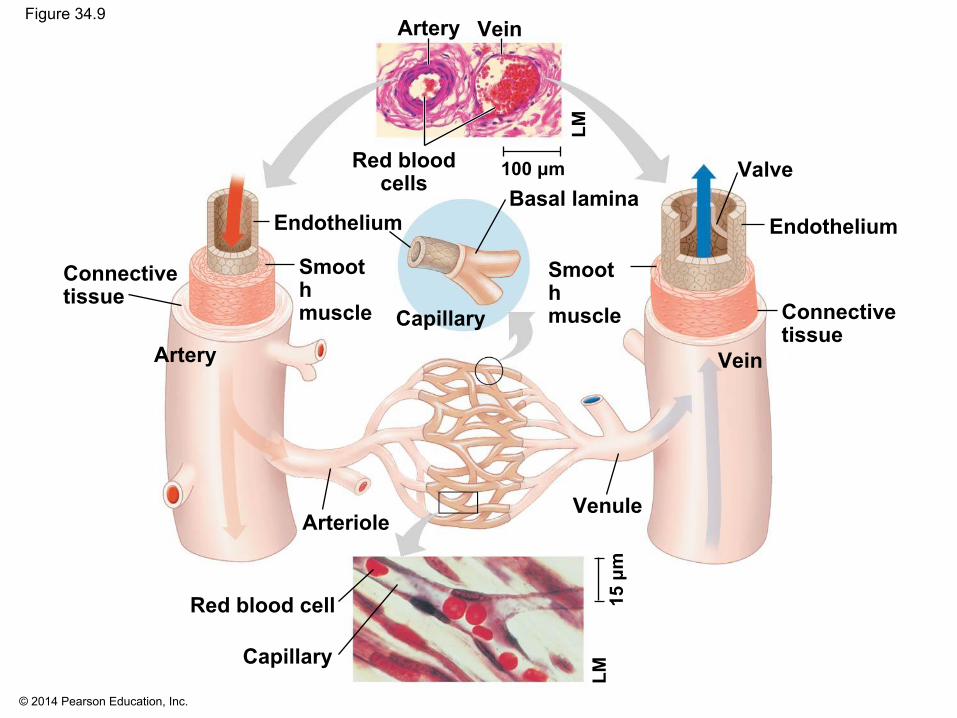
© 2014 Pearson Education, Inc.
Figure 34.9
Connectivetissue
Smoothmuscle Connective
tissue
Smoothmuscle
Endothelium Endothelium
Artery Vein
Artery Vein
Red bloodcells
Basal lamina
Capillary
Red blood cell
Capillary
ArterioleVenule
Valve100 μm
15 μ
m
LMLM

© 2014 Pearson Education, Inc.
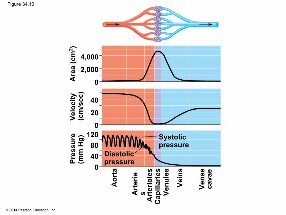
Blood Flow Velocity
▪ Blood vessel diameter influences blood flow▪ Velocity of blood flow is slowest in the capillary
beds, as a result of the high resistance and large total cross-sectional area
▪ Blood flow in capillaries is necessarily slow for exchange of materials
© 2014 Pearson Education, Inc.
Figure 34.10
Systolicpressure
Diastolicpressure
4,000
2,000
120
20
40
8040
0
0
0
Pres
sure
(mm
Hg)
Velo
city
(cm
/sec
)A
rea
(cm
2 )
Aor
ta
Art
erie
s
Vena
eca
vae
Vein
s
Cap
illar
ies
Venu
les
Art
erio
les

© 2014 Pearson Education, Inc.
Blood Pressure
▪ Blood flows from areas of higher pressure to areas of lower pressure
▪ Blood pressure exerts a force in all directions

© 2014 Pearson Education, Inc.
Changes in Blood Pressure During the Cardiac Cycle
▪ Systole is the contraction phase of the cardiac cycle▪ Pressure at the time of ventricle contraction is called
systolic pressure▪ Diastole is the the relaxation phase of the cardiac
cycle; diastolic pressure is lower than systolic

© 2014 Pearson Education, Inc.
Maintenance of Blood Pressure
▪ Blood pressure is determined by cardiac output and peripheral resistance due to constriction of arterioles
▪ Vasoconstriction is the contraction of smooth muscle in arteriole walls; it increases blood pressure
▪ Vasodilation is the relaxation of smooth muscles in the arterioles; it causes blood pressure to fall

© 2014 Pearson Education, Inc.
▪ Vasoconstriction and vasodilation help maintain adequate blood flow as the body’s demands change
▪ Nitric oxide is a major inducer of vasodilation▪ The peptide endothelin is an important inducer of
vasoconstriction
© 2014 Pearson Education, Inc.
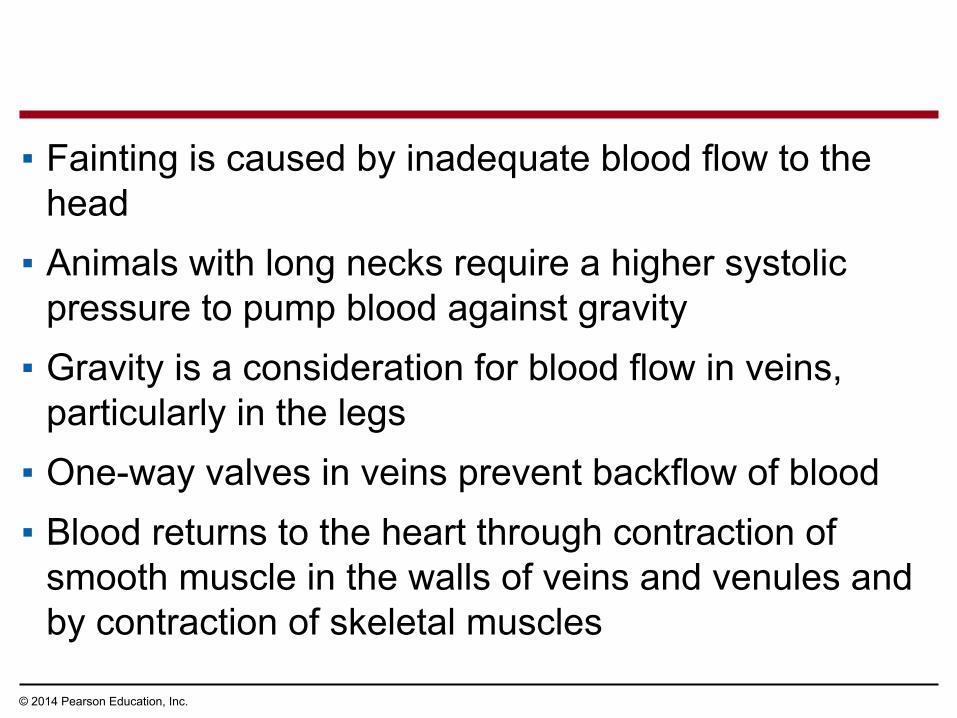
▪ Fainting is caused by inadequate blood flow to the head
▪ Animals with long necks require a higher systolic pressure to pump blood against gravity
▪ Gravity is a consideration for blood flow in veins, particularly in the legs
▪ One-way valves in veins prevent backflow of blood ▪ Blood returns to the heart through contraction of
smooth muscle in the walls of veins and venules and by contraction of skeletal muscles

© 2014 Pearson Education, Inc.
Figure 34.11
Direction of bloodflow in vein(toward heart)
Valve (open)
Valve (closed)
Skeletal muscle

© 2014 Pearson Education, Inc.
Capillary Function
▪ Blood flows through only 5–10% of the body’s capillaries at a time
▪ Capillaries in major organs are usually filled to capacity
▪ Blood supply varies in many other sites

© 2014 Pearson Education, Inc.
▪ Two mechanisms alter blood flow in capillary beds▪ Vasoconstriction or vasodilation of the arteriole that
supplies a capillary bed▪ Precapillary sphincters, rings of smooth muscle at the
capillary bed entrance, open and close to regulate passage of blood
▪ Critical exchange of substances takes place across the thin walls of the capillaries
© 2014 Pearson Education, Inc.
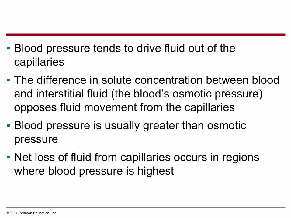
▪ Blood pressure tends to drive fluid out of the capillaries
▪ The difference in solute concentration between blood and interstitial fluid (the blood’s osmotic pressure) opposes fluid movement from the capillaries
▪ Blood pressure is usually greater than osmotic pressure
▪ Net loss of fluid from capillaries occurs in regions where blood pressure is highest
© 2014 Pearson Education, Inc.
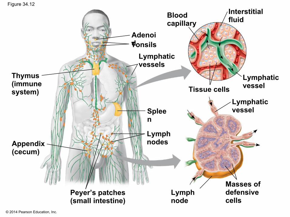
Fluid Return by the Lymphatic System
▪ The lymphatic system returns fluid, called lymph, that leaks out from the capillary beds
▪ Lymph has a very similar composition to interstitial fluid
▪ The lymphatic system drains into veins in the neck▪ Valves in lymph vessels prevent the backflow of
fluid
© 2014 Pearson Education, Inc.
Figure 34.12
Interstitialfluid
Lymphaticvessel
Lymphaticvessel
Bloodcapillary
Tissue cells
Lymph node
Masses ofdefensivecells
Lymphaticvessels
Lymph nodes
Peyer’s patches(small intestine)
Appendix(cecum)
Thymus(immunesystem)
AdenoidTonsils
Spleen

© 2014 Pearson Education, Inc.
▪ Lymph vessels have valves to prevent backflow▪ Lymph nodes are organs that filter lymph and play
an important role in the body’s defense▪ Edema is swelling caused by disruptions in the flow
of lymph▪ The lymphatic system also plays a role in harmful
immune responses, such as those responsible for asthma

© 2014 Pearson Education, Inc.
Concept 34.4: Blood components function in exchange, transport, and defense▪ With open circulation, the fluid that is pumped
comes into direct contact with all cells and has the same composition as interstitial fluid
▪ The closed circulatory systems of vertebrates contain blood, which can be much more highly specialized

© 2014 Pearson Education, Inc.
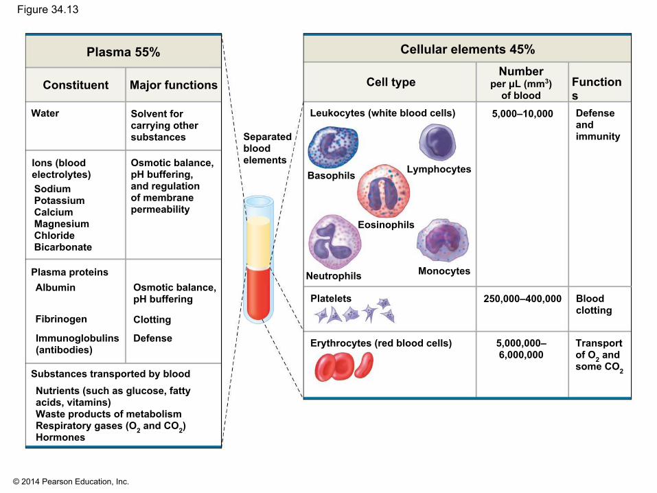
Blood Composition and Function
▪ Blood is a connective tissue consisting of cells suspended in a liquid matrix called plasma
▪ The cellular elements occupy about 45% of the volume of blood
© 2014 Pearson Education, Inc.
Figure 34.13
Separatedbloodelements
Solvent forcarrying othersubstances
Plasma 55% Cellular elements 45%
Constituent Major functions
Osmotic balance,pH buffering,and regulationof membranepermeability
Water
Ions (bloodelectrolytes)SodiumPotassiumCalciumMagnesiumChlorideBicarbonate
Osmotic balance,pH buffering
Clotting
Defense
Fibrinogen
Plasma proteinsAlbumin
Immunoglobulins(antibodies)
Substances transported by bloodNutrients (such as glucose, fattyacids, vitamins)Waste products of metabolismRespiratory gases (O2 and CO2)Hormones
Functions
Leukocytes (white blood cells)
Transportof O2 and some CO2
Cell typeNumber
per μL (mm3)of blood
Basophils Lymphocytes
Eosinophils
Neutrophils Monocytes
Platelets
Erythrocytes (red blood cells)
250,000–400,000
5,000,000– 6,000,000
Bloodclotting
5,000–10,000 Defenseandimmunity
© 2014 Pearson Education, Inc.
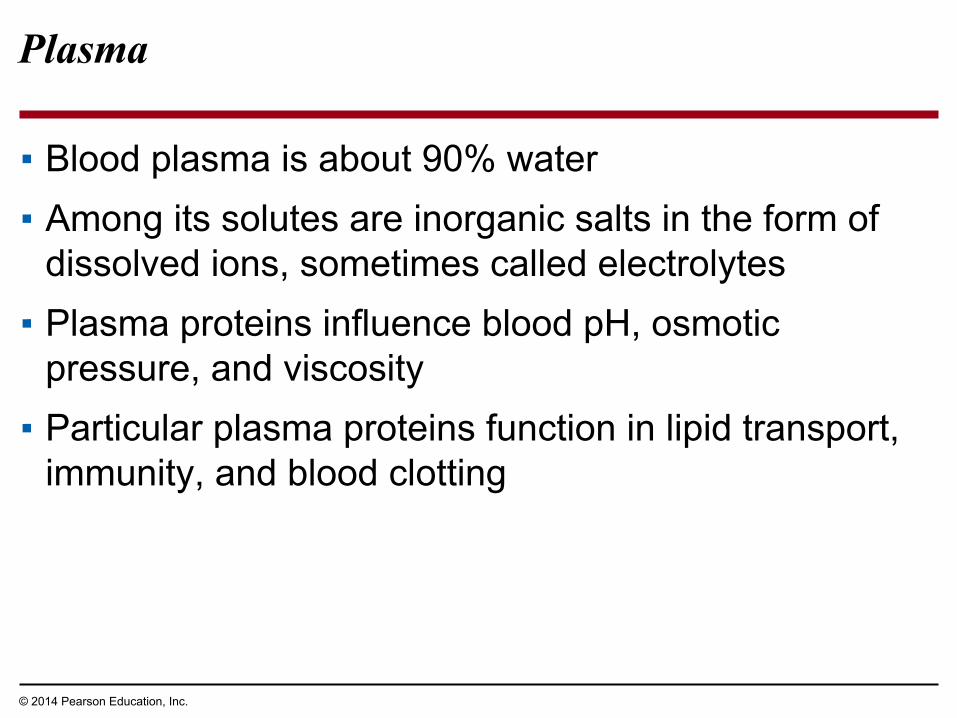
Plasma
▪ Blood plasma is about 90% water▪ Among its solutes are inorganic salts in the form of
dissolved ions, sometimes called electrolytes▪ Plasma proteins influence blood pH, osmotic
pressure, and viscosity▪ Particular plasma proteins function in lipid transport,
immunity, and blood clotting

© 2014 Pearson Education, Inc.
Cellular Elements
▪ Blood contains two classes of cells▪ Red blood cells (erythrocytes) transport O2
▪ White blood cells (leukocytes) function in defense
▪ Platelets, a third cellular element, are fragments of cells that are involved in clotting

© 2014 Pearson Education, Inc.
Figure 34.14
Stem cells(in bone marrow)
Basophils
Lymphocytes
Eosinophils
Neutrophils
MonocytesPlatelets
Erythrocytes
Myeloidstem cells
Lymphoidstem cells
B cells T cells

© 2014 Pearson Education, Inc.
Erythrocytes▪ Red blood cells, or erythrocytes, are by far the most
numerous blood cells▪ They contain hemoglobin, the iron-containing
protein that transports O2
▪ Each molecule of hemoglobin binds up to four molecules of O2
▪ In mammals, mature erythrocytes lack nuclei

© 2014 Pearson Education, Inc.
▪ Sickle-cell disease is caused by abnormal hemoglobin that polymerizes into aggregates
▪ The aggregates can distort an erythrocyte into a sickle shape

© 2014 Pearson Education, Inc.
▪ Through a person’s life, multipotent stem cells replace the worn-out cellular elements of blood
▪ Erythrocytes circulate for about 120 days before they are replaced
▪ Stem cells that produce red blood cells and platelets are located in red marrow of bones like the ribs, vertebrae, sternum, and pelvis

© 2014 Pearson Education, Inc.
Leukocytes▪ There are five major types of white blood cells, or
leukocytes▪ They function in defense by engulfing bacteria and
debris or by mounting immune responses against foreign substances
▪ They are found both in and outside of the circulatory system

© 2014 Pearson Education, Inc.
Platelets▪ Platelets are fragments of cells and function in
blood clotting
© 2014 Pearson Education, Inc.
Blood Clotting
▪ Coagulation is the formation of a solid clot from liquid blood
▪ A cascade of complex reactions converts inactive fibrinogen to fibrin, which forms the framework of a clot
▪ A blood clot formed within a blood vessel is called a thrombus and can block blood flow

© 2014 Pearson Education, Inc.
Figure 34.15
PlateletPlateletplug
Collagenfibers
PlateletsClotting factors from:
Damaged cellsPlasma (factors include calcium, vitamin K)
Fibrin
Thrombin
Fibrinogen
Prothrombin
Enzymatic cascade
Fibrin clot
Fibrin clotformation
Red blood cell 5 μm
1 2 3

© 2014 Pearson Education, Inc.
Cardiovascular Disease
▪ Cardiovascular diseases are disorders of the heart and the blood vessels
▪ Cardiovascular diseases account for more than half the deaths in the United States
▪ Cholesterol, a steroid, helps maintain normal membrane fluidity

© 2014 Pearson Education, Inc.
▪ Low-density lipoprotein (LDL) delivers cholesterol to cells for membrane production
▪ High-density lipoprotein (HDL) scavenges excess cholesterol for return to the liver
▪ Risk for heart disease increases with a high LDL to HDL ratio
▪ Inflammation is also a factor in cardiovascular disease

© 2014 Pearson Education, Inc.
Atherosclerosis, Heart Attacks, and Stroke
▪ One type of cardiovascular disease, atherosclerosis, is caused by the buildup of fatty deposits within arteries
▪ A fatty deposit is called a plaque; as it grows, the artery walls become thick and stiff and the obstruction of the artery increases

© 2014 Pearson Education, Inc.
Figure 34.16
Endothelium
Lumen
Plaque
Blood clot

© 2014 Pearson Education, Inc.
▪ A heart attack, or myocardial infarction, is the death of cardiac muscle tissue resulting from blockage of one or more coronary arteries
▪ Coronary arteries supply oxygen-rich blood to the heart muscle
▪ A stroke is the death of nervous tissue in the brain, usually resulting from rupture or blockage of arteries in the head
▪ Angina pectoris is caused by partial blockage of the coronary arteries and may cause chest pain

© 2014 Pearson Education, Inc.
Risk Factors and Treatment of Cardiovascular Disease
▪ A high LDL to HDL ratio increases the risk of cardiovascular disease
▪ The proportion of LDL relative to HDL is increased by smoking and consumption of trans fats and decreased by exercise
▪ Drugs called statins reduce LDL levels and risk of heart attacks

© 2014 Pearson Education, Inc.
▪ Inflammation plays a role in atherosclerosis and thrombus formation
▪ Aspirin inhibits inflammation and reduces the risk of heart attacks and stroke
▪ Hypertension (high blood pressure) contributes to the risk of heart attack and stroke
▪ Hypertension can be reduced by dietary changes, exercise, medication, or some combination of these

© 2014 Pearson Education, Inc.
Concept 34.5: Gas exchange occurs across specialized respiratory surfaces
▪ Gas exchange is the uptake of molecular O2 from the environment and the discharge of CO2 to the environment

© 2014 Pearson Education, Inc.
Partial Pressure Gradients in Gas Exchange
▪ Partial pressure is the pressure exerted by a particular gas in a mixture of gases
▪ For example, the atmosphere is 21% O2, by volume, so the partial pressure of O2 (PO2) is 0.21 × the atmospheric pressure

© 2014 Pearson Education, Inc.
▪ Partial pressures also apply to gases dissolved in liquid, such as water
▪ When water is exposed to air, an equilibrium is reached in which the partial pressure of each gas is the same in the water and the air
▪ A gas always undergoes net diffusion from a region of higher partial pressure to a region of lower partial pressure
© 2014 Pearson Education, Inc.
Respiratory Media
▪ O2 is plentiful in air, and breathing air is relatively easy
▪ In a given volume, there is less O2 available in water than in air
▪ Obtaining O2 from water requires greater energy expenditure than air breathing
▪ Aquatic animals have a variety of adaptations to improve efficiency in gas exchange

© 2014 Pearson Education, Inc.
Figure 34.17
Coelom
Tube foot
Gills
(b) Sea star(a) Marine worm
Parapodium (functions as gill)

© 2014 Pearson Education, Inc.
Respiratory Surfaces
▪ Gas exchange across respiratory surfaces takes place by diffusion
▪ Respiratory surfaces tend to be large and thin and are always moist
▪ Respiratory surfaces vary by animal and can include the skin, gills, tracheae, and lungs

© 2014 Pearson Education, Inc.
Gills in Aquatic Animals
▪ Gills are outfoldings of the body that create a large surface area for gas exchange
▪ Ventilation is the movement of the respiratory medium over the respiratory surface
▪ Ventilation maintains the necessary partial pressure gradients of O2 and CO2 across the gills
© 2014 Pearson Education, Inc.
▪ Aquatic animals move through water or move water over their gills for ventilation
▪ Fish gills use a countercurrent exchange system, where blood flows in the opposite direction to water passing over the gills
▪ Blood is always less saturated with O2 than the water it meets
▪ Countercurrent exchange mechanisms are remarkably efficient

© 2014 Pearson Education, Inc.
Figure 34.18
Lamella
Water flow
Countercurrent exchange
O2-poor blood
Gill filaments
Operculum
Gillarch
Waterflow
Gill arch
Bloodvessels
O2-rich blood
Blood flow
PO2 (mm Hg) in blood
PO2 (mm Hg) in water
Netdiffusionof O2
140 110 80 50 30
150 120 90 60 30

© 2014 Pearson Education, Inc.
Tracheal Systems in Insects
▪ The tracheal system of insects consists of a network of air tubes that branch throughout the body
▪ The tracheal system can transport O2 and CO2 without the participation of the animal’s open circulatory system
▪ Larger insects must ventilate their tracheal system to meet O2 demands

© 2014 Pearson Education, Inc.
Figure 34.19Tracheoles
Muscle fiberMitochondria
Tracheae
Air sacs
External opening
Airsac Tracheole
Trachea
Air2.
5 μm
Body
cell

© 2014 Pearson Education, Inc.
Lungs
▪ Lungs are an infolding of the body surface, usually divided into numerous pockets
▪ The circulatory system (open and closed) transports gases between the lungs and the rest of the body
▪ The use of lungs for gas exchange varies among vertebrates that lack gills

© 2014 Pearson Education, Inc.
Mammalian Respiratory Systems: A Closer Look
▪ A system of branching ducts conveys air to the lungs▪ Air inhaled through the nostrils is warmed,
humidified, and sampled for odors▪ The pharynx directs air to the lungs and food to the
stomach▪ Swallowing tips the epiglottis over the glottis in the
pharynx to prevent food from entering the trachea
© 2014 Pearson Education, Inc.
▪ Air passes through the pharynx, larynx, trachea, bronchi, and bronchioles to the alveoli, where gas exchange occurs
▪ Exhaled air passes over the vocal cords in the larynx to create sounds
▪ Cilia and mucus line the epithelium of the air ducts and move particles up to the pharynx
▪ This “mucus escalator” cleans the respiratory system and allows particles to be swallowed into the esophagus

© 2014 Pearson Education, Inc.
▪ Gas exchange takes place in alveoli, air sacs at the tips of bronchioles
▪ Oxygen diffuses through the moist film of the epithelium and into capillaries
▪ Carbon dioxide diffuses from the capillaries across the epithelium and into the air space

© 2014 Pearson Education, Inc.
Figure 34.20
Bronchiole
Bronchus
Right lungTrachea(Esophagus)Larynx
Pharynx
(Heart)
Terminalbronchiole
Leftlung
Nasalcavity
Capillaries
Alveoli
Dense capillary bedenveloping alveoli(SEM)
Branch ofpulmonary vein(oxygen-richblood)
Branch of pulmonary artery (oxygen-poorblood)
50 μm
Diaphragm
© 2014 Pearson Education, Inc.
▪ Alveoli lack cilia and are susceptible to contamination▪ Secretions called surfactants coat the surface of
the alveoli▪ Preterm babies lack surfactant and are vulnerable to
respiratory distress syndrome; treatment is provided by artificial surfactants

© 2014 Pearson Education, Inc.
Figure 34.21
Deaths fromother causes
RDS deaths
Body mass of infant<1,200 g >1,200 g
(n = 9) (n = 0) (n = 29)
(n = 9)
Surf
ace
tens
ion
(dyn
es/c
m)
Results
10
20
30
40
0
What causes respiratory distress syndrome?
© 2014 Pearson Education, Inc.
Concept 34.6: Breathing ventilates the lungs
▪ The process that ventilates the lungs is breathing, the alternate inhalation and exhalation of air

© 2014 Pearson Education, Inc.
▪ An amphibian such as a frog ventilates its lungs by positive pressure breathing, which forces air down the trachea
▪ Birds have eight or nine air sacs that function as bellows that keep air flowing through the lungs
▪ Air passes through the lungs of birds in one direction only
▪ Passage of air through the entire system—lungs and air sacs—requires two cycles in inhalation and exhalation
© 2014 Pearson Education, Inc.
How a Mammal Breathes
▪ Mammals ventilate their lungs by negative pressure breathing, which pulls air into the lungs
▪ Lung volume increases as the rib muscles and diaphragm contract
▪ The tidal volume is the volume of air inhaled with each breath

© 2014 Pearson Education, Inc.
Figure 34.22
Inhalation:Diaphragm contracts
(moves down).
Diaphragm
Exhalation:Diaphragm relaxes
(moves up).
Lung
Airinhaled.
Airexhaled.
Rib cageexpands asrib musclescontract.
Rib cage getssmaller asrib musclesrelax.
1 2

© 2014 Pearson Education, Inc.
▪ The maximum tidal volume is the vital capacity▪ After exhalation, a residual volume of air remains
in the lungs▪ Each inhalation mixes fresh air with oxygen-depleted
residual air▪ As a result, the maximum PO2 in alveoli is
considerably less than in the atmosphere

© 2014 Pearson Education, Inc.
Control of Breathing in Humans
▪ In humans, the main breathing control center consists of neural circuits in the medulla oblongata, near the base of the brain
▪ The medulla regulates the rate and depth of breathing in response to pH changes in the cerebrospinal fluid
▪ The medulla adjusts breathing rate and depth to match metabolic demands

© 2014 Pearson Education, Inc.
Figure 34.23-4
Carotidarteries
Response:Signals frommedulla to ribmuscles anddiaphragmincrease rateand depth ofventilation.
Homeostasis:Blood pH of about 7.4
CO2 leveldecreases. Stimulus:
Rising level of CO2in tissues lowers
blood pH.
Sensor/controlcenter:
Aorta
Cerebro-spinalfluid
Medullaoblongata
© 2014 Pearson Education, Inc.
▪ Sensors in the aorta and carotid arteries monitor O2 and CO2 concentrations in the blood
▪ These sensors exert secondary control over breathing
© 2014 Pearson Education, Inc.
Concept 34.7: Adaptations for gas exchange include pigments that bind and transport gases
▪ The metabolic demands of many organisms require that the blood transport large quantities of O2 and CO2

© 2014 Pearson Education, Inc.
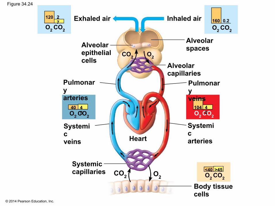
Coordination of Circulation and Gas Exchange
▪ Blood arriving in the lungs has a low PO2 and a high PCO2 relative to air in the alveoli
▪ In the alveoli, O2 diffuses into the blood and CO2 diffuses into the air
▪ In tissue capillaries, partial pressure gradients favor diffusion of O2 into the interstitial fluids and CO2 into the blood
▪ Specialized carrier proteins play a vital role in this process
© 2014 Pearson Education, Inc.
Figure 34.24
Alveolarepithelialcells
Alveolarspaces
Alveolarcapillaries
Inhaled airExhaled air
Pulmonaryveins
Systemicarteries
Pulmonaryarteries
Systemicveins
Systemiccapillaries
Heart
CO2 O2
Body tissuecells
O2 CO2
120 27
O2 CO2
40 45
O2 CO2
160 0.2
O2 CO2
104 40
O2 CO2
<40 >45
O2CO2
© 2014 Pearson Education, Inc.
Respiratory Pigments
▪ Respiratory pigments circulate in blood or hemolymph and greatly increase the amount of oxygen that is transported
▪ A variety of respiratory pigments have evolved among animals
▪ These mainly consist of a metal bound to a protein

© 2014 Pearson Education, Inc.
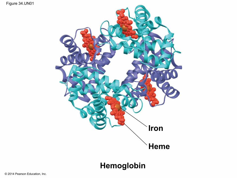
▪ The respiratory pigment of almost all vertebrates and many invertebrates is hemoglobin
▪ A single hemoglobin molecule can carry four molecules of O2, one molecule for each iron- containing heme group
▪ Hemoglobin binds oxygen reversibly, loading it in the gills or lungs and releasing it in other parts of the body
© 2014 Pearson Education, Inc.
Figure 34.UN01
Hemoglobin
Heme
Iron

© 2014 Pearson Education, Inc.
▪ Hemoglobin binds O2 cooperatively▪ When O2 binds one subunit, the others change shape
slightly, resulting in their increased affinity for oxygen▪ When one subunit releases O2, the others release
their bound O2 more readily▪ Cooperativity can be demonstrated by the dissociation
curve for hemoglobin

© 2014 Pearson Education, Inc.
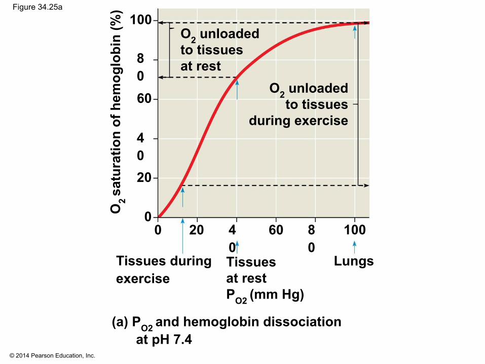
Figure 34.25
pH 7.4
PO2 (mm Hg)
pH 7.2
Hemoglobinretains lessO2 at lower pH(higher CO2concentration
Tissuesat restPO2 (mm Hg)
Tissues duringexercise
Lungs
O2 unloadedto tissues
during exercise
O2 unloadedto tissuesat rest
(b) pH and hemoglobin dissociation(a) PO2 and hemoglobin dissociationat pH 7.4
O2
satu
ratio
n of
hem
oglo
bin
(%)
O2
satu
ratio
n of
hem
oglo
bin
(%) 100
80
60
40
20
0
100
80
60
40
20
0100806040200 100806040200
© 2014 Pearson Education, Inc.
Figure 34.25a
Tissuesat restPO2 (mm Hg)
Tissues duringexercise
Lungs
O2 unloadedto tissues
during exercise
O2 unloadedto tissuesat rest
(a) PO2 and hemoglobin dissociationat pH 7.4
O2
satu
ratio
n of
hem
oglo
bin
(%)
100
8060
4020
01008
0604
0200

© 2014 Pearson Education, Inc.
▪ CO2 produced during cellular respiration lowers blood pH and decreases the affinity of hemoglobin for O2; this is called the Bohr shift
▪ Hemoglobin also assists in preventing harmful changes in blood pH and plays a minor role in CO2 transport

© 2014 Pearson Education, Inc.
Figure 34.25b
pH 7.4
PO2 (mm Hg)
pH 7.2
Hemoglobinretains lessO2 at lower pH(higher CO2concentration
(b) pH and hemoglobin dissociation
O2
satu
ratio
n of
hem
oglo
bin
(%)
100
8060
4020
01008
0604
0200

© 2014 Pearson Education, Inc.
Carbon Dioxide Transport
▪ Most of the CO2 from respiring cells diffuses into the blood and is transported in blood plasma, bound to hemoglobin or as bicarbonate ions (HCO3
–)
© 2014 Pearson Education, Inc.
Respiratory Adaptations of Diving Mammals
▪ Diving mammals have evolutionary adaptations that allow them to perform extraordinary feats
▪ For example, Weddell seals in Antarctica can remain underwater for 20 minutes to an hour
▪ For example, elephant seals can dive to 1,500 m and remain underwater for 2 hours
▪ These animals have a high blood to body volume ratio
© 2014 Pearson Education, Inc.
▪ Deep-diving air breathers can store large amounts of O2
▪ Oxygen can be stored in their muscles in myoglobin proteins
▪ Diving mammals also conserve oxygen by▪ Changing their buoyancy to glide passively▪ Decreasing blood supply to muscles▪ Deriving ATP in muscles from fermentation once
oxygen is depleted