gastointestin al system or digestive system or gut
TRANSCRIPT

GASTOINTESTINAL SYSTEM or
DIGESTIVE SYSTEM or GUT
⚫ Figures in this ppt are from guyton, ganong, best & taylor, tortora and google images
Digestive system
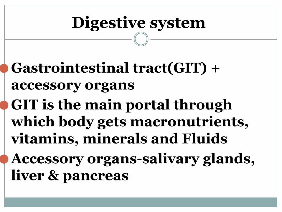
⚫Gastrointestinal tract(GIT) + accessory organs
⚫GIT is the main portal through which body gets macronutrients, vitamins, minerals and Fluids
⚫Accessory organs-salivary glands, liver & pancreas
GIT
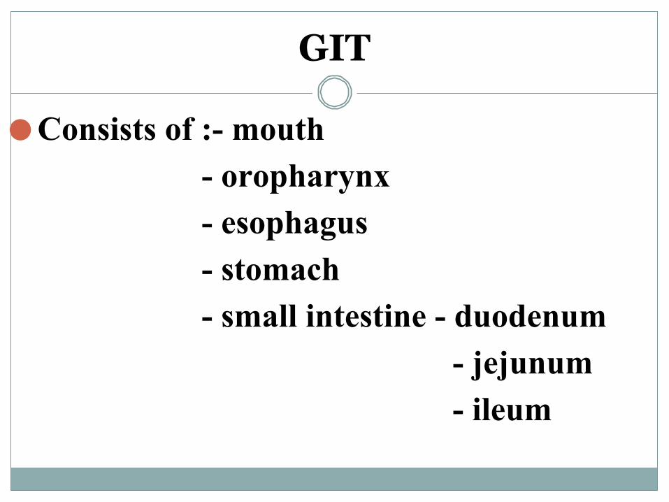
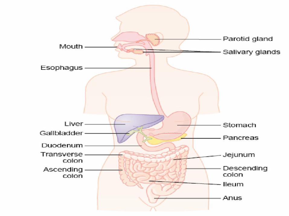
⚫Consists of :- mouth - oropharynx - esophagus - stomach - small intestine - duodenum - jejunum - ileum

GIT
LARGE INTESTINE: - Ceacum - Ascending colon - Transverse colon - Descending colon - Sigmoid colon - Rectum - Anal canal - Anus
Accessory glands
⚫ Salivary glands⚫Pancreas⚫Liver
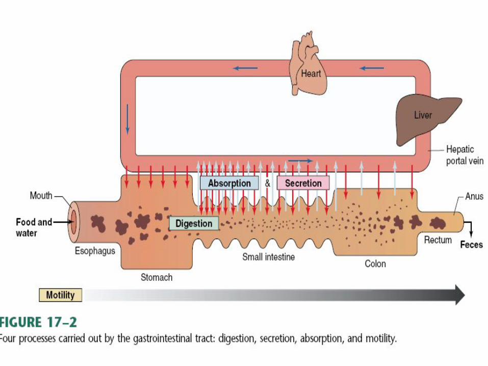
Processes
The GIT breaks down food and supply, the body with water, nutrients and electrolytes needed to sustain life & removes unwanted substances.This is brought about by following processes:
1. ingestion 2. digestion 3. absorption 4. egestion

INGESTION
▪ - Placing food into the mouth▪ - chewing the food into smaller pieces (mastication)▪ - Moistening the food with salivary secretions▪ - swallowing the food (deglutition)
DIGESTION-Mechanical & chemical It is the breaking larger pieces into small ones &
splitting of large chemical compounds into simpler substances that can be used by the body.
Starches -------- monosaccharides (amylases) Proteins --------- dipeptides and amino acids. (proteases)
Fats -------- monosaccharides and FFA (lipases, esterases)
ABSORPTION & ASIMILATION
⚫ Simpler substances produced by digestion as well as nutrients, water and electrolytes are transported from the GIT to the circulation & through portal circulation to liver & then stored or distributed to different tissues for use & storage.
Egestion
⚫ The undigested food particles, unabsorbed food along with various secretions and sloughed-off epithelial cells from tip of villi , constitute faeces which are voided through the anus at periodic intervals.
Functions of the GIT⚫ 1. DIGESTION & ABSORPTION OF FOOD. the primary function of GIT.⚫ 2. EXCRETION OF WASTE MATERIALS.
⚫ 3. FOOD & ELECTOLYTE BALANCE - an adult consumes -1 kg of food -1.5 lit of fluid /day additional - 7 lit secreted in the form of secretions. ⚫ Thus intestine is presented with 8.5 lit of fluid/day.⚫ 99% fluid + 90% of solids are absorbed from SI & LI along with
electrolytes.⚫ So 100ml fluid+ 100g of solids ----excreted/day.⚫ Diarrhoea --- absorption decreases or secretion increases.
Functions of the GIT-contd
4.IMMUNITY
GIT is open at both ends--- organisms enters easilyMALTs or GALTs protect the body from pathogens.
MALTS - payer’s patches - diffuse population of immune cells. acidic secretions of the stomach ---helps killing
organisms..
Functions of the GIT-contd
5. INTESTINAL BACTERIAL FLORA-non pathogenic bacteria inhabit at intestine, form the intestinal bacterial floraIt has many essential intestinal functions-
1 - Epi. Cell permeability to electrolytes &water. 2 - Synthesis & absorption of vitamins 3 - Stimulation of enzymatic activity, peristalsis & mucus secretion # decreased flora- impairs intestinal functions # Increased flora- increases susceptibility to diarrhoea & steatorrhoea.
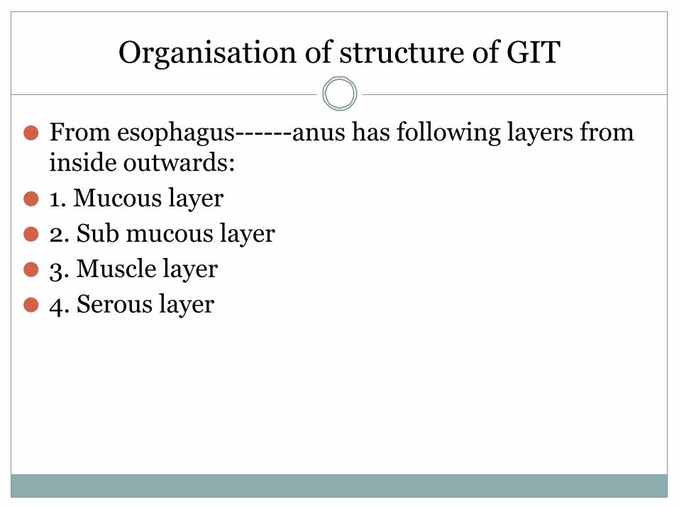
Organisation of structure of GIT
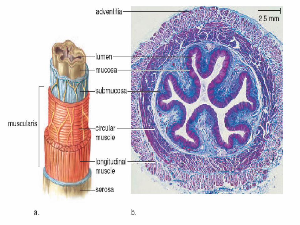
⚫ From esophagus------anus has following layers from inside outwards:
⚫ 1. Mucous layer⚫ 2. Sub mucous layer⚫ 3. Muscle layer⚫ 4. Serous layer
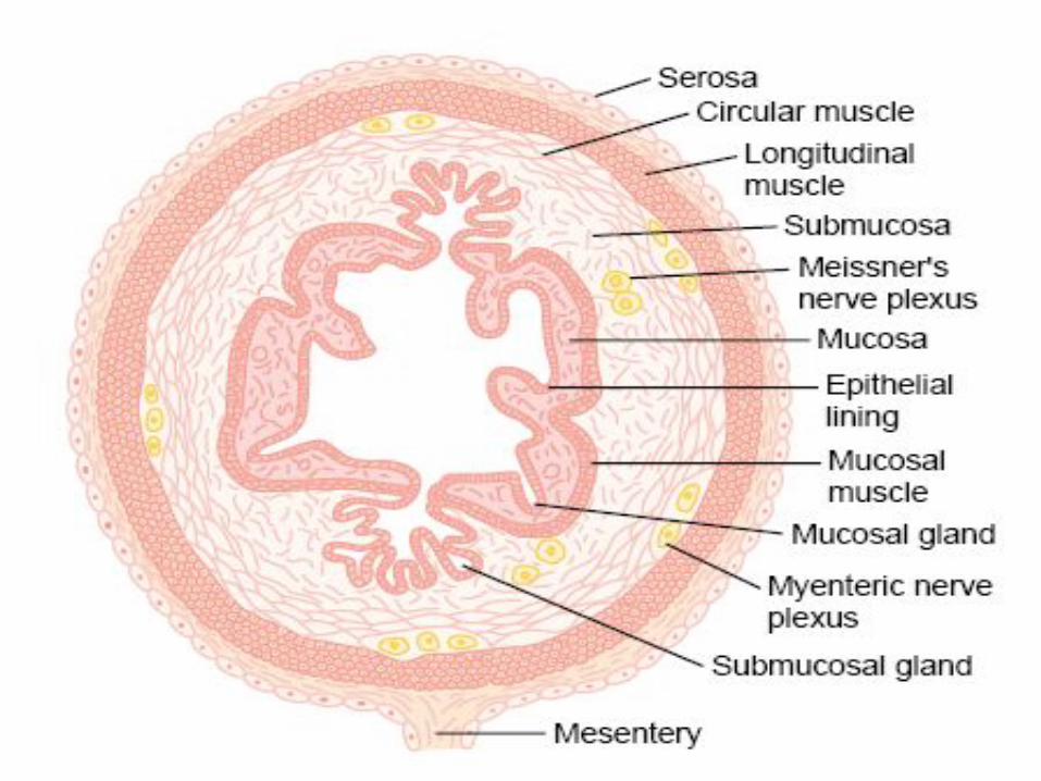
1. Mucous layer
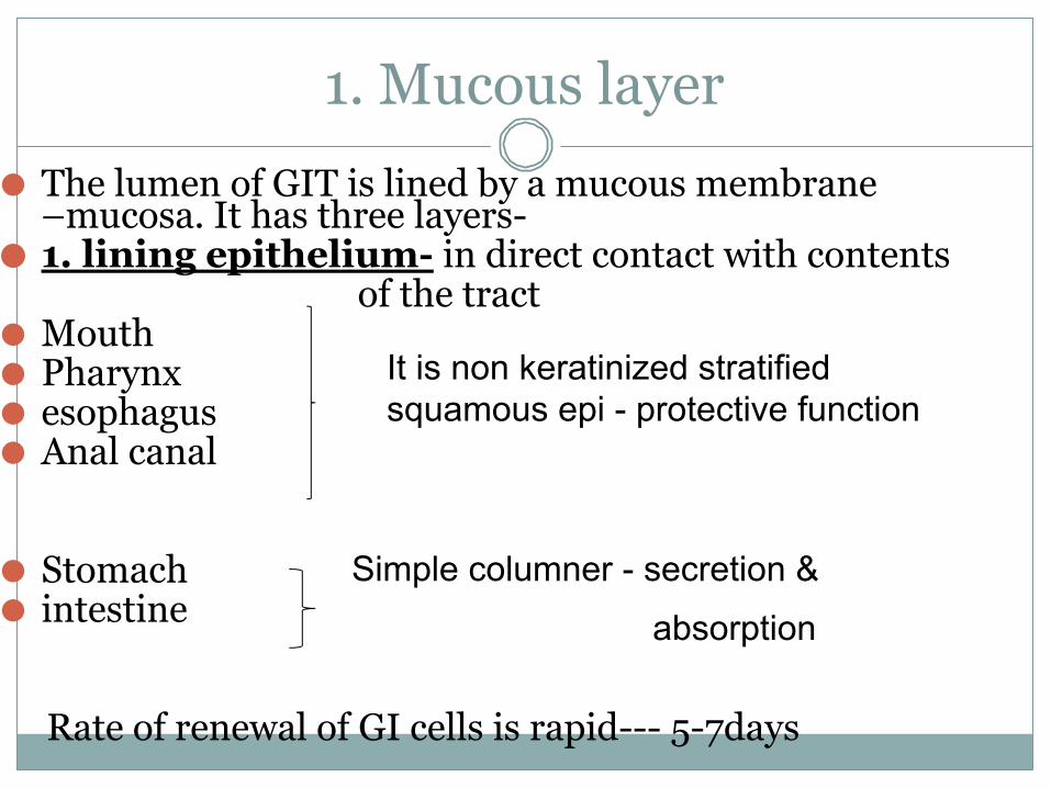
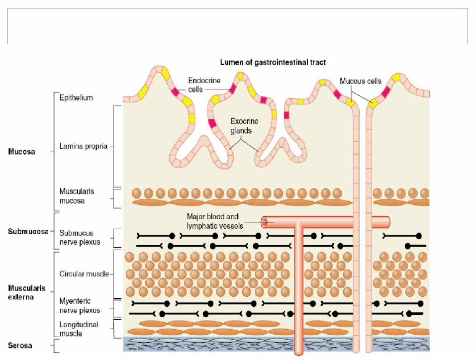
⚫ The lumen of GIT is lined by a mucous membrane –mucosa. It has three layers-
⚫ 1. lining epithelium- in direct contact with contents of the tract⚫ Mouth⚫ Pharynx ⚫ esophagus⚫ Anal canal
⚫ Stomach⚫ intestine
Rate of renewal of GI cells is rapid--- 5-7days
It is non keratinized stratified squamous epi - protective function
Simple columner - secretion &
absorption
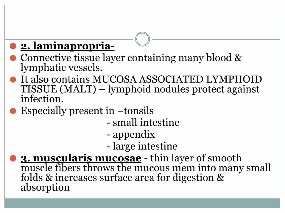
⚫ 2. laminapropria-⚫ Connective tissue layer containing many blood &
lymphatic vessels.⚫ It also contains MUCOSA ASSOCIATED LYMPHOID
TISSUE (MALT) – lymphoid nodules protect against infection.
⚫ Especially present in –tonsils - small intestine - appendix - large intestine⚫ 3. muscularis mucosae - thin layer of smooth
muscle fibers throws the mucous mem into many small folds & increases surface area for digestion & absorption
2. Sub mucous layer
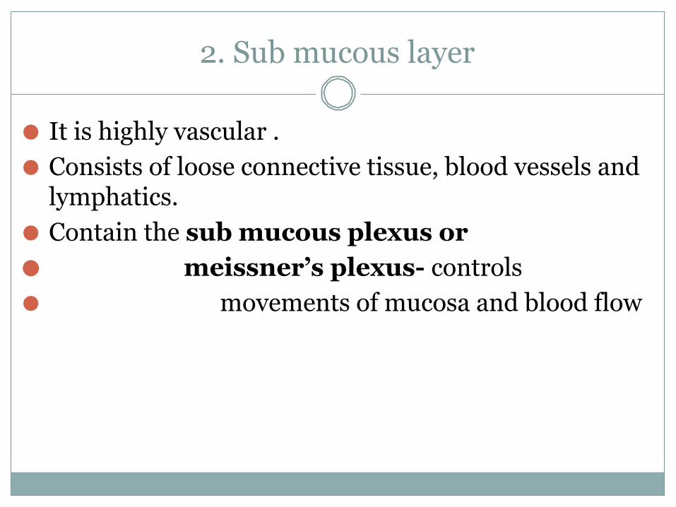
⚫ It is highly vascular .⚫ Consists of loose connective tissue, blood vessels and
lymphatics.⚫ Contain the sub mucous plexus or⚫ meissner’s plexus- controls⚫ movements of mucosa and blood flow
3. Muscular layer
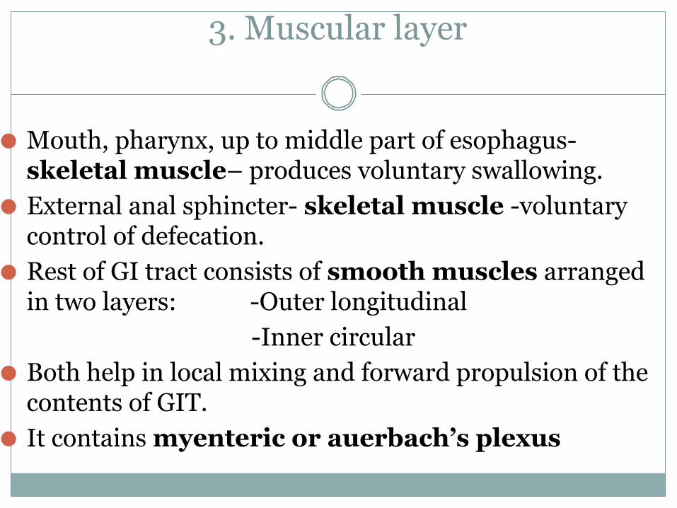
⚫ Mouth, pharynx, up to middle part of esophagus- skeletal muscle– produces voluntary swallowing.
⚫ External anal sphincter- skeletal muscle -voluntary control of defecation.
⚫ Rest of GI tract consists of smooth muscles arranged in two layers: -Outer longitudinal
-Inner circular⚫ Both help in local mixing and forward propulsion of the
contents of GIT.⚫ It contains myenteric or auerbach’s plexus
4. Serous layer
⚫ Helps in attachment of the gut to the surrounding structures.
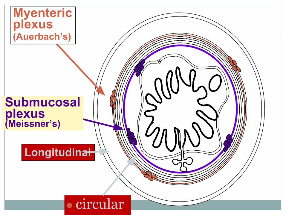
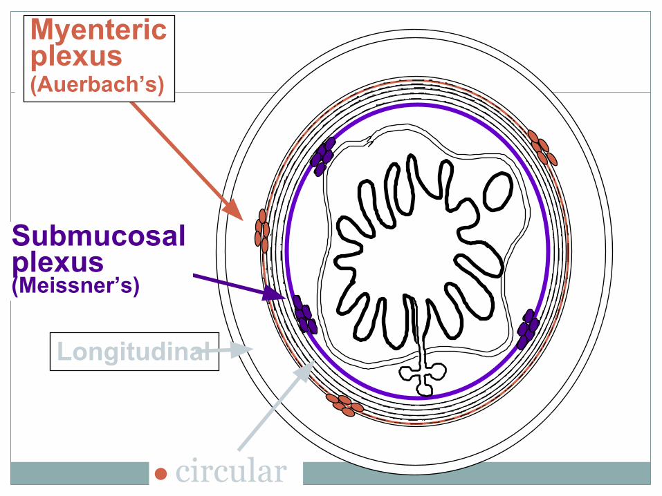
● circular
Longitudinal
Submucosalplexus(Meissner’s)
Myentericplexus(Auerbach’s)
Innervations of GIT
⚫ There are two major networks of nervous system that innervate the GIT:
⚫ INTRINSIC – Enteric nervous system⚫ EXTRINSIC – from ANS
Innervation of the GI tract
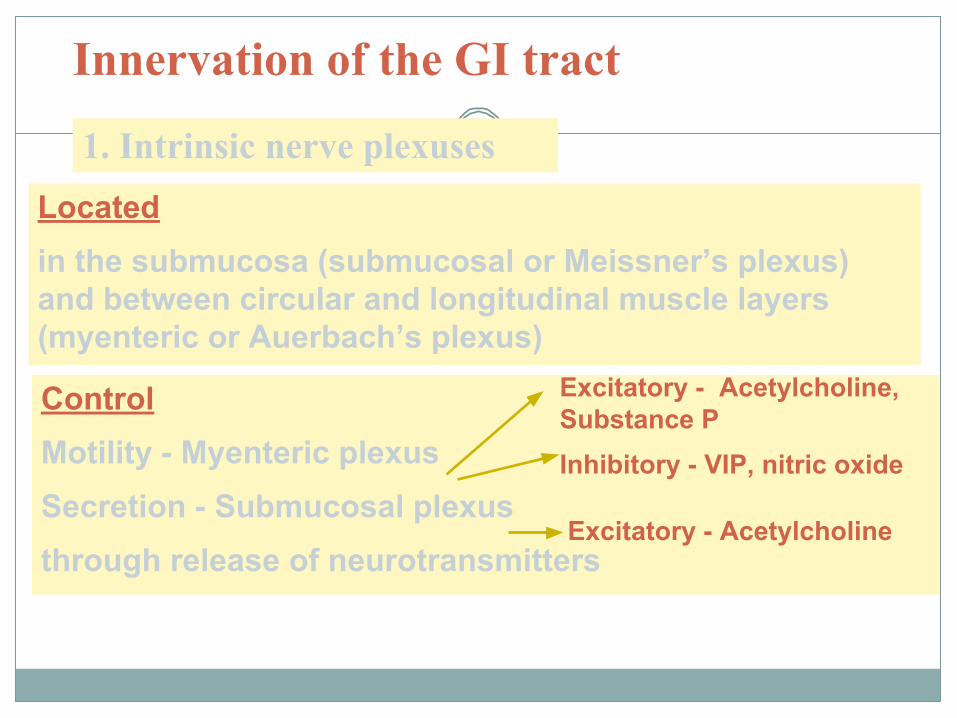
1. Intrinsic nerve plexusesLocated in the submucosa (submucosal or Meissner’s plexus) and between circular and longitudinal muscle layers (myenteric or Auerbach’s plexus)
ControlMotility - Myenteric plexusSecretion - Submucosal plexusthrough release of neurotransmitters
Excitatory - Acetylcholine, Substance PInhibitory - VIP, nitric oxide
Excitatory - Acetylcholine
● circular
Longitudinal
Submucosalplexus(Meissner’s)
Myentericplexus(Auerbach’s)
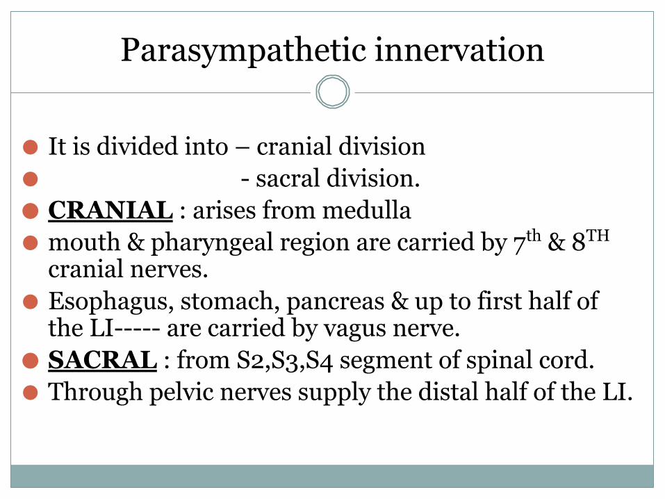
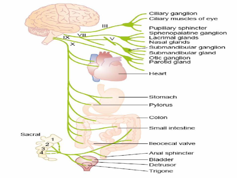
Parasympathetic innervation
⚫ It is divided into – cranial division⚫ - sacral division.⚫ CRANIAL : arises from medulla⚫ mouth & pharyngeal region are carried by 7th & 8TH
cranial nerves.⚫ Esophagus, stomach, pancreas & up to first half of
the LI----- are carried by vagus nerve.⚫ SACRAL : from S2,S3,S4 segment of spinal cord.⚫ Through pelvic nerves supply the distal half of the LI.
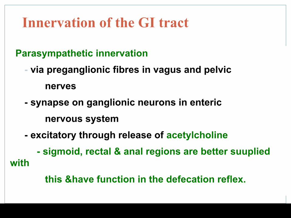
Innervation of the GI tract
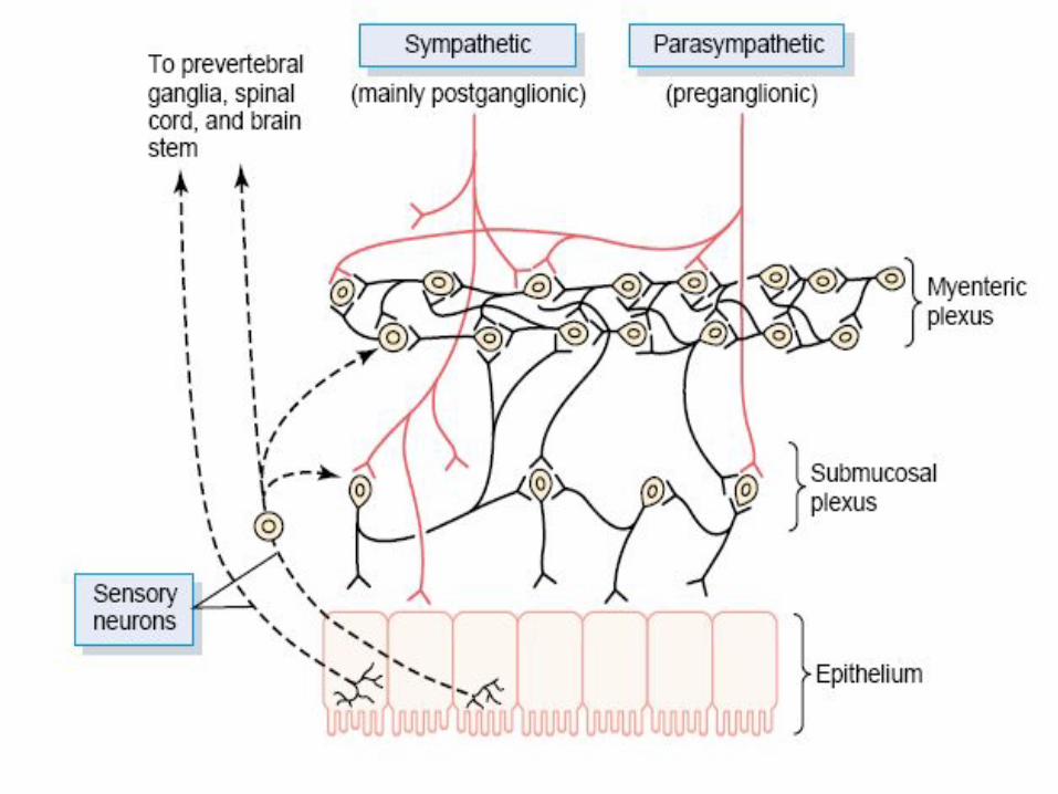
Parasympathetic innervation- via preganglionic fibres in vagus and pelvic
nerves- synapse on ganglionic neurons in enteric
nervous system- excitatory through release of acetylcholine
- sigmoid, rectal & anal regions are better suuplied with this &have function in the defecation reflex.
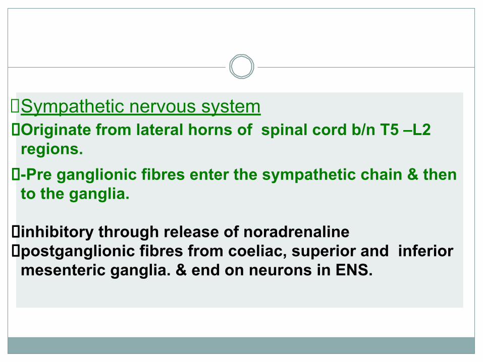
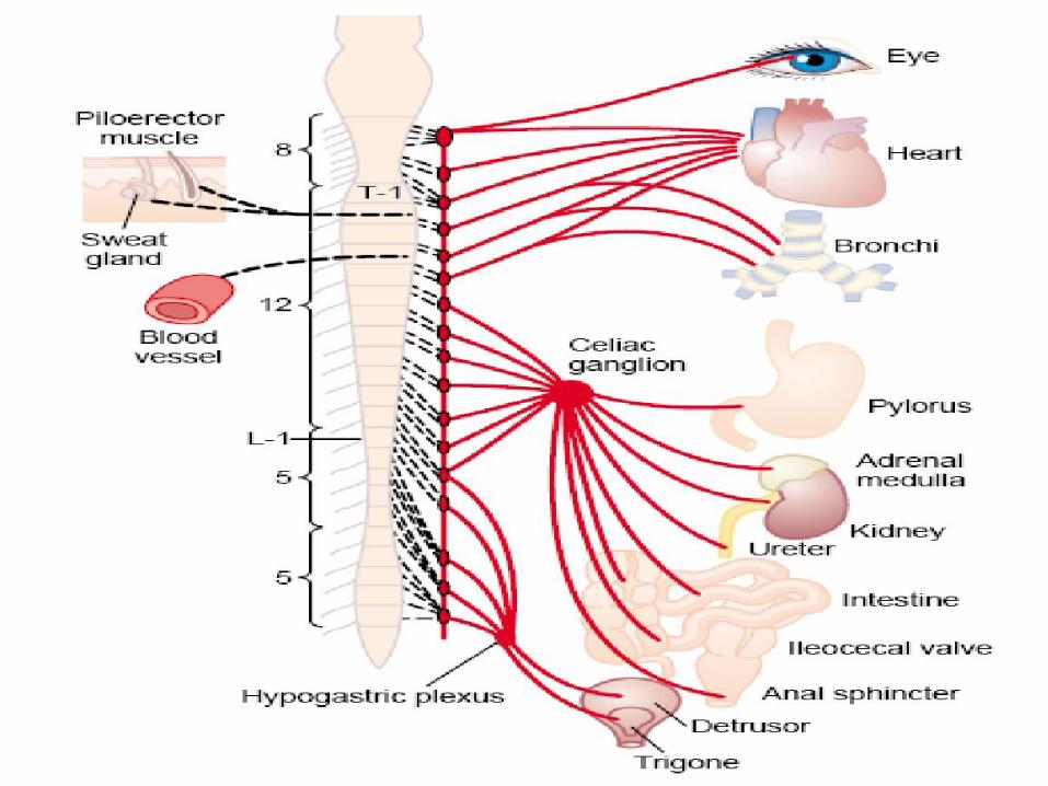
Sympathetic nervous systemOriginate from lateral horns of spinal cord b/n T5 –L2 regions.-Pre ganglionic fibres enter the sympathetic chain & then to the ganglia. inhibitory through release of noradrenalinepostganglionic fibres from coeliac, superior and inferior mesenteric ganglia. & end on neurons in ENS.
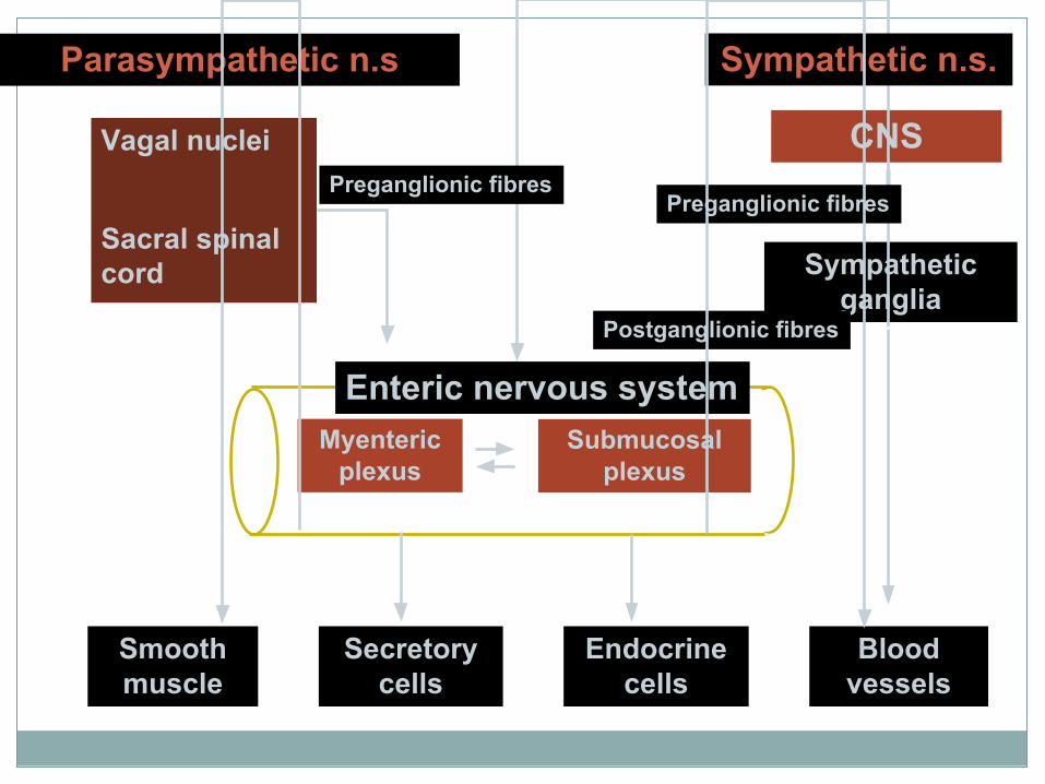
Myenteric plexus
Submucosal plexus
Enteric nervous system
CNS
Sympathetic ganglia
Vagal nuclei
Sacral spinal cord
Preganglionic fibres
Postganglionic fibres
Preganglionic fibres
Parasympathetic n.s Sympathetic n.s.
Smooth muscle
Secretory cells
Blood vessels
Endocrine cells