gastric cancer and adenocarcinoma tumorigenesis · gastritis and metaplasia •h. pylori is often...
TRANSCRIPT

Gastric Cancer and Adenocarcinoma Tumorigenesis:
cellular plasticity and metaplasia in cancer and repair
Jason Mills
Washington University School of Medicine
Some Terms:
• Carcinoma/Adenocarcinoma

Some Terms:
• Carcinoma/Adenocarcinoma
• Metaplasia

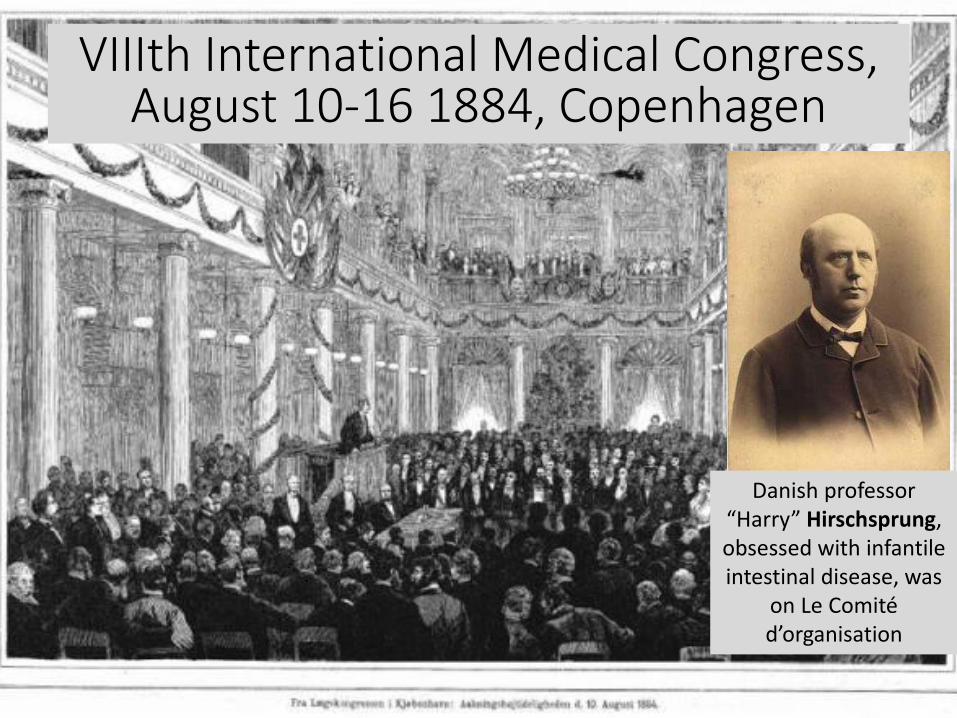
VIIIth International Medical Congress, August 10-16 1884, Copenhagen
Danish professor “Harry” Hirschsprung, obsessed with infantile intestinal disease, was
on Le Comitéd’organisation

Rudolph Virchow

Rudolph Virchow

Rudolph Virchow
•Basically founded the field of pathology
•Prolific in many fields (eg, Virchow’s node, Virchow’s triad)
•Coined many terms (leukemia, embolism, etc.) and started multiple fields
•Key theory (which he popularized from François-Vincent Raspail) was Omnis cellulae cellula
•Thus, if you have weird cells arising in adults, then they must come from existing cells
•Thought Charles Darwin was an “ignoramus” and people who believed in evolution were “fools”

Rudolph Virchow
Take homes from his lecture introducing metaplasia:
•If all cells derive from existing cells, then how do you explain when normal-looking cells show up in the wrong place (not neoplasia, not hyperplasia)
•There are a lot of such “plastischen Prozessen” in the body
•He proposed calling these “Metaplasias”
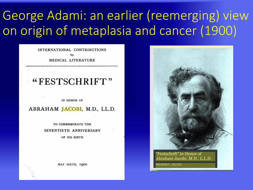
George Adami: an earlier (reemerging) view on origin of metaplasia and cancer (1900)

Adami’s Laws on metaplasia and reversion (slightly paraphrased): 1900
1. “The fully differentiated cells of a tissue proper never arise from cells that arethemselves fully differentiated.”
2. In normal adult tissues, differentiated cells arise by division from “mother”(stem) cells. However, more rarely, functional cells, “by reversion to a moreembryonal type, take on the properties of mother cells.”
3. “Under abnormal conditions, the fully differentiated functioning cells of certaintissues are capable of proliferation and giving rise to cells of like nature, but thisis only after a preliminary reversion to a simpler, more embryonic type.” The fullydifferentiated cell does not normally proliferate.
4. The “energy stored up by the [differentiated] cell may be expended in one of twodirections…either in functional activity or in preparation for proliferation”.Changes in energy usage and differentiation state correspond to structural ormorphological changes in the cell.
5. The more highly differentiated a cell, the more functional it is, the more complexits structure will be. And the more structure it has to scale down the less liable itwill be to undergo reversion.

Adami proposed metaplasia/tumors came from:
1. Embryonic stem cell “rests” which remain latent until they are awoken
2. The mother cells, which remain undifferentiated and maintain activeproliferation
3. “Differentiated cells which reverting to a simpler, more embryonic type, with thisreversion gain the capacity for active and excessive proliferation.

Some Terms:
• Carcinoma/Adenocarcinoma
• Metaplasia
• Stem Cells/Differentiation

1930s-2005: Waddington Landscape predominates

Back to the present: Terms and Concepts OR the metaplasia field
“reverts” back to Adami!
Perspectives:

Key, re(!?)emerging concepts:
• Yamanaka: Differentiated postmitotic cells can reprogram to become proliferative, progenitor cells
• This happens following damage in organs without constitutive stem cells
• Or in organs with stem cells when the constitutive stem cell is insufficient for repair (stomach, intestines)
• The process may be the source of metaplasia, dysplasia, and cancer in adult cancers

Scaling: Even after the cell chooses its final fate, it scales up specific architectural features to perform its physiological function
Mills and Taghert,
Bioessays, 2012

Dedifferentiation/Reversion: A differentiated cell downscales then reverts to an earlier, less differentiated phenotype (becomes a stem cell again)

Transdifferentiation: A differentiated cell downscales, then converts to another differentiated cell type
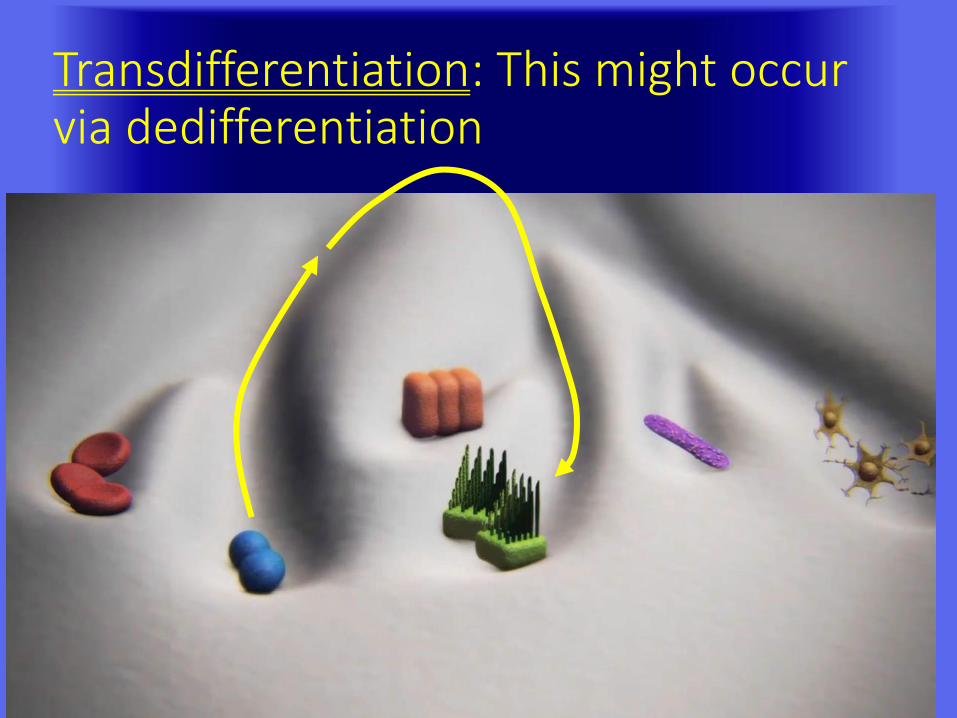
Transdifferentiation: This might occur via dedifferentiation
I propose a Cyclical Hit Threshold Model of cancer initiation: Cycles of metaplasia may allow mutation
storage and unmasking (label-retaining cells are mutation-
retaining cells)
Speculation:

Cycles of dedifferentiation and redifferentiation may lead to accumulation of mutations that cause cancer in adult organs
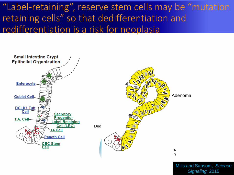
Mills and Sansom, Science
Signaling, 2015
+3d+60d
Mills and Sansom, Science
Signaling, 2015
Inflammation causes
CBC stem cell death
DedifferentiationMutation
blocks
differentiation
Adenoma
“Label-retaining”, reserve stem cells may be “mutation retaining cells” so that dedifferentiation and redifferentiation is a risk for neoplasia

Pancreas: the case has become quickly established that metaplasia arises from differentiated cells that
reenter the cell cycle
Perspectives:
Mills and Sansom, Science
Signaling, 2015
Reviewed in:
Roy and Hebrok, Dev Cell,
2015
Ziv et al., Dev Cell, 2013
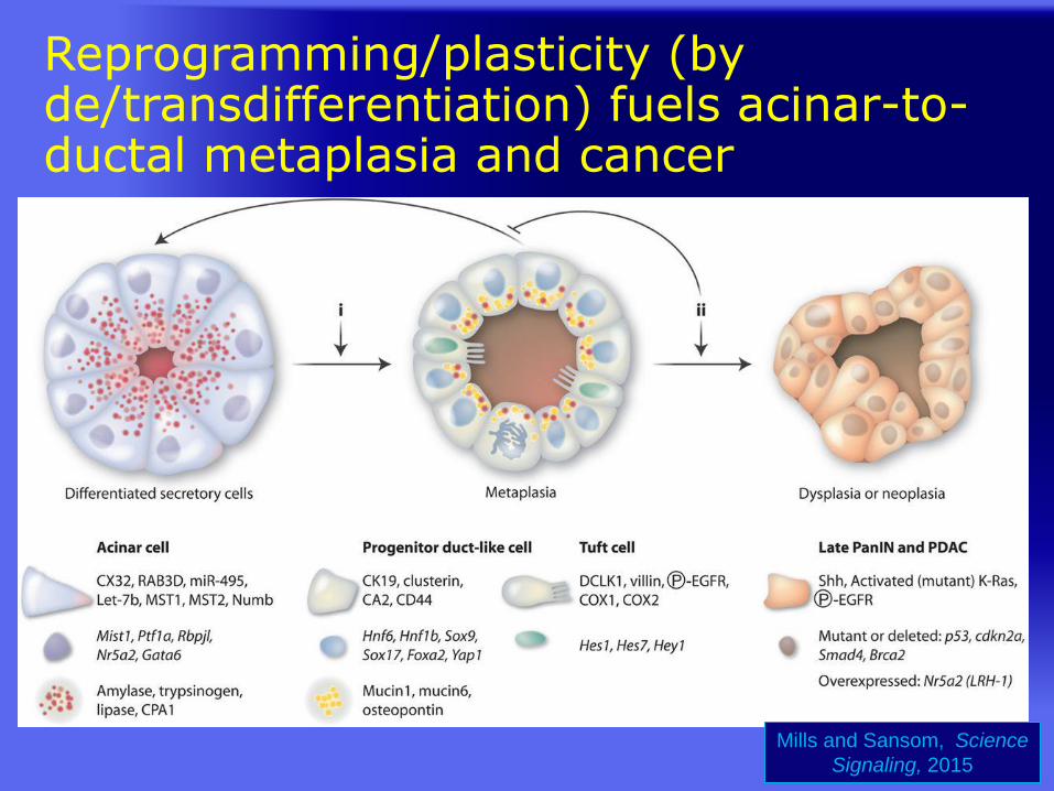
Reprogramming/plasticity (by de/transdifferentiation) fuels acinar-to-ductal metaplasia and cancer
Mills and Sansom, Science
Signaling, 2015

“Downscaling” differentiated cellular features for cell to refocus energy on proliferation: Focus on a key scaling factor
Mills and Sansom, Science
Signaling, 2015

“ I wish to point out what a fruitful field awaits the investigator who wishes to study, with the aid of our present methods, the pathological conditions of the digestive organs and their treatment. Such an investigation is all the more desirable, because clinical study of the same subject (notwithstanding the zeal devoted to it during the last ten years and the results derived therefrom), has to contend with serious difficulties.”
Pavlov, “The Work of the Digestive Glands” J.P. Lipincott & Co., Philadelphia, 1902
What Pavlov was really studying
© Mark Stivers, 2003


Stomach epithelial cell differentiation and disease
Acid-pumping cellDigestive enzyme secreting cell

Stem Cells in the corpus/fundus of the
Stomach: Canonical
Regions:
Cell Type:
Isthmus
Stem Cell
Pit
Pit Cell
Neck
Parietal Cell
Neck Cell
Base
Zymogenic
(Chief) Cell
Gastric Unit
MIST1 zone

Parietal cell atrophy causes regenerative metaplasia, a precursor lesion for gastric cancer
control
Ablation of parietal
cells

Cell of origin for metaplasia?
control
Ablation of parietal
cells
Where do these metaplastic, proliferative basal cells come from?
Some new isthmal stem cell pattern?
The chief cell lineage via dedifferentiation?
(there is evidence for both mechanisms)
Mist1+
Mist1–

Downscaling and Reversion in human
metaplasia
Mouse knockouts are great, but what
about humans?

Carcinoma of the Stomach 3% of cancer deaths in the USA
Pathogenesis• Dietary factors
• Genetic factors
• H. Pylori
• Low socioeconomic status
Hot Spots
Epidemiology:
• Decline in incidence in US relative to colorectal CA and worldwide relative to lung CA
• Still a common cause of cancer death in US, especially male minorities, 3rd most common cause worldwide, and rates are rising again
• Third worst 5 year survival in US (much better in Japan)

Potential Roles of H. pylori and Antecedent Gastritis and Metaplasia
• H. pylori is often termed “necessary but not sufficient” for induction of most gastric CAs
• H. pylori is the most common cause of chronic atrophic gastritis
• Chronic atrophy (via HP or autoimmune gastritis) always associates with metaplasia and is a clear carcinoma precursor state
Cancer

H. Pylori is completely fascinating, but…. I know it can be confusing
• Most colonized people (>1/2 the world population) have no symptoms or occasional gastritis
• ~25% will have a peptic ulcer at some point, treatable by H. pylori eradication
• 1-10% will develop gastric cancer eventually
• (This is why H. pylori occurs multiple times in this lecture!)

modified after Yuasa, Nat Rev Cancer, 2003 Aug 3(8):592-600; Nozaki et al., Gastroenterol., 2008; 134:511-522
Normal
gastric
mucosa
Intestinal-type
gastric Adenocarcinoma
Diffuse-type
gastric carcinoma
Hereditary diffuse-type
gastric carcinoma
Gastric Carcinogenic Pathway
Chronic
gastritis
Atrophic
gastritis
Intestinal
metaplasia
SPEM
Adenoma/
Dysplasia
H. pylori
hereditary
E-
cadherin
mutations
sporadic
E-
cadherin
mutations

Intestinal Metaplasia from Autoimmune Gastritis or H pylori
Parietal cell
Mucous goblet cells
Paneth cells
Proliferative cells

Human metaplasias
Mills lab (Lennerz et al., 2010)

Gastric CarcinomaGross Pathology
Three major growth patterns (different etiologies)Polypoid or fungating
• Resemble right colon carcinomas
• Most common in body and along greater curvature
Ulcerating • Most common in antrum and cardia
• Can be difficult to distinguish from peptic ulcers
Infiltrative (different mechanisms) • Diffuse (linitis plastica)

Gastric CarcinomaUlcerated

Gastric CarcinomaPolypoid/Exophytic/Fungating
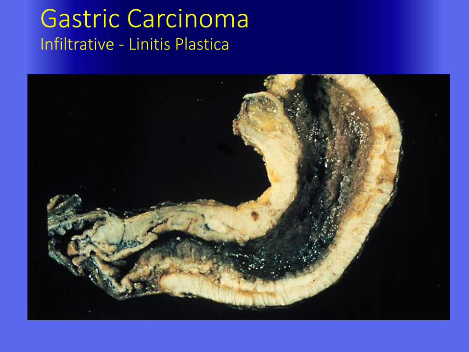
Gastric CarcinomaInfiltrative - Linitis Plastica

Gastric CarcinomaHistological Types and Patterns, molecular mechanisms
Vast majority are adenocarcinomas (>90%)
Two major architectural patterns:• Intestinal: cells look like colorectal cancer cells
and often arises in association with intestinal metaplasia
• Diffuse: signet ring cells (E-cadherin mutations predominate, some familial forms)

Gastric Adenocarcinomaintestinal type

Gastric Adenocarcinomaintestinal type

Loss of E-cadherin junction proteins in Hereditary Diffuse Gastric Cancer (HDGC)
Humar et al., Cancer Res. 2007 67:2480

Model for DGC and HDGC development
Humar et al., Cancer Res. 2007 67:2480

Gastric AdenocarcinomaDiffuse, poorly differentiated, signet ring cells
Normal mucous
neck cells


Unleashing the WUCCI: FIB/SEM 3-D imaging and the tools
we have to study metaplasia
Washington University Center for Cellular Imaging (thanks, Paul! and thanks, Duy Tran for introducing
FIB/SEM)

3D Nanoscale – FIB-SEM

FIB-SEM 3-D imaging reveals substantial architectural changes induced by MIST1

Disease: metaplasia (and cancer)
Where do metaplasias come from?
What is the cell-of-origin?
If cancers arise from metaplasia, then that cell might be the origin of cancer, too.

MIST1: is lost early in reprogramming

Cell reprogramming seems to be a conserved, cellular process, like apoptosis or mitosis with distinct stages
Mills and Sansom, Science
Signaling, 2015
Scaling Events

H Pylori-infected Humans with chronic atrophic gastritis: MIST1+ chief cells seem to scale down and acquire TFF2 expression
10/31 biopsy specimens showed focal MIST1/TFF2 colabeling
TFF2 (Neck Cells/SPEM-type
metaplasia)
MIST1
(Mature Chief
Cells, plasma
cells)
Lennerz et al., Amer J
Pathol 2010
~1% of gastric cancers are strongly MIST1+ (n=400 samples)
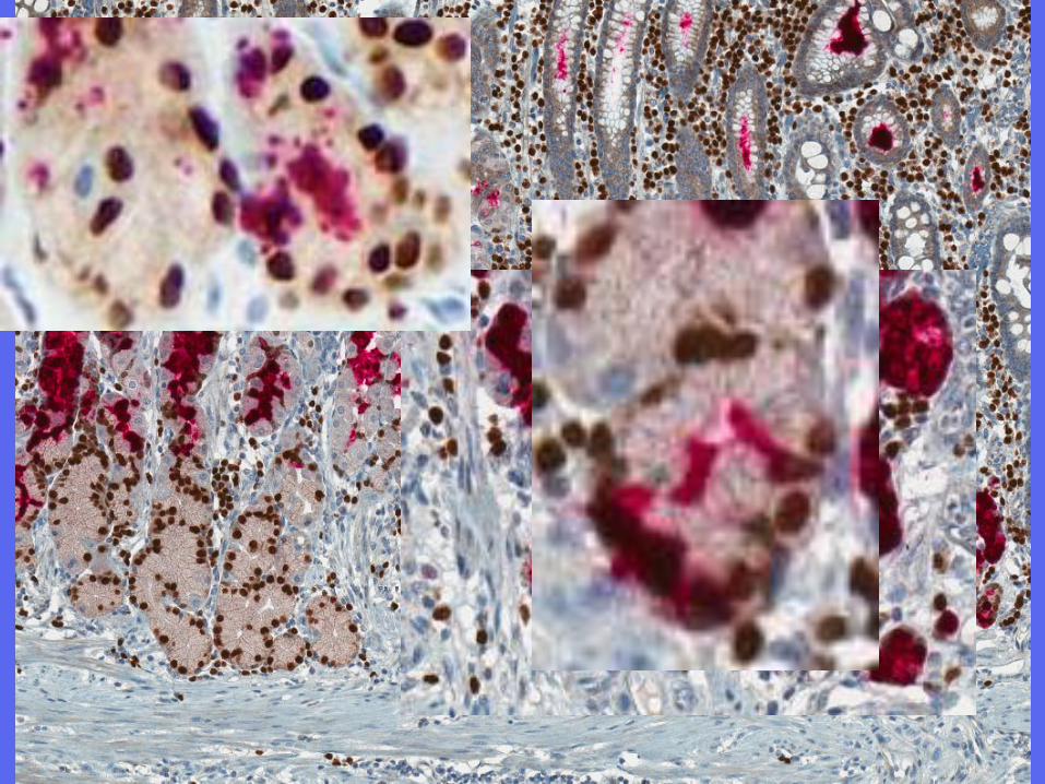

MIST1 and SPEM and Intestinal Metaplasia

Loss of MIST1 from chief cells undergoing metaplasia (downscaling)
Red MIST1
Brown CDX2

MIST1 in intestinal metaplasia Paneth cells
Red MIST1
Brown CDX2
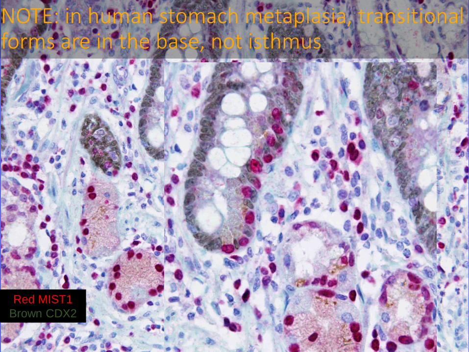
NOTE: in human stomach metaplasia, transitional forms are in the base, not isthmus
Red MIST1
Brown CDX2

The first stage of metaplasia
Mills and Sansom, Science
Signaling, 2015
Scaling Events

Back to mice: Normal gastric chief cells have abundant granules but few lysosomes
A BLAMP1CathepsinDPGC
Mills lab, Unpublished

LAMP1
As MIST1 is lost, lysosomes attack secretory granules
LAMP1CathepsinDPGC
LAMP1CathepsinDPGC
LAMP1CathepsinD
LAMP1PGII
CathepsinD
% Cell cytoplasm occupied
by lysosomes

At 12 hours after Tamoxifen: lysosomes “attack” secretory granules; 3-D projection: Crinophagy vs. Macroautophagy?
LAMP1CathepsinDPGC

SPEM-type metaplasia requires functional lysosome trafficking
Gnptab–/–
mice:Stuart
Kornfeld,
Wash U

Reprogramming of differentiated cells back into the cell cycle is a gene-dependent, conserved cellular process with distinct stages
Mills and Sansom, Science
Signaling, 2015
A note about Pavlov….
© Mark Stivers, 2003

Pavlov Citations over time (Google nGram)
Digestive Disease Studies
Lenin initiates secret funds for studies of how humans might be
conditioned (brainwashed)

Mills Lab

AcknowledgmentsCollaborators
UT Southwestern Ray
MacDonald
Purdue: Steve
Konieczny
Digestive
Disease Center
Wash U: IVIC/AITAC Funding
NIDDK, ACS, AGA
Funderburg Award for
Gastric Cancer, Siteman
Cancer Center
Wash U: Paul Taghert,
Stuart Kornfeld, James
Fitzpatrick, Matt Joens,
Fumi Urano, Indira
Mysorekar
Innsbruck Medical
Institute: Lukas
Huber, Ilja Vietor
Shenyang First
Medical: Zhen-
ning Wang
NIH: Frank
Gonzalez