gastric cancer: introduction
TRANSCRIPT
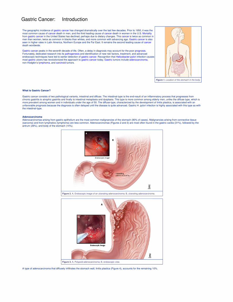
Figure 1. Location of the stomach in the body.
Gastric Cancer: Introduction
The geographic incidence of gastric cancer has changed dramatically over the last few decades. Prior to 1950, it was the
most common cause of cancer death in men, and the third leading cause of cancer death in women in the U.S. Mortality
from gastric cancer in the United States has declined, perhaps due to dietary changes. This cancer is twice as common in
men than women, twice as common in blacks than whites, and more common with advancing age. Gastric cancer is also
seen in higher rates in Latin America, Northern Europe and the Far East. It remains the second leading cause of cancer
death worldwide.
Gastric cancer peaks in the seventh decade of life. Often, a delay in diagnosis may account for the poor prognosis.
Fortunately, dedicated research into its pathogenesis and identification of new risk factors, treatment, and advanced
endoscopic techniques have led to earlier detection of gastric cancer. Recognition that Helicobacter pylori infection causes
most gastric ulcers has revolutionized the approach to gastric cancer today. Gastric tumors include adenocarcinoma,
non-Hodgkin’s lymphoma, and carcinoid tumors.
What is Gastric Cancer?
Gastric cancer consists of two pathological variants, intestinal and diffuse. The intestinal-type is the end-result of an inflammatory process that progresses from
chronic gastritis to atrophic gastritis and finally to intestinal metaplasia and dysplasia. This type is more common among elderly men, unlike the diffuse type, which is
more prevalent among women and in individuals under the age of 50. The diffuse-type, characterized by the development of linitis plastica, is associated with an
unfavorable prognosis because the diagnosis is often delayed until the disease is quite advanced. Gastric H. pylori infection is highly associated with this type as with
the intestinal-type.
Adenocarcinoma
Adenocarcinomas arising from gastric epithelium are the most common malignancies of the stomach (90% of cases). Malignancies arising from connective tissue
(sarcoma) and from lymphatics (lymphoma) are less common. Adenocarcinomas (Figures 2 and 3) are most often found in the gastric cardia (31%), followed by the
antrum (26%), and body of the stomach (14%).
Figure 2. A, Endoscopic image of an ulcerating adenocarcinoma; B, ulcerating adenocarcinoma.
Figure 3. A, Polypoid adenocarcinoma; B, endoscopic view.
A type of adenocarcinoma that diffusely infiltrates the stomach wall, linitis plastica (Figure 4), accounts for the remaining 10%.
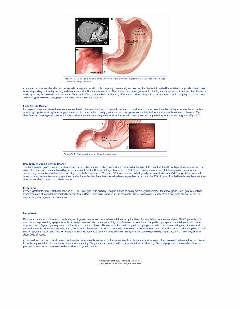
Figure 4. A, CT image of linitis plastica (arrows denote a thickened gastric wall); B, endoscopic image;
C, corresponding illustration.
Adenocarcinomas are classified according to histology and location. Histologically, these malignancies may be divided into well-differentiated and poorly differentiated
types, depending on the degree of gland formation and ability to secrete mucus. Most tumors are heterogeneous in histological appearance; therefore, classification is
made by noting the predominant structures. Thus, well-differentiated tubular and poorly differentiated signet-ring cell carcinoma make up the majority of tumors. Less
common types are mucinous, papillary and undifferentiated carcinoma.
Early Gastric Cancer
Early gastric cancers, where tumor cells are confined to the mucosa (the most superficial layer of the stomach), have been identified in Japan where there is active
screening of patients at high-risk for gastric cancer. In these patients, early gastric cancer may appear as a subtle lesion, usually less than 2 cm in diameter. The
identification of early gastric cancer is important because it is potentially amenable to endoscopic therapy and accompanied by an excellent prognosis (Figure 5).
Figure 5. A, Early gastric cancer; B, endoscopic view.
Hereditary (Familial) Gastric Cancer
The term, familial gastric cancer, has been used to describe families in which several members under the age of 40 have had the diffuse type of gastric cancer. The
criteria for diagnosis, as established by the International Gastric Cancer Linkage Consortium (IGCLC), are: two or more cases of diffuse gastric cancer in first- or
second-degree relatives, with at least one diagnosed before the age of 50 years; OR three or more pathologically documented cases of diffuse gastric cancer in first-
or second-degree relatives of any age. One third of these families have been found to have a germline mutation of the CDH1 gene. Affected family members are also
at increased risk for breast and colon cancer
Lymphoma
Primary gastrointestinal lymphoma may be of B- or T-cell type, with primary Hodgkin’s disease being extremely uncommon. Most low-grade B-cell gastrointestinal
lymphomas are of mucosal associated lymphoid tissue (MALT) and arise primarily in the stomach. These lymphomas usually have a favorable clinical course, but
may undergo high-grade transformation.
Symptoms
Most patients are asymptomatic in early stages of gastric cancer and have advanced disease by the time of presentation. In a review of over 18,000 patients, the
most common presenting symptoms included weight loss and abdominal pain. Epigastric fullness, nausea, loss of appetite, dyspepsia, and mild gastric discomfort
may also occur. Dysphagia may be a prominent symptom for patients with tumors in the cardia or gastroesophageal junction. In patients with pyloric tumors and
tumors located in the antrum, vomiting and gastric outlet obstruction may occur. Unusual presentations may include acute appendicitis, musculoskeletal pain, and the
sudden appearance of seborrheic keratosis and freckles, accompanied by pruritis and dermatomyositis. Gastrointestinal bleeding is uncommon, and only seen in
about 20% of cases.
Abdominal pain occurs in most patients with gastric lymphoma; however, symptoms may vary from those suggesting peptic ulcer disease to advanced gastric cancer.
Patients may complain of weight loss, nausea and vomiting. They may also present with overt gastrointestinal bleeding. Gastric lymphoma is more often found in
younger females when compared to the incidence of gastric cancer.
© Copyright 2001-2013 | All Rights Reserved.
600 North Wolfe Street, Baltimore, Maryland 21287
Gastric Cancer: Anatomy
The stomach is located in the upper part of the abdomen just beneath the diaphragm. The size, shape, and position may vary with posture and with content because
it is distensible and on a free mesentery. An empty stomach is roughly the size of an open hand. It can fill much of the upper abdomen when distended with food and
may descend into the lower abdomen or pelvis upon standing. The duodenum extends from the pylorus to the ligament of Treitz in a sharp curve that almost
completes a circle. It is so named because it is about equal in length to the breadth of 12 fingers, or about 25 cm. It is largely retroperitoneal and the position is
relatively fixed. The stomach and duodenum are closely related in function and in pathogenesis and manifestation of disease.
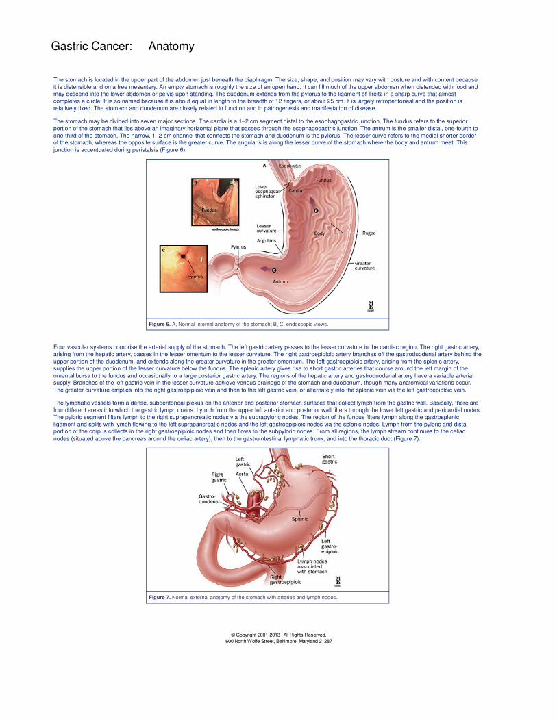
The stomach may be divided into seven major sections. The cardia is a 1–2 cm segment distal to the esophagogastric junction. The fundus refers to the superior
portion of the stomach that lies above an imaginary horizontal plane that passes through the esophagogastric junction. The antrum is the smaller distal, one-fourth to
one-third of the stomach. The narrow, 1–2-cm channel that connects the stomach and duodenum is the pylorus. The lesser curve refers to the medial shorter border
of the stomach, whereas the opposite surface is the greater curve. The angularis is along the lesser curve of the stomach where the body and antrum meet. This
junction is accentuated during peristalsis (Figure 6).
Figure 6. A, Normal internal anatomy of the stomach; B, C, endoscopic views.
Four vascular systems comprise the arterial supply of the stomach. The left gastric artery passes to the lesser curvature in the cardiac region. The right gastric artery,
arising from the hepatic artery, passes in the lesser omentum to the lesser curvature. The right gastroepiploic artery branches off the gastroduodenal artery behind the
upper portion of the duodenum, and extends along the greater curvature in the greater omentum. The left gastroepiploic artery, arising from the splenic artery,
supplies the upper portion of the lesser curvature below the fundus. The splenic artery gives rise to short gastric arteries that course around the left margin of the
omental bursa to the fundus and occasionally to a large posterior gastric artery. The regions of the hepatic artery and gastroduodenal artery have a variable arterial
supply. Branches of the left gastric vein in the lesser curvature achieve venous drainage of the stomach and duodenum, though many anatomical variations occur.
The greater curvature empties into the right gastroepiploic vein and then to the left gastric vein, or alternately into the splenic vein via the left gastroepiploic vein.
The lymphatic vessels form a dense, subperitoneal plexus on the anterior and posterior stomach surfaces that collect lymph from the gastric wall. Basically, there are
four different areas into which the gastric lymph drains. Lymph from the upper left anterior and posterior wall filters through the lower left gastric and pericardial nodes.
The pyloric segment filters lymph to the right suprapancreatic nodes via the suprapyloric nodes. The region of the fundus filters lymph along the gastrosplenic
ligament and splits with lymph flowing to the left suprapancreatic nodes and the left gastroepiploic nodes via the splenic nodes. Lymph from the pyloric and distal
portion of the corpus collects in the right gastroepiploic nodes and then flows to the subpyloric nodes. From all regions, the lymph stream continues to the celiac
nodes (situated above the pancreas around the celiac artery), then to the gastrointestinal lymphatic trunk, and into the thoracic duct (Figure 7).
Figure 7. Normal external anatomy of the stomach with arteries and lymph nodes.
© Copyright 2001-2013 | All Rights Reserved.
600 North Wolfe Street, Baltimore, Maryland 21287
Gastric Cancer: Causes
Environmental Risk Factors
The continued identification of risk factors for gastric cancer may one day lead to the global development of early detection programs that will change the clinical
history of this disease. Environmental factors appear to be related to the intestinal type of gastric cancer. Socioeconomic status is inversely correlated with the
incidence of this disease. Factors associated with low socioeconomic status, such as poor sanitation, poor nutrition, and inadequate handling and preservation of food
and water, are involved. Diets high in fresh fruit, leafy vegetables, ascorbic acid, and beta-carotene are associated with reduced risk. The literature also reports that
decreased use of nitrites in prepared foods has also resulted in a decreased incidence. Though cigarette smoking may increase pre-malignant lesions and gastric
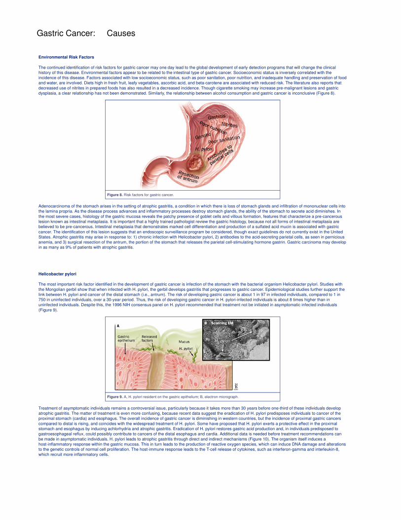
dysplasia, a clear relationship has not been demonstrated. Similarly, the relationship between alcohol consumption and gastric cancer is inconclusive (Figure 8).
Figure 8. Risk factors for gastric cancer.
Adenocarcinoma of the stomach arises in the setting of atrophic gastritis, a condition in which there is loss of stomach glands and infiltration of mononuclear cells into
the lamina propria. As the disease process advances and inflammatory processes destroy stomach glands, the ability of the stomach to secrete acid diminishes. In
the most severe cases, histology of the gastric mucosa reveals the patchy presence of goblet cells and villous formation, features that characterize a pre-cancerous
lesion known as intestinal metaplasia. It is important that a highly trained pathologist review the gastric histology, because not all forms of intestinal metaplasia are
believed to be pre-cancerous. Intestinal metaplasia that demonstrates marked cell differentiation and production of a sulfated acid mucin is associated with gastric
cancer. The identification of this lesion suggests that an endoscopic surveillance program be considered, though exact guidelines do not currently exist in the United
States. Atrophic gastritis may arise in response to: 1) chronic infection with Helicobacter pylori, 2) antibodies to the acid-secreting parietal cells, as seen in pernicious
anemia, and 3) surgical resection of the antrum, the portion of the stomach that releases the parietal cell-stimulating hormone gastrin. Gastric carcinoma may develop
in as many as 9% of patients with atrophic gastritis.
Helicobacter pylori
The most important risk factor identified in the development of gastric cancer is infection of the stomach with the bacterial organism Helicobacter pylori. Studies with
the Mongolian gerbil show that when infected with H. pylori, the gerbil develops gastritis that progresses to gastric cancer. Epidemiological studies further support the
link between H. pylori and cancer of the distal stomach (i.e., antrum). The risk of developing gastric cancer is about 1 in 97 in infected individuals, compared to 1 in
750 in uninfected individuals, over a 30-year period. Thus, the risk of developing gastric cancer in H. pylori-infected individuals is about 8 times higher than in
uninfected individuals. Despite this, the 1996 NIH consensus panel on H. pylori recommended that treatment not be initiated in asymptomatic infected individuals
(Figure 9).
Figure 9. A, H. pylori resident on the gastric epithelium; B, electron micrograph.
Treatment of asymptomatic individuals remains a controversial issue, particularly because it takes more than 30 years before one-third of these individuals develop
atrophic gastritis. The matter of treatment is even more confusing, because recent data suggest the eradication of H. pylori predisposes individuals to cancer of the
proximal stomach (cardia) and esophagus. The overall incidence of gastric cancer is diminishing in western countries, but the incidence of proximal gastric cancers
compared to distal is rising, and coincides with the widespread treatment of H. pylori. Some have proposed that H. pylori exerts a protective effect in the proximal
stomach and esophagus by inducing achlorhydria and atrophic gastritis. Eradication of H. pylori restores gastric acid production and, in individuals predisposed to
gastroesophageal reflux, could possibly contribute to cancers of the distal esophagus and cardia. Additional data is needed before treatment recommendations can
be made in asymptomatic individuals. H. pylori leads to atrophic gastritis through direct and indirect mechanisms (Figure 10). The organism itself induces a
host-inflammatory response within the gastric mucosa. This in turn leads to the production of reactive oxygen species, which can induce DNA damage and alterations
to the genetic controls of normal cell proliferation. The host-immune response leads to the T-cell release of cytokines, such as interferon-gamma and interleukin-8,
which recruit more inflammatory cells.
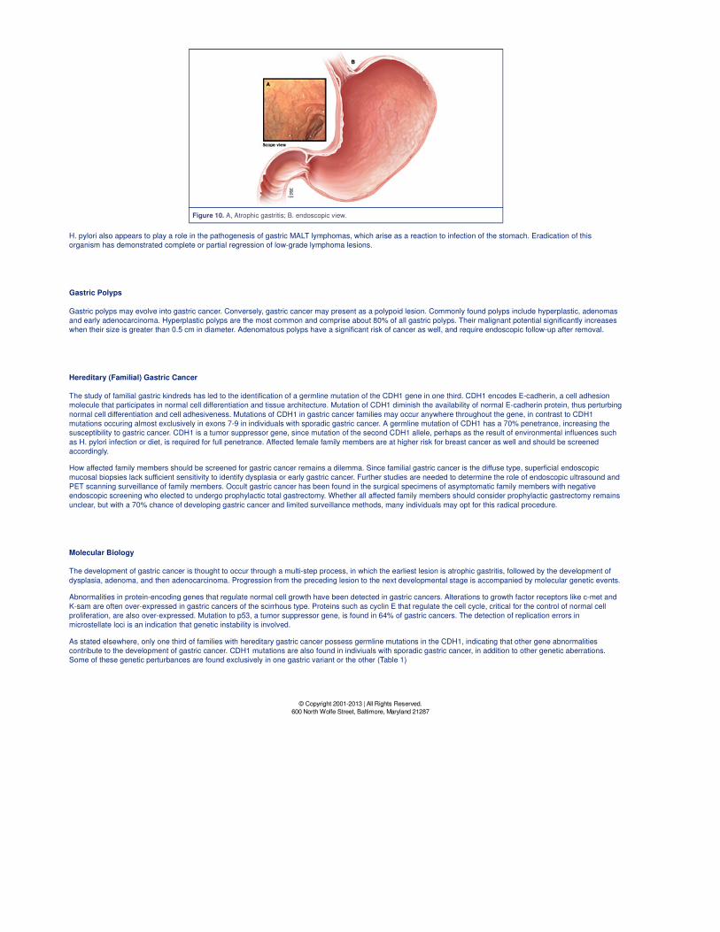
Figure 10. A, Atrophic gastritis; B. endoscopic view.
H. pylori also appears to play a role in the pathogenesis of gastric MALT lymphomas, which arise as a reaction to infection of the stomach. Eradication of this
organism has demonstrated complete or partial regression of low-grade lymphoma lesions.
Gastric Polyps
Gastric polyps may evolve into gastric cancer. Conversely, gastric cancer may present as a polypoid lesion. Commonly found polyps include hyperplastic, adenomas
and early adenocarcinoma. Hyperplastic polyps are the most common and comprise about 80% of all gastric polyps. Their malignant potential significantly increases
when their size is greater than 0.5 cm in diameter. Adenomatous polyps have a significant risk of cancer as well, and require endoscopic follow-up after removal.
Hereditary (Familial) Gastric Cancer
The study of familial gastric kindreds has led to the identification of a germline mutation of the CDH1 gene in one third. CDH1 encodes E-cadherin, a cell adhesion
molecule that participates in normal cell differentiation and tissue architecture. Mutation of CDH1 diminish the availability of normal E-cadherin protein, thus perturbing
normal cell differentiation and cell adhesiveness. Mutations of CDH1 in gastric cancer families may occur anywhere throughout the gene, in contrast to CDH1
mutations occuring almost exclusively in exons 7-9 in individuals with sporadic gastric cancer. A germline mutation of CDH1 has a 70% penetrance, increasing the
susceptibility to gastric cancer. CDH1 is a tumor suppressor gene, since mutation of the second CDH1 allele, perhaps as the result of environmental influences such
as H. pylori infection or diet, is required for full penetrance. Affected female family members are at higher risk for breast cancer as well and should be screened
accordingly.
How affected family members should be screened for gastric cancer remains a dilemma. Since familial gastric cancer is the diffuse type, superficial endoscopic
mucosal biopsies lack sufficient sensitivity to identify dysplasia or early gastric cancer. Further studies are needed to determine the role of endoscopic ultrasound and
PET scanning surveillance of family members. Occult gastric cancer has been found in the surgical specimens of asymptomatic family members with negative
endoscopic screening who elected to undergo prophylactic total gastrectomy. Whether all affected family members should consider prophylactic gastrectomy remains
unclear, but with a 70% chance of developing gastric cancer and limited surveillance methods, many individuals may opt for this radical procedure.
Molecular Biology
The development of gastric cancer is thought to occur through a multi-step process, in which the earliest lesion is atrophic gastritis, followed by the development of
dysplasia, adenoma, and then adenocarcinoma. Progression from the preceding lesion to the next developmental stage is accompanied by molecular genetic events.
Abnormalities in protein-encoding genes that regulate normal cell growth have been detected in gastric cancers. Alterations to growth factor receptors like c-met and
K-sam are often over-expressed in gastric cancers of the scirrhous type. Proteins such as cyclin E that regulate the cell cycle, critical for the control of normal cell
proliferation, are also over-expressed. Mutation to p53, a tumor suppressor gene, is found in 64% of gastric cancers. The detection of replication errors in
microstellate loci is an indication that genetic instability is involved.
As stated elsewhere, only one third of families with hereditary gastric cancer possess germline mutations in the CDH1, indicating that other gene abnormalities
contribute to the development of gastric cancer. CDH1 mutations are also found in indiviuals with sporadic gastric cancer, in addition to other genetic aberrations.
Some of these genetic perturbances are found exclusively in one gastric variant or the other (Table 1)
© Copyright 2001-2013 | All Rights Reserved.
600 North Wolfe Street, Baltimore, Maryland 21287
Gastric Cancer: Diagnosis
Physical Examination
Physical examination may provide clues to diagnose gastric cancer. The presence of anemia, occult blood in the stool, and weight loss may suggest a malignancy. A
midepigastric palpable mass or nodular liver may be helpful in localizing the process to the abdomen.
The patient may appear completely healthy on physical exam. Additional findings may include: abdominal mass, liver metastases, gastric distention, weight loss,
supraclavicular adenopathy (Virchow’s node), rectal mass (Blumer’s shelf), enlarged ovary (Krukenberg’s syndrome), or umbilical metastases (Sister Mary Joseph’s
node). Migratory phlebitis (Trousseau’s syndrome), seborrheic keratosis and freckles (Leser-Trélat sign), muscle weakness, splenomegaly, ascites, obstructive
jaundice, and peritoneal carcinomatosis may be noted in more advanced disease.
Immunohistochemical techniques are used to differentiate low-grade lymphomas of MALT from benign reactive lymphoid hyperplasia. Lymphomas show
monoclonality of the lymphoid proliferation. Clinicians should be aware of the possibility that non-Hodgkin’s lymphoma of the stomach may be linked to acquired
immunodeficiency syndrome (AIDS).
Genetic Screening
Genetic screening has been advocated in family members of young patients with the diffuse-type of gastric cancer. There are no mutational hotspots, so screening for
CDH1 mutations requires a survey of the entire gene. Prophylactic gastrectomy has been performed on carriers of truncating germ-line CDH1 mutations. Remarkably,
even asymptomatic individuals who had normal upper endoscopies have demonstrated malignant cells in their surgical resection specimens, suggesting that this
could be a viable therapeutic option for highly selected individuals. Genetic counseling is necessary for all family members considering genetic testing and
prophylactic gastrectomy. Women in these families who have a germline mutation of CDH1 are at an increased risk for developing lobular breast cancer and should
be screened accordingly.
Resources: The reader is referred to two excellent reviews on hereditary diffuse gastric cancer:
Graziano, F. et al. Annals of Oncology 14:1705-1713, 2003.
Nardone, G. Alimentary Pharmacology Therapeutics 2003: 17 (suppl.2):75-81.
More information about E-cadherin mutations and prophylactic gastrectomy can be found in these two references:
Huntsman, DG, et al. NEJM 2001 344:1904-1909.
Guilford, P, et al. Nature 1998, 392:402-404.
Radiological Diagnosis
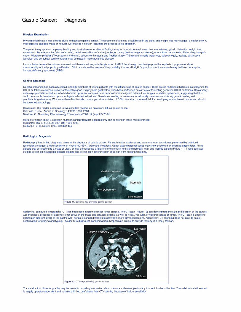
Radiography has limited diagnostic value in the diagnosis of gastric cancer. Although better studies (using state-of-the-art techniques performed by practiced
technicians) suggest a high sensitivity of x-rays (80–95%), there are limitations. Upper gastrointestinal series may show thickened or enlarged gastric folds, filling
defects that correspond to a mass or ulcer, or may demonstrate a failure of the stomach to distend normally to air and instilled barium (Figure 11). These contrast
studies do not aid in accurate disease staging and do not allow differentiation of benign from malignant lesions.
Figure 11. Barium x-ray showing gastric cancer.
Abdominal computed tomography (CT) has been used in gastric cancer tumor staging. The CT scan (Figure 12) can demonstrate the size and location of the cancer,
wall thickness, presence or absence of fat between the mass and adjacent organs, as well as nodal, vascular, or visceral spread of tumor. The CT scan is unable to
distinguish different layers of the gastric wall; hence, it cannot differentiate early from more advanced lesions. Additionally, CT scanning does not provide tissue
confirmation for grading and typing. The ability to distinguish carcinoma from lymphoma is crucial to provide therapy in a timely fashion.
Figure 12. CT image showing gastric cancer.
Transabdominal ultrasonography may be useful in providing information about metastatic disease, particularly that which affects the liver. Transabdominal ultrasound
is largely operator-dependent and has more limited usefulness than CT scanning because of its low sensitivity.
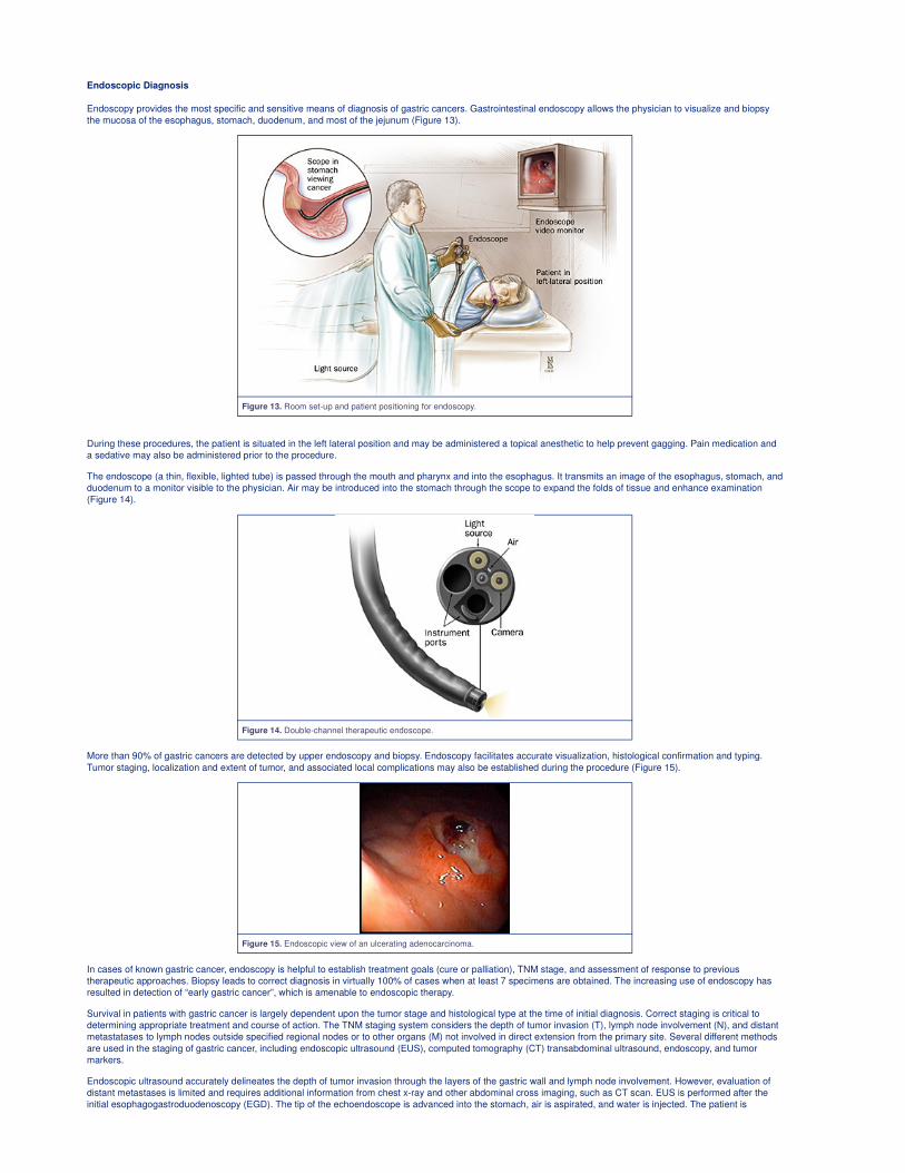
Endoscopic Diagnosis
Endoscopy provides the most specific and sensitive means of diagnosis of gastric cancers. Gastrointestinal endoscopy allows the physician to visualize and biopsy
the mucosa of the esophagus, stomach, duodenum, and most of the jejunum (Figure 13).
Figure 13. Room set-up and patient positioning for endoscopy.
During these procedures, the patient is situated in the left lateral position and may be administered a topical anesthetic to help prevent gagging. Pain medication and
a sedative may also be administered prior to the procedure.
The endoscope (a thin, flexible, lighted tube) is passed through the mouth and pharynx and into the esophagus. It transmits an image of the esophagus, stomach, and
duodenum to a monitor visible to the physician. Air may be introduced into the stomach through the scope to expand the folds of tissue and enhance examination
(Figure 14).
Figure 14. Double-channel therapeutic endoscope.
More than 90% of gastric cancers are detected by upper endoscopy and biopsy. Endoscopy facilitates accurate visualization, histological confirmation and typing.
Tumor staging, localization and extent of tumor, and associated local complications may also be established during the procedure (Figure 15).
Figure 15. Endoscopic view of an ulcerating adenocarcinoma.
In cases of known gastric cancer, endoscopy is helpful to establish treatment goals (cure or palliation), TNM stage, and assessment of response to previous
therapeutic approaches. Biopsy leads to correct diagnosis in virtually 100% of cases when at least 7 specimens are obtained. The increasing use of endoscopy has
resulted in detection of “early gastric cancer”, which is amenable to endoscopic therapy.
Survival in patients with gastric cancer is largely dependent upon the tumor stage and histological type at the time of initial diagnosis. Correct staging is critical to
determining appropriate treatment and course of action. The TNM staging system considers the depth of tumor invasion (T), lymph node involvement (N), and distant
metastatases to lymph nodes outside specified regional nodes or to other organs (M) not involved in direct extension from the primary site. Several different methods
are used in the staging of gastric cancer, including endoscopic ultrasound (EUS), computed tomography (CT) transabdominal ultrasound, endoscopy, and tumor
markers.
Endoscopic ultrasound accurately delineates the depth of tumor invasion through the layers of the gastric wall and lymph node involvement. However, evaluation of
distant metastases is limited and requires additional information from chest x-ray and other abdominal cross imaging, such as CT scan. EUS is performed after the
initial esophagogastroduodenoscopy (EGD). The tip of the echoendoscope is advanced into the stomach, air is aspirated, and water is injected. The patient is
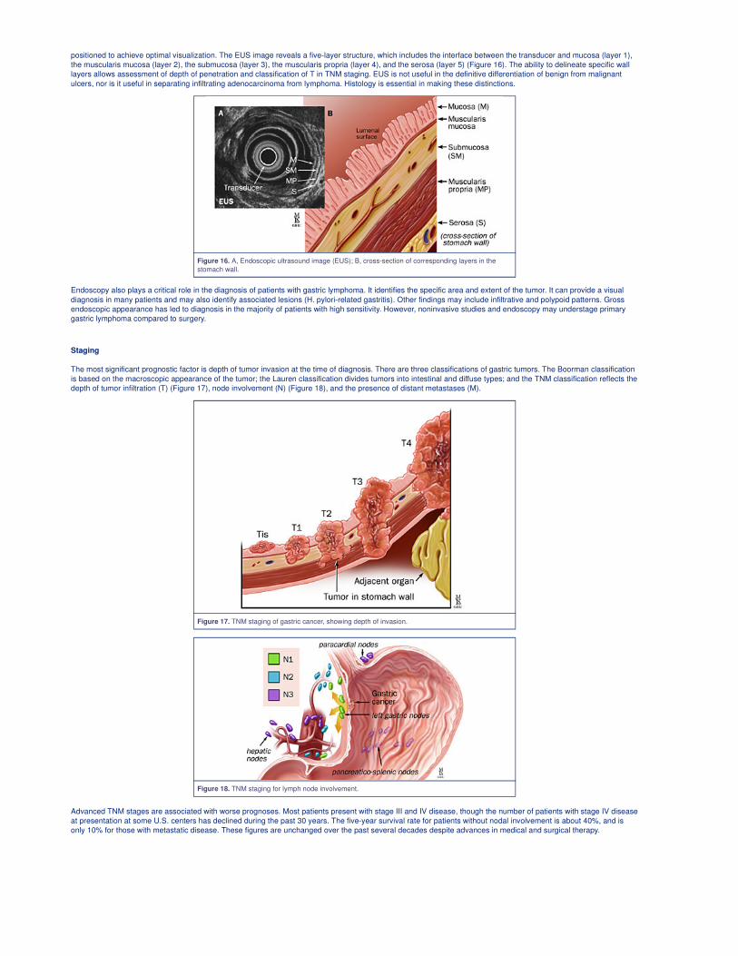
positioned to achieve optimal visualization. The EUS image reveals a five-layer structure, which includes the interface between the transducer and mucosa (layer 1),
the muscularis mucosa (layer 2), the submucosa (layer 3), the muscularis propria (layer 4), and the serosa (layer 5) (Figure 16). The ability to delineate specific wall
layers allows assessment of depth of penetration and classification of T in TNM staging. EUS is not useful in the definitive differentiation of benign from malignant
ulcers, nor is it useful in separating infiltrating adenocarcinoma from lymphoma. Histology is essential in making these distinctions.
Figure 16. A, Endoscopic ultrasound image (EUS); B, cross-section of corresponding layers in the
stomach wall.
Endoscopy also plays a critical role in the diagnosis of patients with gastric lymphoma. It identifies the specific area and extent of the tumor. It can provide a visual
diagnosis in many patients and may also identify associated lesions (H. pylori-related gastritis). Other findings may include infiltrative and polypoid patterns. Gross
endoscopic appearance has led to diagnosis in the majority of patients with high sensitivity. However, noninvasive studies and endoscopy may understage primary
gastric lymphoma compared to surgery.
Staging
The most significant prognostic factor is depth of tumor invasion at the time of diagnosis. There are three classifications of gastric tumors. The Boorman classification
is based on the macroscopic appearance of the tumor; the Lauren classification divides tumors into intestinal and diffuse types; and the TNM classification reflects the
depth of tumor infiltration (T) (Figure 17), node involvement (N) (Figure 18), and the presence of distant metastases (M).
Figure 17. TNM staging of gastric cancer, showing depth of invasion.
Figure 18. TNM staging for lymph node involvement.
Advanced TNM stages are associated with worse prognoses. Most patients present with stage III and IV disease, though the number of patients with stage IV disease
at presentation at some U.S. centers has declined during the past 30 years. The five-year survival rate for patients without nodal involvement is about 40%, and is
only 10% for those with metastatic disease. These figures are unchanged over the past several decades despite advances in medical and surgical therapy.
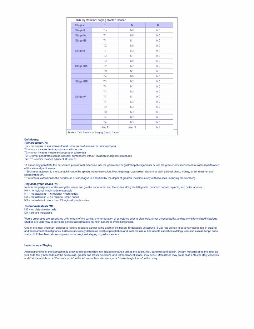
Table 1. TNM System for Staging Gastric Cancer
Definitions:
Primary tumor (T):
Tis = carcinoma in situ: intraepithelial tumor without invasion of lamina propria
T1 = tumor invades lamina propria or submucosa
T2 = tumor invades muscularis propria or subserosa
T3* = tumor penetrates serosa (visceral peritoneum) without invasion of adjacent structures
T4**,*** = tumor invades adjacent structures
*A tumor may penetrate the muscularis propria with extension into the gastrocolic or gastrohepatic ligaments or into the greater or lesser omentum without perforation
of the visceral peritoneum.
**Structures adjacent to the stomach include the spleen, transverse colon, liver, diaphragm, pancreas, abdominal wall, adrenal gland, kidney, small intestine, and
retroperitoneum.
***Intramural extension to the duodenum or esophagus is classified by the depth of greatest invasion in any of these sites, including the stomach).
Regional lymph nodes (N):
Include the perigastric nodes along the lesser and greater curvatures, and the nodes along the left gastric, common hepatic, splenic, and celiac arteries.
N0 = no regional lymph node metastasis
N1 = metastasis to 1–6 regional lymph nodes
N2 = metastasis in 7–15 regional lymph nodes
N3 = metastasis in more than 15 regional lymph nodes
Distant metastasis (M):
M0 = no distant metastasis
M1 = distant metastasis
Worse prognoses are associated with tumors of the cardia, shorter duration of symptoms prior to diagnosis, tumor unresectability, and poorly differentiated histology.
Studies are underway to correlate genetic abnormalities found in tumors to overall prognosis.
One of the most important prognostic factors in gastric cancer is the depth of infiltration. Endoscopic ultrasound (EUS) has proven to be a very useful tool in staging
and assessment of malignancy. EUS can accurately determine depth of penetration and, with the use of fine-needle aspiration cytology, can also assess lymph node
status. EUS has been shown superior for locoregional staging of gastric cancers.
Laparoscopic Staging
Adenocarcinoma of the stomach may grow by direct extension into adjacent organs such as the colon, liver, pancreas and spleen. Distant metastases to the lung, as
well as to the lymph nodes of the celiac axis, greater and lesser omentum, and retroperitoneal space, may occur. Metastases may present as a “Sister Mary Joseph's
node” at the umbilicus, a “Virchow’s node” in the left supraclavicular fossa, or a "Krukenberg's tumor" in the ovary.
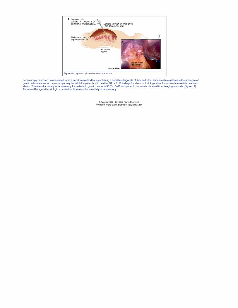
Figure 19. Laparoscopic evaluation of metastasis.
Laparoscopy has been demonstrated to be a sensitive method for establishing a definitive diagnosis of liver and other abdominal metastases in the presence of
gastric adenocarcinoma. Laparoscopy may be helpful in patients with positive CT or EUS findings for which no histological confirmation of metastasis has been
shown. The overall accuracy of laparoscopy for metastatic gastric cancer is 96.5%, 5–20% superior to the results obtained from imaging methods (Figure 19).
Abdominal lavage with cytologic examination increases the sensitivity of laparoscopy.
© Copyright 2001-2013 | All Rights Reserved.
600 North Wolfe Street, Baltimore, Maryland 21287
Gastric Cancer: Therapy
Overview
There are essentially three modes of therapy for the treatment of gastric cancer. Curative resection, including endoscopic resection, appears the most effective.
Surgical resection entails the removal of the primary tumor and regional lymph nodes with resection margins free of tumor. Gastric cancer has not been shown to
respond successfully to radiation alone. Chemotherapy has demonstrated limited success with multi-drug regimens.
Surgical Therapy
The prognosis following surgical resection depends on the stage at presentation. Early tumors confined to the stomach lining have higher cure rates than cases in
which disease has already spread to distant sites or regional lymph nodes. Cure rates have improved in the past 30 years, particularly in Japan. These improvements
can be attributed mainly to an increase in early detection rates.
The type of surgery performed depends on the extent and location of tumor; therefore, preoperative evaluation is critical. Initial staging may be established by
endoscopy with biopsy. Endoscopic ultrasound should follow. Endoscopic Ultrasound (EUS) has a sensitivity of 85% in assessing depth of tumor invasion and
detecting nodal involvement prior to surgery. Laparoscopic staging prior to surgical resection is also advocated and has impacted preoperative treatment decisions.
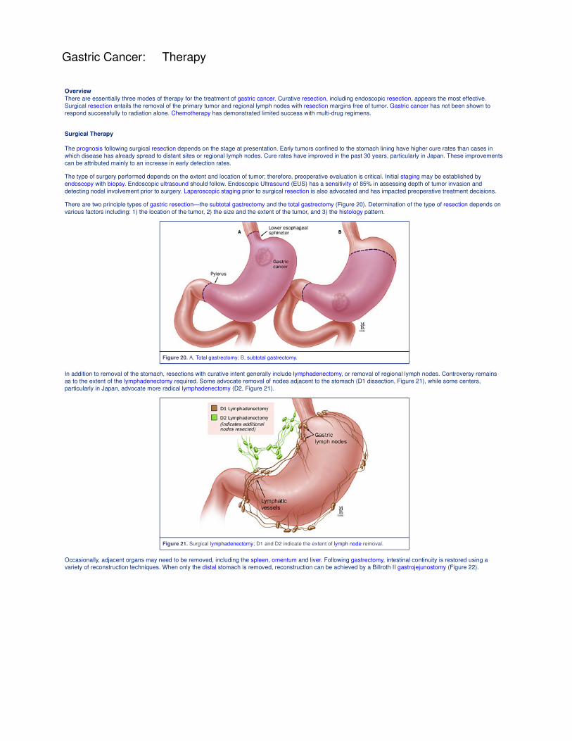
There are two principle types of gastric resection—the subtotal gastrectomy and the total gastrectomy (Figure 20). Determination of the type of resection depends on
various factors including: 1) the location of the tumor, 2) the size and the extent of the tumor, and 3) the histology pattern.
Figure 20. A, Total gastrectomy; B, subtotal gastrectomy.
In addition to removal of the stomach, resections with curative intent generally include lymphadenectomy, or removal of regional lymph nodes. Controversy remains
as to the extent of the lymphadenectomy required. Some advocate removal of nodes adjacent to the stomach (D1 dissection, Figure 21), while some centers,
particularly in Japan, advocate more radical lymphadenectomy (D2, Figure 21).
Figure 21. Surgical lymphadenectomy; D1 and D2 indicate the extent of lymph node removal.
Occasionally, adjacent organs may need to be removed, including the spleen, omentum and liver. Following gastrectomy, intestinal continuity is restored using a
variety of reconstruction techniques. When only the distal stomach is removed, reconstruction can be achieved by a Billroth II gastrojejunostomy (Figure 22).
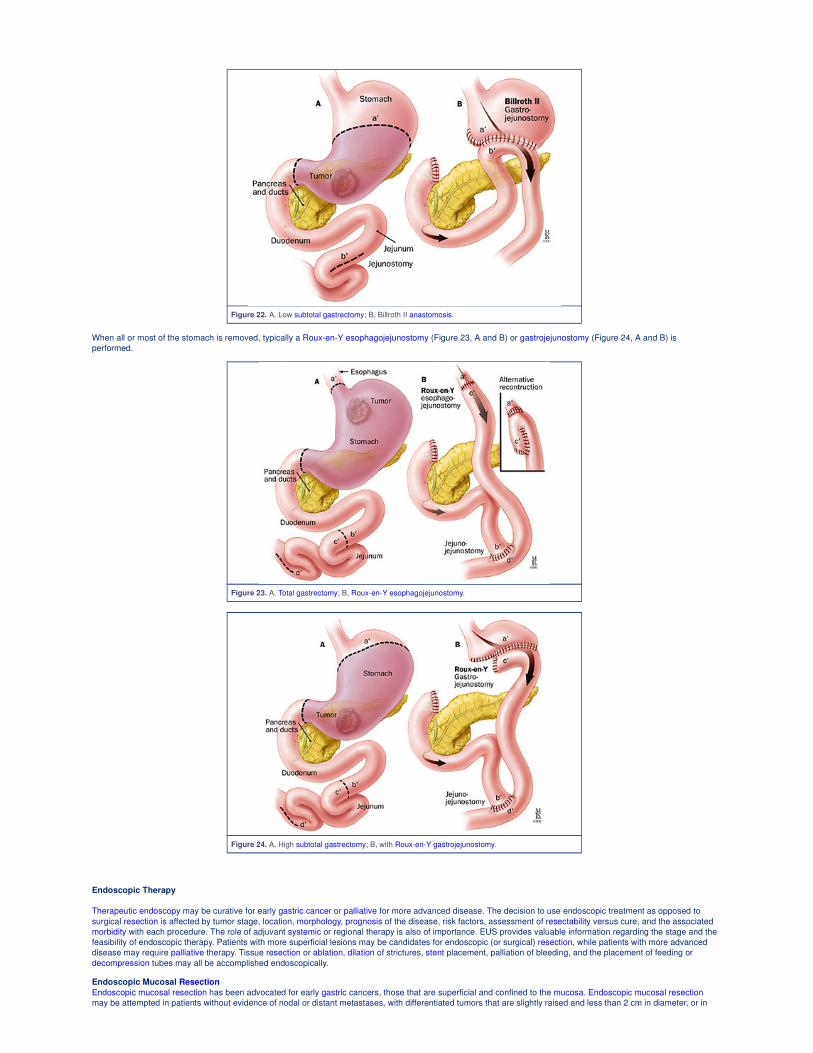
Figure 22. A, Low subtotal gastrectomy; B, Billroth II anastomosis.
When all or most of the stomach is removed, typically a Roux-en-Y esophagojejunostomy (Figure 23, A and B) or gastrojejunostomy (Figure 24, A and B) is
performed.
Figure 23. A, Total gastrectomy; B, Roux-en-Y esophagojejunostomy.
Figure 24. A, High subtotal gastrectomy; B, with Roux-en-Y gastrojejunostomy.
Endoscopic Therapy
Therapeutic endoscopy may be curative for early gastric cancer or palliative for more advanced disease. The decision to use endoscopic treatment as opposed to
surgical resection is affected by tumor stage, location, morphology, prognosis of the disease, risk factors, assessment of resectability versus cure, and the associated
morbidity with each procedure. The role of adjuvant systemic or regional therapy is also of importance. EUS provides valuable information regarding the stage and the
feasibility of endoscopic therapy. Patients with more superficial lesions may be candidates for endoscopic (or surgical) resection, while patients with more advanced
disease may require palliative therapy. Tissue resection or ablation, dilation of strictures, stent placement, palliation of bleeding, and the placement of feeding or
decompression tubes may all be accomplished endoscopically.
Endoscopic Mucosal Resection
Endoscopic mucosal resection has been advocated for early gastric cancers, those that are superficial and confined to the mucosa. Endoscopic mucosal resection
may be attempted in patients without evidence of nodal or distant metastases, with differentiated tumors that are slightly raised and less than 2 cm in diameter, or in
differentiated tumors that are ulcerated and less than 1 cm in diameter. The most commonly employed methods of endoscopic mucosal resection include strip biopsy,
double-snare polypectomy, resection with combined use of highly concentrated saline and epinephrine, and resection using a cap. The prognosis after treatment is
comparable to that of surgical resection for early gastric cancer. Five-year survival rates for individuals undergoing endoscopic mucosal resection of early gastric
cancers have been reported to be as high as 95%.
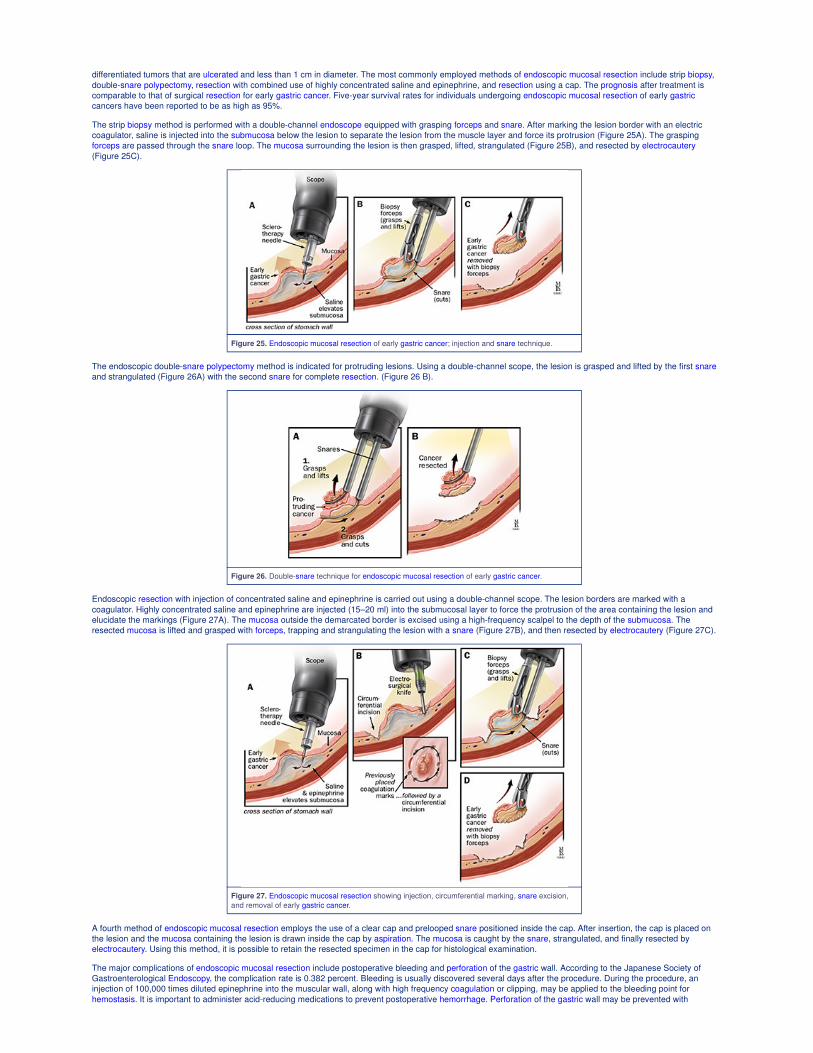
The strip biopsy method is performed with a double-channel endoscope equipped with grasping forceps and snare. After marking the lesion border with an electric
coagulator, saline is injected into the submucosa below the lesion to separate the lesion from the muscle layer and force its protrusion (Figure 25A). The grasping
forceps are passed through the snare loop. The mucosa surrounding the lesion is then grasped, lifted, strangulated (Figure 25B), and resected by electrocautery
(Figure 25C).
Figure 25. Endoscopic mucosal resection of early gastric cancer; injection and snare technique.
The endoscopic double-snare polypectomy method is indicated for protruding lesions. Using a double-channel scope, the lesion is grasped and lifted by the first snare
and strangulated (Figure 26A) with the second snare for complete resection. (Figure 26 B).
Figure 26. Double-snare technique for endoscopic mucosal resection of early gastric cancer.
Endoscopic resection with injection of concentrated saline and epinephrine is carried out using a double-channel scope. The lesion borders are marked with a
coagulator. Highly concentrated saline and epinephrine are injected (15–20 ml) into the submucosal layer to force the protrusion of the area containing the lesion and
elucidate the markings (Figure 27A). The mucosa outside the demarcated border is excised using a high-frequency scalpel to the depth of the submucosa. The
resected mucosa is lifted and grasped with forceps, trapping and strangulating the lesion with a snare (Figure 27B), and then resected by electrocautery (Figure 27C).
Figure 27. Endoscopic mucosal resection showing injection, circumferential marking, snare excision,
and removal of early gastric cancer.
A fourth method of endoscopic mucosal resection employs the use of a clear cap and prelooped snare positioned inside the cap. After insertion, the cap is placed on
the lesion and the mucosa containing the lesion is drawn inside the cap by aspiration. The mucosa is caught by the snare, strangulated, and finally resected by
electrocautery. Using this method, it is possible to retain the resected specimen in the cap for histological examination.
The major complications of endoscopic mucosal resection include postoperative bleeding and perforation of the gastric wall. According to the Japanese Society of
Gastroenterological Endoscopy, the complication rate is 0.382 percent. Bleeding is usually discovered several days after the procedure. During the procedure, an
injection of 100,000 times diluted epinephrine into the muscular wall, along with high frequency coagulation or clipping, may be applied to the bleeding point for
hemostasis. It is important to administer acid-reducing medications to prevent postoperative hemorrhage. Perforation of the gastric wall may be prevented with
sufficient saline injection to raise the mucosa containing the lesion. The “non-lifting sign” and complaints of pain with snare strangulation of the lesion are
contraindications to endoscopic mucosal resection. When perforation is recognized immediately after a procedure, clips should be applied to close the perforation,
followed by abdominocentesis and aspiration of air from the abdominal cavity. Surgery should be considered in cases of endoscopic closure failure.
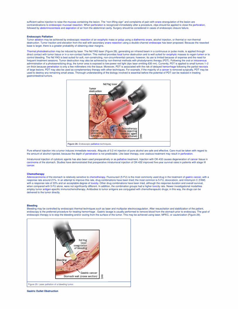
Endoscopic Palliation
Tumor ablation may be achieved by endoscopic resection of an exophytic mass or polyp using a diathermic snare, alcohol injection, or thermal or non-thermal
destruction. Tumor traction and elevation from the wall with secondary snare resection using a double-channel endoscope has been proposed. Because the resected
base is larger, there is a greater probability of obtaining clear margins.
Thermal photodestruction may be induced by laser. The Nd:YAG laser (Figure 28), generating an infrared beam in a continuous or pulse mode, is applied through
direct contact with tumor tissue or in a non-contact fashion. This method provides focal tumor destruction and is well suited for exophytic masses to regain lumen or to
control bleeding. The Nd:YAG is best suited for soft, non-constricting, non-circumferential cancers; however, its use is limited because of expense and the need for
frequent treatment sessions. Tumor destruction may also be achieved by non-thermal methods with photodynamic therapy (PDT). Following the oral or intravenous
administration of a photosensitizing drug, the tumor area is exposed to low-power red light (dye laser emitting 630 nm). Currently, PDT is applied to small tumors 1–2
cm thick because penetration is only a few millimeters into the tissue. Moreover, PDT is associated with the risk of delayed hemorrhage following the partial necrosis
of large lesions. PDT may also be used as complementary therapy with other techniques. For example, if the majority of a cancer is removed surgically, PDT may be
used to destroy any remaining small areas. Thorough understanding of the biology involved is essential before the potential of PDT can be realized in treating
gastrointestinal tumors.
Figure 28. Endoscopic palliative techniques.
Pure ethanol injection into a tumor induces immediate necrosis. Aliquots of 0.2 ml injection of pure alcohol are safe and effective. Care must be taken with regard to
the amount of alcohol injected, because the depth of penetration is not predictable. Like laser therapy, over zealous treatment may result in perforation.
Intratumoral injection of cytotoxic agents has also been used preoperatively or as palliative treatment. Injection with OK-432 causes degeneration of cancer tissue in
carcinoma of the stomach. Studies have demonstrated that preoperative intratumoral injection of OK-432 improved five-year survival rates in patients with stage III
cancer.
Chemotherapy
Adenocarcinoma of the stomach is relatively sensitive to chemotherapy. Fluorouracil (5-FU) is the most commonly used drug in the treatment of gastric cancer, with a
response rate around 21%. In an attempt to improve this rate, drug combinations have been tried; the most common is 5-FU, doxorubicin, and mitomycin C (FAM)
with a response rate of 33% and an acceptable degree of toxicity. Other drug combinations have been tried, although the response duration and overall survival,
when compared with 5-FU alone, were not significantly different. In addition, the combination groups had a higher toxicity rate. Newer investigational modalities
employ tumor antigen-specific immunochemotherapy. Antibodies to tumor antigens are conjugated with chemotherapeutic drugs; in this way, the drugs can be
delivered to the tumor directly.
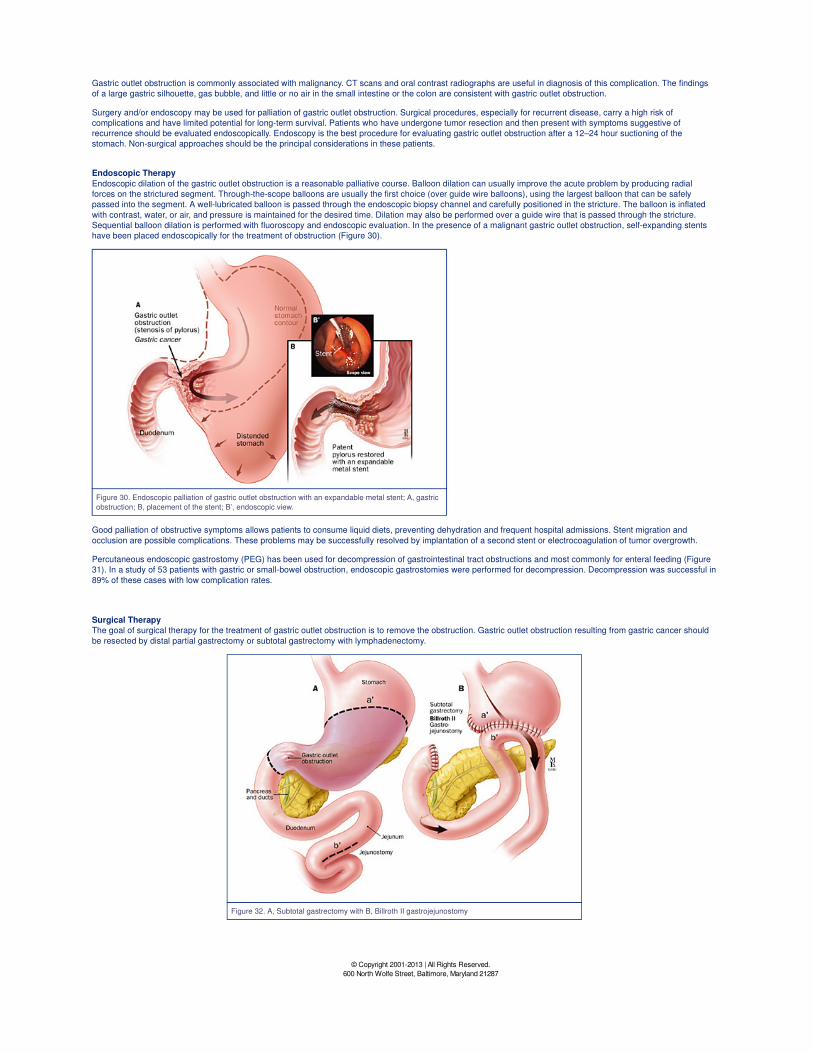
Bleeding
Bleeding may be controlled by endoscopic thermal techniques such as laser and multipolar electrocoagulation. After resuscitation and stabilization of the patient,
endoscopy is the preferred procedure for treating hemorrhage . Gastric lavage is usually performed to remove blood from the stomach prior to endoscopy. The goal of
endoscopic therapy is to stop the bleeding and/or oozing from the surface of the tumor. This may be achieved using laser, MPEC, or cauterization (Figure 29).
Figure 29. Laser palliation of a bleeding tumor.
Gastric Outlet Obstruction
Gastric outlet obstruction is commonly associated with malignancy. CT scans and oral contrast radiographs are useful in diagnosis of this complication. The findings
of a large gastric silhouette, gas bubble, and little or no air in the small intestine or the colon are consistent with gastric outlet obstruction.
Surgery and/or endoscopy may be used for palliation of gastric outlet obstruction. Surgical procedures, especially for recurrent disease, carry a high risk of
complications and have limited potential for long-term survival. Patients who have undergone tumor resection and then present with symptoms suggestive of
recurrence should be evaluated endoscopically. Endoscopy is the best procedure for evaluating gastric outlet obstruction after a 12–24 hour suctioning of the
stomach. Non-surgical approaches should be the principal considerations in these patients.
Endoscopic Therapy
Endoscopic dilation of the gastric outlet obstruction is a reasonable palliative course. Balloon dilation can usually improve the acute problem by producing radial
forces on the strictured segment. Through-the-scope balloons are usually the first choice (over guide wire balloons), using the largest balloon that can be safely
passed into the segment. A well-lubricated balloon is passed through the endoscopic biopsy channel and carefully positioned in the stricture. The balloon is inflated
with contrast, water, or air, and pressure is maintained for the desired time. Dilation may also be performed over a guide wire that is passed through the stricture.
Sequential balloon dilation is performed with fluoroscopy and endoscopic evaluation. In the presence of a malignant gastric outlet obstruction, self-expanding stents
have been placed endoscopically for the treatment of obstruction (Figure 30).
Figure 30. Endoscopic palliation of gastric outlet obstruction with an expandable metal stent; A, gastric
obstruction; B, placement of the stent; B’, endoscopic view.
Good palliation of obstructive symptoms allows patients to consume liquid diets, preventing dehydration and frequent hospital admissions. Stent migration and
occlusion are possible complications. These problems may be successfully resolved by implantation of a second stent or electrocoagulation of tumor overgrowth.
Percutaneous endoscopic gastrostomy (PEG) has been used for decompression of gastrointestinal tract obstructions and most commonly for enteral feeding (Figure
31). In a study of 53 patients with gastric or small-bowel obstruction, endoscopic gastrostomies were performed for decompression. Decompression was successful in
89% of these cases with low complication rates.
Surgical Therapy
The goal of surgical therapy for the treatment of gastric outlet obstruction is to remove the obstruction. Gastric outlet obstruction resulting from gastric cancer should
be resected by distal partial gastrectomy or subtotal gastrectomy with lymphadenectomy.
Figure 32. A, Subtotal gastrectomy with B, Billroth II gastrojejunostomy
© Copyright 2001-2013 | All Rights Reserved.
600 North Wolfe Street, Baltimore, Maryland 21287